Next Article in Journal
Skitour
Previous Article in Journal
Percutaneous Implantation of an ASD Occluder with Intracardiac Ultrasound
 
 
Cardiovascular Medicine is published by MDPI from Volume 28 Issue 1 (2025). Previous articles were published by another publisher in Open Access under a CC-BY (or CC-BY-NC-ND) licence, and they are hosted by MDPI on mdpi.com as a courtesy and upon agreement with Editores Medicorum Helveticorum (EMH).
Interesting Images

Acute Leriche Syndrome in an 83-Year-Old Man with Non NSTEMI After Cardiac Resuscitation Because of Ventricular Fibrillation

by
Daniel Sürder
1,*,
Jos C. van den Berg
2,
Tiziano Moccetti
1 and
Giovanni B. Pedrazzini
1
1
Cardiocentro Ticino, Division of Cardiology, Lugano, Switzerland
2
Ospedale Civico, Division of Interventional Radiology, Lugano, Switzerland
*
Author to whom correspondence should be addressed.
Cardiovasc. Med. 2010, 13(9), 292; https://doi.org/10.4414/cvm.2010.01521
Submission received: 15 June 2010 / Revised: 15 July 2010 / Accepted: 15 August 2010 / Published: 15 September 2010

Case description

An 83-year-old man on permanent haemodialysis was admitted to the emergency department in cardiogenic shock after prolonged electromechanical resuscitation because of ventricular fibrillation. ECG revealed complete left bundle branch block.
The patient received 5000 units of non-fractionated heparin before undergoing coronary angiography. The invasive blood pressure was monitored using a 4 French sheath in the right femoral artery, while the angiography was performed with a 6 French system through the left femoral artery.
Coronary angiography showed a severe ostial left circumflex disease, which was treated after a further 5000 units of heparin with V-Stenting technique of the distal left main artery (Figure 1A). Despite high doses of catecholamine, the blood pressure did not exceed 90/60 mm Hg.
At the end of the coronary procedure, the femoral pressure (monitored on each femoral side) dropped dramatically. A peripheral angiography, performed with a 6F pigtail catheter, showed an acute thrombotic occlusion of both common iliac arteries (Figure 1B).
The activated clotting time at this moment was 200 seconds and a further 5000 units of heparin was added. A prolonged aspiration with a 6 French multipurpose-shape guiding catheter was performed (Figure 1C) and 30 ml of thrombotic material (Figure 1D) was removed from both arterial axes. At the end of the manipulation, the circulation was restored on both vessels (Figure 1E,F).
In this old patient on chronic haemodialysis, we suppose that the acute thrombotic occlusion of the iliac axis was related to the association of severe and prolonged haemodynamic instability, pre-existing peripheral artery disease and a hypercoagulable state. Owing to severe multi-organ failure, the patient died 48 h after acute PCI.
A similar finding in a patient following coronary artery bypass grafting and extracorporeal circulation [1], was published some years ago.
To the best of our knowledge, this is the first documented case of acute Leriche Syndrome presenting during PCI.

Comments

Classic Leriche Syndrome, first described by the French surgeon René Leriche (1879–1955), is caused by obstruction of the terminal aorta. It usually occurs in males and is characterised by fatigue in the hips, thighs, or calves while exercising, absence of pulsation in the femoral arteries, impotence, and often pallor and coldness of the lower limbs [2]. An acute form of the disease is rare and mostly seen in pre-existing, severe atherosclerosis of the distal abdominal aorta and the iliac arteries.

Conflicts of Interest

The authors have no conflict of interest to disclose.

References

  1. Wiesenack, C.; Kerschbaum, G.; Keyser, A.; Kobuch, R.; Taeger, K. Acute Leriche’s syndrome in a patient undergoing coronary artery bypass grafting with extracorporeal circulation. Anaesthesist. 2001, 50(1), 32–36. [Google Scholar] [CrossRef] [PubMed]
  2. Dorland’s Illustrated Medical Dictionary 27th edition.
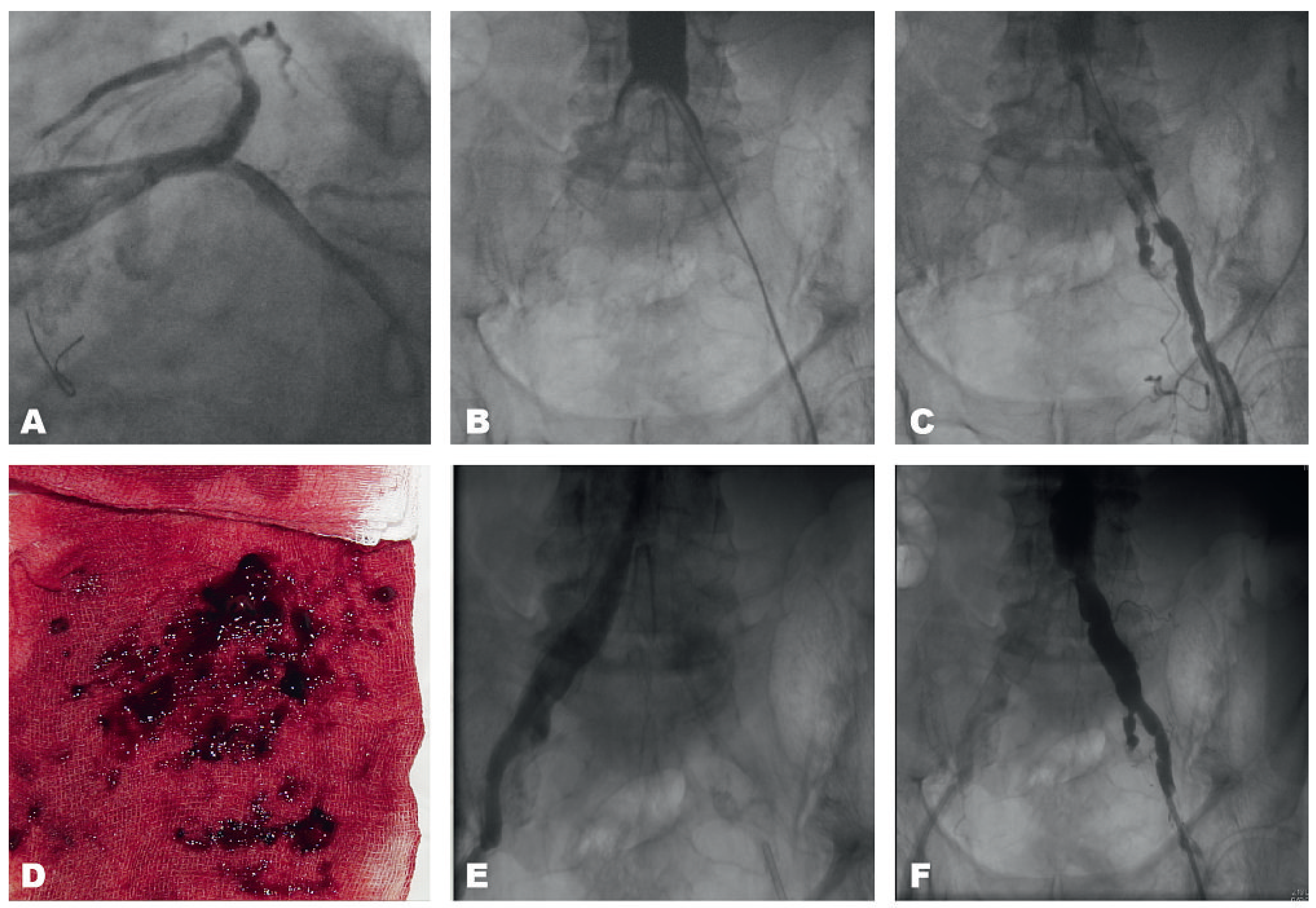
Figure 1. (A) Final result after successful V-Stenting of the biforcation LAD/LCX (RAO caudal). (B) Acute bilateral occlusion of the distal aorta (AP). (C) Massive thrombus burden in the left common iliac artery (AP, left). (D) Aspiration of a large amount of thrombotic material. (E,F) Restored blood flow in both common iliac arteries following thrombus aspiration (AP).
Cardiovascmed 13 00292 g001