Next Article in Journal
Research Prize of the Swiss Heart Foundation
Previous Article in Journal
Impact of Comorbidities on Clinical Presentation, Management and Outcome of Patients with Acute Coronary Syndrome
 
 
Cardiovascular Medicine is published by MDPI from Volume 28 Issue 1 (2025). Previous articles were published by another publisher in Open Access under a CC-BY (or CC-BY-NC-ND) licence, and they are hosted by MDPI on mdpi.com as a courtesy and upon agreement with Editores Medicorum Helveticorum (EMH).
Article

Muscular Ventricular Septal Defect After Mitral and Aortic Valve Replacement

by
Augusto Aragão
*,
Dorothea Vogel
and
Christoph Schmidt
Department of Cardiac Rehabilitation, Clinic Barmelweid, Barmelweid, Switzerland
*
Author to whom correspondence should be addressed.
Cardiovasc. Med. 2010, 13(5), 167; https://doi.org/10.4414/cvm.2010.01500
Submission received: 26 February 2010 / Revised: 26 March 2010 / Accepted: 26 April 2010 / Published: 26 May 2010

Abstract

We describe a case series of five patients who were referred to our cardiac rehabilitation department after mitral or aortic valve replacement, and whose transthoracic echocardiographic studies showed postoperative muscular septal defects.

Introduction to Case Series

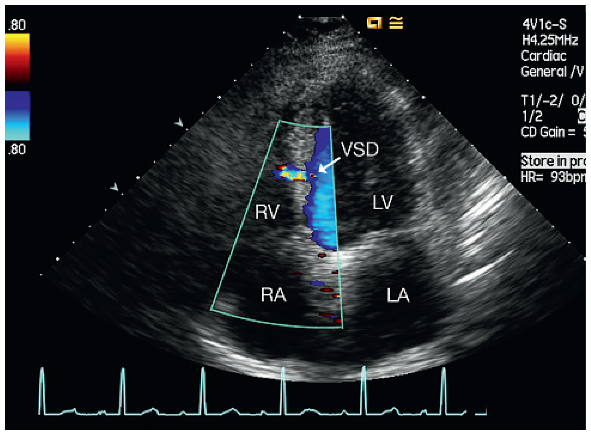
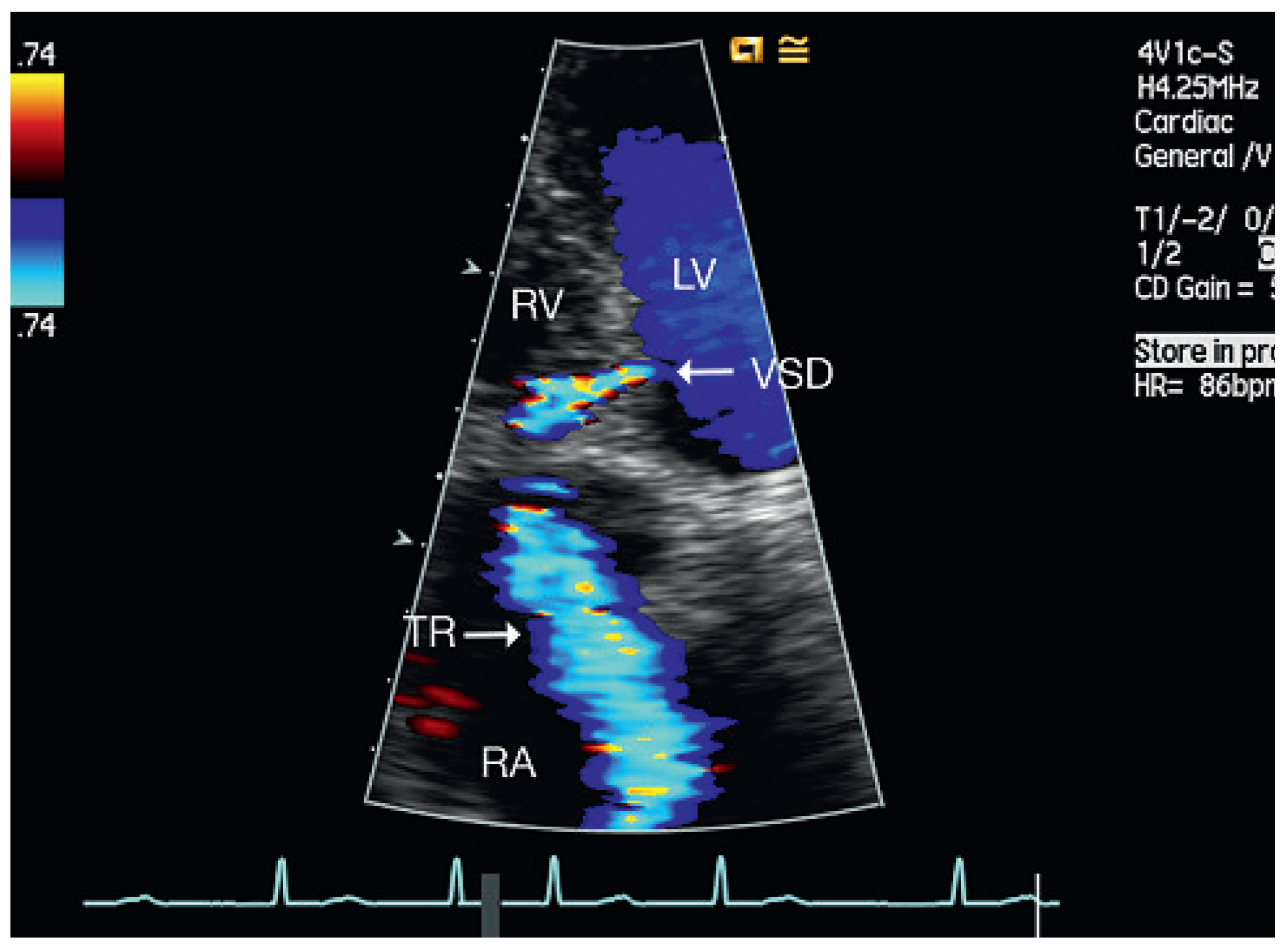
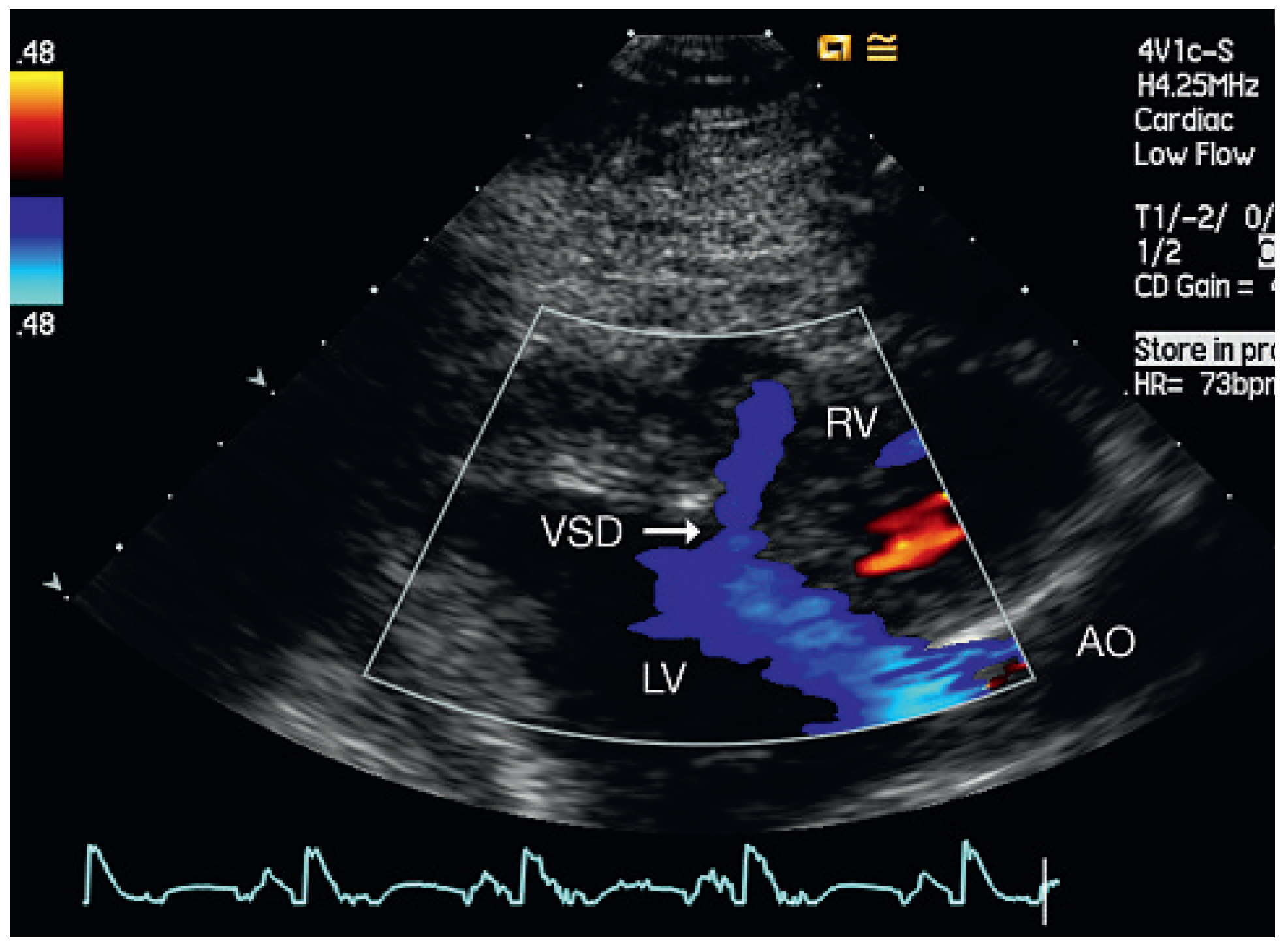
From September 2007 to December 2008, five patients aged 54–69 years were admitted to our institution for cardiac rehabilitation after surgical cardiac valve replacement (Table 1). Successful replacement of the aortic valve was performed in four of the patients and mitral valve replacement in the other. All patients received mechanical prosthetic valves (ATS medical) and had a history of dyspnoea, fatigue and exercise limitation. Physical examination on admission did not reveal signs of congestion in any of the patients. The admission electrocardiogram showed sinus rhythm or atrial fibrillation (patient 4) and signs of left ventricular hypertrophy in 3 of 5 patients. Doppler echocardiography revealed a restrictive muscular ventricular septal defect (Figure 1, Figure 3, Figure 4, Figure 5 and Figure 6) with Qp/Qs < 1.4 in all of the patients. The function of the prosthetic valves was normal and in one patient (patient 5) systolic pump function was moderately reduced.
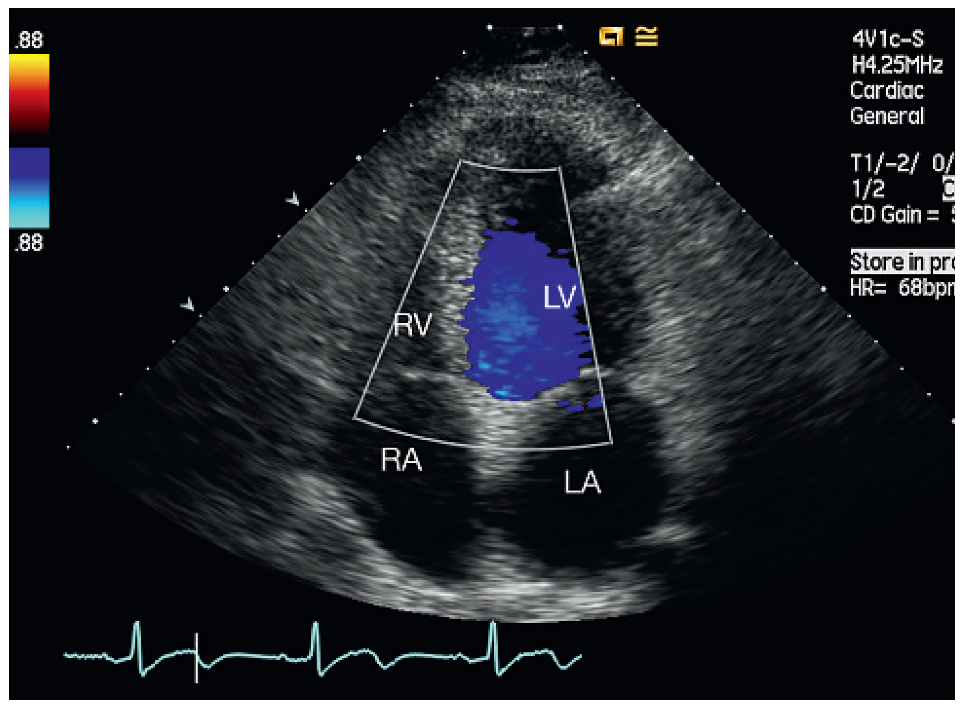
Echocardiographic follow-up revealed spontaneous closure of the septal perforation in four of the five patients (Figure 2). In one patient the septal defect persisted for one and a half years after surgery, but he remained asymptomatic without deterioration of left ventricular function or increased pulmonary artery systolic pressure.
Figure 1. Patient 1, at admission. Four-chamber view: muscular VSD mid-septal. LA = left atrium; LV = left ventricle; RA = right atrium; RV = right ventricle; VSD = ventricular septal defect.
Cardiovascmed 13 00167 g001
Figure 2. Patient 1, five months later. Four-chamber view: no evidence of residual VSD. LA = left atrium; LV = left ventricle; RA = right atrium; RV = right ventricle.
Cardiovascmed 13 00167 g002
Figure 3. Patient 2, at admission. Parasternal long-axis view (LV outflow): muscular VSD anteroseptal basal. AO = aorta; LA = left atrium; LV = left ventricle; RV = right ventricle; VSD = ventricular septal defect.
Cardiovascmed 13 00167 g003
Figure 4. Patient 3, at admission. Four-chamber view: muscular VSD mid-septal. LV = left ventricle; RA = right atrium; RV = right ventricle; TR = tricuspid regurgitation; VSD = ventricular septal defect.
Cardiovascmed 13 00167 g004
Figure 5. Patient 4, at admission. Four-chamber view: muscular VSD mid-septobasal. LV = left ventricle; RA = right atrium; RV = right ventricle; TR = tricuspid regurgitation; VSD = ventricular septal defect.
Cardiovascmed 13 00167 g005
Figure 6. Patient 5, at admission. Parasternal long-axis view (LV outflow): muscular VSD mid-anteroseptal. AO = aorta; LV = left ventricle; RV = right ventricle; VSD = ventricular septal defect.
Cardiovascmed 13 00167 g006

Discussion

Ventricular septal defect (VSD) may be of congenital origin, but also acquired as a result of myocardial infarction, penetrating trauma, blunt trauma, postsurgery or iatrogenic. VSD is the most common congenital cardiac abnormality in infants and children. It is estimated that 25–40% of such defects have closed spontaneously by the time the child is two years old; 90% of those that eventually close do so by the time the child is ten years old [1,2].
Ventricular septal defect complicating acute myocardial infarction is uncommon, with a reported incidence of 0.2% of all myocardial infarctions. The outcome remains extremely poor, with mortality of 50% in patients undergoing surgical repair and 95% in those treated medically [3]. This is not unexpected, given the advanced age, severity of coronary artery disease, haemodynamic instability and technical challenges associated with the surgical procedure.
Posttraumatic VSD is infrequent, with clinical sequelae ranging from imminent death to complete spontaneous resolution. It is unclear what the most appropriate management strategy is. Careful observation has been advocated in the management of asymptomatic patients with small, restrictive shunts. Percutaneous or surgical VSD closure is required for large defects or symptomatic patients [4,5,6].
Septal perforation has also been described as a rare complication of some cardiac interventions, such as transvenous pacemaker-electrode implantation [7], and after percutaneous coronary interventions [8], hypertrophic obstructive cardiomyopathy treated by left ventricular myotomy and myectomy, or alcohol-induced septal ablation [9].
Development of an iatrogenic VSD following mechanical aortic or mitral valve replacement is a rare complication. Such VSDs are typically membranous in location and small in size, with little haemodynamic significance. These defects result not only from technical surgical complications but also from postoperative endocarditis [10,11,12].
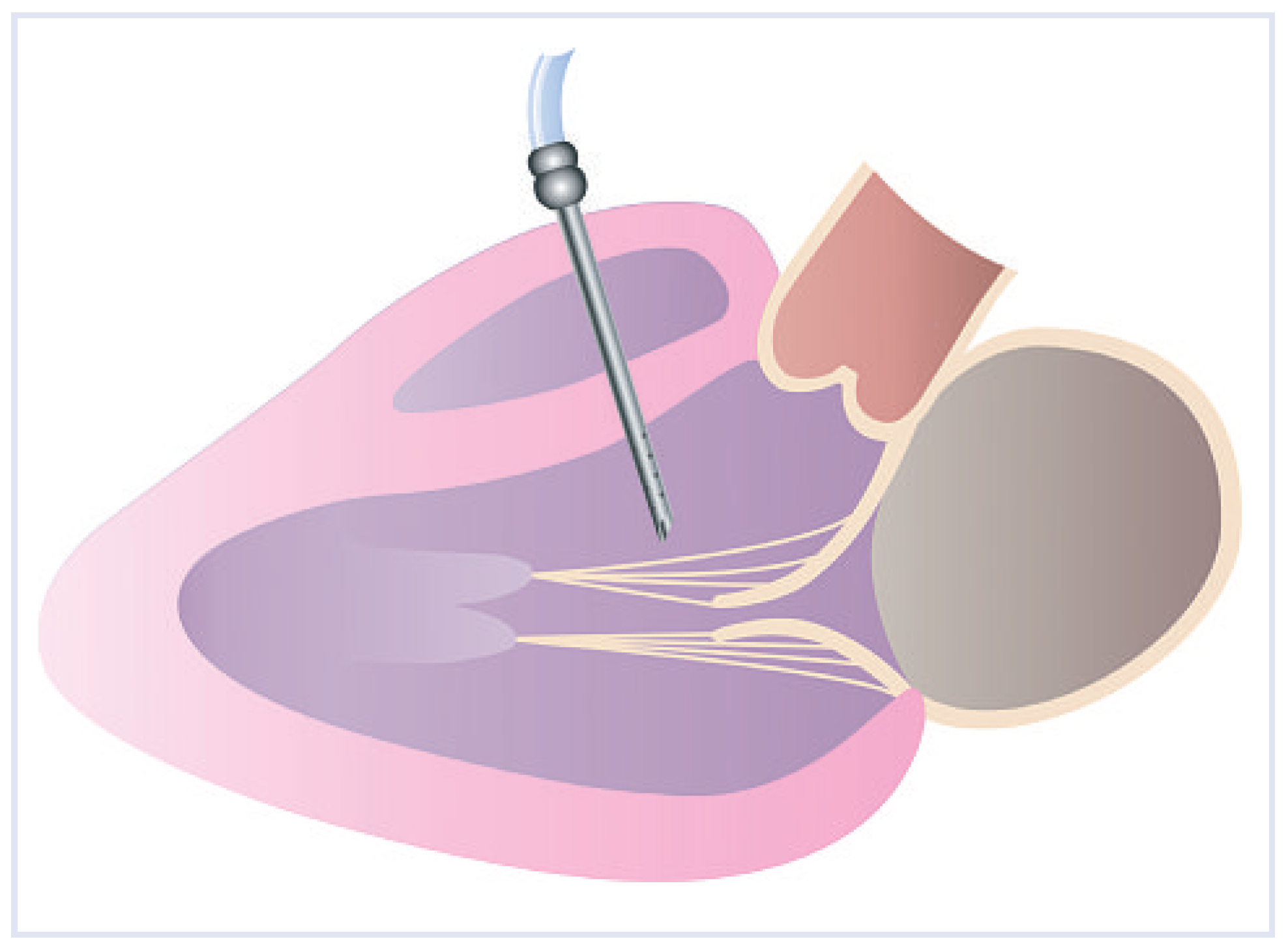
In the case of our patients the muscular septal defects were probably secondary to left ventricular transseptal venting during surgical valve replacement (Figure 7 and Figure 8). Left ventricular venting has been used since the early years of open-heart surgery with a heart-lung machine [13,14], and is frequently used to prevent air embolism and ventricular distension during systemic cooling [15].
Figure 7. Long-axis view: venting cannula in position. Cannula dimensions: lenght 70 mm, diameter 3.5 mm.
Cardiovascmed 13 00167 g007
Figure 8. Venting cannula (arrow) in position during aortic valve replacement.
Cardiovascmed 13 00167 g008
There are several techniques for venting during open-heart surgery: cannulation of the left ventricle apex, the aortic root, the left atrium, the pulmonary artery or through direct puncture of the free wall of the right ventricle and ventricular septum by a metal cannula with multiple perforations near the tip (Figure 7). This method was used in all the patients of our series.
Complications after ventricular venting are uncommon, but e.g., bleeding and left ventricular [16] or aortic false aneurysms [17] have been documented. To the best of our knowledge left ventricular venting complicated by iatrogenic muscular septal defect has not so far been described as a complication after surgical valve replacement.
The natural history of iatrogenic VSD may be variable and include spontaneous closure, as in the case of four of our five patients. An observational follow-up should therefore be recommended for asymptomatic patients with small defects. All patients in the present report had valve replacement with mechanical valves (ATS) requiring antibiotic prophylaxis for endocarditis [18]. The ACC/AHA 2008 Guidelines for the Management of Adults with Congenital Heart Disease recommend surgical closure of a ventricular septal defect if there is a Qp/Qs (pulmonary-to-systemic blood flow ratio) of 2.0 or more and clinical evidence of LV volume overload (Class I Indication, Level of Evidence: B), and also if the patient has a history of infective endocarditis (Class I Indication, Level of Evidence: C). Percutaneous closure of VSD (Class IIb, Level of Evidence: C) offers an attractive alternative to surgical management in patients with increased surgical risk factors, multiple previous cardiac surgical interventions, poorly accessible muscular VSDs, or “Swiss cheese”-type VSDs. Indications for catheter device closure of VSDs include iatrogenic artefacts after surgical replacement of the aortic valve and residual defects after prior attempts at surgical closure or trauma [19].

Conclusions

In this report we describe a case series of five patients who developed muscular VSDs after left ventricular venting during the mitral or aortic valve replacement procedure. To our knowledge this rare complication has not been reported previously. In our series most of these small VSDs closed spontaneously, and therefore an observational approach may be appropriate. Percutaneous or surgical closure is indicated for symptomatic patients or patients with haemodynamically relevant defects. In addition we recommend the use of other venting routes to avoid such complications.

Conflicts of Interest

No conflict of interest to disclose.

References

  1. Weidman WH, DuShane JW, Ellison RC. Clinical course in adults with ventricular septal defect. Circulation 1977, 56 (Suppl. I), 178–179.
  2. Brickner ME, Hillis LD, Lang RA. Congenital Heart Disease in Adults. N Engl J Med. 2000, 342, 256–262. [CrossRef] [PubMed]
  3. Crenshaw BS, Granger CB, Birnbaum Y; et al. GUSTO-I (Global Utilization of Streptokinase and TPA for Occluded Coronary Arteries) Trial Investigators. Risk factors, angiographic patterns, and outcomes in patients with ventricular septal defect complicating acute myocardial infarction. Circulation 2000, 101, 27–32.
  4. Dehghani P, Ibrahim R, Collins N, Latter D, CheemaAN, Chisholm RJ. Post-traumatic ventricular septal defects—Review of the literature and a novel technique for percutaneous closure. J Invasive Cardiol. 2009, 21, 483–487.
  5. Berry C, Hillis WS, Knight WB. Transcatheter closure of a ventricular septal defect resulting from knife stabbing using the Amplatzer muscular VSD occluder. Catheter Cardiovasc Interv. 2006, 68, 153–156.
  6. Martinez MW, Mookadam F, Sun Y, Hagler DJ. Transcatheter closure of ischemic and post-traumatic ventricular septal ruptures. Catheter Cardiovasc Interv. 2007, 69, 403–407.
  7. Villanueva FS, Heinsiner JA, Burkman MH, Fonanapazir L, Halvorsen RA, Jr, Chen JT. Echocardiographic detection of perforation of the cardiac ventricular septum by a permanent pacemaker lead. Am J Cardiol. 1987, 59, 370–371. [CrossRef] [PubMed]
  8. Park JY, Park SH, Oh JY, Kim IJ, Lee YH, Park SH, Kwon KH. Delayed ventricular septal rupture after percutaneous coronary intervention in acute myocardial infarction. Korean J Intern Med. 2005, 20, 243–246. [CrossRef] [PubMed]
  9. Nishimura RA, Holmes, Jr. DR. Hypertrophic obstructive cardiomyopathy. N Engl J Med. 350, 1320–1327. [CrossRef] [PubMed]
  10. Dodos F, Fehske W, Hoppe U, Sreeram N. Thranscatheter closure of iatrogenic perimembranous ventricular septal defect following prosthetic aortic valve replacement. Clin Res Cardiol. 2008, 97, 53–55. [CrossRef] [PubMed]
  11. Noble S, Ibrahim R. Transcatheter membranous ventricular septal defect closure through a mechanical aortic prosthesis using the Amplatzer membranous ventricular septal defect occluder. Catheter Cardiovasc Interv. 2009, 73, 167–172.
  12. Klein AJ, Garcia JA, Carrol JD. Percutaneous closure of an iatrogenic ventricular septal defect following mechanical aortic valve replacement using transseptal technique. Catheter Cardiovasc Interv. 2007, 70, 1018–1024.
  13. Hedlund, K.D. A tribute to Frank F. Allbritten, Jr. Texas Heart Inst J. 2001, 28, 292–296. [Google Scholar]
  14. Hedlund, K.D. Origin of the left ventricular vent. Texas Heart Inst J. 2004, 31, 107–108. [Google Scholar]
  15. Groves LK, Effler DB. Needle-Vent safeguard against systemic air embolus in open-heart surgery. J Thorac Cardiovasc Surg. 1964, 47, 349–355. [CrossRef]
  16. Sa MI, Lli Wie, Sheppard MN, Kilner PJ. Mycotic left ventricular false aneurysm at the site of an apical vent presenting 24 years after aortic valve surgery. Circulation 2008, 118, e501–e503.
  17. Strassano P, DeAmicis V, Gagliardi C, Di Lello F, Stampinato N. False aneurysm from the aortic vent site. J Cardiovasc Surg (Torino) 1982, 23, 401–402.
  18. Wilson W, Taubert KA, Gewitz M, Lockhart PB, Baddour LM, Levison M; et al.; American Heart Association Rheumatic Fever, Endocarditis, and Kawasaki Disease Committee; American Heart Association Council on Cardiovascular Disease in the Young; American Association Council on Clinical Cardiology; American Heart Association Council on Cardiovascular Surgery andAnesthesia; Quality of Care and Outcomes Research Interdisciplinary Working Group. Prevention of infective endocarditis: Guidelines from the American Heart Association: Heart Association Rheumatic Fever, Endocarditis, and Kawasaki Disease Committee; American Heart Association Council on Cardiovascular Disease in the Young; American Association Council on Clinical Cardiology; American Heart Association Council on Cardiovascular Surgery and Anesthesia; Quality of Care and Outcomes Research Interdisciplinary Working Group. Circulation 2007, 116, 1736–1754, Erratum in: Circulation 2007, 116, e376–e377.
  19. Warnes CA, Williams RG, Bashore TM, Child JS, Connolly HM, Dearani JA; et al. ACC/AHA 2008 Guidelines for the management of adults with congenital heart disease: A report of the American College of Cardiology/American Heart Association Task Force on Practice Guidelines (writing committee to develop guidelines on the management of adults with congenital heart disease). Circulation 2008, 118, e714–e833.
Table 1. Patients’ characteristics.
AgeGenderDiagnosisValveSize ofNYHA ClassVSDLVDD,LAD,
ProsthesisProsthesisat AdmissionLocationmmmm
54MaleCombined aortic stenosis with regurgitationATS23 mmIIMid-septal4640
59MaleSevere aortic regurgitationATS23 mmIIAnteroseptal basal5046
59MaleSevere mitral regurgitation due to chordal ruptureATS31 mmIMid-basal septum4845
69FemaleCombined aortic stenosis with regurgitationATS20 mmIIMid-septobasal5054
61MaleSevere aortic stenosisATS22 mmIIAnteroseptal5560
LVDD = left ventricular diastolic diameter; LAD = left atrial diameter; VSD = ventricular septal defect.