Next Article in Journal
Cardiovascular Pathology in the Land of the Doges
Previous Article in Journal
Wo Sind Sie Schweizer Geblieben?
 
 
Cardiovascular Medicine is published by MDPI from Volume 28 Issue 1 (2025). Previous articles were published by another publisher in Open Access under a CC-BY (or CC-BY-NC-ND) licence, and they are hosted by MDPI on mdpi.com as a courtesy and upon agreement with Editores Medicorum Helveticorum (EMH).
Article

Mechanical Versus Manual Chest Compression During CPR in a Cardiac Catherization Setting

by
Christophe A. Wyss
1,*,
Julia Fox
2,
Fabian Franzeck
1,
Marco Moccetti
4,
Alfons Scherrer
3,
Jens P. Hellermann
4 and
Thomas F. Lüscher
1
1
CardioVascular Centre, Cardiology, University Hospital Zürich, CH-8091 Zurich, Switzerland
2
Hirslanden Accident and Emergency Centre Zürich, Zürich, Switzerland
3
Institute of Anaesthesiology, University Hospital Zürich, Zürich, Switzerland
4
Spitalregion Rheintal Werdenberg Sarganserland, Spital Altstätten, Altstätten, Switzerland
*
Author to whom correspondence should be addressed.
Cardiovasc. Med. 2010, 13(3), 92; https://doi.org/10.4414/cvm.2010.01480
Submission received: 24 December 2009 / Revised: 24 January 2010 / Accepted: 24 February 2010 / Published: 24 March 2010

Abstract

Background: Cardiopulmonary resuscitation (CPR) during treatment-resistant cardiac arrest in the catheterisation laboratory is exceedingly difficult, for practical reasons and on grounds of efficacy and safety. Mechanical chest compression devices may provide more constant CPR in this setting. Methods: Using a CPR training manikin we simulated a prolonged resuscitation situation during cardiac catheterisation requiring continuous chest compressions over ten minutes. We compared a team of three experienced resuscitators with the performance of a mechanical chest compression device (LUCASTM). Results: No overall differences in compression rate could be documented between manual and mechanical chest compression rate during ten minutes of continuous CPR (105/min vs. 101/min; p = ns), but there was considerable variation with manual compression (88/min–121/min). Mechanical chest compression resulted in more constant and correct depth of chest compressions compared to manual performance (98% vs. 70%; p < 0.01), even if a device failure occurred (95% vs. 70%; p < 0.01). Resuscitator fatigue was reflected in a mean of 21% (range 0–45%) too shallow compressions in the manual CPR group (vs 1% in the device CPR group; p < 0.05). We documented comparable time frames of overall interruption of CPR due to installation of the mechanical device (10 s, range 9–11 s) and the overall time due to resuscitator changes (9 s, range 8–12 s) over ten minutes of continuous CPR. Manual CPR resulted in too deep compression in 8% of the compressions, whereas no deep compressions were noted for mechanical CPR (p = ns). Conclusion: Mechanical CPR devices may offer some advantages over manual CPR in the setting of prolonged resuscitation during cardiac catheterisation in treatment-resistant cardiac arrest requiring continuous chest compressions during PCI.

Introduction

In the infrequent but dramatic cases of therapy-resistant cardiac arrest during cardiac catheterisation, e.g., during primary percutaneous intervention (PCI) in acute myocardial infarction, defibrillation alone is often unable to restore spontaneous and sufficient circulation. In these situations, effective chest compressions are crucial in ensuring appropriate cerebral and cardiac perfusion. Minimally interrupted, regular and appropriate CPR (cardiopulmonary resuscitation) is mandatory to improve outcome in cardiac arrest [1,2].
However, administering manual CPR to a patient lying on the catheterisation laboratory table is exceedingly difficult: first, access to the patient is constrained by the cardiac angiography system and the operating interventional cardiologist. Second, the catheterisation laboratory table is quite flexible and therefore does not offer a firm basis, thus making chest compression physically more demanding and less effective. Third, CPR during therapy-resistant cardiac arrest in the catheterisation laboratory is often prolonged (mean compression time 105 ± 60 min, range 45–240 min) [3], resulting in premature exhaustion of the resuscitators. Finally, there are concerns regarding the radiation exposure risk for the resuscitators.
Mechanical chest compression devices have therefore been proposed [4]. There is a growing volume of data on the safety and efficacy of these devices in the catheterisation laboratory, but mainly limited to case reports or series [2,3,5]. In Switzerland two chest compression devices are available: LUCAS™ (Jolife AB, Lund, Sweden) is a sternal compression device (gasdriven or electric), incorporating a suction cup for active decompression, and the Autopulse™ (ZOLL, Chelmsford, MA, USA), which is an automated load-distributing band resuscitation device.
The aim of the present study was to compare manually administered CPR with LUCAS™ CPR in the setting of cardiac catheterisation.

Methods

Study setting

We simulated a prolonged resuscitation situation during cardiac catheterisation (e.g., primary percutaneous intervention; PCI) in treatment-resistant cardiac arrest requiring continuous and prolonged chest compressions. Access to the patient was limited by the x-ray tubes of a biplane cardiac angiography system in lateral or 20° LAO/30° cranial position with centred table height. To keep the scenario simple and avoid confounding factors, we did not integrate defibrillation, ventilation, administration of medication or airway management processes in the scenario.
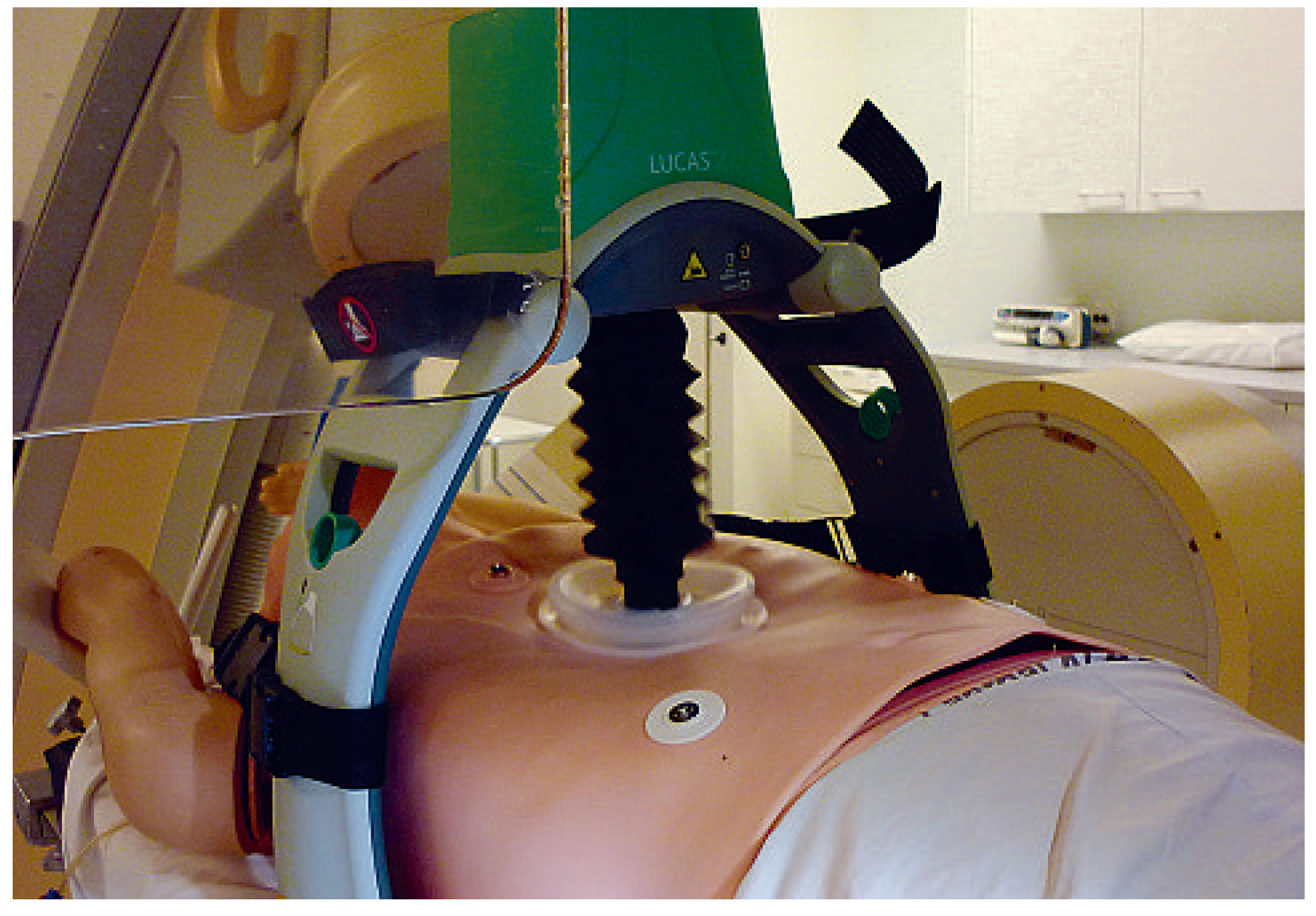
In one setting a team of three experienced resuscitators administrated continuous manual chest compressions for 10 min, changing every minute (after approximately 100 compressions) (Figure 1). In a second setting, two resuscitators started manual CPR and installed a mechanical chest compression device (LUCASTM, Jolife, Lund, Sweden) providing 10 min’ continuous CPR (Figure 2).

Data acquisition

To assess chest compression performance we used a CPR training manikin (ALS Skillmaster/HeartSim4000-Software, Laerdal, Stavanger, Norway). The software recorded the absolute number and frequency of chest compressions and classified chest compressions as correct (sternum compression 4–5 cm), too deep (>5 cm) or not deep enough (<4 cm). Time intervals with no chest compression on the manikin (changing of the resuscitator, installation of the device) were recorded.

Device characteristics

The LUCASTM1 device is a pneumatic system requiring a gas port for supply with compressed air. It performed two sequences without technical problems. The newer recently released LUCASTM2 system has an electric motor with integrated engine-cooling system. It performed two sequences without technical problems, but in the third run the device stopped after 7.5 min, probably due to overheating, and the sequence was completed after a delay of 45 s with manual CPR by one resuscitator.

Statistics

Data are presented as mean and range. For descriptive statistics, continuous variables were compared using ANOVA. The study has a power of 0.9 to detect a difference of 2 cm in compression depth, given an alpha error of 0.05 and an error standard deviation of 1 cm.

Results

Manual chest compression

Six sequences were performed manually and a total of 6323 compressions were analysed. Two different resuscitation teams consisting of three resuscitators each performed three scenarios of continuous chest compressions over 10 min, changing every minute (resulting in changes of the resuscitator after approximately 100 compressions).

Resuscitator characteristics

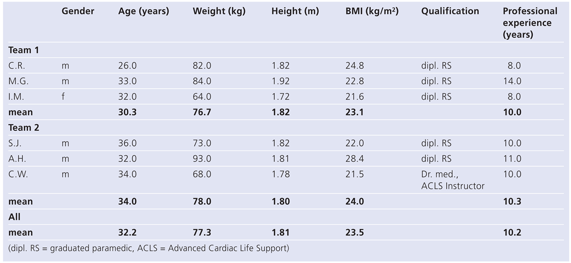
Resuscitator characteristics are shown in Table 1. The two teams (six resuscitators; five male, one female) consisted of well trained professionals with a mean professional experience of 10 years (range 8–14 years). Mean age was 32 years (range 26–36 years). All resuscitators were rather athletic in stature (mean BMI 23.5 kg/m2).

Manual CPR performance

CPR performance results are shown in Table 2. Mean manual compression rate over 10 min of continuous chest compression was 105/min, which is in the range of current recommendations. Nevertheless, there was a considerable range in the different sequences (88/min–121/min). Over the six sequences, a mean of 70% of all compressions was classified as correct (defined as sternal compression depth 4–5 cm) with a range from 54 to 91% in the different sequences. 8% of all compressions were classified as too deep (range 0–44%) and 21% as too shallow (range 0–45%). When changing the resuscitator chest compression was interrupted for mean 9 s (range 8–12 s).

Mechanical chest compression

Three sequences of 10 min of continuous chest compressions were performed using the LUCASTM2 system with electric power supply. Two sequences were performed using the LUCASTM1 system with pneumatic gas-driven sternal compression. A total of 4957 mechanical compressions were analysed.

Mechanical CPR performance

CPR performance results are shown in Table 2. In the present section of the manuscript we show only the results of the sequences without technical problems (two sequences LUCASTM1 CPR and two sequences LUCASTM2 CPR), but we will discuss the effect of device failure in the comparison of manual versus mechanical CPR. The mean manual compression rate over 10 min of continuous chest compression was 101/min. There was a very narrow range in the different sequences (99/min–102/min). Over the four sequences, a mean 98% of all compressions were classified as correct (defined as sternal compression depth 4–5 cm) with a range from 97 to 99% in the different sequences. No compressions were classified as too deep and 1% as too shallow (range 0–2%). A mean time of ten seconds (range 9–11 s) was noted for interruption of chest compression over ten minutes due to installation of the device.

Comparison: mechanical versus manual chest compression

Due to device failure during the ninth sequence we decided to analyse the mechanical compression performance separately for scenarios with a properly working LUCASTM system and for overall mechanical compression performance including the sequence with device failure.

Performance with no device failure

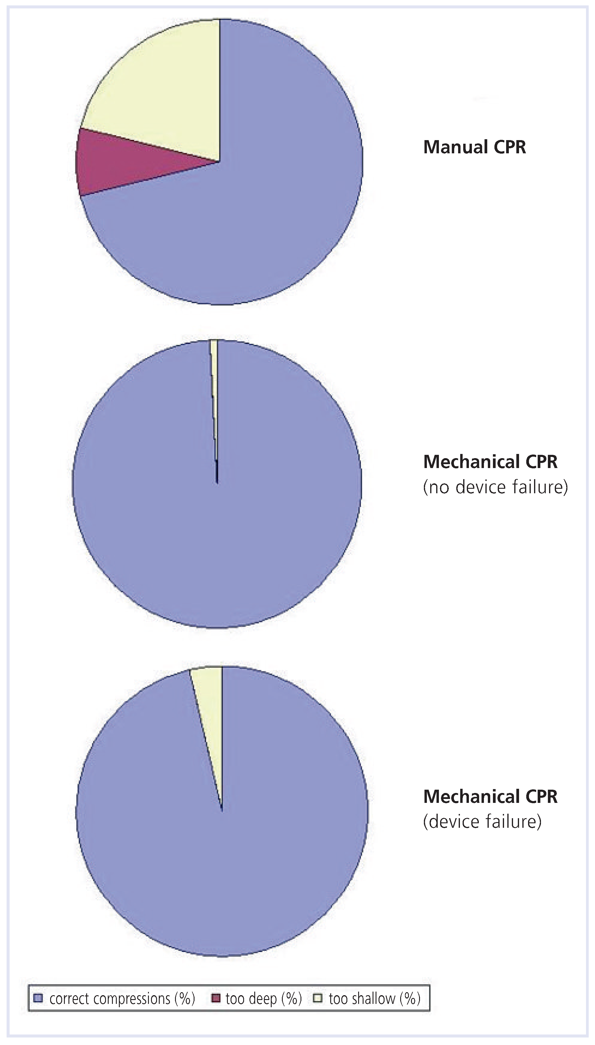
We compared four sequences of proper mechanical CPR (4957 compressions) with six sequences of manual CPR (6323 compressions). There were no statistically significant differences regarding compression rate during manual versus mechanical CPR (105/min vs. 101/min; p = 0.6192). The proportion of correct compression depth was significantly better during mechanical CPR (98% vs. 70% during manual CPR; p = 0.0049) (Figure 3). Manual CPR resulted in too deep compression in 8% of the compressions, whereas no deep compressions were noted for mechanical CPR (p = 0.4120). Too shallow compressions occurred more often during manual CPR compared to mechanical CPR (21% vs. 1%; p = 0.0465) (Figure 3).

Device failure

In a clinical setting, failure of the mechanical CPR device would require manual CPR to be started. We therefore analysed the influence of a device failure on the overall mechanical performance separately by including sequence 9 (device failure after 7.5 min and manual continuation of CPR after a delay of 45 s) in the comparison. We compared five sequences (one with device failure) of mechanical CPR (5869 compressions) with six sequences of manual CPR (6323 compressions). There were no statistically significant differences regarding compression rate during manual versus mechanical CPR (105/min vs. 101/min; p = 0.5590). The proportion of correct compression depth was still significantly better during mechanical CPR (95% vs. 70% during manual CPR; p = 0.0054); too shallow compressions occurred less often in the mechanical CPR group compared to purely manual CPR (4% vs. 21%; p = 0.0595).

Discussion

In cardiopulmonary resuscitation, administration of effective and uninterrupted chest compressions, sometimes for prolonged periods of time, is crucial. The proper return of spontaneous circulation is correlated with achieved coronary perfusion pressure [6]. Because there is a delayed rise in coronary perfusion pressure during continuous chest compressions [7], every interruption of chest compressions should be avoided. Unfortunately, in real life situations interruptions of chest compression during CPR are common [8]. Rescuer fatigue is an important issue; indeed, compression depth decay already becomes evident after 90 s of CPR [9]. Furthermore, efficient CPR during treatment-resistant cardiac arrest in the catheterisation laboratory is very difficult due to limited access to the patient and is physically more demanding and often prolonged, resulting in substantial manpower requirement and often inadequate CPR quality.
In the present experimental study we compared the performance of a mechanical chest compression device (LUCASTM) with performance of manual chest compression in the setting of a prolonged resuscitation situation during cardiac catheterisation. No overall differences in compression rate were documented between manual and mechanical chest compression rate during ten minutes of continuous CPR, but during manual compression there was considerable variability which was obviously avoided with mechanical CPR. Furthermore, mechanical chest compression using the LUCASTM device resulted in more constant and appropriate depth of chest compressions compared to manual performance, even if a device failure occurred. Resuscitator fatigue was reflected by the fact that one fifth of all compressions were too shallow in the manual CPR group, while the devices provided appropriate compressions in almost all circumstances.
In the past there has been concern about CPR-related injuries using a mechanical CPR device [10]. Interestingly, in the present study there were no deep compressions with the mechanical device, but even with experienced resuscitators such as in this study such potentially hazardous events occurred in almost half of the compressions during manual CPR. A recent autopsy study was unable to detect differences in CPRrelated injuries between the LUCASTM device and manual CPR [11]. Another often discussed issue is the time delay to effective CPR due to installation of the mechanical device. We documented comparable time delays of overall interruption of CPR due to installation of the mechanical device (10 s) and the overall time due to resuscitator changes (9 s) over ten minutes of continuous CPR.
Several limitations are inherent in the current study. We did not investigate hard clinical endpoints or experimental circulatory parameters. Certainly, compression depth, compression rate and interruption of CPR are surrogate markers of effective CPR. Nevertheless, lower compression rates have been associated with a worse outcome [12] and sufficient compression depth can prevent post-resuscitation brain damage [13]. It has to be noted that previously published data on mechanical CPR with the LUCASTM device demonstrated improved coronary [14] and cerebral [15] blood flow compared to manual CPR. Another limitation is the clinical setting, which in reality may not be present in the setting of this study: the use of biplane angiography equipment limited access to the patient more than monoplane equipment, and the table height could not be adapted to a comfortable resuscitation position. On the other hand, we invited only highly trained resuscitators. Furthermore, they could concentrate fully on chest compressions and had no other tasks to perform such as ventilation or administration of medication during the resuscitation process. Last but not least, we did not investigate the way of compression release, which is also an important feature of haemodynamically efficient CPR.

Conclusion

Mechanical CPR devices may offer some advantages over manual CPR in the setting of prolonged resuscitation during cardiac catheterisation in treatment-resistant cardiac arrest requiring continuous and prolonged chest compressions during PCI. The LUCASTM system provided more reliable and appropriate chest compressions than manual CPR.

Acknowledgments

The authors would like to thank Isabella Müller, Sascha Janssen, André Hug, Christian Rupp, Mario Gabler and Andreas Igel for their assistance.

Conflicts of Interest

None of the authors reports a conflict of interest for the present study. In particular, the study was not funded by the producer of the LUCASTM system nor did any of the authors receive honoraria from the company.

References

  1. Ramaraj, R.; Ewy, G.A. Rationale for Continuous Chest Compression Cardiopulmonary Resuscitation. Heart. 2009. [Google Scholar] [CrossRef] [PubMed]
  2. Grogaard, H.K.; Wik, L.; Eriksen, M.; Brekke, M.; Sunde, K. Continuous mechanical chest compressions during cardiac arrest to facilitate restoration of coronary circulation with percutaneous coronary intervention. J Am Coll Cardiol. 2007, 50, 1093–1094. [Google Scholar] [CrossRef] [PubMed][Green Version]
  3. Larsen, A.I.; Hjornevik, A.S.; Ellingsen, C.L.; Nilsen, D.W. Cardiac arrest with continuous mechanical chest compression during percutaneous coronary intervention. A report on the use of the LUCAS device. Resuscitation. 2007, 75, 454–459. [Google Scholar] [CrossRef] [PubMed]
  4. Risom, M.; Jorgensen, H.; Rasmussen, L.S.; Sorensen, A.M. Resuscitation, Prolonged Cardiac Arrest, and an Automated Chest Compression Device. J Emerg Med. 2009. [Google Scholar] [CrossRef] [PubMed]
  5. Agostoni, P.; Cornelis, K.; Vermeersch, P. Successful percutaneous treatment of an intraprocedural left main stent thrombosis with the support of an automatic mechanical chest compression device. Int J Cardiol. 2008, 124, e19–21. [Google Scholar] [CrossRef] [PubMed]
  6. Paradis, N.A.; Martin, G.B.; Rivers, E.P.; Goetting, M.G.; Appleton, T.J.; Feingold, M.; Nowak, R.M. Coronary perfusion pressure and the return of spontaneous circulation in human cardiopulmonary resuscitation. JAMA. 1990, 263, 1106–1113. [Google Scholar] [CrossRef] [PubMed]
  7. Ewy, G.A. Cardiocerebral resuscitation: the new cardiopulmonary resuscitation. Circulation. 2005, 111, 2134–2142. [Google Scholar] [CrossRef] [PubMed]
  8. Valenzuela, T.D.; Kern, K.B.; Clark, L.L.; Berg, R.A.; Berg, M.D.; Berg, D.D.; Hilwig, R.W.; Otto, C.W.; Newburn, D.; Ewy, G.A. Interruptions of chest compressions during emergency medical systems resuscitation. Circulation. 2005, 112, 1259–1265. [Google Scholar] [CrossRef] [PubMed]
  9. Sugerman, N.T.; Edelson, D.P.; Leary, M.; Weidman, E.K.; Herzberg, D.L.; Vanden Hoek, T.L.; Becker, L.B.; Abella, B.S. Rescuer fatigue during actual in-hospital cardiopulmonary resuscitation with audiovisual feedback: a prospective multicenter study. Resuscitation. 2009, 80, 981–984. [Google Scholar] [CrossRef] [PubMed]
  10. Baubin, M.; Rabl, W.; Pfeiffer, K.P.; Benzer, A.; Gilly, H. Chest injuries after active compression-decompression cardiopulmonary resuscitation (ACD-CPR) in cadavers. Resuscitation. 1999, 43, 9–15. [Google Scholar] [CrossRef] [PubMed]
  11. Smekal, D.; Johansson, J.; Huzevka, T.; Rubertsson, S. No difference in autopsy detected injuries in cardiac arrest patients treated with manual chest compressions compared with mechanical compressions with the LUCAS device—a pilot study. Resuscitation. 2009, 80, 1104–1107. [Google Scholar] [CrossRef] [PubMed]
  12. Abella, B.S.; Alvarado, J.P.; Myklebust, H.; Edelson, D.P.; Barry, A.; O’Hearn, N.; Vanden Hoek, T.L.; Becker, L.B. Quality of cardiopulmonary resuscitation during in-hospital cardiac arrest. JAMA. 2005, 293, 305–310. [Google Scholar] [CrossRef] [PubMed]
  13. Wu, J.Y.; Li, C.S.; Liu, Z.X.; Wu, C.J.; Zhang, G.C. A comparison of 2 types of chest compressions in a porcine model of cardiac arrest. Am J Emerg Med. 2009, 27, 823–829. [Google Scholar] [CrossRef] [PubMed]
  14. Steen, S.; Liao, Q.; Pierre, L.; Paskevicius, A.; Sjoberg, T. Evaluation of LUCAS, a new device for automatic mechanical compression and active decompression resuscitation. Resuscitation. 2002, 55, 285–299. [Google Scholar] [CrossRef] [PubMed]
  15. Rubertsson, S.; Karlsten, R. Increased cortical cerebral blood flow with LUCAS; a new device for mechanical chest compressions compared to standard external compressions during experimental cardiopulmonary resuscitation. Resuscitation. 2005, 65, 357–363. [Google Scholar] [CrossRef] [PubMed]
Figure 1. Three resuscitators performing manual CPR during cardiac catheterisation.
Cardiovascmed 13 00092 g001
Figure 2. LUCASTM2 performing mechanical CPR during cardiac catheterisation.
Cardiovascmed 13 00092 g002
Figure 3. Comparison of manual vs. mechanical CPR during cardiac catheterisation. Mechanical chest compression using the LUCASTM device resulted in more constant and correct depth of chest compressions compared to manual performance (98% vs. 70%; p < 0.05), even if a device failure occurred (95% vs. 70%; p < 0.05). Manual CPR resulted in too deep compression in 8% of the compressions, whereas no deep compressions were noted for mechanical CPR (p < 0.05). Too shallow compressions occurred more often during manual CPR compared to mechanical CPR (21% vs. 1%; p < 0.05).
Cardiovascmed 13 00092 g003
Table 1. Resuscitator characteristics.
Cardiovascmed 13 00092 i001
Table 2. CPR performance.
Cardiovascmed 13 00092 i002