We report the case of a 79-year-old lady with severe symptomatic mitral regurgitation who successfully underwent a novel technique of percutaneous edge-to-edge valve reconstruction using a metallic clip.
The patient, with a history of aortic valve replacement 4 years prior and known mitral regurgitation for the last 2 years, was referred to us due to worsening symp-toms of heart failure over the 3 months prior. She presented with dyspnoea NYHA class III, despite medical treatment with an ACE-inhibitor, a thiazide and a loop diuretic. The patients’ history was also characterised by an ischemic stroke with partial right leg hemiparesis, osteoarthritis and renal function impairment (creatinin clearance 36 ml/min).
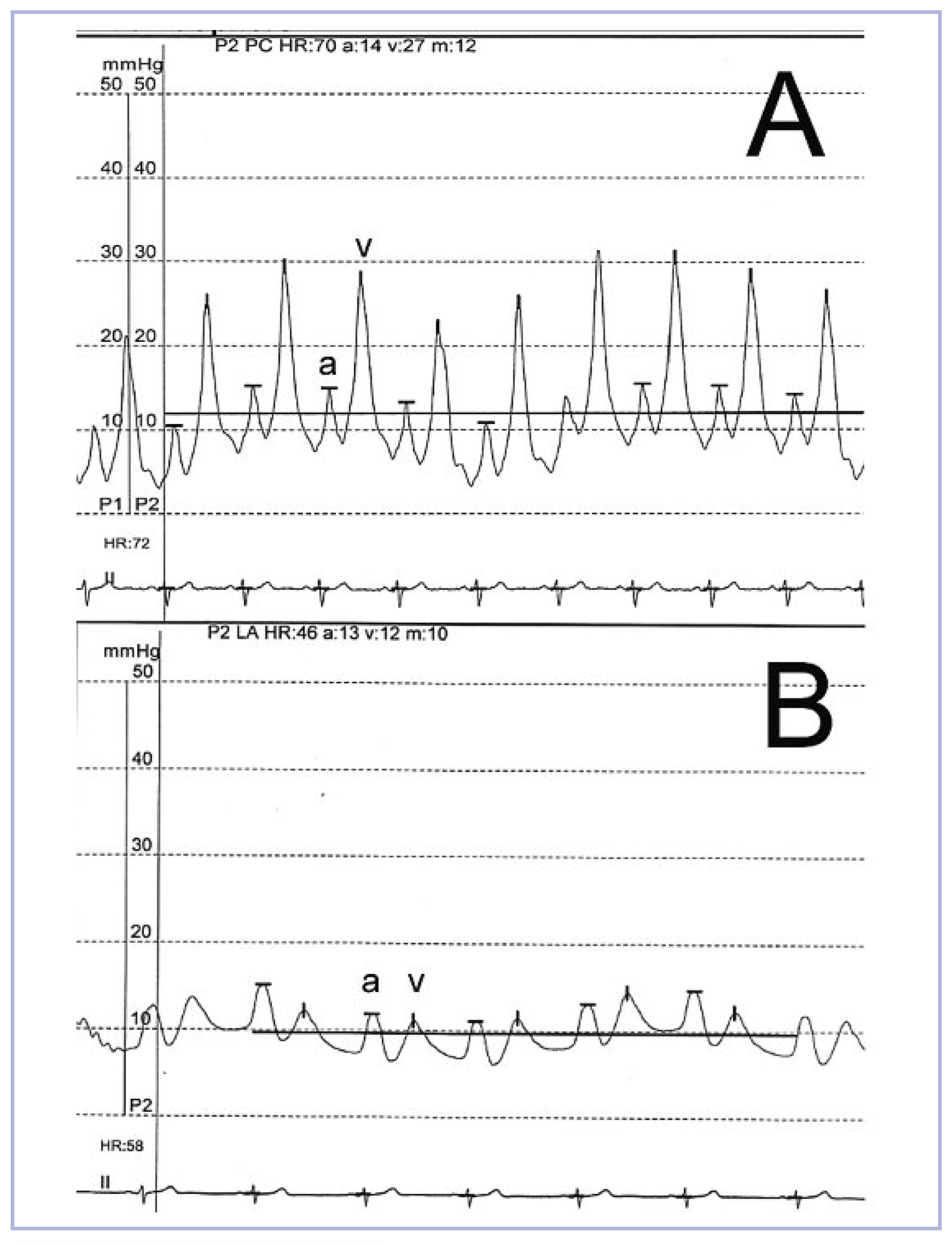
Due to her co-morbidities and the advanced age of the patient, the calculated surgical risk scores were greatly elevated (Morbidity or Mortality according to the Society of Thoracic Surgeons Score was 27.8% and the EuroSCORE 30-day-mortality was 33.8%). Therefore, we opted for a percutaneous mitral valve reconstruction using the MitraClip
® system (
fig. 2).
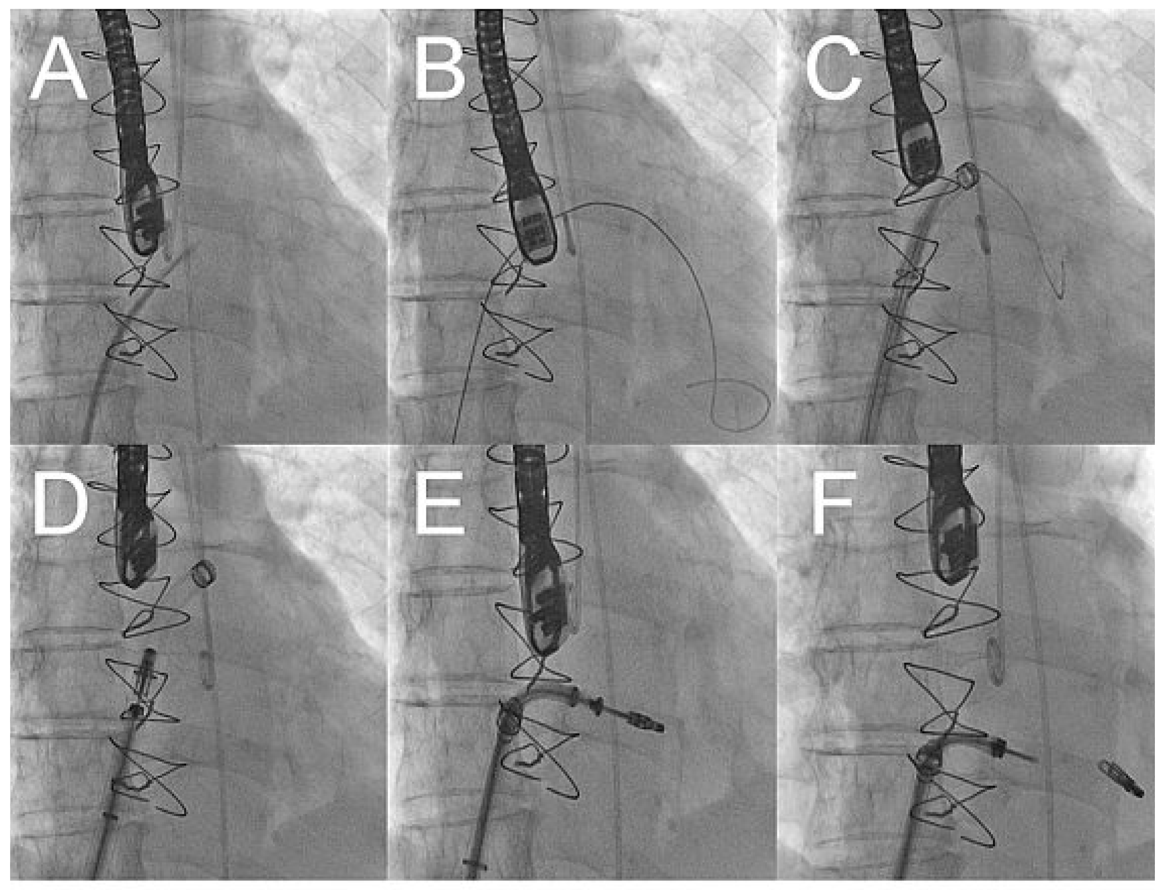
After gaining access through an 8 french sheath via the right femoral vein, transseptal puncture was performed under TEE and fluoroscopic-guidance, and an Amplatz-Super-Stiff
® catheter was advanced into the left atrium (
fig. 3A–C). Changing to a MitraClip-guide catheter, the clip delivery system was advanced via the left atrium (
fig. 3D) into mitral position (
fig. 3E). The system allows repositioning of the device under echocardiographic control until optimal placement has been found. This meant, before releasing the Clip (
fig. 3F), that it could be repositioned until echocardiographic control showed the correct position of the device with mild residual regurgitation (
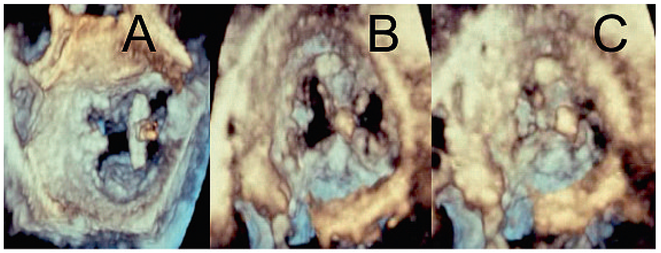
fig. 4). 3-dimensional echocardiographic reconstruction images allowed the positioning and adjusting of the clip (
fig. 5A) and the open and closed mitral valve after MitraClip
® placement (
fig. 5B and 5C) to be seen. The patient could be extubated in the catheter lab directly after clip implantation. The in-hospital recovery was uneventful and the patient could be discharged home three days after the intervention. During her four-week follow-up, the patient was in excellent health and reported a substantial improvement of her symptoms, reducing her exertional dyspnoea symptoms down to NYHA I–II. Echocardiographic control showed the MitraClip
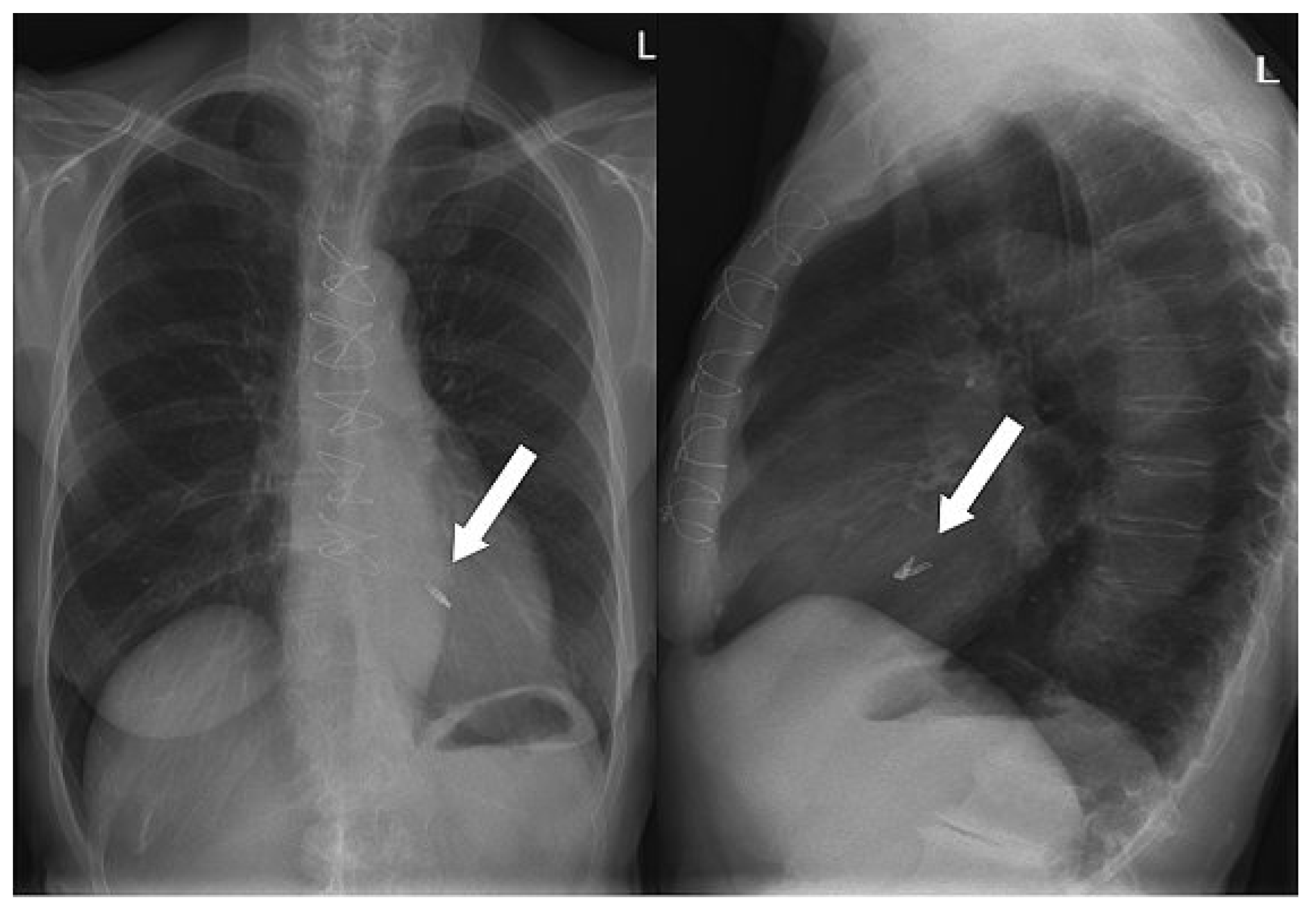
® in an optimal place with a residual mild-moderate regurgitation jet. On the chest x-ray film, the metallic clip can easily be identified (
fig. 6).


