Introduction
Takotsubo cardiomyopathy (TC), also called transient left ventricular apical ballooning, stress-induced cardiomyopathy or broken heart syndrome, is a newly described cardiomyopathy mimicking acute myocardial infarction and is characterised, in its typical form, by a transient systolic dysfunction of the left ventricle (LV) with apical ballooning without significant coronary artery disease [
1,
2,
3,
4,
5,
6,
7,
8,
9,
10,
11,
12,
13,
14,
15,
16,
17,
18,
19,
20,
21,
22,
23,
24,
25,
26,
27,
28,
29,
30,
31,
32,
33,
34,
35,
36]. The condition was named by Japanese authors who first described this entity in 1991 [
1]. Takotsubo is a pot with a round bottom and a narrow neck used by fishermen to catch octopuses, which closely reproduces the systolic shape of the LV in TC.
In the present study, all the TC cases collected in our hospital over six years were analysed, integrating their clinical and paraclinical characteristics, their complications, in-hospital course and recurrence rate, as well as the prevalence of the disease. The cases were compared with published, international data in order to provide possible additional data to improve the knowledge of this still under-recognised disease. We also explored the question of whether a non invasive approach can obtain a high probability of TC diagnosis after gaining experience with the clinical presentation of this syndrome.
Methods
Between July 2003 and February 2009, we systematically looked for TC in patients hospitalised in the medical department of our hospital, as well as among patients in other services for whom a medical consultation was required. The diagnosis was initially based on the criteria proposed by Tsuchihashi in 2001 [
2], which was then revised by Gianni in 2006 [
3]:
- –
New ST segment elevation or depression or T wave inversion on the electrocardiogram (ECG).
- –
Transient akinesis or dyskinesis of the apical and midventricular segments in association with regional wall motion abnormalities that extend beyond the distribution of a single epicardial coronary vessel on the echocardiography.
- –
Absence of evidence of obstructive coronary artery disease.
- –
Absence of recent significant head trauma, intracranial bleeding, phecochromocytoma, myocarditis or hypertrophic cardiomyopathy.
Patients were recruited by a single cardiologist, who was head of the medical department and a consultant in the other services of the hospital. Whenever the diagnosis was suspected, based on suggestive ECG modifications following an acute stress-full event, an echocardiography was performed and cardiac enzyme levels, including troponin I, creatine kinase (CK) and brain natriuretic peptide (BNP), were obtained and repeated during the hospital stay. Urgent or delayed coronary angiography was performed according to the clinical presentation, and required transfer to the University Hospital of Lausanne.
All men underwent a coronary angiography, because stress cardiomyopathy mainly affects women and the prevalence of coronary artery disease is higher in men. All women with chest pain for less than 24 hours and ST-segment elevation underwent an angiography, as well as six other particular cases for whom this exam was clinically required. Reasons for not practicing this investigation were typical clinical presentation for TC and evolution in women less than 50-years or more than 80-years old without major cardiac risk factors and without chest pain during the course of the disease. An echocardiogram was repeated as clinically indicated, but at least after 1 and 3 months, until complete recovery of LV function was achieved. In the same way, follow-up ECGs were obtained until complete normalisation of the tracing occurred. Patients were all clinically followed by the same cardiologist and controlled annually after normalisation of all the parameters.
Prevalence was estimated by comparing the number of definitive TC cases during the study period among the number of patients with a diagnosis of either acute coronary syndrome, acute heart failure, acute pulmonary edema or cardiogenic shock based on codes recorded in the discharge letter. To compare with percentages cited in the literature, we also determined the prevalence of TC in our patients presenting with chest pain among the total number of patients with acute coronary syndrome during the study period.
Statistical analysis
Categorical data are presented as numbers (or percentages) and continuous data as mean ± standard deviation or range. An unpaired t-test (GraphPad Prism software, 5.02 version) was used to compare patients with and without right ventricle (RV) involvement according to their peak BNP levels and initial LV ejection fraction. The difference was considered as statistically significant for p values <0.05.
Results
Clinical characteristics
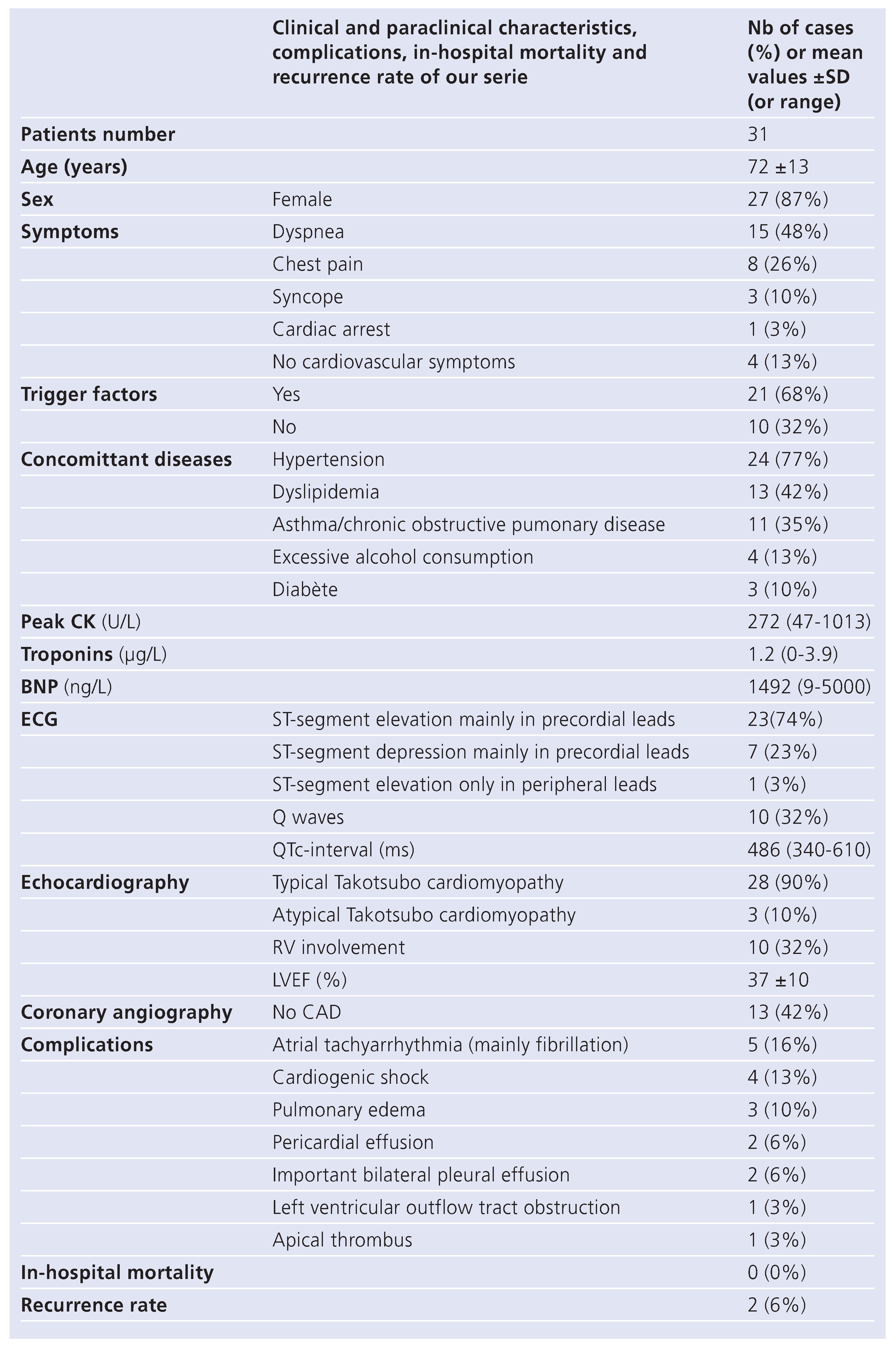
The study included 31 patients with a mean age of 72 ± 13 years (
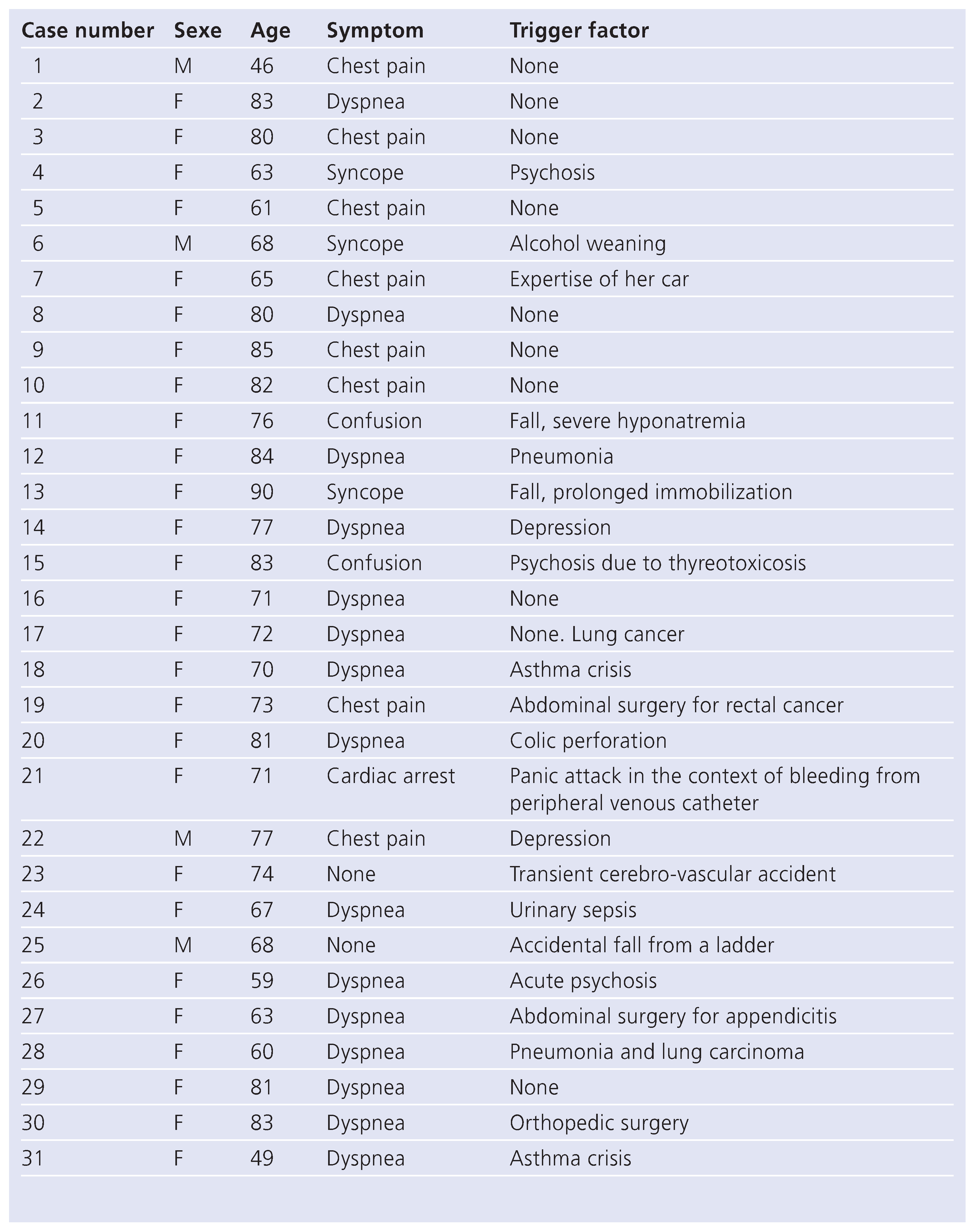
Table 1). In 87% of cases, patients were females. In 68% of cases, a psychological or a somatic trigger factor precipitated the TC (
Table 2). Three patients had an acute psychosis, among which 1 case was due to thyreotoxicosis, 2 patients were suffering from depression and 1 patient was afraid of driving her car. A surgical procedure was the initiating factor in 3 patients and 3 patients had suffered a fall: 1 patient had accidentally fallen from a ladder, another had fallen due to severe hyponatremia and the last patient had fallen at home due to an incapacity to stand up. One patient had had a panic attack due to bleeding from a peripheral venous catheter. An acute infectious process was present in 3 patients (1 urinary sepsis and 2 pneumonias). Two patients developed apical ballooning after a severe asthma crisis necessitating an endotracheal intubation. One patient suffered from alcohol weaning and another from an abdominal disease. There was no identified precipitating factor in 32% of cases. Significant head trauma or intracranial bleeding were excluded in a patient presenting with a transient cerebral attack and in the patients who had suffered a fall. None of the patients exhibited clinical characteristics suggestive of pheochromocytoma.
Dyspnea was the main presenting symptom in 48% of the cases, followed by chest pain mimicking an acute coronary syndrome in 26% of the patients. Three patients exhibited a syncope. One patient experienced a cardiac arrest after a panic attack following bleeding from a peripheral venous catheter and was successfully resuscitated. Four patients had no cardiovascular symptoms at the initial presentation and were investigated after the discovery of an abnormal ECG.
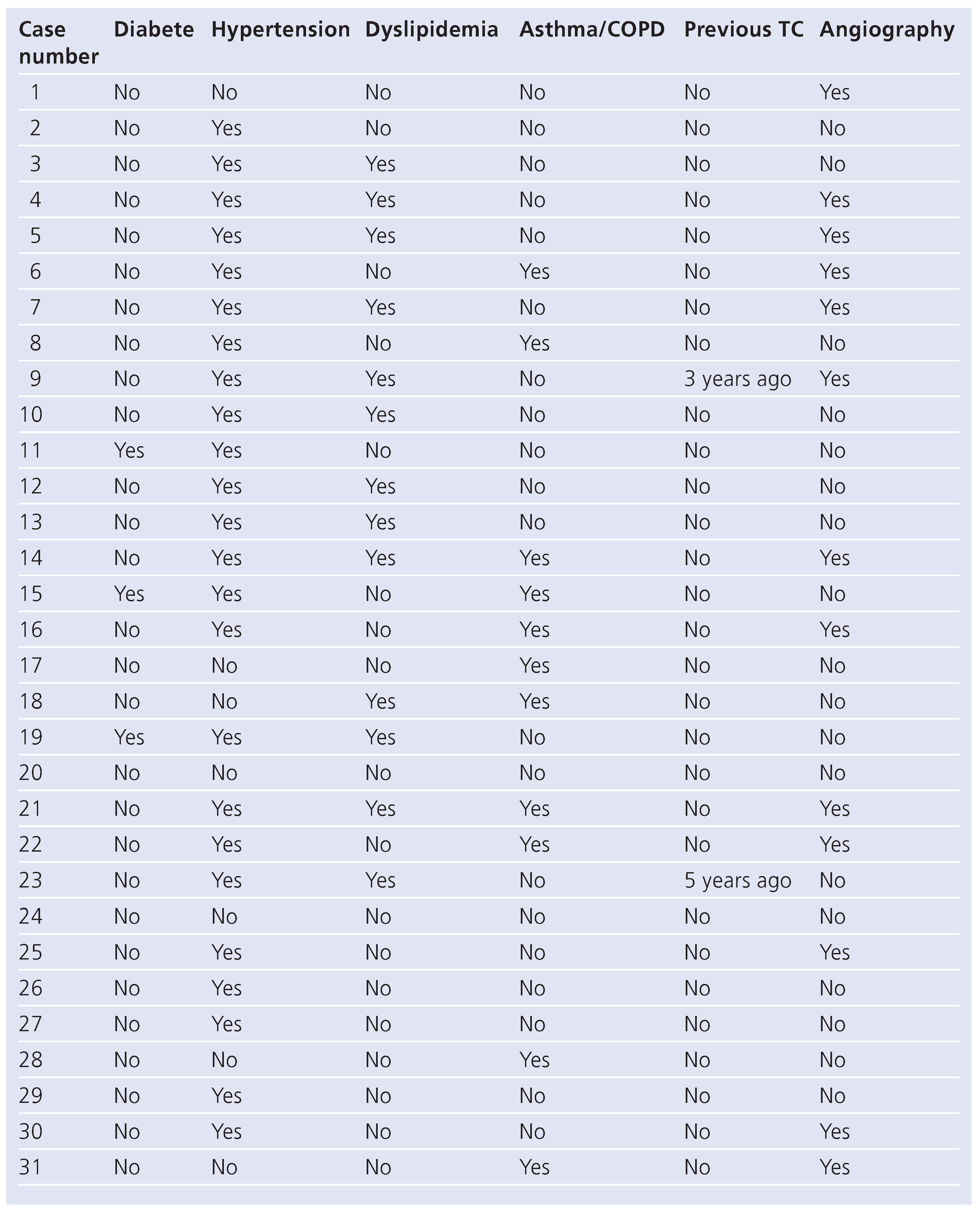
The most frequent concomitant diseases (
Table 3) were hypertension (77%) followed by asthma or chronic obstructive pulmonary disease (35%). Excessive alcohol consumption was noted in 13% of cases, dyslipidemia in 42% and diabetes in 10%. Finally, 2 patients had a pulmonary carcinoma and 1 had a rectal adenocarcinoma when diagnosed with a TC. One patient developed a pulmonary carcinoma during follow-up.
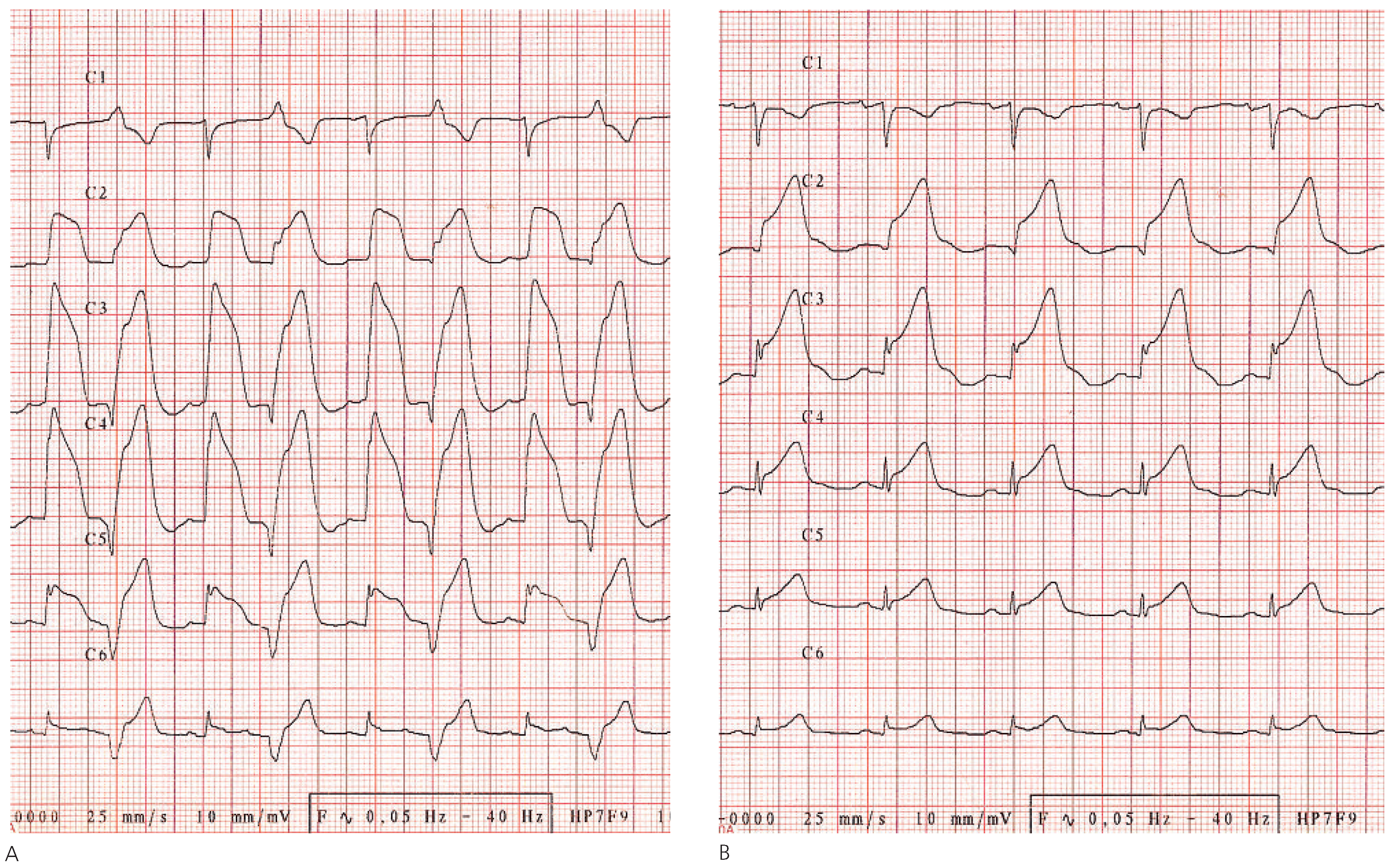
ECG modifications are essential because, when added to the clinical picture, they, most of the time, point to the syndrome. The most frequently encountered abnormalities were ST-segment elevation in precordial leads in 74% of cases and a prolongation of the QTc-interval during follow-up tracings (77% of QTc >440 ms). Sometimes, ST elevation can be grotesque as illustrated in figure 1. One patient had a ST-segment elevation only in the peripheral leads (I and AVL) with an atypical midventricular form of TC. Other ECG modifications included ST-segment depression (23%) mainly in the precordial leads and the presence of an abnormal Q wave (32%). The ECG evolution generally showed a progressive deepening of the T waves with often a marked prolongation of the QTcinterval (maximal 610 ms), before normalisation of this interval and progressive correction of all ECG abnormalities in 1 to 6 months. Interestingly, ECG normalisation occurred later than resolution of wall motion abnormalities on the echocardiogram.
Biological characteristics (Table 1)
The mean value of peak creatine kinase, measured in 27 patients (87%), was mildly raised to 272 U/l with a range of 47 to 1013 U/l (N <170 U/l). One patient had a peak CK value of 19367 U/l secondary to a rhabdomyolysis, after a fall and a prolonged immobilisation. Her troponin I value was 2 mcg/l. Troponin I was measured in 29 patients (94%) and was abnormal in 90% of the cases. Elevation was mild with a mean value of 1.2 mcg/L and a range of 0 to 3.9 mcg/l (N <0.1 mcg/L). Three patients, all women, had no elevation of troponine I, one presenting with chest pain, one with dyspnea and one with no cardiovascular symptoms. BNP values were obtained in 19 patients (61%) and were raised in all but 3. The mean peak value was 1492 ng/L (N <100 ng/L) with a range of 9 to >5000 ng/L. The rate of BNP decline was generally rapid within a few days after the diagnosis. Its maximal value was not always correlated to the severity of the LV dysfunction, but mean values were significantly higher in patients with right ventricular involvement (2297 ng/L vs 686 ng/L, p = 0.01).
Echocardiography and coronary angiography (Table 1)
The majority of the patients presented a moderate to severe LV systolic dysfunction at the first echocardiogram with a mean LVEF of 37 ± 10% with a range of 15 to 55%. The dysfunction was secondary to an apical LV akinesis with an apical ballooning and a compensatory hypercontraction of the base in the typical forms of TC (90% of cases). In the four patients with cardiogenic shock, only a small annular basal ring was contracting and one patient exhibited a severe left ventricular outflow tract obstruction. Right ventricular involvement was present in 32% of cases. It was associated with a significantly lower left ventricular ejection fraction (31% vs 40%, p = 0.01). Atypical forms included isolated mid-ventricular akinesia in three patients (fig. 2). All the patients completely recovered their cardiac function in one week to three months. None of our patients exhibited the features of an asymmetrical hypertrophic cardiomyopathy.
Coronary angiography was performed in 13 patients (42% of cases) and did not reveal any significant coronary artery lesions explaining the symptomatology. This investigation was performed in all men with a presumed diagnosis of TC independently of their mode of presentation and in all women with chest pain lasting for less than 24 hours, except one 82 year old lady in a poor general condition with typical transient ECG and echographic modification with mild LV dysfunction. Interestingly, one patient had an urgent coronary angiography for an ongoing myocardial infarction with ST-segment elevation, and then underwent an angioplasty due to a significant stenosis in the mid right coronary artery. ECG modifications in the antero-lateral leads and echocardiography later confirmed the diagnosis of a TC. One patient (case 19) who experienced a post-operative mid-ventricular ballooning did not have a coronary angiography because of the typical echographic pattern associated with a negative preoperative dobutamine stress echocardiography. Coronary angiography was performed in 6 women without chest pain, 3 with dyspnea, 1 with syncope, 1 with cardiogenic shock and 1 following a cardiac arrest and discarded significant coronary artery disease.
Complications and recurrence
The most frequent complications were atrial tachyarrhythmia (16%), mainly atrial fibrillation or multifocal atrial tachycardia, and cardiogenic shock (13%). Patients with atrial fibrillation regained a normal sinus rhythm either spontaneously or after a typical loading dose of amiodarone intravenously followed by oral treatment. Patients in cardiogenic shock required vaso-active amines, which was associated to an intraaortic balloon pump for one patient. One of the 4 patients with cardiogenic shock exhibited a severe dynamic obstruction of the left ventricular outflow tract with a systolic anterior motion of the mitral valve after hip surgery, partially due to hypovolemia secondary to blood losses and required a particularly challenging management. Three patients presented acute cardiac insufficiency with pulmonary oedema and two had significant bilateral pleural effusion. A pericardial effusion without haemodynamic consequences was discovered incidentally in two patients. One patient had an apical thrombus which completely resolved as well as the apical ballooning after six months of anticoagulation.
Follow-up was complete for the whole cohort with a mean duration of 36.3 ± 18.7 months. No patient died during the hospital stay but two died later in the course of follow up, both due to carcinomas of the lung. Interestingly, both had RV involvement in their initial presentation. Two patients (6%) experienced a recurrent episode of apical ballooning, one after 3 years and the other one after 5 years of follow-up.
Prevalence
When considering patients with the diagnosis of acute coronary syndrome including myocardial infarctions with or without ST-segment elevation and unstable angina, acute heart failure, acute pulmonary oedema and cardiogenic shock, the prevalence of TC during the study period was 3%. When taking TC presenting as acute coronary syndrome compared to the whole population with acute coronary syndrome, the prevalence was 1.6%.
Discussion
Stress cardiomyopathy essentially affects postmenopausal women (85–95% of cases) [
1,
2,
3,
6,
9,
11,
16,
17,
18,
19,
20,
21,
22,
24,
27,
29,
30,
31,
34,
35] with a mean age of 60–75 years [
2,
3,
4,
5,
6,
10,
16,
19,
20,
21,
22,
23,
24,
25,
28,
31,
32,
33,
34,
36] and often develops after psychological or somatic stress (>50% of the cases) [
2,
3,
4,
6,
9,
10,
15,
16,
19,
20,
21,
23,
24,
25,
28,
31,
33,
34]. In our study, 87% of patients were female with a mean age of 72 years and presented following a trigger factor in 65% of cases.
Our results are well matched to the data in the literature in terms of ECG manifestations, biological characteristics, echocardiographic appearance, complications and recurrence rate. They also confirm the relatively high percentage (26% in the literature versus 32% in our study) of RV apical ballooning reported [
4,
5], its association with more extensive LV dysfunction and higher BNP values, and consequently the more complicated in-hospital course of these patients. An association with malignancies (found in 10% of our patients) has already been described [
6,
7,
8], but the contribution of cancer to the development of TC remains hypothetical.
Most studies collected cases either retrospectively by searching in catheterisation laboratory databases for patients with acute coronary syndromes and normal coronary arteries fulfilling the other criteria of TC or by prospectively recruiting the same type of patients. In our series, patients were collected based on clinical presentation associated with suggestive ECG modifications and typical echocardiographic appearance. Complete recovery of LV and RV segmental abnormalities, as well as ECG modifications, were documented in all cases during a careful follow-up. Using this approach, we found more patients presenting with dyspnea without chest pain, or even patients without cardiovascular symptoms less well described in the literature. We found an association with hypertension (77% of the patients) and chronic obstructive disease (35% of the patients) which was greater than the usual prevalence of these diseases in the general population. We did not perform coronary angiography to confirm the absence of significant coronary artery disease in all patients, but the investigation was done in all patients, except one, with chest pain as the presenting symptom. In this category of patients the prevalence of TC in our study (1.6%) was comparable to the usual reported rate of 1 to 2% [
1,
3,
7,
9,
10,
14,
19,
20,
25,
26,
27,
33]. Therefore the higher general prevalence of TC observed in our study was mostly due to patients presenting with symptoms other than chest pain, where attention to the pathology was mainly raised by an abnormal ECG or echocardiography in the presence of unusual dyspnea. This category of patients deserves more recognition from the medical community, even though the course of TC is frequently benign. With regard to atypical forms of TC, they can reach up to 40% of TC (10% of our cases) and it is possible that we initially missed some atypical TC because this entity was less well known and described [
9,
10,
11]. Concerning the prognosis, the largest study [
6] included 100 TC patients followed during four years and survival was identical to the general population, in-hospital mortality was 2% (none in the current sample), 4 to 5 times lower than the mortality of myocardial infarction, and the recurrence rate was 11% (6% in our study). Finally, the complication of LV thrombus is a rare condition and was only documented in one case (3%), a proportion which is similar to the literature [
5,
12,
13].
Figure 1.
(A). Very significant ST-segment elevation in the precordial leads with an intermittent right bundle block concerning an 83 year old woman who underwent a hip replacement with a total prosthesis and presented with acute post-operative dyspnea. (B). The same patient, 30 minutes after the ECG cited above. The ST-segment elevation is less accentuated but remains nevertheless high in the precordial leads. The right bundle block disappeared.
The pathogenesis of TC is not completely understood, but several arguments plead in favour of a hyperactivity of the sympathetic system to which women are more prone than men and that is responsible for cellular and vascular toxicity [
7,
14]. Formerly, the catecholamine-induced myocardial toxicity was known under the entity “stunned myocardium” in cases of intracranial haemorrhages or paroxysms of pheochromocytoma [
15,
16,
17,
18]. Indeed, many studies demonstrated a dysfunction of the sympathetic system located at the apical level using I123-MIBG myocardial scintigraphy [
14,
19], an increase of plasma catecholamines levels [
3,
14,
15,
16,
19,
20,
21,
22] out of proportion compared to myocardial necrosis and typical histological lesions with the presence of contraction-band necrosis in endomyocardial biopsies [
7,
14,
16,
20,
23,
24]. Furthermore, some authors showed a global dysfunction of the microcirculation provoked by multifocal non epicardial coronary spasms visualised by Doppler flow measurements during coronary angiography, by myocardial scintigraphy, by fluorodeoxyglucose – positron emission tomography studies or more recently by myocardial contrast echocardiography [
3,
7,
10,
20,
21,
25,
26]. Some investigators postulated a dynamic left ventricular outflow tract obstruction [
2,
7,
28] caused by the basal hyperdynamism in patients with geometrical predispositions. This can be responsible for a systolic anterior motion of the mitral valve during stress or a postoperative hypovolemia [
2,
20,
24,
25,
28,
29,
30,
31]. One case of our series illustrated this mechanism after a hip replacement. Finally, myocarditis [
3,
20] has been excluded by endomyocardial biopsies [
16,
22,
23,
24], cardiac magnetic resonance imaging [
23,
28] and negative viral serologies [
19,
22,
23].
Figure 2.
(A.) Diastolic frame in a 4 chamber view showing a normal shape and appearance of the left ventricle. (B). In systole, there is a complete akinesia of the middle portion of the ventricle characteristic of mid ventricular form of Takotsubo cardiomyopathy (arrows).
Figure 2.
(A.) Diastolic frame in a 4 chamber view showing a normal shape and appearance of the left ventricle. (B). In systole, there is a complete akinesia of the middle portion of the ventricle characteristic of mid ventricular form of Takotsubo cardiomyopathy (arrows).
Limitations
Only 42% of our patients underwent a coronary angiography. As stated in the method, this investigation was routinely done in all male patients with a suspicion of TC because this entity is rare in men and coronary artery disease is much more frequent. All the women, except one, with chest pain for less than 24 hours and six particular cases without chest pain also had a coronary angiography. The investigation was withheld in women presenting with dyspnea, typical ECG modifications and echocardiographic apical or midventricular ballooning after emotional or physical stress, and was followed with complete recovery.
Myocarditis was not formally discarded in our patients. At the time of the study, we did not have access to cardiac MRI which is the non invasive gold standard for this purpose.


