Next Article in Journal
Annual Report 2008 of the Working Group “Interventional Cardiology and Acute Coronary Syndroms”
Previous Article in Journal
Oldest Survivors After Repair of Tetralogy of Fallot
 
 
Cardiovascular Medicine is published by MDPI from Volume 28 Issue 1 (2025). Previous articles were published by another publisher in Open Access under a CC-BY (or CC-BY-NC-ND) licence, and they are hosted by MDPI on mdpi.com as a courtesy and upon agreement with Editores Medicorum Helveticorum (EMH).
Proceeding Paper

Inflammation and Stroke †

by
Corinne Benakis
1,
Lorenz Hirt
1 and
Renaud A. Du Pasquier
1,2,*
1
Service of Neurology, Centre Hospitalier Universitaire Vaudois (CHUV), Lausanne, Switzerland
2
Service of Immunology and Allergy, Centre Hospitalier Universitaire Vaudois (CHUV), Lausanne, Switzerland
*
Author to whom correspondence should be addressed.
This article summarises a lecture at the annual meeting of the Swiss Society of Cardiology in Berne, May 2008.
Cardiovasc. Med. 2009, 12(5), 143; https://doi.org/10.4414/cvm.2009.01424
Submission received: 22 February 2009 / Revised: 22 March 2009 / Accepted: 22 April 2009 / Published: 22 May 2009

Summary

It is well-known that inflammation plays a major role in the genesis of the atherosclerotic plaque and thus favours the occurrence of stroke. But, inflammation is also involved after an ischaemic event affecting the brain. In this case, one can observe the infiltration of numerous inflammatory cells at the site of the lesion, the activation of the microglia and of pro-inflammatory cytokines, etc. It is usually thought that this poststroke inflammation is rather deleterious, as suggested by the fact that, after an experimentally-induced ischaemic stroke, blockade of the inflammatory response improves the neurological condition of mice. Nevertheless, until now, the application of such experimental treatments in humans has revealed unsuccessful. One of the possible explanations for this phenomenon might be that inflammation also has some beneficial effects, such as clearance of damaged tissue, promotion of angiogenesis, or still tissue remodelling and regeneration. After this first “basic science” part, we will briefly review some clinical aspects of the most significant inflammatory diseases that can cause stroke, i.e., the vasculitis. Among them, Takayasu’s arteritis, giant cell temporal arteritis (Horton’s disease), and primary angiitis of the central nervous system will be discussed. We will also shortly address the question of the antiphospholipid syndrome.

Inflammatory mechanisms related to an ischaemic stroke Brain ischaemia

Stroke is a leading cause of long-term disability and remains the third cause of death in developed countries (World Health report 2007, World Health Organisation). While haemorrhagic stroke triggers cerebral oedema and inflammation, this short review focuses on the more common ischaemic stroke. Importantly, while ischaemia at first induces only a loss of function at its very early stages, structural damage appears rapidly thereafter and progresses as minutes and hours go by. Reduction of cerebral blood flow leads to a lack of oxygen and glucose supply to the brain parenchyma. This nutrient deficiency triggers multiple events including a dramatic depletion of ATP, perturbation of the cellular ionic homeostasis, neurotransmitter release and activation of many cytotoxic enzymes. The release of the excitatory neurotransmitter glutamate leads to excessive excitotoxic stimulation of glutamate receptors in energy deprived neurons. Excitotoxicity is a major mechanism in the early stages of the progression of ischaemic brain injury. Other detrimental events include peri-infarct depolarisation, apoptosis and inflammation (Figure 1) [1,2].
Several therapeutic strategies aimed at decreasing the effect of these ischaemia-induced phenomena have been successful in animal stroke models, but not so far in stroke patients. The only successful treatment in the acute phase for stroke patients is thrombolysis, the goal of which is to restore the blood flow to the brain [3,4]. As it needs to be administered intra-venously within 3 hours to 4.5 hours or intra-arterially within 6 hours from symptom onset it is limited to a small number of patients [5]. Beyond this time-window restoring cerebral blood flow is no longer beneficial and there is an increased risk of developing a symptomatic intracerebral haemorrhage [6]. Furthermore, the recombinant tissue plasminogen activator (rtPA) enhances excitotoxicity and increases the lesion volume in our mouse MCAo model [7,8]. Thus, there is a need for new treatments for the patients who can not be thrombolysed.
An important delayed mechanism beginning within hours from the onset of ischaemia is the robust inflammatory response in the ischaemic tissue. There is increasing evidence showing a detrimental effect of the post-ischaemic inflammatory reaction [9,10]. Therefore therapeutic strategies targeting the delayed inflammatory response could inhibit the progression of the tissue damage providing an extended therapeutic window for neuroprotection.

Neuroinflammatory response after ischaemia

The brain immune defence system is essential to protect neurons against infectious agents. In cerebral ischaemia, the inflammatory response plays a role in the clearance of cell debris; however it also enhances the damage to the tissue.
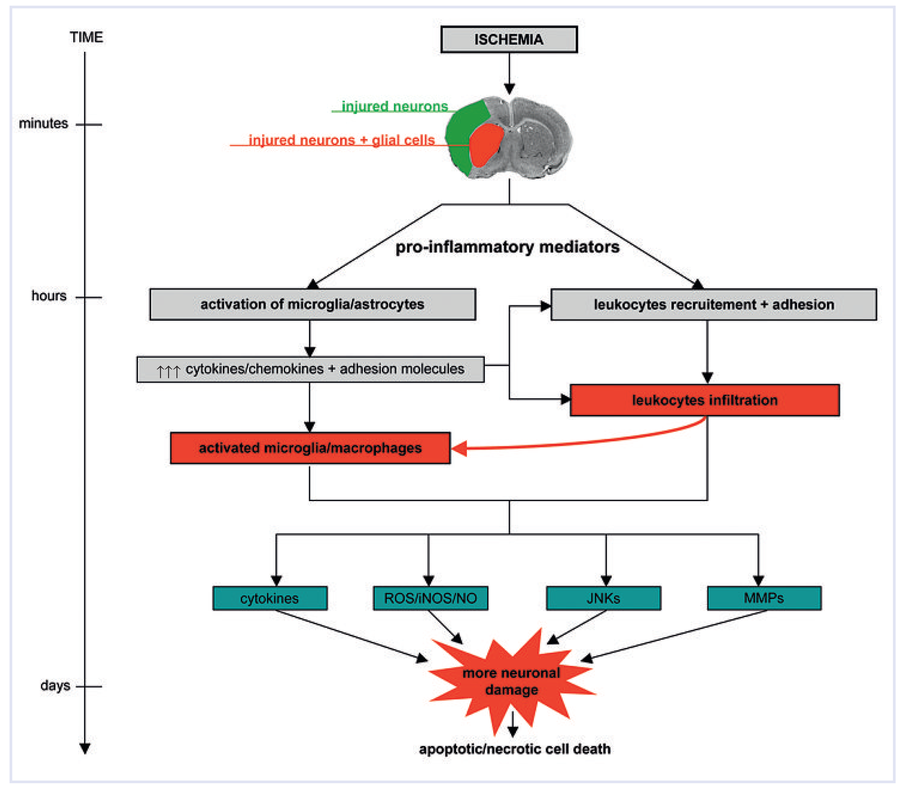
An immune response at the site of injury is characterised by the infiltration, accumulation and activation of inflammatory cells. Within hours after the onset of focal cerebral ischaemia peripheral leukocytes adhere to the cerebral endothelium, cross the vessel wall and invade the damaged parenchyma [11,12]. At the same time astrocytes and microglia become activated. These cellular events depend on the secretion of inflammatory mediators which are produced by neural and glial cells in response to an ischaemic insult. Once activated in the site of injury inflammatory cells start to secrete a large variety of cytotoxic agents such as cytokines, chemokines and promote the expression of adhesion molecules, matrix metalloproteinases (MMPs) with an increased production of free radicals. Stress signalling pathways, such as the c-Jun N-terminal kinases (JNKs) pathway are also activated. Both the JNK pathway and pro-inflammatory mediators further potentiate the brain tissue injury and lead within hours and days to apoptotic and necrotic cell death of the potential viable tissue [13,14,15]. Figure 2 provides a schematic diagram illustrating the inflammatory response after ischaemic stroke.
Table 1. Classification of different vasculitis types.
Cardiovascmed 12 00143 i001

Glial cell activation

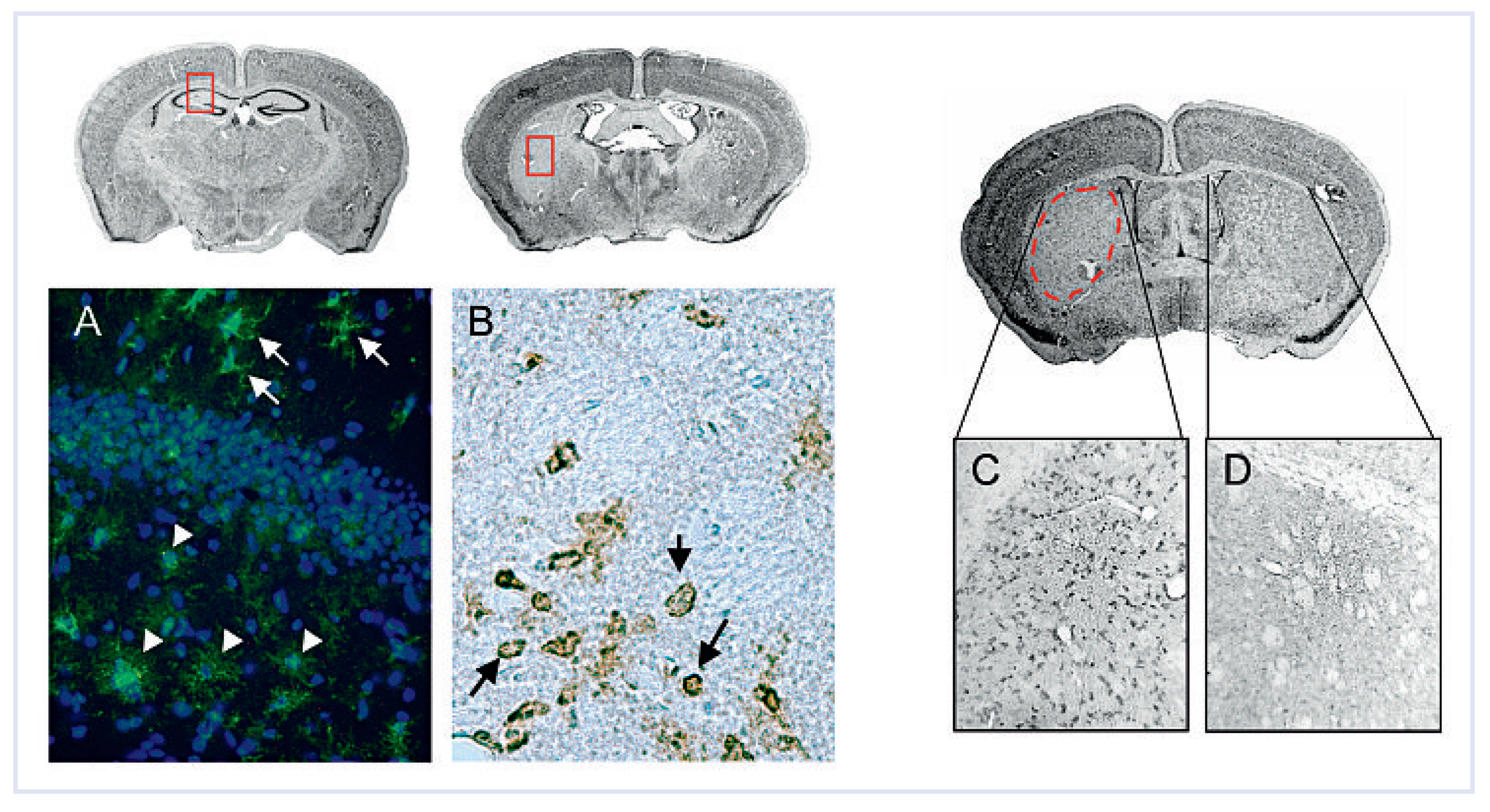
Microglia are the resident macrophages of the brain. They are very sensitive to subtle alterations in their neuronal microenvironment. Resting microglia has a very ramified cytoplasm, covering a territory of 30–40 mm in diameter. In response to an injury, they quickly become activated and undergo morphological transformations as well as functional changes. They start to retract their long processes and their shape becomes rounded – so called phagocytotic, amoeboid microglia [16,17,18]. The degree of microglial activation depends on the severity of neuronal injury. The mildest injuries may only cause ramification of microglia with a bushy appearance, whereas in acute pathological cases microglia are characterised by hypertrophic bodies and less arborised processes. If neurons die, microglia transform into brain macrophages. These stimulated cells rapidly proliferate to focal sites of injury due to an increasing expression of immunoreactive cell surface molecules and to the secretion of various inflammatory molecules such as chemotactic factors which induce the recruitment of other microglial cells. Figure 3 shows the changes in microglial morphology and accumulation of activated microglia in a mouse stroke model (48 hours after transient 30 minutes middle cerebral artery occlusion). This phenomenon is accompanied by an increased expression of cytokines: interleukins (IL-1b, IL-4, IL-6, IL-10), tumor necrosis factora (TNFa), interferons and chemokines such as MCP-1 [19,20]. The surrounding astrocytes are sensitive to the increased release of these immunomodulatory peptides and therefore severe ischaemia also compromises astrocytic function. Astrocytes modulate the phagocytic functions of microglia and promote the expression of adhesion molecules in the neurovascular unit on endothelial cells and circulating leukocytes [21,22]. These early inflammatory processes are likely to be deleterious for neuron survival.

Leukocyte infiltration

Shortly after the onset of injury the blood brain barrier opens by the disruption of the endothelia tight junctions [23]. The release of inflammatory mediators from activated glial cells induces the expression of proteins on the outer cell membrane of vascular endothelial cells and leukocytes. The opening of the barrier and the release of inflammatory mediators leads to the migration of circulating leukocytes to the site of injury. Infiltration of leukocytes occurs in three steps: rolling on the surface of endothelial cells, adhesion to the endothelial wall and migration or diapedesis. The initial capture and migration is mediated by three main groups of cell adhesion molecules: selectins (P-, E- and L-selectins), immunoglobulins (VCAM-1, ICAM-1) and integrins (CD11a-c) [11,24]. Circulating monocytes/macrophages are also recruited at the site of injury and will penetrate into the parenchyma. The proportion of invading macrophages in the ischaemic tissue can not be discriminate from resident activated microglia.
Table 2.
Cardiovascmed 12 00143 i002
Activation and accumulation of leukocytes (granulocytes, monocytes/macrophages, lymphocytes) at the site of injury results in further damage. Current evidence suggests a detrimental role of inducible or immunological nitric oxide synthase (iNOS) and cyclooxygenase-2 (COX-2) from neutrophils and vascular cells in the ischaemic brain [25,26]. They induce the formation of reactive oxygen species: nitric oxide (NO) and superoxide respectively, leading to the generation of peroxinitrite [14,27] which is a powerful oxidant which triggers damage to DNA and other cell constituents [1]. Up-regulation of pro-inflammatory molecules and reactive oxygen species after cerebral ischaemia are not the only cause of secondary injury. Expression of metalloproteinase genes is related to the presence of inflammatory cells in ischemic tissue. A recent study has demonstrated the infiltration of metalloproteinase-9 positive (MMP-9+) neutrophils after human stroke [28]. MMP-9 is involved in the degradation of components of the extracellular matrix and basal lamina which may potentiate haemorrhagic complications after ischaemic stroke.

Transcriptional regulation of inflammation

The secretion of inflammatory molecules in cerebral ischaemia triggers the activation of several transcription factors involved in the inflammatory response. The nuclear factor-kappa B (NF-kB) when activated, induces the expression of genes encoding cell adhesion molecules, cell surface receptors and cytokines [29,30]. Iadecola and colleagues have shown an attenuation of the inflammatory response induced by focal cerebral ischaemia in mice with a null mutation in the CD36 receptor which recognises pathogens and induces an inflammatory response through the activation of NFkB [31]. Interference with the nuclear factor-kappa B activation may therefore be beneficial to secondary ischaemic injury.
Mitogen activated protein kinases (MAPKs) are a family of key proteins which are activated in response to stress signals. MAPK signalling pathways positively regulate transcription of inflammatory genes, such as those coding for TNF-a, IL-1b, IL-6, IL-8, COX-2 (for review [9,32]). D-JNKi-1, a specific inhibitor of the JNK pathway, was shown to induce a strong neuroprotective effect in a range of experimental cerebral ischaemia model [33,34]. Besides, the inhibition of MAP kinases, especially p38 and JNK, could lead to a reduction in pro-inflammatory molecule production by inflammatory cells, especially microglia/macrophages in which the MAPK cascades are highly activated after an ischaemic injury [35,36].We are therefore currently investigating the effect of D-JNKi-1 on the inflammatory response and the progression of the tissue damage after ischaemia.

Neuroprotective approaches targeting inflammation

Different strategies aimed at preventing the inflammatory response after cerebral ischaemia have been successful in rodent models. Ischaemic damage was shown to be attenuated, together with systemic leukocyte depletion by preventing the expression of, or by blocking adhesion molecules, by inhibiting pro-inflammatory cytokines or by diminishing the free radical generating enzymes iNOS or COX-2 (for review [1,37]). Attempts to translate therapeutic interventions to stroke patients have been more disappointing than promising. For instance the selective IL-1 receptor antagonist (IL-1ra) which limits the pro-inflammatory action of IL-1, has been tested in randomised patients with acute stroke [38]. Despite a conclusive phase II study no recent publications have reported IL-ra as a therapeutic agent for acute stroke.
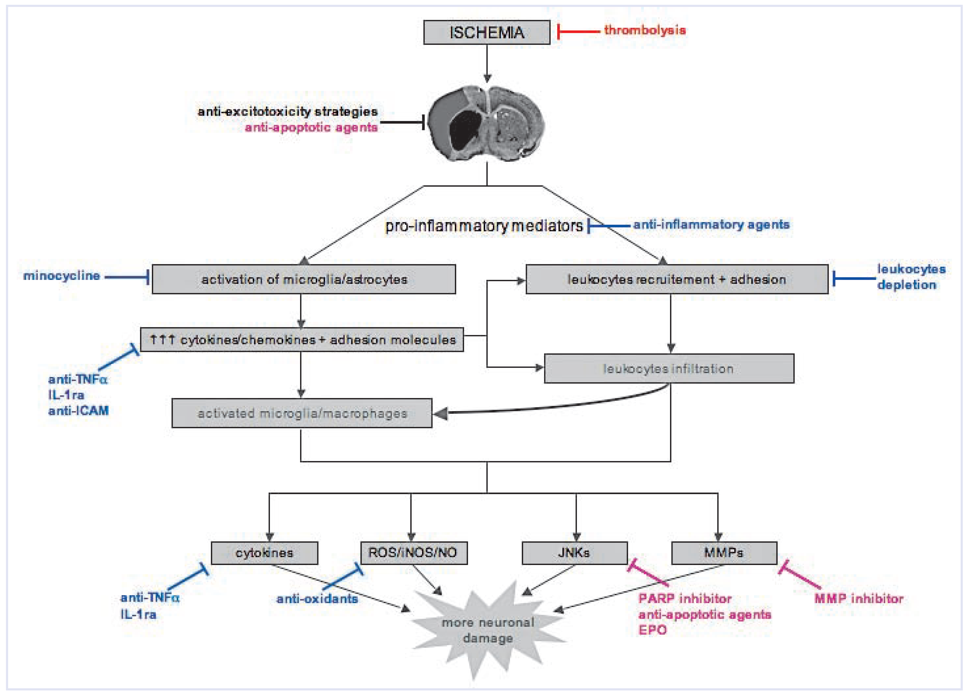
More than a thousand of potential agents underwent clinical evaluation [39]. However none of these drugs have demonstrated benefit in stroke clinical trials as for instance the application of murine monoclonal anti-ICAM-1 in Enlimomab trial [40] and XY059, a nitrone-based free radical trapping agent [41]. A non-exhaustive list of therapeutic strategies targeting especially inflammation after ischaemic stroke is shown in Figure 4 and Table 2.

Experimental studies to clinical trials: lost in translation

Reasons for failure have been discussed before [42] and include morphological and functional differences between rodents and humans, timing of the intervention, evaluation of efficacy, pharmacokinetic issues (plasma concentration of drugs) and side effects. Anti-inflammatory strategies have also in some cases promoted deleterious infections and fever [40]
Until now, researchers have focused mostly on the negative role of inflammation after stroke and thus have looked to therapeutic means to inhibit post-stroke inflammation. Nevertheless, there is evidence suggesting that inflammation might also be beneficial in stroke: it is a crucial mechanism to clear damaged tissue after an infarction, it promotes angiogenesis, tissue remodelling and regeneration [1,43]. Therefore, there is clearly the need to better understand the subtle balance between the beneficial and deleterious effects of inflammation in stroke. Furthermore, experimental studies on cerebral ischaemia have mostly target one cell type, i.e., neurons, while endothelium, astrocytes and microglia have been considerably neglected. Future research on experimental stroke models should considered the important role of non-neuronal cells and the bivalent function of inflammation. A better insight in these aspects is important before planning future clinical trials.

Autoimmune and infectious aetiologies of stroke

We have discussed on the inflammatory response after cerebral ischaemia and its consequences. In this second part of this review we will illustrate how a disorder affecting the immune system could lead to stroke. Therefore, we will briefly review the vasculitis which can cause stroke. Primary vasculitis is classified into three categories, depending on the size of the affected arteries. This classification is relevant for the clinician. Indeed, magnetic resonance angiography or even conventional arteriography can provide good information on large-size arteries, are of variable value in middlesize arteries vasculitis and are useless in small-size arteries vasculitis, since in the latter case, the lesions are below the threshold of detection (Table 1). In other terms, a normal neuro-imaging study does not rule out a middle-size or a small-size arteries vasculitis. A meningeal and brain parenchymal biopsy might thus be warranted. Another important fact is that most vasculitis that can cause strokes are systemic diseases and thus other organs, including the heart, are frequently involved, for instance the Takayasu arteritis (TA) which affects the large arterial trunks. TA must always be ruled out in young patients with stroke, especially if they are female and of Asian descent. Stroke, either ischaemic or haemorrhagic occurs in 20–30% of TA. Treatment consists in a combination of corticosteroids, immunosuppressive therapies and surgery [44].
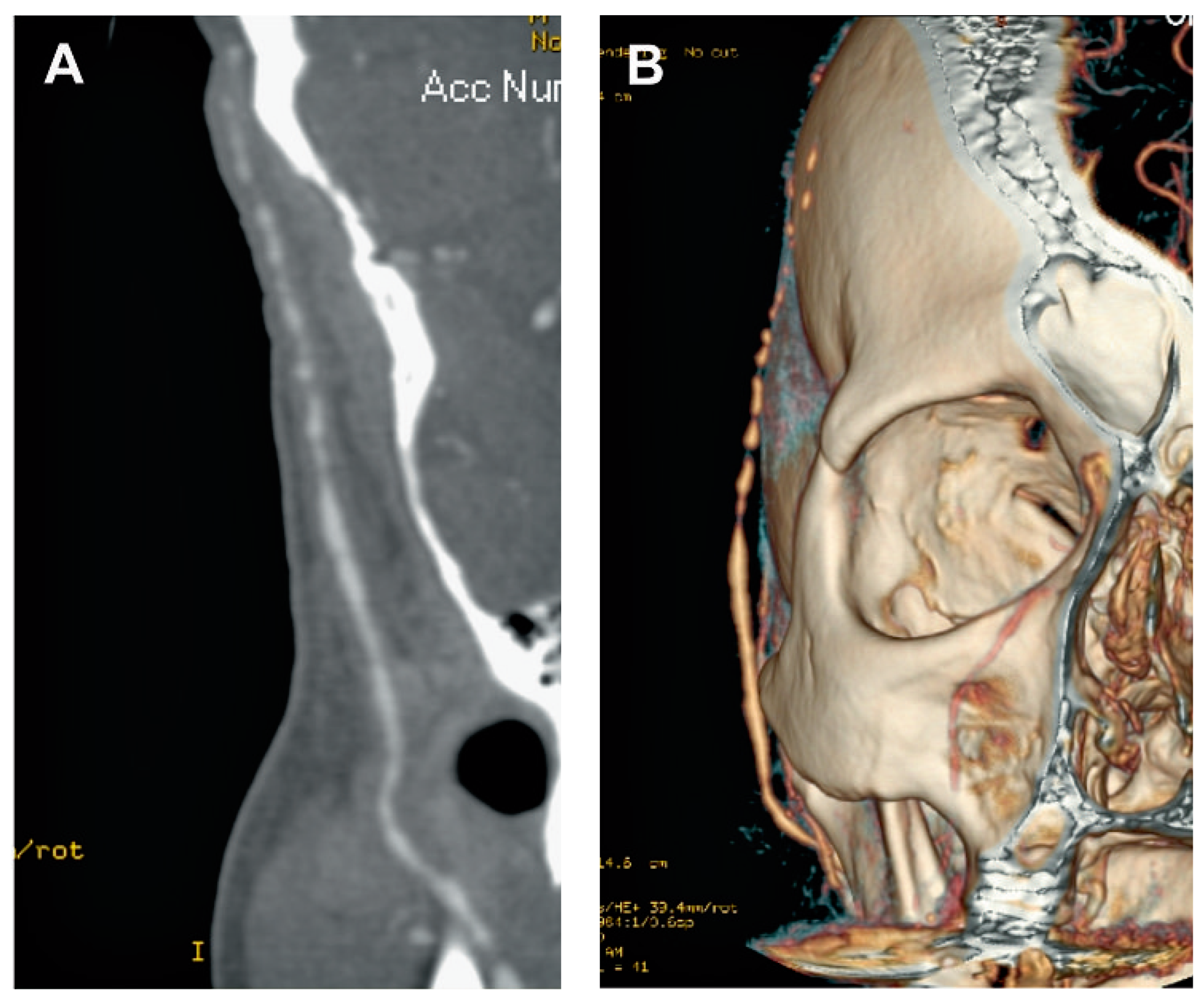
By contrast, temporal arteriitis (Horton’s disease) affects elderly people (usually >60 years old) and is characterised by headaches and a high erythrocyte sedimentation rate (>50 mm/hour) (Figure 5). When suspecting Horton’s disease, one must immediately administer high doses of corticosteroids, since there is a risk of occlusion of the central artery of the retina, leading to definite loss of vision. Stroke may occur in 10% of patients with Horton’s disease. Other manifestations include fever, fatigue, jaw claudication and its very frequent association with polymyalgia rheumatica. Of importance, temporal artery biopsy, which is the gold standard diagnostic procedure, remains positive up to 7–10 days after corticosteroids were started. The duration of corticosteroid therapy depends on the erythrocyte sedimentation rate and the clinical symptoms. Usually, the treatment lasts a minimum of one year [45].
Primary angiitis of the central nervous system (PACNS) is a rare condition, but very difficult to diagnose. Indeed, in contrast to other vasculitis, PACNS affects only the vessels of the brain (middle-size arteries), without any other systemic manifestations. Its mode of presentation is variable including, in addition to ischaemic or haemorrhagic strokes, confusion, cognitive deterioration, headaches, etc. Extensive blood tests are not contributory. Cerebrospinal fluid examination is abnormal in 50–90% of cases, but of little help, since it shows only aspecific findings, i.e., a mild to moderate increase in proteins, and/or leucocytes, and/or erythrocytes.
Nevertheless, if a PACNS is suspected, it is of importance to establish the diagnosis as the treatment is heavy, consisting in long-term corticosteroids and cyclophosphamide. Therefore, a meningeal and parenchymal brain biopsy is often necessary [46].
We will not address here the other primary vasculitis causing stroke (Table 1), as strokes are relatively rare in those vasculitis and, usually, other organs are affected before the central nervous system. The same is true for secondary vasculitis. Nevertheless, it is worth to mention here the problem of the anti-phospholipid syndrome (aPLs). Indeed, this condition is found in 40% of the cases of systemic lupus erythematosus, but can also be primary, i.e., not associated with any underlying condition. aPLs may cause arterial and venous thrombosis. Typical manifestations include: spontaneous abortion beyond 10 week of gestation, cardiac valve abnormalities, thrombocytopenia, haemolytic anaemia. A neurological involvement is frequent, consisting in strokes, migraine-like phenomenon, chorea, and transverse myelopathy. The diagnosis of aPLs is based on the Sapporo criteria established in 1999 and revised in 2004 in Sydney. Detailing these diagnostic criteria would be well beyond the purpose of this small review and we advise the avid reader to consult the following references: [47,48,49].
From a neurological standpoint, it is recommended to rule out an aPLs in patients younger than 45 yearsold who present with an ischaemic stroke. But it is crucial to follow the above-mentioned modified Sapporo criteria in order to avoid over-diagnosis of aPLs. Indeed, this diagnosis often implies a long-term anticoagulation treatment [50].
Conclusions
Our knowledge on pathophysiology of cerebral ischaemia has greatly improved because of experimental in vitro and in vivo studies. A large number of drugs have been developed in the purpose to inhibit the complex cascade taking place after stroke including excitotoxicity and inflammation. Despite those efforts none of the potential therapeutic agent has been successful in clinical trials so far.
In this short review we have shown that inflammation can cause an occlusion of a brain artery and therefore drive to cerebral ischaemia, as well as be the direct consequence of stroke and exacerbate damage. Furthermore, stroke induces immunodepression and favors opportunistic infections such as bronchopneumonia [51].
Therefore, in stroke therapy there is a great need to identify new approaches which could block the detrimental inflammatory response as well as inducing neuroprotection or perhaps in combination with thrombolysis.

Funding

L. Hirt was supported by the CTI grant # 8901.1 and R. A. Du Pasquier was supported by the SNF PP-00B-106716.

Acknowledgments

We thank Melanie Price for helpful comments.

References

  1. Dirnagl, U.; Iadecola, C.; Moskowitz, M.A. Pathobiology of ischaemic stroke: an integrated view. Trends Neurosci. 1999, 22, 391–397. [Google Scholar] [CrossRef]
  2. Mehta, S.L.; Manhas, N.; Raghubir, R. Molecular targets in cerebral ischemia for developing novel therapeutics. Brain Res. Rev. 2007, 54, 34–66. [Google Scholar] [CrossRef] [PubMed]
  3. The National Institute of Neurological Disorders and Stroke rt-PA Stroke Study Group. Tissue plasminogen activator for acute ischemic stroke. N. Engl. J. Med. 1995, 333, 1581–1587. [Google Scholar] [CrossRef] [PubMed]
  4. Furlan, A.; Higashida, R.; Wechsler, L.; Gent, M.; Rowley, H.; Kase, C.; et al. Intra-arterial prourokinase for acute ischemic stroke. The PROACT II study: a randomized controlled trial. Prolyse in acute cerebral thromboembolism. JAMA 1999, 282, 2003–2011. [Google Scholar] [CrossRef] [PubMed]
  5. Hacke, W.; Kaste, M.; Bluhmki, E.; Brozman, M.; Davalos, A.; Guidetti, D.; et al. Thrombolysis with alteplase 3 to 4.5 hours after acute ischemic stroke. N. Engl. J. Med. 2008, 359, 1317–1329. [Google Scholar] [CrossRef]
  6. The NINDS t-PA Stroke Study Group. Intracerebral hemorrhage after intravenous t-PA therapy for ischemic stroke. Stroke 1997, 28, 2109–18. [Google Scholar] [CrossRef]
  7. Nicole, O.; Docagne, F.; Ali, C.; Margaill, I.; Carmeliet, P.; MacKenzie, E.T.; et al. The proteolytic activity of tissue-plasminogen activator enhances NMDA receptor-mediated signalling. Nat. Med. 2001, 7, 59–64. [Google Scholar] [CrossRef]
  8. Wiegler, K.; Bonny, C.; Coquoz, D.; Hirt, L. The JNK inhibitor XG-102 protects from ischemic damage with delayed intravenous administration also in the presence of recombinant tissue plasminogen activator. Cerebrovasc. Dis. 2008, 26, 360–366. [Google Scholar] [CrossRef]
  9. Barone, F.C.; Feuerstein, G.Z. Inflammatory mediators and stroke: new opportunities for novel therapeutics. J. Cereb. Blood Flow. Metab. 1999, 19, 819–834. [Google Scholar] [CrossRef]
  10. del Zoppo, G.J.; Becker, K.J.; Hallenbeck, J.M. Inflammation after stroke: is it harmful? Arch. Neurol. 2001, 58, 669–672. [Google Scholar] [CrossRef]
  11. Petty, M.A.; Wettstein, J.G. Elements of cerebral microvascular ischaemia. Brain Res. Brain Res. Rev. 2001, 36, 23–34. [Google Scholar] [CrossRef]
  12. Wang, Q.; Tang, X.N.; Yenari, M.A. The inflammatory response in stroke. J. Neuroimmunol. 2007, 184, 53–68. [Google Scholar] [CrossRef] [PubMed]
  13. Ishikawa, M.; Zhang, J.H.; Nanda, A.; Granger, D.N. Inflammatory responses to ischemia and reperfusion in the cerebral microcirculation. Front. Biosci. 2004, 9, 1339–1347. [Google Scholar] [CrossRef] [PubMed]
  14. del Zoppo, G.J.; Ginis, I.; Hallenbeck, J.M.; Iadecola, C.; Wang, X.; Feuerstein, GZ. Inflammation and stroke: putative role for cytokines, adhesion molecules and iNOS in brain response to ischemia. Brain Pathol. 2000, 10, 95–112. [Google Scholar] [CrossRef] [PubMed]
  15. Feuerstein, G.Z.; Wang, X.; Barone, F.C. The role of cytokines in the neuropathology of stroke and neurotrauma. Neuroimmunomodulation 1998, 5, 143–59. [Google Scholar] [CrossRef]
  16. Raivich, G.; Bohatschek, M.; Kloss, C.U.; Werner, A.; Jones, L.L.; Kreutzberg, G.W. Neuroglial activation repertoire in the injured brain: graded response, molecular mechanisms and cues to physiological function. Brain Res. Brain Res. Rev. 1999, 30, 77–105. [Google Scholar] [CrossRef]
  17. Ladeby, R.; Wirenfeldt, M.; Garcia-Ovejero, D.; Fenger, C.; Dissing-Olesen, L.; Dalmau, I.; et al. Microglial cell population dynamics in the injured adult central nervous system. Brain Res. Brain Res. Rev. 2005, 48, 196–206. [Google Scholar] [CrossRef]
  18. Streit, W.J.; Walter, S.A.; Pennell, N.A. Reactive microgliosis. Prog. Neurobiol. 1999, 57, 563–581. [Google Scholar] [CrossRef]
  19. Garden, G.A.; Moller, T. Microglia biology in health and disease. J. Neuroimmune Pharmacol. 2006, 1, 127–137. [Google Scholar] [CrossRef]
  20. Hanisch, U.K. Microglia as a source and target of cytokines. Glia 2002, 40, 140–155. [Google Scholar] [CrossRef]
  21. Bezzi, P.; Domercq, M.; Brambilla, L.; Galli, R.; Schols, D.; De, C.E.; et al. CXCR4-activated astrocyte glutamate release via TNFalpha: amplification by microglia triggers neurotoxicity. Nat. Neurosci. 2001, 4, 702–710. [Google Scholar] [CrossRef]
  22. Kimelberg, H.K. Astrocytic swelling in cerebral ischemia as a possible cause of injury and target for therapy. Glia 2005, 50, 389–397. [Google Scholar] [CrossRef]
  23. del Zoppo, G.J.; Hallenbeck, JM. Advances in the vascular pathophysiology of ischemic stroke. Thromb. Res. 2000, 98, 73–81. [Google Scholar] [CrossRef]
  24. Sughrue, M.E.; Mehra, A.; Connolly, E.S., Jr.; D‘Ambrosio, A.L. Anti-adhesion molecule strategies as potential neuroprotective agents in cerebral ischemia: a critical review of the literature. Inflamm. Res. 2004, 53, 497–508. [Google Scholar] [CrossRef]
  25. Zhu, D.Y.; Deng, Q.; Yao, H.H.; Wang, D.C.; Deng, Y.; Liu, G.Q. Inducible nitric oxide synthase expression in the ischemic core and penumbra after transient focal cerebral ischemia in mice. Life Sci. 2002, 71, 1985–1996. [Google Scholar] [CrossRef]
  26. Iadecola, C.; Forster, C.; Nogawa, S.; Clark, H.B.; Ross, M.E. Cyclooxygenase2 immunoreactivity in the human brain following cerebral ischemia. Acta Neuropathol. 1999, 98, 9–14. [Google Scholar] [CrossRef] [PubMed]
  27. Iadecola, C. Bright and dark sides of nitric oxide in ischemic brain injury. Trends Neurosci. 1997, 20, 132–139. [Google Scholar] [CrossRef] [PubMed]
  28. Rosell, A.; Cuadrado, E.; Ortega-Aznar, A.; Hernandez-Guillamon, M.; Lo, E.H.; Montaner, J. MMP-9-positive neutrophil infiltration is associated to blood-brain barrier breakdown and basal lamina type IV collagen degradation during hemorrhagic transformation after human ischemic stroke. Stroke 2008, 39, 1121–1126. [Google Scholar] [CrossRef] [PubMed]
  29. Baeuerle, P.A.; Henkel, T. Function and activation of NF-kappa B in the immune system. Annu. Rev. Immunol. 1994, 12, 141–179. [Google Scholar] [CrossRef]
  30. Stephenson, D.; Yin, T.; Smalstig, E.B.; Hsu, M.A.; Panetta, J.; Little, S.; et al. Transcription factor nuclear factor-kappa B is activated in neurons after focal cerebral ischemia. J. Cereb. Blood Flow. Metab. 2000, 20, 592–603. [Google Scholar] [CrossRef]
  31. Kunz, A.; Abe, T.; Hochrainer, K.; Shimamura, M.; Anrather, J.; Racchumi, G.; et al. Nuclear factor-kappaB activation and postischemic inflammation are suppressed in CD36-null mice after middle cerebral artery occlusion. J. Neurosci. 2008, 28, 1649–1658. [Google Scholar] [CrossRef]
  32. Kaminska, B. MAPK 14 pathways as molecular targets for antiinflammatory therapy – from molecular mechanisms to therapeutic benefits. Biochim. Biophys. Acta 2005, 1754, 253–262. [Google Scholar] [CrossRef]
  33. Borsello, T.; Clarke, P.G.; Hirt, L.; Vercelli, A.; Repici, M.; Schorderet, D.F.; et al. A peptide inhibitor of c-Jun N-terminal kinase protects against excitotoxicity and cerebral ischemia. Nat. Med. 2003, 9, 1180–1186. [Google Scholar] [CrossRef]
  34. Hirt, L.; Badaut, J.; Thevenet, J.; Granziera, C.; Regli, L.; Maurer, F.; et al. DJNKI1, a cell-penetrating c-Jun-N-terminal kinase inhibitor, protects against cell death in severe cerebral ischemia. Stroke 2004, 35, 1738–43. [Google Scholar] [CrossRef] [PubMed]
  35. Waetzig, V.; Czeloth, K.; Hidding, U.; Mielke, K.; Kanzow, M.; Brecht, S.; et al. c-Jun N-terminal kinases (JNKs) mediate pro-inflammatory actions of microglia. Glia 2005, 50, 235–246. [Google Scholar] [CrossRef] [PubMed]
  36. Hidding, U.; Mielke, K.; Waetzig, V.; Brecht, S.; Hanisch, U.; Behrens, A.; et al. The c-Jun N-terminal kinases in cerebral microglia: immunological functions in the brain. Biochem. Pharmacol. 2002, 64, 781–788. [Google Scholar] [CrossRef]
  37. Iadecola, C.; Alexander, M. Cerebral ischemia and inflammation. Curr. Opin. Neurol. 2001, 14, 89–94. [Google Scholar] [CrossRef]
  38. Emsley, H.C.; Smith, C.J.; Georgiou, R.F.; Vail, A.; Hopkins, S.J.; Rothwell, N.J.; et al. A randomised phase II study of interleukin-1 receptor antagonist in acute stroke patients. J. Neurol. Neurosurg. Psychiatry 2005, 76, 1366–1372. [Google Scholar] [CrossRef]
  39. O‘Collins, V.E.; Macleod, M.R.; Donnan, G.A.; Horky, L.L.; van der Worp, B.H.; Howells, D.W. 1026 experimental treatments in acute stroke. Ann. Neurol. 2006, 59, 467–477. [Google Scholar]
  40. Use of anti-ICAM-1 therapy in ischemic stroke: results of the Enlimomab Acute Stroke Trial. Neurology 2001, 57, 1428–1434. [CrossRef]
  41. Lees, K.R.; Zivin, J.A.; Ashwood, T.; Davalos, A.; Davis, S.M.; Diener, H.C.; et al. NXY-059 for acute ischemic stroke. N. Engl. J. Med. 2006, 354, 588–600. [Google Scholar] [CrossRef]
  42. Dirnagl, U. Bench to bedside: the quest for quality in experimental stroke research. J. Cereb. Blood Flow. Metab. 2006, 26, 1465–1478. [Google Scholar] [CrossRef]
  43. Chamorro, A.; Hallenbeck, J. The harms and benefits of inflammatory and immune responses in vascular disease. Stroke 2006, 37, 291–293. [Google Scholar] [CrossRef]
  44. Ringleb, P.A.; Strittmatter, E.I.; Loewer, M.; Hartmann, M.; Fiebach, J.B.; Lichy, C.; et al. Cerebrovascular manifestations of Takayasu arteritis in Europe. Rheumatology 2005, 44, 1012–1015. [Google Scholar] [CrossRef] [PubMed]
  45. Salvarani, C.; Cantini, F.; Boiardi, L.; Hunder, G.G. Polymyalgia rheumatica and giant-cell arteritis. N. Engl. J. Med. 2002, 347, 261–271. [Google Scholar] [CrossRef] [PubMed]
  46. Salvarani, C.; Brown, R.D.; Jr Calamia, K.T.; Christianson, T.J.; Weigand, S.D.; Miller, D.V.; et al. Primary central nervous system vasculitis: analysis of 101 patients. Ann. Neurol. 2007, 62, 442–451. [Google Scholar] [CrossRef] [PubMed]
  47. Miyakis, S.; Lockshin, M.D.; Atsumi, T.; Branch, D.W.; Brey, R.L.; Cervera, R.; et al. International consensus statement on an update of the classification criteria for definite antiphospholipid syndrome (APS). J. Thromb. Haemost. 2006, 4, 295–306. [Google Scholar] [CrossRef]
  48. Bobba, R.S.; Johnson, S.R.; Davis, A.M. A review of the sapporo and revised Sapporo criteria for the classification of antiphospholipid syndrome. Where do the revised sapporo criteria add value? J. Rheumatol. 2007, 34, 1522–1527. [Google Scholar]
  49. Bart, P.A. When should consider antiphospholipid syndrome? Rev. Med. Suisse 2008, 4, 97–99. [Google Scholar]
  50. Lim, W.; Crowther, M.A.; Eikelboom, J.W. Management of antiphospholipid antibody syndrome: a systematic review. JAMA 2006, 295, 1050–1057. [Google Scholar] [CrossRef]
  51. Dirnagl, U.; Klehmet, J.; Braun, J.S.; Harms, H.; Meisel, C.; Ziemssen, T.; et al. Stroke-induced immunodepression: experimental evidence and clinical relevance. Stroke 2007, 38 (Suppl. S2), 770–773. [Google Scholar] [CrossRef] [PubMed]
  52. Wintermark, M.; Reichhart, M.; Thiran, J.P.; Maeder, P.; Chalaron, M.; Schnyder, P.; et al. Prognostic accuracy of cerebral blood flow measurement by perfusion computed tomography, at the time of emergency room admission, in acute stroke patients. Ann. Neurol. 2002, 51, 417–432. [Google Scholar] [CrossRef] [PubMed]
  53. Ikonomidou, C.; Turski, L. Why did NMDA receptor antagonists fail clinical trials for stroke and traumatic brain injury? Lancet Neurol. 2002, 1, 383–386. [Google Scholar] [CrossRef] [PubMed]
  54. Iadecola, C.; Zhang, F.; Xu, X. Inhibition of inducible nitric oxide synthase ameliorates cerebral ischemic damage. Am. J. Physiol. 1995, 268 Pt 2, R286–R292. [Google Scholar] [CrossRef][Green Version]
  55. Parmentier, S.; Bohme, G.A.; Lerouet, D.; Damour, D.; Stutzmann, J.M.; Margaill, I.; et al. Selective inhibition of inducible nitric oxide synthase prevents ischaemic brain injury. Br. J. Pharmacol. 1999, 127, 546–552. [Google Scholar] [CrossRef]
  56. Veldhuis, W.B.; Floris, S.; van der Meide, P.H.; Vos, I.M.; de Vries, H.E.; Dijkstra, C.D.; et al. Interferon-beta prevents cytokine-induced neutrophil infiltration and attenuates blood-brain barrier disruption. J. Cereb. Blood Flow. Metab. 2003, 23, 1060–1069. [Google Scholar] [CrossRef]
  57. Yang, G.Y.; Gong, C.; Qin, Z.; Ye, W.; Mao, Y.; Bertz, A.L. Inhibition of TNFalpha attenuates infarct volume and ICAM-1 expression in ischemic mouse brain. Neuroreport 1998, 9, 2131–2134. [Google Scholar] [CrossRef]
  58. Yrjanheikki, J.; Tikka, T.; Keinanen, R.; Goldsteins, G.; Chan, P.H.; Koistinaho, J. A tetracycline derivative, minocycline, reduces inflammation and protects against focal cerebral ischemia with a wide therapeutic window. Proc. Natl. Acad. Sci. USA 1999, 96, 13496–13500. [Google Scholar] [CrossRef]
  59. Asahi, M.; Wang, X.; Mori, T.; Sumii, T.; Jung, J.C.; Moskowitz, M.A.; et al. Effects of matrix metalloproteinase-9 gene knock-out on the proteolysis of blood-brain barrier and white matter components after cerebral ischemia. J. Neurosci. 2001, 21, 7724–7732. [Google Scholar] [CrossRef]
  60. Ding, Y.; Zhou, Y.; Lai, Q.; Li, J.; Gordon, V.; Diaz, F.G. Long-term neuroprotective effect of inhibiting poly(ADP-ribose) polymerase in rats with middle cerebral artery occlusion using a behavioral assessment. Brain Res. 2001, 915, 210–217. [Google Scholar] [CrossRef]
  61. Moroni, F. Poly(ADP-ribose)polymerase 1 (PARP-1) and postischemic brain damage. Curr. Opin. Pharmacol. 2008, 8, 96–103. [Google Scholar] [CrossRef]
  62. Endres, M.; Namura, S.; Shimizu-Sasamata, M.; Waeber, C.; Zhang, L.; Gomez-Isla, T.; et al. Attenuation of delayed neuronal death after mild focal ischemia in mice by inhibition of the caspase family. J. Cereb. Blood Flow. Metab. 1998, 18, 238–247. [Google Scholar] [CrossRef]
  63. Hara, H.; Friedlander, R.M.; Gagliardini, V.; Ayata, C.; Fink, K.; Huang, Z.; et al. Inhibition of interleukin 1beta converting enzyme family proteases reduces ischemic and excitotoxic neuronal damage. Proc. Natl. Acad. Sci. USA 1997, 94, 2007–2012. [Google Scholar] [CrossRef]
  64. Ehrenreich, H.; Hasselblatt, M.; Dembowski, C.; Cepek, L.; Lewczuk, P.; Stiefel, M.; et al. Erythropoietin therapy for acute stroke is both safe and beneficial. Mol. Med. 2002, 8, 495–505. [Google Scholar] [CrossRef]
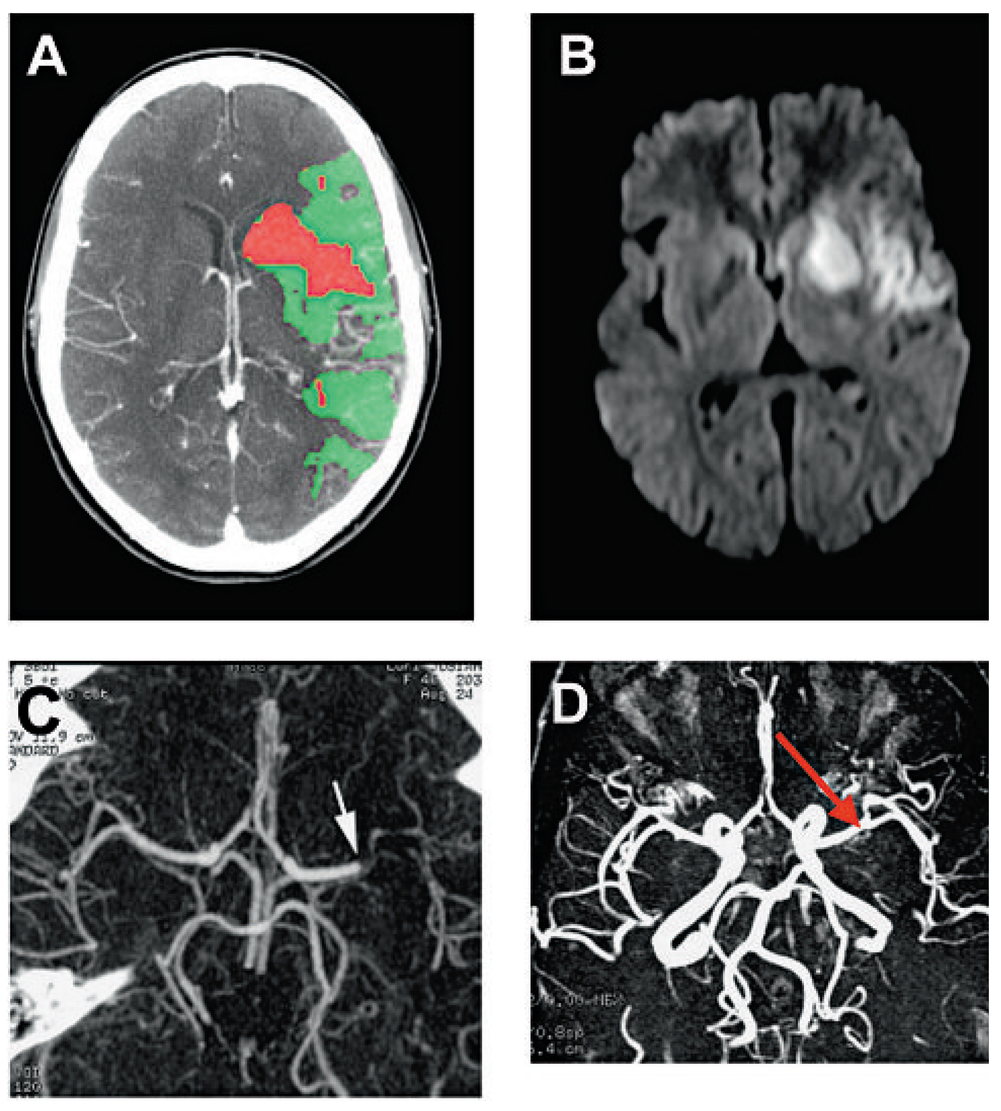
Figure 1. Ischaemic stroke in the left middle cerebral artery. A. Perfusion cerebral computerised tomography (CT) shows an ischaemic core (red) surrounded by penumbra (green) [52]. The core corresponds to the irreversible ischaemic insult (infarction), whereas the penumbra represents brain parenchyma which is suffering from ischaemia but which can be rescued if the blood perfusion is rapidly restored. B. The diffusion sequences on brain MRI show a hyperintensity in the territory of the left middle cerebral artery. Of note, only the infracted territory, which corresponds to the core in (A) is visible. C. Angiographic sequences of cerebral CT show a sub-occlusion of the left middle cerebral artery (arrow). D. The patient benefited from an intra-venous thrombolysis with recombinant tissue plasminogen activator (rtPA). Few days later, angiographic sequences on brain MRI showed a complete repermeabilisation of the left middle cerebral artery, which paralleled a favourable clinical outcome. (With courtesy of Dr A. Croquelois and the Division of Neuroradiology, Lausanne, Switzerland.).
Cardiovascmed 12 00143 g001
Figure 2. Diagram showing inflammation after stroke. Within minutes after cerebral ischaemia excitotoxic mechanisms are activated in neurons in the territory of the occluded artery which lead to apoptotic cell death independently of inflammatory mediators. These injured neurons both in the core (shown in red) and periphery (shown in green) of the lesion and glial cells in the core are producing pro-inflammatory mediators (such as cytokines and reactive oxygen species) which highly activate glial cells, leading to more cytokines and chemokines being released from activated microglia and astrocytes, and to an upregulation of adhesion molecules in vascular endothelial cells. Adhesion molecules and chemokines mediate the recruitement of circulating leukocytes to the vessel wall. Their infiltration or diapedesis into the ischaemic tissue occurs via highly specific receptor-ligand interactions between endothelial cells and leukocytes. In the next few hours after injury, once activated in the parenchyma, leukocytes and microglia (red boxes) produce more inflammatory mediators such as cytokines, nitric oxide (NO) through inducible nitric oxide synthase (iNOS), reactive oxygen species (ROS) and metalloproteinases (MMPs). Stress signalling pathways (JNKs) are also up-regulated and induce transcription of cytotoxic and inflammatory genes (blue boxes). This local inflammatory response contributes to secondary injury to potentially viable tissue and leads within hours and days to apoptotic or necrotic neuronal cell death (adapted from [12]).
Cardiovascmed 12 00143 g002
Figure 3. Activation and accumulation of microglia after cerebral ischaemia. Shown are examples of immunohistochemical staining for CD11b, an integrin surface marker on microglial cells, after 48 hours of reperfusion following 30 minutes middle cerebral artery occlusion (MCAo) in mice. Hyper-ramified (A, arrow heads) or activated microglia (A, white arrows) can be detected in the hippocampus (nuclei are stained in blue with DAPI). Phagocytotic microglia are present in the injured brain tissue (B, black arrows). Cresyl violet stained sections of ischemic brains are shown above, red boxes locate the immunolabelled ischaemic tissue in the hippocampus and the striatum shown in A and B. Magnification 20×. Accumulation of activated amoeboid microglia is found in the ischaemic striatum (C), while the contralateral healthy region shows no CD11b positive staining (D). The dashed red circle outlines the ischaemic tissue. Magnification 5×.
Cardiovascmed 12 00143 g003
Figure 4. Therapeutic interventions targeting the inflammatory response after ischaemia. Experimental and clinically tested approaches to reduce brain damage and inflammation after stroke. In red is shown thrombolysis used to dissolve the blood clot [3]. In black, strategies to block excitotoxic pathways, such as the inhibition of glutamate receptors [53] and JNK stress signaling pathway [33,34]. In blue are shown therapeutic interventions targeting inflammation, such as inhibitors of free radicals [41,54,55] and anti-inflammatory molecules [38,40,56,57,58]. And in pink other potential targets such as MMPs [59], PARP-1 [60,61], inhibitors of caspase-1 or caspase-3 [62,63] and erythropoietin (EPO) as a neuroprotective agent [64].
Cardiovascmed 12 00143 g004
Figure 5. Giant cell temporal arteriitis (Horton’s disease). A Angiographic CT of the head and neck showing skipped stenosis of the right temporal artery in a patient suffering from Horton’s disease. B Three-dimensional reconstruction precising the anatomy and the lesions of this artery. (With courtesy of the Division of Neuroradiology, Lausanne, Switzerland.).
Cardiovascmed 12 00143 g005
Disclaimer/Publisher’s Note: The statements, opinions and data contained in all publications are solely those of the individual author(s) and contributor(s) and not of MDPI and/or the editor(s). MDPI and/or the editor(s) disclaim responsibility for any injury to people or property resulting from any ideas, methods, instructions or products referred to in the content.