Next Article in Journal
La Pratique de L’échocardiographie en Suisse
Previous Article in Journal
Osborn Waves Due to Severe Hypothermia
 
 
Cardiovascular Medicine is published by MDPI from Volume 28 Issue 1 (2025). Previous articles were published by another publisher in Open Access under a CC-BY (or CC-BY-NC-ND) licence, and they are hosted by MDPI on mdpi.com as a courtesy and upon agreement with Editores Medicorum Helveticorum (EMH).
Communication

Recommendations for Quality Maintenance in Echocardiography

by
Cédric Vuille
1,*,
Christina Attenhofer Jost
2,
Peter Buser
3,
Xavier Jeanrenaud
4,
René Lerch
5,
Christian Seiler
6,
Pedro Trigo Trindade
2,
Michel Zuber
7 and
Writing Committee for the Swiss Society of Cardiology
1
Cardiac Imaging of the Swiss Society of Cardiology, Av. Viollier 8, CH-1260 Nyon, Switzerland
2
HerzGefässZentrum Im Park, Hirslanden Klinik Im Park, 8038 Zürich, Switzerland
3
Department of Cardiology, University Hospital, Basel, Switzerland
4
Lausanne University Hospital, Lausanne, Switzerland
5
Division of Cardiology, University Hospital, Geneva, Switzerland
6
Department of Visceral Surgery and Medicine, Universitätsspital Bern, Bern, Switzerland
7
Medecin adjoint Kardiologie, Kantonsspital, Luzern, Switzerland
*
Author to whom correspondence should be addressed.
Cardiovasc. Med. 2009, 12(1), 22; https://doi.org/10.4414/cvm.2009.01384
Submission received: 23 October 2008 / Revised: 23 November 2008 / Accepted: 23 December 2008 / Published: 23 January 2009

Introduction

Echocardiography is one of the most frequently performed examinations in cardiology, both in ambulatory care and in hospitals. The present recommendations are an update of the 1999 Guidelines for quality maintenance in echocardiography published by the Working Group of Echocardiography of the Swiss Society of Cardiology [1]. The following recommendations focus on transthoracic echocardiography in adults (Table 1). Transoesophageal echocardiography and stress echocardiography require additional training and proof of competence which is summarised in the last paragraph. Echocardiography in paediatric cardiology and perioperative transoesophageal echocardiography require special training which are not detailed in this document.

Equipment and imaging protocol

Echocardiography must be performed with up-to-date equipment with specific settings for cardiac examinations including harmonic imaging as well as continuous, pulsed and color Doppler. Digital recording is the preferred mode of documentation. Three-dimensional echocardiography, detailed echocardiographic assessment of cardiac resynchronisation (CRT) and myocardial perfusion imaging are considered “advanced techniques”.
A standard echocardiographic examination is performed using two-dimensional and M-mode imaging, colour flow Doppler and spectral Doppler. It includes parasternal long and short axis views; apical 4-chamber, 2-chamber, “5-chamber” and long axis views; subcostal and suprasternal views; with Doppler analysis of all four valves. Left ventricular size and wall thickness, left atrial size, aortic root size, aortic valve flow velocity, tricuspid regurgitation velocity and mitral valve flow profile should be measured in each patient. Additional measurements should be made for the assessment of severity of pathologic findings in compliance with published recommendations of the European Association of Echocardiography and American Society of Echocardiography [2]. Left ventricular contrast may be needed to improve endocardial border delineation [3]. Saline contrast should be available to detect right-to-left shunt flow.
The examinations must be adequately documented and archived digitally (VCR tapes, if digital storage not yet available). An acquisition protocol with 33 loops and 10 still frames is provided as an example by the American Society of Echocardiography [4]. A structured report must be established for each echocardiographic examination. Each report includes the indication for the study, quality of the study, height, weight, body surface area, blood pressure, heart rate and rhythm of the patient. It provides a description of the four chambers, the four valves, the pericardium and all additional relevant findings. It includes measurements of left ventricular size and function, wall thickness, left atrial and aortic root size and relevant Doppler measurements. The conclusion must include the pathological findings, interpret the results and answer the question of the request. A clinical comment may be added when appropriate. The report is signed by the physician responsible for interpretation of the examination.

Training in echocardiography

Echocardiography should be performed by a cardiologist, since correct, detailed and complete interpretation of echocardiography requires advanced knowledge and training in cardiology. Therefore training in echocardiography, in Switzerland, is an integral part of training in cardiology. During the 4-year specialised training in cardiology the trainee has to perform and interpret at least 500 transthoracic examinations. The examinations should be performed in teaching echocardiography laboratories with a case load of at least 2000 studies per year. Knowledge and practical skills in echocardiography are evaluated during the board examination (one of the three parts of the examination).
Physicians who received post-graduate training in cardiology and echocardiography abroad should provide proof of their training fulfilling these requirements.

Maintenance of competence

In order to maintain their echocardiographic skills cardiologists should perform at least 200 complete transthoracic examinations per year. It is recommended to be a member of a regional group of the Working Group of Echocardiography and Cardiac Imaging. In addition 10 hours of continuing medical education in echocardiography should be completed annually, at meetings dedicated to echocardiography and recommended by the Swiss Society of Cardiology or by other national or international cardiology societies, such as the annual scientific session of the Working Group of Echocardiography and Cardiac Imaging.
Each cardiologist keeps records of the studies she/he performed, and of the meetings she/he attended.
Non cardiologists performing echocardiography according to their “acquired rights” (registered for TARMED) should meet the same requirements of maintenance of competence as cardiologists.

Transoesophageal and stress echocardiography

Training in transoesophageal and stress echocardiography requires complete training in transthoracic echocardiography.
For the performance of transoesophageal echocardiography the trainee has to perform at least 75 studies under supervision in a training laboratory performing at least 200 transoesophageal studies per year. For maintenance of competence at least 50 examinations should be performed per year.
Training in stress echocardiography should include the performance of at least 75 studies under supervision. The training laboratory should have a case load of at least 150 stress echocardiograms per year. To ascertain maintenance of competence at least 50 studies should be performed per year.

References

  1. Groupe de travail Echocardiographie; Société Suisse de Cardiologie. Directives pour la garantie de qualité en échocardiographie. Richtlinien zur Qualitätssicherung in der Echokardiographie. Kardiovaskuläre Medizin. 1999, 2, 50–55. [Google Scholar]
  2. Lang, R.M.; Bierig, M.; Devereux, R.B.; et al. Recommendations for chamber quantification: a report from the American Society of Echocardiography’s Guidelines and Standards Committee and the Chamber Quantification Group, developed in conjunction with the European Association of Echocardiography, a branch of the European Society of Cardiology. J Am Soc Echocardiogr. 2005, 18, 1440–1463. [Google Scholar] [PubMed]
  3. Mulvagh, S.L.; DeMaria, A.N.; Feinstein, S.B.; et al. Contrast echocardiography: current and future applications. J Am Soc Echocardiogr. 2000, 13, 331–342. [Google Scholar] [CrossRef] [PubMed]
  4. Writing Committee. Thomas, J.D.; Adams, D.B.; De Vries, S.; et al. Guidelines and recommendations for digital echocardiography. A Report from the Digital Echocardiography Committee of the American Society of Echocardiography. J Am Soc Echocardiogr. 2005, 18, 287–297. [Google Scholar]
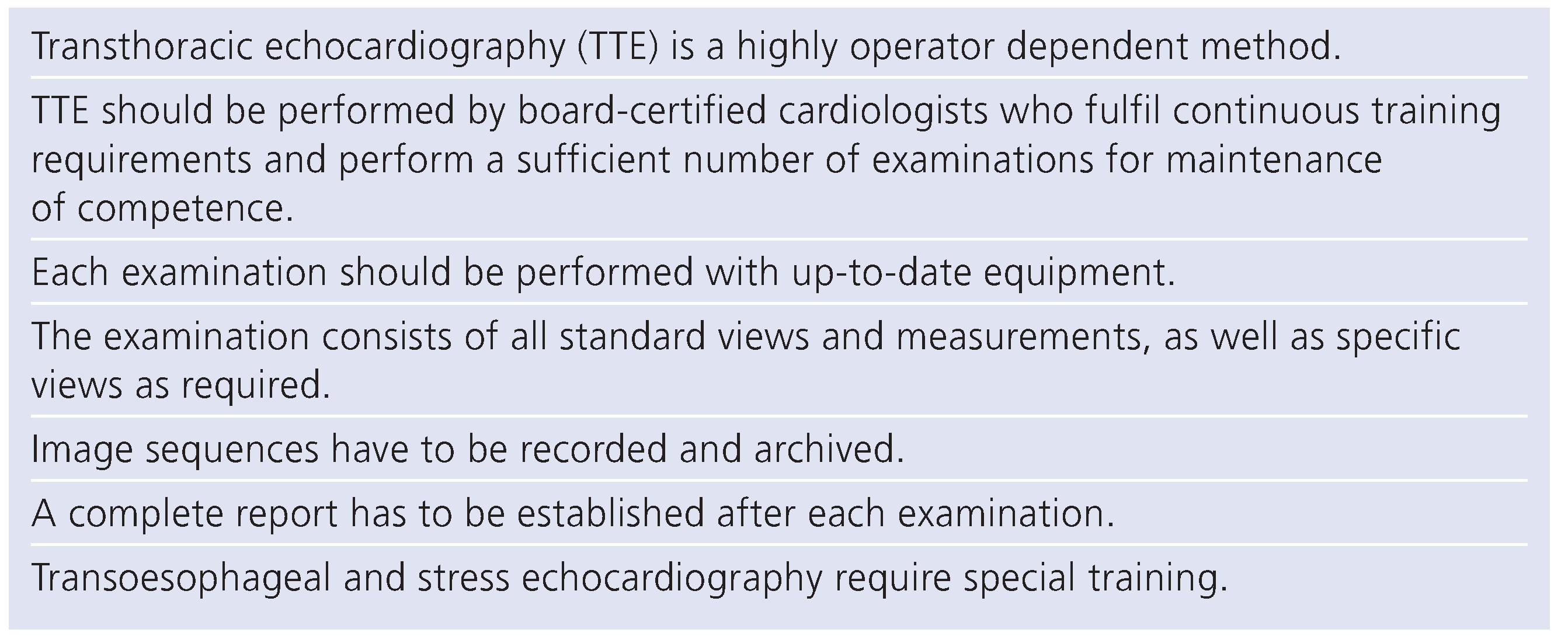
Table 1. Key issues for quality care in echocardiography.
Cardiovascmed 12 00022 i001