Next Article in Journal
Jahresbericht 2007 der Arbeitsgruppe «Herzschrittmacher und Elektrophysiologie»
Previous Article in Journal
La Pratique de L’échocardiographie en Suisse
 
 
Cardiovascular Medicine is published by MDPI from Volume 28 Issue 1 (2025). Previous articles were published by another publisher in Open Access under a CC-BY (or CC-BY-NC-ND) licence, and they are hosted by MDPI on mdpi.com as a courtesy and upon agreement with Editores Medicorum Helveticorum (EMH).
Editorial

Pulse: From Very Young to Very Old †

by
Jean-Pierre Pfammatter
University Children’s Hospital, Inselspital, Berne, Switzerland
This article summarises a lecture at the annual meeting of the Swiss Society of Cardiology in Berne, May 2008.
Cardiovasc. Med. 2009, 12(1), 13; https://doi.org/10.4414/cvm.2009.01382
Submission received: 23 October 2008 / Revised: 23 November 2008 / Accepted: 23 December 2008 / Published: 23 January 2009

Abstract

Types of arrhythmias are the same throughout all age groups. There are however differences with regard to the epidemiology of arrhythmias, clinical tolerance, therapeutic strategies and comorbidities. One typical difference is the predominance of accessory pathways as the causative mechanism for supraventricular tachycardias in infancy and early childhood compared to the clear preponderance of AV-nodal re-entrant tachycardias in adult patients. Another important difference is the rarity of ventricular arrhythmias in children whereas in adults, ventricular tachycardias largely predominate, mainly due to an increase in the incidence of coronary artery disease. The overall prognosis of arrhythmias is better in children first due to the relative rarity of ventricular arrhythmias and second due to an improved clinical tolerance to arrhythmias in children because of the lack of the multiple comorbid conditions typically found in adult patients.

Introduction

This review will not deal with philosophical or physiological aspects of the human heartbeat in the different age groups but will focus on irregularities of the pulse and highlight the divergent or common features of arrhythmias through the process of human aging.

The physiological basis

As an organ, the cardiac conduction system is established anatomically as well as functionally at birth. Thus there are almost no developmental variations in the different forms of disturbances of cardiac conduction, this means that the types of arrhythmias that are clinically observed, basically are the same throughout all age groups. The differences that are observed between age groups first are due to differences in the predominance of specific types of arrhythmias at various stages of human growth and aging. Then there are differences in the underlying anatomic substrate resulting in differences in the risk profile of arrhythmias and a different clinical tolerance of arrhythmias and this is widely influenced by co-morbidities acquired during life and their impact on cardiac function.

Distribution of types of arrhythmias trough all ages

Arrhythmias are caused by different intrinsic anomalies or extrinsic factors influencing the conduction system, these are:
  • congenital malformations of the conduction system
  • acquired disturbances of the conduction system
  • predisposing genetic disease (channelopathies, cardiomyopathies)
  • alterations in cardiac haemodynamics
  • aging of the heart itself
  • co-morbidities
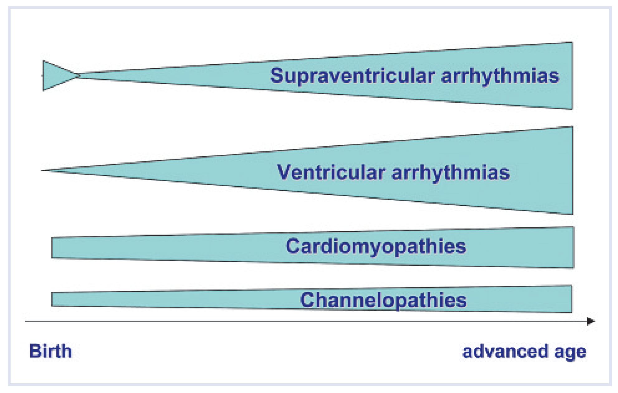
These factors at different ages have a specific impact on the cardiac conduction system and contribute to the different spectrum of arrhythmias encountered at the various stages of human life. First there are congenital malformations of the conduction system such as accessory atrioventricular (AV) pathways or ectopic foci acting as a concurrent pacemaker. The conduction axis may be damaged throughout life mechanically (perfusion deficit, cardiac surgery), and all the comorbidities acquired during a lifetime (e.g., hypertension, diabetes, coronary artery disease ) may further either be the cause of arrhythmias or by impairing heart function lead to a worse clinical tolerance of any given arrhythmia at a later age. The process of aging of the heart itself, by a variety of mechanisms such as decreased protein expression (connexins and calcium channels) [1, 2] or myocardial fibrosis [3] leads to impairment of electrophysiological parameters [4] and thus aging of the heart itself literally is a proarrhytmic process of “aging to arrhythmias” as shown in Figure 1 and Figure 2. It has been found that by the age of 100 years, otherwise healthy individuals rarely (women) or never (men) had a normal resting ECG [5].
Based on these various influences which act progressively during a lifetime the frequency of specific arrhythmias varies considerably between age groups. This is best shown by the increase of either supraventricular and even more ventricular arrhythmias at increasing age.

Supraventricular arrhythmias in the different age groups
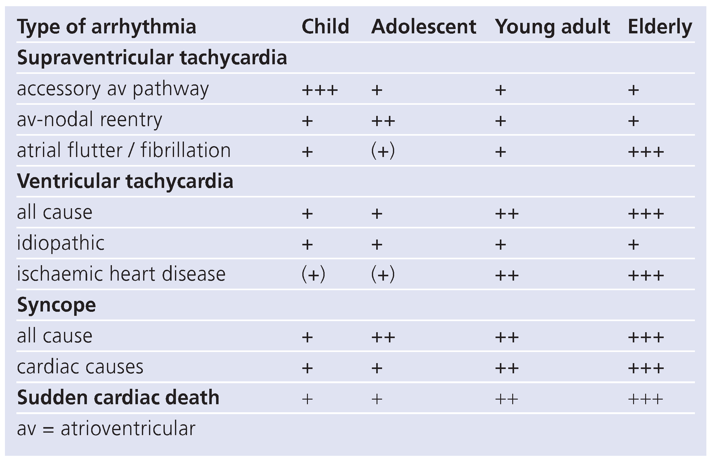
A unique feature is the observation of arrhythmias already in intrauterine life, where supraventricular tachycardia (SVT) due to atrial flutter or due to accessory av-pathways are observed in approximately one out of 2000 pregnancies [6]. Except for neonatal atrial flutter, whose aetiology still remains to be elucidated, atrial flutter or fibrillation is exceedingly rare in childhood. SVT mediated by av-reentry on the other hand is by far the most frequently encountered arrhythmia in infancy and childhood. SVT in infancy has a characteristic epidemiology due to specific features of maturation of the conduction system. As it was shown in experimental models [7, 8]. The electrical isolation of the atrioventricular junction is an ongoing process from intrauterine life to the perinatal period and early infancy with gradual loss of electrical properties of accessory av-connections. This explains the large predominance of SVT due to accessory av-pathways in this age group [9] with only a small minority of children having avnodal reentry as the mechanism underlying the clinical SVT. This also explains the excellent prognosis that is shared by most of the neonates and infants with SVT early in life [10]. The clinical observation of “outgrowing” of the SVT-episodes as it is very frequently observed after neonatal SVT is thus based on the delayed maturation of av-accessory pathways that increasingly loose their electrical properties after birth leaving the affected baby without an anatomic substrate for recurrencies of the SVT. The proportion of SVT patients having AV-nodal reentry increases with age and already in the teenage group is substantial, even more so in adulthood. This predominance of av-nodal reentry is even more pronounced due to the fact, that nowadays we witness an “unnatural” history of accessory pathways as most of the patients with preexitation undergo curative ablation already during childhood.
A notable difference between childhood and adult SVT lies in the fact, that clinical tolerance of tachycardia is better in children as compared to adults due to often coexisting organic heart disease in the latter. In older age groups symptoms due to SVT occur earlier and often are more severe with for instance a high rate of syncopes which has been observed in about 40% of elderly patients due to SVT [11].
The antiarrhythmic substances used are the same whether you treat a newborn or an adult patient. The therapeutic strategy differs, especially in the infant age group. As SVT often remains undetected until an infant becomes symptomatic, it is the rule to give an oral antiarrhythmic prophylaxis to any newborn or infant that has experienced SVT usually until the end of the first year of life. Due to the benign spontaneous course of the disease, curative ablation in the age group up to about 5 years usually is only an option in the rare case where SVT cannot be controlled medically, but when indicated can be performed safely and with high efficacy even in premature neonates [12, 13]. Ablation in paediatric patients has the same success rates and the same low complication rate as in adult patients except for small patients where a slightly higher complication rate is observed [14]. At the other end of the spectrum, also in the elderly patients, ablation offers a safe and efficient curative therapy [15].

Ventricular arrhythmias in the different age groups

Ventricular tachycardia (VT) is a very rare observation in a paediatric population. A recent study has found that in an unselected paediatric population spontaneously occuring VT was seen on only 1/100 000 children per year [16]. The factor of most prognostic importance in the presence of VT is, whether VT is occurring in a patient with a normal heart or in the context of underlying heart disease. So called idiopathic VT in patients with structurally normal hearts is encountered in all paediatric and adult age groups, but in childhood, idiopathic VT accounts for a high proportion of about half of all cases of spontaneous VT [16, 17]. Prognosis of childhood idiopathic VT generally was reported to be very good [18]. Prognosis was much worse for VT in patients with any kind if underlying heart disease with mortality already in childhood of up to 36% [16, 17]. The main difference between children and adults is that the proportion of patients with VT due to organic heart disease is clearly increasing in with increasing age, mainly due to ischaemic heart disease [19]. Together with the worse clinical tolerance of VT in a polymorbid adult patient, overall prognosis of VT in an adult population is worse than it is for children, especially due to the significantly increased proportion of patients with organic heart disease underlying VT. Besides the treatment of the underlying organic heart disease, the antiarrhythmic treatment strategies do not differ much between paediatric and adult patients, be it for medical treatment or for curative ablation and device therapy.
Table 1. Overview of relative frequency of different types / aetiologies of arrhythmias in the different age groups.
Cardiovascmed 12 00013 i001

Rhythmologic emergencies in the different age groups

Real emergencies due to arrhythmias are relatively rare in paediatrics. Urgent situations may arise with intrauterine arrhythmias especially bradycardia due to total av-block or tachycardia – induced cardiac failure (hydrops) in SVT. Intrauterine treatment of arrhythmias is complicated by the surrounding mother and there is a considerable mortality associated with hydropic presentation of intrauterine arrhythmias [20]. Neonatal / infant SVT may present as an emergency in case the tachycardia was detected late and only by the occurrence of symptoms, in which context a baby may present with advanced cardiac failure. Ventricular arrhythmias presenting as emergencies are quite rare in childhood and generally limited to patients with cardio-myopathy or channelopathies or else children with operated congenital heart disease.
In adult patients ventricular arrhythmias presenting as emergencies represent a substantial number of all arrhythmia patients, with ischaemic heart disease as the leading cause [19] but with the whole spectrum of organic heart disease being represented. Worse clinical tolerance of arrhythmias translates into significant number of adult patients presenting with syncopal episodes as the leading symptom of SVT [11]. Again a significant number of patients present with syncopes due to bradycardia in sinoatrial nodal dysfunction or high grade av-block, entities only exceptionally encountered in a paediatric population.

Syncope in childhood and old age

The incidence of syncope significantly increases with advancing age with relevant differences in the underlying causes leading to syncopal events.
In young children (below 10 years of age) neurocardiogenic syncopes are the exception and other causes definitely have to be ruled out (epileptic seizures, breath-holding spells, cardiac syncope). In the teenage age group in contrast, neurocardiogenic syncopes largely predominate with a significant female preponderance [21] and represent 75% of all syncopal events. In clear contrast to what is true for adult and elderly patients, a cardiac cause of a syncope in childhood is found in only 2–5% of instances [22]. This proportion of underlying cardiac cause for a syncope rises to 12% in young adults and to over 30% in elderly people [23, 24]. In children the underlying causes mainly are cardiomyopathies and channelopathies or more rarely congenital coronary artery anomalies, whereas in adults coronary artery disease, sinoatrial nodal dysfunction, SVT, av-block and valvar heart disease add to this list.

Sudden cardiac death throughout all ages

The only difference between syncope of cardiac origin and sudden cardiac death is that in the latter you don’t wake up again. Thus most of the above discussion on syncope applies also in this section and needs not to be repeated. Again sudden cardiac death is a very rare event in young people and coronary artery disease which by numbers is only an irrelevant cause of death in children, becomes largely predominant in the adult and elderly population [24]. Prevention strategies basically do not differ in the various age groups. In case of a high-risk profile in a paediatric patient the implantation of an internal defibrillator in already small children is feasible and effective [25] just as in any adult patient. Whereas the main indication in adults is ischaemic heart disease, in children the indication usually is in a child with a high risk for sudden death due to cardiomyopathy, channelopathy or operated congenital heart disease.
An interesting study recently found that in a place where the different generations usually meet in daily life, in schools, only 10% of sudden deaths observed, occurred in children but 90% affecting teachers and other adults, reflecting nicely the different risk profile in different age groups for the occurrence of sudden cardiac death [26].

Conflicts of Interest

There is no conflict of interest.

References

  1. Jones, S.A.; Boyett, M.R.; Lancaster, M.K. Declining into failure: the age dependent loss of L-type calcium channel within the sinoatrial node. Circulation. 2007, 115, 1183–1190. [Google Scholar] [CrossRef]
  2. Kostin, S.; Klein, G.; Szalay, Z.; Hein, S.; Bauer, E.P.; Schaper, J. Structural correlate of atrial fibrillation in human patients. Cardiovasc Res. 2002, 54, 361–379. [Google Scholar] [CrossRef]
  3. Stein, M.; Noorman, M.; van Veen, T.; Herold, E.; Engelen, M.A.; Boulaksil, M.; et al. Dominant arrhythmia vulnerability of the right ventricle in senescent mice. Heart Rhythm. 2008, 5, 438–448. [Google Scholar] [CrossRef]
  4. Kojodjojo, P.; Kanagaratnam, P.; Markides, V.; Davies, W.; Peters, N. Age related changes in human left and right atrial conduction. J Cardiovasc Electrophysiol. 2006, 17, 120–127. [Google Scholar] [CrossRef]
  5. Lakireddy, D.R.; Clark, R.A.; Mohiuddin, S.M. Electrocardiographic findings in patients >100 years of age without clinical evidence of cardiac disease. Am J Cardiol. 2003, 92, 1249–1251. [Google Scholar] [CrossRef]
  6. Hornberger, L.K.; Sahn, D.J. Rhythm abnormalities of the fetus. Heart. 2007, 93, 1294–1300. [Google Scholar] [CrossRef]
  7. Kolditz, D.P.; Wijffels, M.C.; Blom, N.A.; van der Larse, A.; Markwald, R.R.; Schalij, M.J.; et al. Persistence of functional atrioventricular accessory pathways in postseptated embryonic avian hearts. Circulation. 2007, 115, 17–26. [Google Scholar] [CrossRef]
  8. Hahurij, N.D.; Gittenberger de Groot, A.C.; Kolditz, D.P.; Bökenkamp, R.; Schalij, M.J.; Poelmann, R.E.; et al. Accessory atrioventricular myocardial connections in the developing human heart. Circulation 2008, 117, 2850–2858. [Google Scholar] [CrossRef]
  9. Ko, J.K.; Deal, B.J.; Strasburger, J.F.; Benson, W.D. Supraventricular tachycardia mechanisms and their age distribution in pediatric patients. Am J Cardiol. 1992, 69, 1028–1032. [Google Scholar] [CrossRef]
  10. Tortoriello, T.A.; Snyder, C.S.; Smith, E.; Fenrich, A.L.; Friedman, R.A.; Kertesz, N.J. Frequency of recurrence among infants with supraventricular tachycardia and comparison of recurrence rates among those with and without preexcitation. Am J Cardiol. 2003, 92, 1045–1049. [Google Scholar] [CrossRef]
  11. Kalusche, D.; Ott, P.; Arentz, T.; Stockinger, J.; Betz, P.; Roskamm, H. AV nodal reentry tachycardia in elderly patients: clinical presentation and results of radiofrequency catheter ablation. Coron Artery Dis. 1998, 9, 359–363. [Google Scholar] [CrossRef]
  12. Blaufox, A.D.; Paul, T.; Saul, J.P. Radiofrequency catheter ablation in small children. PACE. 2004, 27, 224–229. [Google Scholar] [CrossRef]
  13. Kolditz, D.P.; Blom, N.A.; Bökenkamp, R.; Schalij, M.J. Low-energy radiofrequency catheter ablation as therapy for supraventricular tachycardia in a premature neonate. Eur J Pediatr. 2005, 164, 559–562. [Google Scholar] [CrossRef]
  14. Bauersfeld, U.; Pfammatter, J.P.; Jaeggi, E. Treatment of supraventricular tachycardias in the new millenium: drugs or radiofrequency catheter ablation? Eur J Pediatr. 2001, 160, 1–9. [Google Scholar] [CrossRef]
  15. Meiltz, A.; Zimmermann, M. Atrioventricular nodal reentrant tachycardia in the elderly: efficacy and safety of radiofrequency catheter ablation. PACE. 2007, 30 (Suppl), S103–S107. [Google Scholar] [CrossRef]
  16. Roggen, A.; Pavlovic, M.; Pfammatter, J.P. Frequency of spontaneous ventricular tachycardia in a pediatric population. Am J Cardiol. 2008, 101, 852–854. [Google Scholar] [CrossRef]
  17. Davis, A.M.; Gow, R.M.; McCrindle, B.W.; Hamilton, R.M. Clinical spectrum, therapeutic management and follow-up of ventricular tachycardia in infants and young children. Am Heart J. 1996, 131, 186–191. [Google Scholar] [CrossRef]
  18. Pfammatter, J.P.; Paul, T. Idiopathic ventricular tachycardia in infancy and childhood. J Am Coll Cardiol. 1999, 33, 2067–2072. [Google Scholar] [CrossRef]
  19. Henkel, D.M.; Witt, B.J.; Gersh, B.J.; Jacobsen, S.J.; Weston, S.A.; Meverden, R.A.; et al. Ventricular arrhythmias after acute myocardial infarction: a 20 year community study. Am Heart J. 2006, 151, 806–812. [Google Scholar] [CrossRef]
  20. Simpson, J.M.; Garland, G.K. Fetal tachycardias: management and outcome of 127 consecutive cases. Heart. 1998, 79, 576–581. [Google Scholar] [CrossRef]
  21. Driscoll, D.J.; Jacobsen, S.J.; Porter, C.J.; Wollan, P.C. Syncope in children and adolescents. J Am Coll Cardiol. 1997, 29, 1039–1045. [Google Scholar] [CrossRef]
  22. Massin, M.M.; Bourguignont, A.; Coremans, C.; Comte, L.; Lepage, P.; Gerard, P. Syncope in pediatric patients presenting to an emergency department. J Pediatr. 2004, 145, 223–228. [Google Scholar] [CrossRef]
  23. Del Rosso, A.; Alboni, P.; Brignole, M.; Menozzi, C.; Raviele, A. Relation of clinical presentation of syncope to the age of patients. Am J Cardiol. 2005, 96, 1431–1435. [Google Scholar]
  24. Zipes, D.P.; Wellens, H.J.J. Sudden cardiac death. Circulation. 1998, 98, 2334–2351. [Google Scholar]
  25. Bauersfeld, U.; Tomaske, M.; Dodge-Khatami, A.; Rahn, M.; Kellenberger, C.J.; Pretre, R. Initial experience with implantable cardioverter defibrillator systems using epicardial and pleural electrodes in pediatric patients. Ann Thorac Surg. 2007, 84, 303–305. [Google Scholar] [CrossRef]
  26. Lotfi, K.; White, L.; Rea, T.; Cobb, L.; Copass, M.; Yin, L.; et al. Cardiac arrests in school. Circulation 2007, 116, 1374–1379. [Google Scholar] [CrossRef]
Figure 1. Schematic drawing showing the overall increase in the arrhythmia burden through human growing and aging.
Cardiovascmed 12 00013 g001
Figure 2. Schematic drawing showing the increase in the burden of arrhythmias separated for different aetiologies throughout human aging.
Cardiovascmed 12 00013 g002