Next Article in Journal
Jugendlicher Mit Einer Breitkomplextachykardie
Previous Article in Journal
CETP-Inhibitoren (Torcetrapib und JTT-705)
 
 
Cardiovascular Medicine is published by MDPI from Volume 28 Issue 1 (2025). Previous articles were published by another publisher in Open Access under a CC-BY (or CC-BY-NC-ND) licence, and they are hosted by MDPI on mdpi.com as a courtesy and upon agreement with Editores Medicorum Helveticorum (EMH).
Interesting Images

Six Simultaneously Employed Methods to Gauge the Coronary Collateral Flow of the Decade

by
Steffen Gloekler
,
Tobias Rutz
and
Christian Seiler
*
Department of Cardiology, University Hospital, CH-3000 Bern, Switzerland
*
Author to whom correspondence should be addressed.
Cardiovasc. Med. 2007, 10(9), 298; https://doi.org/10.4414/cvm.2007.01264
Submission received: 28 June 2007 / Revised: 28 July 2007 / Accepted: 28 August 2007 / Published: 28 September 2007

Case report

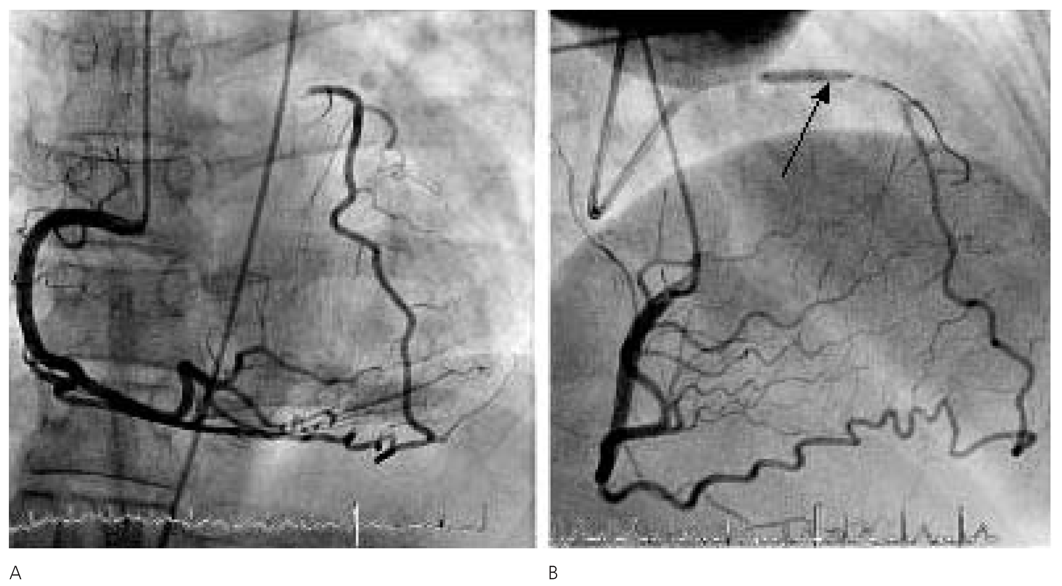
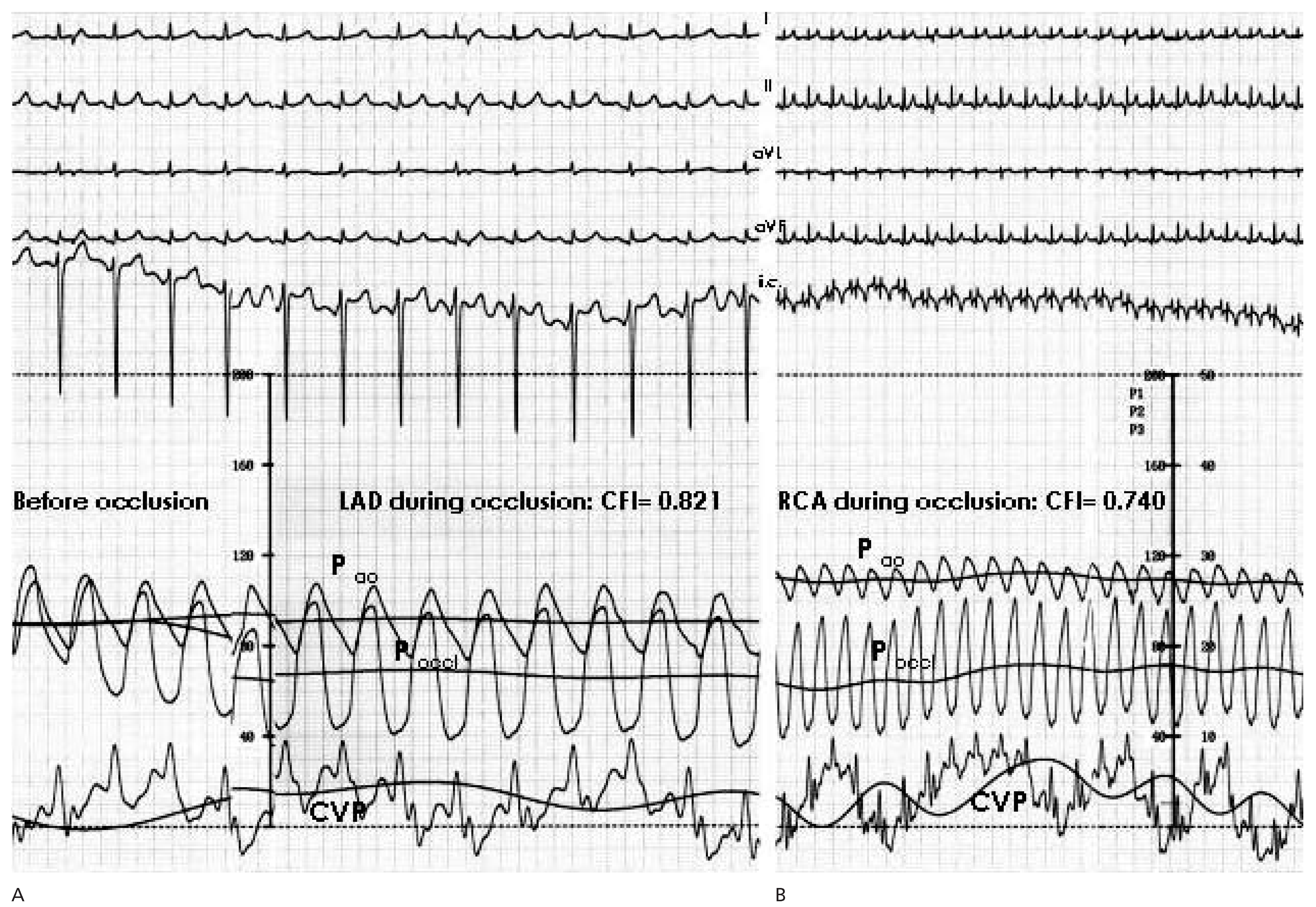
A 59-year-old woman with bronchial asthma and obesity underwent coronary angiography because of exertional dyspnea and atypical chest pain both at rest and sometimes during exertion. Coronary angiography revealed a chronic total occlusion (CTO) of the proximal left anterior descending (LAD) artery (Figure 1A). Systolic left ventricular (LV) function was normal (Figure 1B,C). Angiography of the right coronary artery showed a large collateral artery to the LAD and some smaller septal collateral vessels (Figure 2A). The very well developed coronary collateral circulation protected the patient entirely from a large anterior myocardial infarction. Moreover, it was likely sufficient to prevent angina pectoris even under physical exertion. There are more than half a dozen methods for collateral assessment, six of them performed in this particular patient. The presence of normal LV anterior wall motion in the presence of a CTO of the LAD qualifies collaterals as relevant enough to prevent myocardial infarction (Figure 1B,C). The coronary angiographic assessment, first described by Rentrop et al., qualifies naturally or artificially (Figure 2B) occluded coronary arteries according to the degree (0–3) of retrograde filling by collateral vessels (grade 3 as in the present case with entire filling of the balloon-occluded LAD) [1]. Two further methods for the characterisation of well developed collaterals during a brief coronary balloon occlusion are the absence of ST-segment changes in the intracoronary ECG (Figure 3), and the lack of angina pectoris. Measurement of invasively derived collateral flow index (CFI) or contrast-echocardiography-obtained collateral perfusion index (CPI) provide quantitative measures of the human coronary collateral circulation [2,3]. Both methods employ the described coronary artery balloon-occlusion model. Distal to the occlusion, coronary wedge pressure (poccl) is determined by a pressure sensor-tipped guide wire. This value minus the central venous pressure divided by mean aortic pressure (pao) minus central venous backpressure is equal to CFI (Figure 3A,B) [2]. Simultaneously, absolute myocardial blood flow can be determined by myocardial contrast echocardiography (MCE) in the collateralised territory and in an adjacent normal region (ie, the ratio is equal to CPI) [3].
In the present case, all six methods to gauge coronary collateral channels have been employed and provide evidence for their outstanding structure and function. Absolute flow in the collateralised anterior myocardial area was 1.003 mL/min/g at rest and 1.210 mL/min/g during adenosine induced hyperaemia (= coronary flow reserve in the collateralised region of 1.20).
Coronary collateral resistance (Rcoll) to flow (equal to pao minus poccl divided by the myocardial blood flow) in the present case was 53.17 dyn × sec × cm−5 in the collateralised LAD territory, and it was 105.61 dyn × sec × cm−5 in the balloon-occluded RCA area. CFI and CPI in the collateral-receiving LAD territory were 0.821, respectively 0.891. In the collateral-supplying RCA territory CFI and CPI were 0.740, respectively 0.651. Compared to our database including approximately 1100 CFI measurements, only 4 values were larger than 0.780, and the largest CFI value obtained so far using intracoronary Doppler measurement was 0.870.

Conflicts of Interest

The authors declare no conflict of interest.

References

  1. Rentrop, K.P.; Cohen, M.; Blanke, H.; Phillips, R.A. Changes in collateral channel filling immediately after controlled coronary artery occlusion by an angioplasty balloon in human subjects. J Am Coll Cardiol. 1985, 5, 587–592. [Google Scholar] [CrossRef] [PubMed]
  2. Seiler, C.; Fleisch, M.; Garachemani, A.; Meier, B. Coronary collateral quantitation in patients with coronary artery disease using intravascular flow velocity or pressure measurements. J Am Coll Cardiol. 1998, 32, 1272–1279. [Google Scholar] [CrossRef] [PubMed]
  3. Vogel, R.; Zbinden, R.; Indermuhle, A.; Windecker, S.; Meier, B.; Seiler, C. Collateral-flow measurements in humans by myocardial contrast echocardiography: Validation of coronary pressure-derived collateral-flow assessment. Eur Heart J. 2006, 27, 157–165. [Google Scholar] [CrossRef] [PubMed]
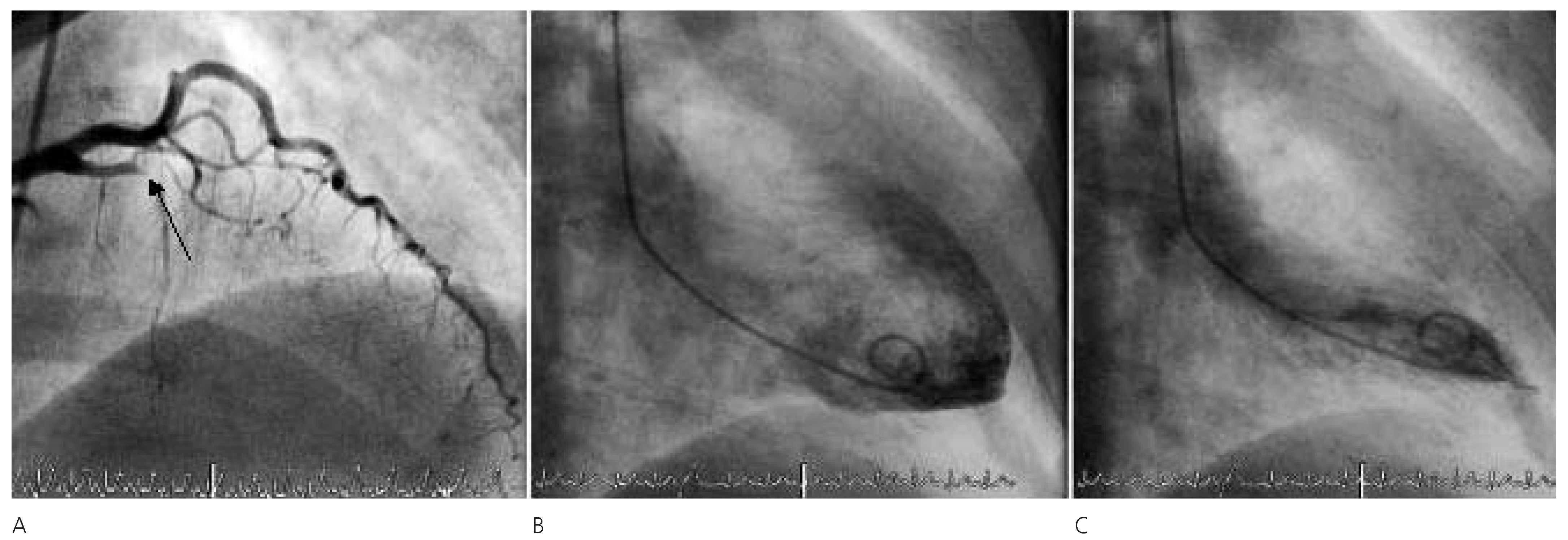
Figure 1. (A) Chronic total occlusion (arrow) of left anterior descending artery (LAD). (B) End-diastolic left ventricular angiogram. (C) End-systolic left ventricular angiogram.
Cardiovascmed 10 00298 g001
Figure 2. (A) Right coronary artery (RCA) with large collateral branch to the chronically occluded left anterior descending artery (LAD). (B) Complete filling of the balloon-occluded left anterior descending artery (arrow) during simultaneous dye injection into the right coronary artery via a second coronary catheter.
Cardiovascmed 10 00298 g002
Figure 3. Determination of collateral function in the left anterior descending artery (LAD) (A), and in the right coronary artery (RCA) territory after recanalisation of the chronic LAD occlusion (B). Collateral flow index (CFI) is calculated as mean distal coronary occlusive pressure (poccl, mm Hg; scales 0–200 mm Hg). minus central venous pressure (CVP, mm Hg; scale 0–50 mm Hg) divided by mean aortic pressure (pao, mm Hg; scale 0–200 mg Hg), minus CVP. CFI in the collateral-receiving (LAD) as well as supplying vessel is sufficient to prevent myocardial ischaemia during six minutes and 20 s of coronary balloon occlusion (no ST segment elevations on the intracoronary [i.c.] as well as peripheral ECG leads).
Cardiovascmed 10 00298 g003