Article

Mushroom β-Glucan as a Novel Prebiotic: Enhancing Recovery of the Post-Antibiotic Gut Microbiota over 60 Days


by
Emanuel Vamanu
1,2,*,
Laura Dorina Dinu
1,
Elisabeta-Irina Geană
3,
Corina Teodora Ciucure
3,
Alexandru Cristian Grosu
1,
Răzvan Roșca
2 and
Florentina Gatea
4
1
Department of Industrial Biotechnology, Faculty of Biotechnology, University of Agricultural Sciences and Veterinary Medicine, 59, Marasti Blvd., 011464 Bucharest, Romania
2
Anoom Laboratories SRL, 077190 Voluntari, Romania
3
National Research & Development Institute for Cryogenics and Isotopic Technologies (ICSI Rm. Valcea), 4th Uzinei Street, 240050 Râmnicu Vâlcea, Romania
4
Ecotoxicology, Physiology and Biotechnology Laboratory, Department of Agricultural Biotechnologies and Bioresources, Research Development Institute for Plant Protection Bucharest, 8 Ion Ionescu de la Brad Blvd., District 1, 013813 Bucharest, Romania
*
Author to whom correspondence should be addressed.
Nutraceuticals 2026, 6(3), 54; https://doi.org/10.3390/nutraceuticals6030054
Submission received: 3 June 2026 / Revised: 9 July 2026 / Accepted: 31 July 2026 / Published: 17 August 2026

Abstract

Antibiotic-induced dysbiosis can cause persistent alterations in gut microbial composition and fermentative metabolism, yet the long-term role of mushroom β-glucan-based prebiotics in supporting post-antibiotic microbiota modulation remains poorly defined. To address this gap, the present study evaluated the modulatory effects of ColonX, a mushroom β-glucan-based formulation, during a 60-day in vitro simulation of post-antibiotic gut microbiota modulation. Quantitative PCR (qPCR) was used to monitor key bacterial groups, while UHPLC-DAD analysis was applied to characterize fermentation-derived organic acids. ColonX administration produced a selective, time-dependent increase in Bifidobacterium spp., with limited effects on Lactobacillus spp. and no stimulation of opportunistic bacteria such as Escherichia coli. This response became more evident after prolonged administration, suggesting progressive adaptation of the dysbiotic microbiota. Metabolomic analysis showed increased production of short-chain fatty acids and other fermentation-derived organic acids, indicating enhanced saccharolytic activity and functional metabolic remodeling. The accumulation of succinic acid further suggested ongoing microbial metabolic restructuring during recovery, while comparison with individual excipients indicated that resistant dextrin contributed to the fermentative response. Overall, this study addresses an important gap by linking prolonged mushroom β-glucan administration with both taxonomic modulation and functional metabolic recovery markers in a post-antibiotic dysbiosis model. These findings support ColonX as a promising nutraceutical strategy to promote gut microbiota restoration following antibiotic exposure.

1. Introduction

Antibiotics are indispensable in medical practice; however, their use frequently disrupts the gut microbiome, leading to dysbiosis [1]. Microbial balance is the target, but antibiotic use can reduce beneficial populations, promote opportunistic microorganisms, and alter metabolic activity. These effects often persist, characterized by recurrent bloating, irregular bowel habits, and reduced food tolerance, indicating incomplete recovery of the microbiota [2]. Nutritional strategies to support post-antibiotic recovery have attracted growing attention, and prebiotics have emerged as a significant option in this context [3].
Prebiotics are selectively utilized by gut microorganisms, leading to the production of metabolites relevant to colonic health. A primary mechanism is the fermentation of non-digestible carbohydrates, which produces short-chain fatty acids (SCFAs) [4]. These compounds regulate colonic pH and promote an ecological balance favorable to beneficial microbial communities [5]. Prebiotic effects depend on physicochemical characteristics such as chemical structure and solubility, which shape fermentability, microbial selectivity, and the persistence of their modulatory action [6,7].
Mushroom β-glucans are polysaccharides composed of β-(1→3) and β-(1→6) linkages, which define the specific connections between sugar units. These structural characteristics confer resistance to digestion, resulting in a fermentation profile beneficial to the colon [8]. Derived from edible species such as Boletus edulis, these fibers are increasingly recognized for their potential to support recovery after antibiotic exposure by restoring dominant microbial groups and key metabolic functions. As naturally sourced ingredients, they also meet consumer demand for clean-label nutritional products supported by scientific evidence [9].
The rationale for selecting ColonX is therefore based on both its bioactive composition and the current literature on prebiotic strategies for dysbiosis management. Antibiotic-induced dysbiosis is associated not only with reduced microbial diversity but also with impaired production of short-chain fatty acids, decreased colonization resistance, and increased susceptibility to opportunistic microorganisms [2,3]. Recent evidence indicates that dietary prebiotics can support microbiome recovery after antibiotic exposure by enhancing fermentative metabolism and limiting opportunistic expansion [7]. In this context, mushroom-derived β-glucans are particularly relevant because their β-(1→3)/(1→6)-linked fungal polysaccharide structure resists host digestion, reaches the colon, and can be fermented by selected microbial groups, thereby promoting short-chain fatty acid production and microbiota-mediated effects on gut homeostasis [8,10,11]. In vitro studies with β-glucan-rich edible mushrooms have further reported bifidogenic and lactogenic responses, increased short-chain fatty acid formation, and species-dependent modulation of aging or dysbiotic microbiota [8]. ColonX was therefore chosen as a β-glucan-rich mushroom formulation that combines a biologically plausible prebiotic substrate with a delivery matrix containing resistant dextrin, allowing assessment of whether prolonged administration can support selective microbial and metabolic modulation in a post-antibiotic dysbiosis model [12].
This study investigates, using in vitro simulations, the potential of a mushroom-derived prebiotic to support post-antibiotic microbiota modulation after antibiotic-induced dysbiosis over 60 days. Microbial changes and functional markers of the colonic environment, including pH variation and fermentative metabolites, were monitored to provide a comprehensive understanding of intestinal modulation following prebiotic administration.

2. Materials and Methods

2.1. The Biological Samples and Ethical Approval

The biological matrix used for all in vitro simulations consisted of dysbiotic human fecal microbiota obtained from a previous investigation after antibiotic treatment and preserved at −80 °C in glycerol until use [13,14]. Before inclusion in the present experiments, the frozen inoculum was thawed under controlled laboratory conditions and used as the starting microbiota for all treatment and control variants, ensuring that each experimental condition originated from the same dysbiotic microbial source.
The study was conducted in accordance with the Declaration of Helsinki and was approved by the Institutional Ethics Committee of the University of Agricultural Sciences and Veterinary Medicine (ColHumB Approval No. 1418, 23 November 2017). The study protocol, recruitment procedures, and data collection methods were reviewed and approved prior to participant enrollment, and informed consent was obtained for the use and storage of microbial fingerprint.

2.2. Tested Substrates and Experimental Formulation

The tested materials were ColonX, xanthan gum, acacia gum, resistant dextrin, and HPMC (hydroxypropyl methylcellulose). Xanthan gum was included as a thickening agent, acacia gum as a plant-derived soluble fiber, resistant dextrin as a non-digestible carbohydrate, and HPMC as a semi-synthetic dietary fiber. All excipients were supplied by Hypericum Impex SRL in collaboration with Anoom Laboratories SRL. ColonX was supplied by Anoom Laboratories SRL as a dried powder in gastro-resistant capsules containing 250 mg/capsule.
The quantities tested reflected the amount achievable through supplementation. ColonX was administered at 250 mg/day, corresponding to one capsule, whereas the individual excipients were tested at the standard per-capsule amount of 17.5 mg/day to evaluate their direct contribution to microbiota modulation. To assess the direct influence of each substrate on the microbiota profile, no additional carbon sources were added. All experiments were performed in a peptone-water-based medium with minor modifications, as previously described [13].

2.3. In Vitro Simulation Design and Sampling

The simulations were conducted using the GIS1 in vitro system (www.gissystems.ro, accessed on 2 June 2026) in a reduced simulation environment while preserving the relevant technological characteristics of the model. The thawed dysbiotic microbiota was inoculated into the peptone-water-based medium, after which the treatment variants were established as follows: untreated dysbiotic control, ColonX-treated microbiota, xanthan gum, acacia gum, resistant dextrin, and HPMC. The untreated control received no supplementation.
The experimental simulation lasted 60 days. ColonX was evaluated after 1 month and 2 months of administration to assess time-dependent modulation, whereas the excipient groups were evaluated after 60 days. Samples collected at the defined endpoints were used for microbiological analysis by qPCR and for metabolomic analysis of fermentation-derived organic acids by UHPLC-DAD (Thermo Fisher Scientific and Dionex, Bremen, Germany). This sequence was used to connect changes in bacterial groups with functional fermentation markers.
All treatment variants were processed under the same experimental conditions, using the same dysbiotic inoculum source and medium composition. This design allowed direct comparison between the complete ColonX formulation, the untreated dysbiotic control, and the individual excipients, while preserving the focus on reproducibility of substrate dose, exposure duration, and analytical endpoints.

2.4. qPCR Analysis

For qPCR analysis, genomic DNA was extracted from collected fermentation samples using the Quick-DNA Miniprep Plus kit (Zymo Research, Irvine, CA, USA), following the manufacturer’s instructions for bacterial DNA extraction. DNA concentration was measured using a NanoDrop 8000 spectrophotometer (Thermo Fisher Scientific, Waltham, MA, USA).
All qPCR assays were performed as previously described on a Rotor-Gene 6000 5plex HRM system (Qiagen–Corbett Life Science, Clayton, Australia). The software was used to generate standard curves and quantify microbial groups [15]. For each assay, standard curves were generated from serial dilutions of control DNA using primer sets targeting conserved gene regions [11,12]. Quantification of Lactobacillus spp. and the Firmicutes/Bacillota phylum was performed using a mixed DNA template of Lactobacillus acidophilus, L. plantarum, and L. rhamnosus with the Lac-1F/Lac-2R and Firm-934F/Firm-1060R primer sets, respectively (R2 = 0.9918). Bifidobacterium spp. quantification employed a mixed template of Bifidobacterium animalis and B. bifidum using the g-BIFID-F/g-BIFID-R primer set (R2 = 0.9995). For the Bacteroidetes/Bacteroidota phylum, Bacteroides fragilis ATCC 25285 was used with the Bac-960F/Bac-1100R primer set (R2 = 0.9682). Each reaction was performed in a total volume of 25 μL containing 1 μL of template DNA, 12.5 μL of Maxima SYBR Green Master Mix (Thermo Fisher Scientific, Waltham, MA, USA), and 0.5 μL of each primer. The thermal cycling protocol included initial denaturation at 95 °C for 10 min, followed by 40 cycles of 95 °C for 15 s, 60 °C for 30 s, and 72 °C for 30 or 45 s, depending on amplicon length. Reaction specificity was confirmed by melting curve analysis. All reactions were performed in triplicate.

2.5. Determination of Organic Acids by UHPLC-DAD Analysis

Quantitative analysis of organic acids (formic, lactic, acetic, succinic, propionic, and butyric acids) in fermentation broth was performed using an UltiMate 3000 UHPLC (ThermoFisher Scientific, Bremen, Germany) with DAD detection at 210 nm. Chromatographic separation was achieved on a Hypersil Gold aQ column (250 × 4.6 mm, 5 μm; Thermo Fisher Scientific, Waltham, MA, USA) maintained at 40 °C under isocratic elution at a flow rate of 0.8 mL/min using two mobile phases: solvent A—ultrapure water with 5 nM H2SO4 and solvent B—methanol. Identification and quantification of phenolic compounds were performed by comparison with external standard solutions, and instrument calibration was conducted over a concentration range between 0.25 and 2.5 g/L for each organic acid by serial dilution of the stock standard solution in water. All stock and working standard solutions were stored at 4 °C until analysis. Calibration curves generated from duplicate injections showed excellent linearity, with correlation coefficients (R2) greater than 0.9999. Samples were filtered through a 0.2 µm hydrophilic membrane filter and then analyzed instrumentally. Instrument control, data acquisition, and data processing were performed using Chromeleon software (version 7.2). All reagents and analytical standards were of HPLC grade and were purchased from Merck (Darmstadt, Germany) [14].

2.6. Determination of Glucan Content

1,3- and 1,6-β-D-glucans, as well as α-glucans, were solubilized in 12 M H2SO4, followed by hydrolysis in 2 M H2SO4 at 100 °C. The D-glucose released after hydrolysis was measured using the GOPOD reagent (Megazyme, Bray, County Wicklow, Ireland). α-Glucans (present in starch and maltodextrins), sucrose, and trehalose were degraded in the presence of specific enzymes. Free glucose and the glucose released during hydrolysis were quantified using the GOPOD reagent. For these determinations, a Megazyme enzymatic kit was used. (https://www.megazyme.com/beta-glucan-assay-kit-yeast-mushroom, accessed on 2 June 2026) [15,16].

2.7. Statistical Analysis

All measurements were performed in triplicate, and results are expressed as mean ± SD. Data were analyzed in IBM SPSS v23 using two-way ANOVA with Dunnett’s post hoc test. Statistical significance was set at p ≤ 0.05, p ≤ 0.01, p ≤ 0.001, and p ≤ 0.0001, indicated by letters a–d. Principal component analysis (PCA) was performed using Microsoft Excel 2010 (Microsoft, Redmond, WA, USA) with the XLSTAT add-in, version 15.March 5, 3707 (Addinsoft, New York, NY, USA) [17].

3. Results and Discussion

The product’s nutraceutical activity is primarily attributed to its β-glucan content, the most bioactive component. The present work extends a previous study [9] that examined only one month of ColonX administration. In Figure 1, which illustrates the modulatory effect of ColonX, the product was atomized with resistant dextrin to exclude any secondary influence from the processing excipient. The results suggest that the current ColonX formulation has a favorable modulatory effect on the microbiota, particularly on Bifidobacterium species, supporting its relevance as a nutraceutical with prebiotic potential.
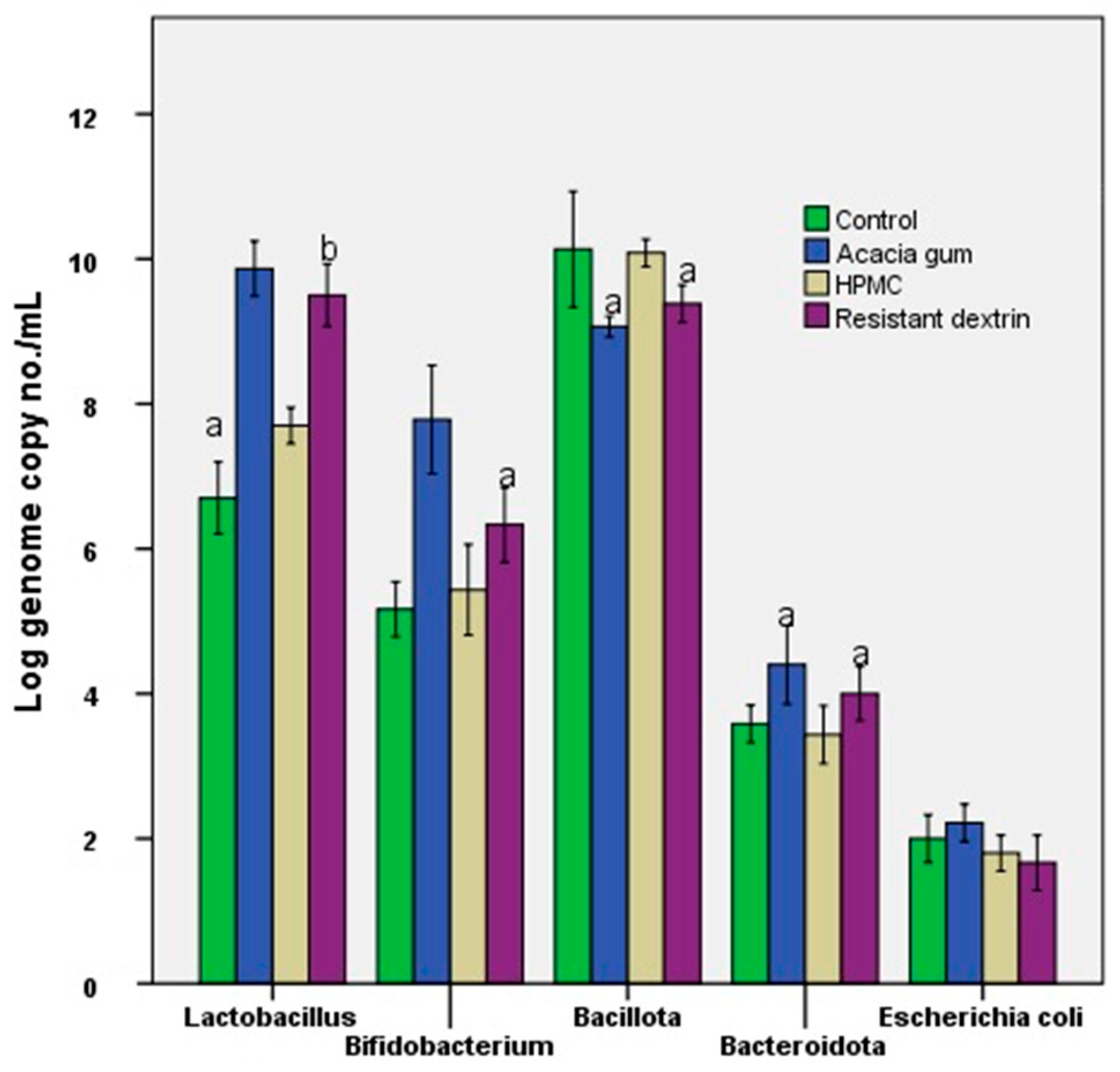
For Bifidobacterium, a gradual increase was observed from the control group to the Control CX 1M variant, then to the Control CX 2M variant, indicating a favorable response with increasing treatment duration. In contrast, Lactobacillus remained relatively stable, with only minor variation across groups, suggesting that ColonX’s effect on this genus was limited or less selective. At the Bacillota level, values were high in both the control group and at 1M but declined at 2M, indicating that the modulation was not uniformly stimulatory across all dominant bacterial groups. In Bacteroidota, a moderate increase was observed, especially after 2 months, although the magnitude of this effect was lower than that seen in Bifidobacterium. At the same time, Escherichia coli did not show a meaningful increase and even tended to decline slightly at 2M, which is favorable for microbiota balance. Overall, the data suggest that the main microbiota-modulating effect of ColonX after 60 days of administration was a selective bifidogenic response (Figure 2). ColonX combined with resistant dextrin was associated with stimulation of a bacterial group considered a marker of a healthy gut microbiota, without a parallel expansion of opportunistic bacteria such as E. coli.
For example, the pattern shown in Figure 1 indicates that the most evident change was not a generalized increase across all bacterial targets, but a selective enrichment of Bifidobacterium after prolonged ColonX exposure. This is supported by Figure 2 and Table 1, where the Bifidobacterium/Enterobacteriaceae ratio increased from 2.60 in the untreated control to 3.00 after 1 month and 4.61 after 2 months of ColonX administration. The Lactobacillaceae/Enterobacteriaceae ratio followed a similar trend, increasing from 3.40 in the control to 3.73 at 1 month and 4.61 at 2 months, whereas the strict anaerobes/facultative anaerobes ratio changed from 6.90 in the control to 6.50 at 1 month and 7.11 at 2 months. These numerical shifts indicate that prolonged ColonX exposure favored beneficial anaerobic and saccharolytic groups without supporting a proportional expansion of facultative opportunistic bacteria.
Because compositional changes in the microbiota may not directly indicate functional relevance, fermentation-derived metabolites were subsequently examined [18]. This approach enabled assessment of the biological significance of ColonX-induced microbial changes by correlating bacterial patterns with metabolite production.
Table 1 shows that ColonX produced a time-dependent improvement in microbiota balance. The Bifidobacterium/Enterobacteriaceae ratio increased from 2.60 in the control to 3.00 at 1 month and 4.61 at 2 months, while the Lactobacillaceae/Enterobacteriaceae ratio rose from 3.40 to 4.61. The strict anaerobes/facultative anaerobes ratio changed modestly, from 6.90 to 7.11 after 2 months, indicating selective bifidogenic modulation rather than broad community restructuring. Among the excipients, resistant dextrin showed the highest Lactobacillaceae/Enterobacteriaceae and strict anaerobes/facultative anaerobes ratios, 5.59 and 7.88, respectively, suggesting its contribution to the overall favorable microbial profile.
The ratio data also clarify differences between the complete formulation and the individual excipients. Resistant dextrin produced the highest Lactobacillaceae/Enterobacteriaceae ratio (5.59) and strict anaerobes/facultative anaerobes ratio (7.88), followed by HPMC for the strict anaerobes/facultative anaerobes ratio (7.56) and acacia gum for the Lactobacillaceae/Enterobacteriaceae ratio (4.50). In contrast, the Bifidobacterium/Enterobacteriaceae ratio was highest in the ColonX 2M group (4.61), compared with resistant dextrin (3.76), acacia gum (3.55), HPMC (3.06), and ColonX 1M (3.00). This numerical comparison suggests that resistant dextrin contributed strongly to the general favorable microbial balance, whereas the complete ColonX formulation produced the clearest time-dependent bifidogenic response.
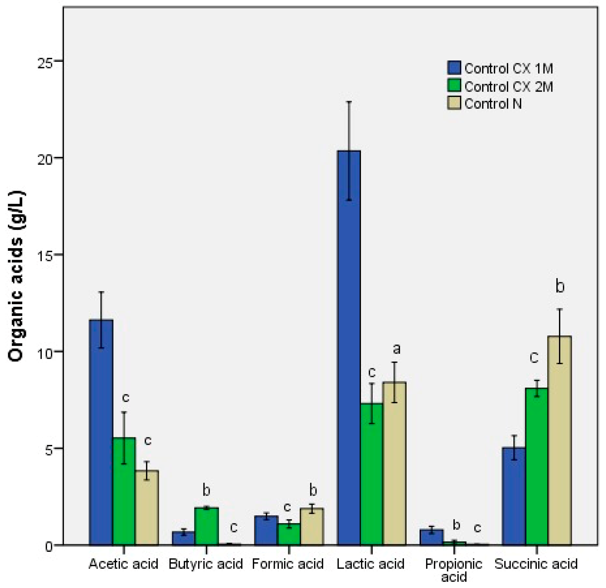
The organic acid profile corroborated and extended the microbiological findings, demonstrating that ColonX-induced modulation encompassed both taxonomic changes and functional metabolic adaptation. Prolonged administration led to a more pronounced production of fermentation-derived metabolites, indicating a progressive increase in microbial activity in response to the formulation (Figure 3).
A marked increase in acetic acid was observed after 1 month of ColonX administration, suggesting intense saccharolytic fermentation during the early phase of microbiota adaptation. Although acetate levels declined after 2 months, they remained above those of the untreated control, indicating that fermentative activity stabilized over time. In parallel, lactic acid production remained elevated in both ColonX-treated variants compared with the control, supporting the view that ColonX promoted carbohydrate fermentation pathways associated with beneficial anaerobic bacteria. These findings are consistent with the selective increase in Bifidobacterium spp. shown in Figure 1 and supports the interpretation that prolonged ColonX administration induced a metabolically active bifidogenic response.
For instance, Figure 3 shows that the early increase in acetate and lactate after 1 month coincided with the first stage of Bifidobacterium enrichment, suggesting rapid utilization of available fermentable substrates. After 2 months, the stronger association with butyrate indicates a shift from primary fermentation products toward downstream metabolites, which may reflect cross-feeding interactions within the adapting dysbiotic microbiota.
Butyric acid concentrations increased steadily throughout the experimental period, peaking after 2 months of administration. Because butyrate is widely regarded as a key biomarker of colonic eubiosis and epithelial health, this finding suggests that prolonged supplementation favored the development of a more functionally mature fermentative ecosystem. Propionic acid levels remained relatively low, particularly after two months, indicating that metabolic conversion pathways were still adapting within the dysbiotic microbiota model.
Among the quantified short-chain fatty acids, acetate increased most prominently after 1 month of ColonX administration and remained above the untreated control after 2 months, whereas butyrate increased progressively and peaked after prolonged administration; propionate remained comparatively low, indicating that downstream fermentative conversion pathways were not fully stabilized. Therefore, these changes should be interpreted as evidence of functional metabolic adaptation and enhanced saccharolytic fermentation rather than complete functional recovery of the microbiota.
The increase in succinic acid may reflect intensified fermentative activity linked to microbiota remodeling during recovery from dysbiosis. As an intermediate metabolite in anaerobic carbohydrate fermentation, succinate accumulation suggests active metabolic turnover before conversion into downstream short-chain fatty acids such as propionate and butyrate [19]. In the present study, succinic acid increased progressively from the control to the ColonX-treated groups, with the highest concentration observed after prolonged administration. This pattern indicates that microbial metabolic activity remained dynamic throughout recovery. The differences between succinic acid and downstream metabolites, particularly propionic and butyric acids, suggest that fermentative conversion had not yet fully stabilized in the in vitro system. For this reason, succinate concentration was interpreted cautiously as a marker of ongoing metabolic restructuring rather than as a beneficial end product of fermentation [20].
Because succinate is a context-dependent intermediate metabolite, its accumulation should not be interpreted as a direct marker of beneficial metabolic activity. Elevated succinate may also occur during dysbiosis or impaired microbial cross-feeding [21]. In the present study, the increase in succinic acid was therefore interpreted with caution as evidence of ongoing fermentative turnover and incomplete stabilization of downstream conversion pathways, particularly because propionate remained comparatively low. Further metagenomic and functional analyses are needed to determine whether succinate accumulation reflects adaptive metabolic restructuring or persistence of dysbiotic fermentation.
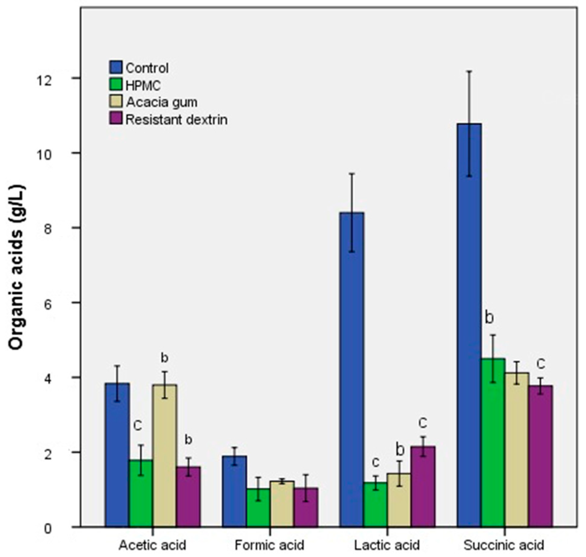
Analysis of the organic acid profile revealed consistent differences among treatments for all four quantified metabolites. The control had the highest concentrations. In contrast, HPMC, acacia gum, and resistant dextrin produced significantly lower levels, as shown in Figure 4. Acetic and formic acids decreased markedly in all fiber-treated samples, indicating reduced primary fermentation. Lactic acid followed the same pattern, with all treatments showing significantly lower concentrations than the control. Succinic acid levels were also reduced across treatments, suggesting an overall attenuation of anaerobic metabolic activity. These results demonstrate that the tested fibers modulate microbial fermentation, leading to a consistent reduction in the production of the measured organic acids.
Figure 4 provides a useful contrast to the ColonX profile, as the individual excipients generally produced lower organic acid concentrations than the untreated control. For example, the reduced acetate, lactate, and succinate levels in the acacia gum, HPMC, and resistant dextrin groups indicate that these components alone did not reproduce the same fermentative pattern observed with ColonX in Figure 3. This supports the interpretation that the complete formulation, rather than any single excipient alone, was responsible for the coordinated microbial and metabolic response.
The modulation of the microbiota induced by ColonX was also reflected in changes to the fermentation system’s simulation environment, particularly a reduction in pH associated with increased organic acid production. The decrease in pH observed after prolonged administration is consistent with enhanced production of acetic, lactic, and butyric acids and supports the establishment of a more active saccharolytic fermentation profile [21]. This acidic environment is considered favorable for beneficial anaerobic bacteria while limiting the expansion of opportunistic microorganisms, including Enterobacteriaceae. The microbiological data correlated well with the metabolomic profile, as the increase in Bifidobacterium spp. was accompanied by higher concentrations of fermentation-derived metabolites, confirming that the observed taxonomic modulation was functionally relevant. At the same time, elevated succinic acid levels suggest that post-antibiotic microbiota modulation. Remained metabolically dynamic, reflecting an active transition between intermediate and terminal fermentation phases [20,22,23]. Overall, the combined microbiological and metabolomic findings indicate that ColonX administration promoted both structural and functional modulation of the dysbiotic microbiota, with progressive stabilization during prolonged administration.
The selective increase in Bifidobacterium spp. may reflect the capacity of this genus to utilize complex non-digestible carbohydrates through saccharolytic pathways, including the bifid shunt. The β-glucan-rich ColonX formulation, together with resistant dextrin, may therefore have preferentially supported bifidobacterial growth and fermentative activity. In contrast, Lactobacillus spp. showed only limited variation, suggesting that the substrate profile or environmental conditions generated in the present in vitro model were not sufficient to promote a comparable lactobacilli expansion [24]. The absence of Escherichia coli stimulation may be related to increased organic acid production and a reduction in pH, which can limit facultative opportunistic bacteria while favoring beneficial anaerobic groups. Nevertheless, because only selected bacterial groups were quantified by qPCR, this explanation should be considered hypothesis-generating and requires confirmation by 16S rRNA sequencing, shotgun metagenomics, and strain-level functional analyses.
The bifidogenic response observed in the present study should be interpreted in the context of the ColonX formulation and the experimental design, rather than as a generalizable effect of all β-glucans. Indeed, previous studies investigating β-glucan prebiotic activity have reported inconsistent effects on Bifidobacterium. β-glucan did not induce a clear bifidogenic effect in an in vitro fermentation model, although it favorably modulated SCFA production [23]. Similarly, studies on β-glucan-rich oats have more frequently reported increases in Lactobacillus, with limited or non-significant effects on Bifidobacterium in vivo [25]. These discrepancies may reflect differences in β-glucan source and structure, including fungal β-(1→3)/(1→6)-glucans versus cereal β-(1→3)/(1→4)-glucans, as well as differences in solubility, molecular weight, branching degree, administered dose, duration of exposure, baseline microbiota composition, and analytical methods. In the present study, the prolonged 60-day exposure, the post-antibiotic dysbiotic microbiota model, and the presence of resistant dextrin in the formulation may have favored Bifidobacterium-associated enrichment. Therefore, the observed increase in Bifidobacterium spp. is best considered a formulation- and model-dependent marker of microbiota modulation, rather than definitive evidence that β-glucan alone exerts a consistent bifidogenic effect [26].
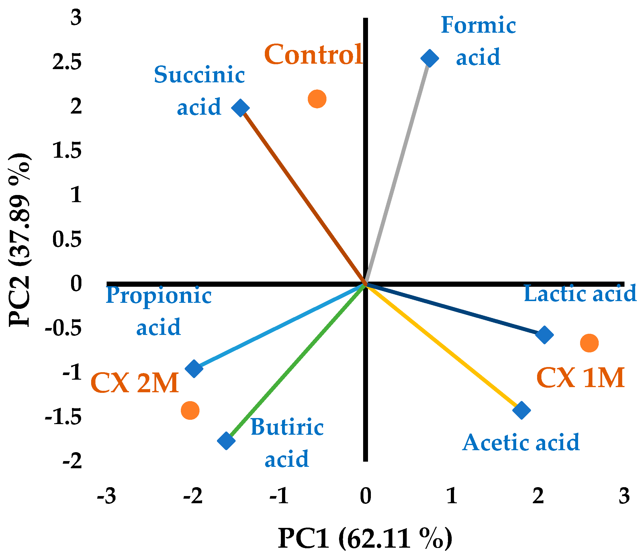
As shown in Figure 5, principal component analysis (PCA) confirmed distinct metabolic profiles between the untreated dysbiotic microbiota and ColonX-treated variants. The first principal component captured most of the total variance and was primarily associated with patterns of organic acid production. One month of ColonX administration was closely associated with increased production of acetic and lactic acids, suggesting early stimulation of saccharolytic fermentation pathways. After two months, the metabolic profile shifted toward increased butyric acid production, indicating progressive maturation and stabilization of microbial fermentation. In contrast, the control group remained associated with elevated succinic acid levels, suggesting incomplete recovery from dysbiosis and the persistence of intermediate fermentation pathways. The PCA therefore supports a correlation between microbiological modulation and functional metabolomic adaptation induced by prolonged ColonX administration [27].
The dietary polysaccharide analysis revealed significant levels of β -glucans, compounds widely recognized for their prebiotic and immunomodulatory properties [28]. These findings support the observed modulation of the microbiota, suggesting that the formulation’s biological effects may be partially mediated by β -glucan-stimulated microbial populations, as indicated by the associated metabolomic data. [28,29]. The balance between β- and α-glucan fractions (Table 2) may contribute to differences in substrate fermentability and short-chain organic acid production observed during the study.
Xanthan gum, gum arabic, resistant dextrin, and HPMC are relevant excipients for older adults because, beyond their technological functions, they also act as soluble, fermentable fibers that may influence intestinal transit, postprandial glycemic response, and lipid metabolism—parameters often affected in age-related conditions such as chronic constipation, metabolic syndrome or type 2 diabetes, dyslipidemia, and frailty [30,31,32]. In particular, resistant dextrin and gum arabic have been investigated as fiber sources with prebiotic and metabolic benefits, supporting bowel regularity and gastrointestinal tolerance through colonic fermentation and the production of short-chain fatty acids [31]. HPMC, as a viscous fiber, has been associated with reduced cholesterol absorption and improved lipid markers, whereas xanthan gum is particularly useful for dysphagia because it increases liquid viscosity and may support safer swallowing in patients with neurological conditions [33,34]. Accordingly, the inclusion of these polysaccharides in formulations intended for geriatric populations is justified not only by their contribution to processing and release characteristics but also by their potential nutritional and physiological benefits, provided that the dose and delivery method are aligned with clinical objectives and patient tolerability [35]. The microbiological and metabolic findings are further supported by the final formulation, which includes excipients that do not increase blood glucose levels and reinforce the product’s microbiota-modulating potential [36].
A limitation of the present study is that the observed effects cannot be attributed exclusively to mushroom β-glucan. Although ColonX contains a high proportion of β-glucans, the formulation also includes resistant dextrin and other excipients that may contribute to fermentation and microbial modulation. Therefore, the findings should be interpreted as effects of the complete β-glucan-rich ColonX formulation, with mushroom β-glucan as the principal bioactive component, rather than as effects of purified β-glucan alone. Future studies comparing purified mushroom β-glucan, resistant dextrin at equivalent doses, and formulation-matched controls are required to clarify the relative contribution of each component to the microbiological and metabolomic responses.
The increase in Bifidobacterium spp. should therefore be interpreted as selective enrichment of a beneficial bacterial group and as a targeted marker of microbiota modulation, rather than as evidence of complete restoration of a healthy microbiome [37]. This interpretation is consistent with the targeted qPCR approach used in the present study and with the accompanying metabolomic data, which indicate fermentative adaptation but do not demonstrate whole-community recovery. Future studies using 16S rRNA sequencing, shotgun metagenomics, and broader functional analyses are required to determine whether these targeted changes correspond to community-level restoration.
Although the 60-day in vitro model is useful for monitoring prolonged microbiota modulation under controlled conditions, it cannot fully reproduce the complexity of in vivo recovery. The host immune responses, intestinal absorption, epithelial barrier interactions, gut motility, bile acid metabolism, and dietary variability are either absent or only partially represented (e.g., mucus layer dynamics). The observed increases in beneficial bacterial groups and fermentation-derived metabolites should be interpreted as indicators of microbiota-modulating potential rather than direct evidence of complete physiological recovery [38]. Future in vivo and clinical studies are required to confirm whether the taxonomic and metabolomic changes observed here translate into improved host–microbiota interactions and intestinal health outcomes.
Selective stimulation of specific bacterial groups should not be interpreted as complete microbiota restoration or as definitive evidence of improved ecosystem balance. In the present study, the increase in beneficial bacterial ratios, together with the absence of stimulation by Escherichia coli and the associated metabolomic adaptation, suggests a favorable direction for microbiota modulation [39]. However, because microbial diversity and whole-community structure were not assessed, the possibility that selective enrichment may affect broader ecosystem balance cannot be ruled out. Therefore, ColonX should be considered a promising nutraceutical candidate for post-antibiotic microbiota modulation, but its efficacy and safety require confirmation in animal models and human clinical trials before claims of complete microbiota recovery can be made.

4. Conclusions

In this in vitro post-antibiotic dysbiosis model, ColonX showed selective microbiota-modulating potential, mainly through a time-dependent increase in Bifidobacterium-associated ratios without stimulation of opportunistic bacteria. These changes were accompanied by shifts in fermentation-derived organic acids, suggesting functional metabolic adaptation during prolonged administration. Because the effects were observed in a controlled in vitro system and may reflect the complete β-glucan-rich formulation, including resistant dextrin, further in vivo and clinical studies are needed before stronger claims regarding microbiota recovery can be made.

Author Contributions

Methodology, E.-I.G., A.C.G. and F.G.; formal analysis, L.D.D., E.-I.G., C.T.C., A.C.G. and F.G.; investigation, L.D.D., E.-I.G., C.T.C., A.C.G. and F.G.; resources, L.D.D., E.-I.G., C.T.C., A.C.G., R.R. and F.G.; data curation, A.C.G.; writing—original draft preparation, E.V.; writing—review and editing, E.V., L.D.D. and E.-I.G.; supervision, E.V.; project administration, E.V.; funding acquisition, R.R. and F.G. All authors have read and agreed to the published version of the manuscript.

Funding

This research received no external funding.

Institutional Review Board Statement

All fecal samples were processed according to the ethical standards of UASVM Bucharest (ColHumB Registration number: 1418/23 November 2017).

Informed Consent Statement

It was obtained from all subjects involved in the study. No participants under 18 years of age. A blank copy of the informed consent form was obtained to preserve the microbiological load from the feces.

Data Availability Statement

The data supporting the findings of this study are available within the article. Additional raw data are not publicly available due to ethical and institutional restrictions related to the use of human-derived biological material.

Acknowledgments

During the preparation of this manuscript, the authors used ChatGPT, version of GPT-5.6 Luna, for the purpose of graphical abstract generation. The authors have reviewed and edited the output and take full responsibility for the content of this publication.

Conflicts of Interest

Author Emanuel Vamanu and Răzvan Roșca were employed and administrator by the company Anoom Laboratories SRL. The remaining authors declare that the research was conducted without any commercial or financial relationships that could be construed as a potential conflict of interest.

References

  1. Ramirez, J.; Guarner, F.; Bustos Fernandez, L.; Maruy, A.; Sdepanian, V.L.; Cohen, H. Antibiotics as Major Disruptors of Gut Microbiota. Front. Cell. Infect. Microbiol. 2020, 10, 572912. [Google Scholar] [CrossRef] [Scilit] [PubMed]
  2. Patangia, D.V.; Ryan, C.A.; Dempsey, E.; Ross, R.P.; Stanton, C. Impact of Antibiotics on the Human Microbiome and Consequences for Host Health. MicrobiologyOpen 2022, 11, e1260. [Google Scholar] [CrossRef] [Scilit] [PubMed]
  3. Roy, S.; Dhaneshwar, S. Role of Prebiotics, Probiotics, and Synbiotics in Management of Inflammatory Bowel Disease: Current Perspectives. World J. Gastroenterol. 2023, 29, 2078–2100. [Google Scholar] [CrossRef] [Scilit] [PubMed]
  4. Al-Habsi, N.; Al-Khalili, M.; Haque, S.A.; Elias, M.; Olqi, N.A.; Al Uraimi, T. Health Benefits of Prebiotics, Probiotics, Synbiotics, and Postbiotics. Nutrients 2024, 16, 3955. [Google Scholar] [CrossRef] [Scilit] [PubMed]
  5. Park, I.; Mannaa, M. Fermented Foods as Functional Systems: Microbial Communities and Metabolites Influencing Gut Health and Systemic Outcomes. Foods 2025, 14, 2292. [Google Scholar] [CrossRef] [Scilit] [PubMed]
  6. Swackhamer, C.; Jang, S.; Park, B.-R.; Hamaker, B.R.; Jung, S.K. Structure-Based Standardization of Prebiotic Soluble Dietary Fibers Based on Monosaccharide Composition, Degree of Polymerization, and Linkage Composition. Carbohydr. Polym. 2025, 367, 123949. [Google Scholar] [CrossRef] [Scilit] [PubMed]
  7. Livesey, G.; Tagami, H. Interventions to Lower the Glycemic Response to Carbohydrate Foods with a Low-Viscosity Fiber (Resistant Maltodextrin): Meta-Analysis of Randomized Controlled Trials. Am. J. Clin. Nutr. 2009, 89, 114–125. [Google Scholar] [CrossRef] [Scilit]
  8. Cerletti, C.; Esposito, S.; Iacoviello, L. Edible Mushrooms and Beta-Glucans: Impact on Human Health. Nutrients 2021, 13, 2195. [Google Scholar] [CrossRef] [Scilit] [PubMed]
  9. Singh, R.P.; Bhardwaj, A. β-Glucans: A Potential Source for Maintaining Gut Microbiota and the Immune System. Front. Nutr. 2023, 10, 1143682. [Google Scholar] [CrossRef] [Scilit] [PubMed]
  10. Amirinia, F.; Jabrodini, A.; Morovati, H.; Ardi, P.; Motamedi, M. Fungal β-Glucans: Biological Properties, Immunomodulatory Effects, Diagnostic and Therapeutic Applications. Infect. Dis. Clin. Microbiol. 2025, 7, 1–16. [Google Scholar] [CrossRef] [Scilit] [PubMed]
  11. Avram, I.; Pelinescu, D.; Gatea, F.; Ionescu, R.; Barcan, A.; Rosca, R.; Zanfirescu, A.; Vamanu, E. Boletus edulis Extract—A New Modulator of Dysbiotic Microbiota. Life 2023, 13, 1481. [Google Scholar] [CrossRef] [Scilit] [PubMed]
  12. Chaikliang, C.; Wichienchot, S.; Youravoug, W.; Graidist, P. Evaluation on prebiotic properties of β-glucan and oligo-β-glucan from mushrooms by human fecal microbiota in fecal batch culture. Funct. Foods Health Dis. 2015, 5, 395–405. [Google Scholar] [CrossRef] [Scilit]
  13. Vamanu, E.; Dinu, L.D. In Vitro Modulation Processes, Prebiotic vs. Postbiotic, of Microbiota Pattern: A Preliminary Study. Nutraceuticals 2025, 5, 30. [Google Scholar] [CrossRef] [Scilit]
  14. Subramaniam, S.; Kamath, S.; Ariaee, A.; Prestidge, C.; Joyce, P. The Impact of Common Pharmaceutical Excipients on the Gut Microbiota. Expert Opin. Drug Deliv. 2023, 20, 1297–1314. [Google Scholar] [CrossRef] [Scilit] [PubMed]
  15. Dinu, L.D.; Gatea, F.; Matei, F.; Banciu, C.; Vamanu, E. Gut Microbiota Modulation by Selenium and Zinc Enrichment Postbiotic Dysbiosis Associated with Hypertension. Curr. Vasc. Pharmacol. 2024, 22, 365–374. [Google Scholar] [CrossRef] [Scilit] [PubMed]
  16. McCleary, B.V.; Draga, A. Measurement of β-Glucan in Mushrooms and Mycelial Products. J. AOAC Int. 2016, 99, 364–373. [Google Scholar] [CrossRef] [Scilit] [PubMed]
  17. Vidal, N.P.; Pham, T.H.; Stewart, P.; Keough, D.; Manful, C.F.; Thomas, R.H. The Use of XLSTAT in Conducting Principal Component Analysis (PCA) When Evaluating the Relationships between Sensory and Quality Attributes in Grilled Foods. MethodsX 2020, 7, 100835. [Google Scholar] [CrossRef] [Scilit] [PubMed]
  18. Morrison, D.J.; Preston, T. Formation of Short Chain Fatty Acids by the Gut Microbiota and Their Impact on Human Metabolism. Gut Microbes 2016, 7, 189–200. [Google Scholar] [CrossRef] [Scilit] [PubMed]
  19. Louis, P.; Flint, H.J. Formation of Propionate and Butyrate by the Human Colonic Microbiota. Environ. Microbiol. 2017, 19, 29–41. [Google Scholar] [CrossRef] [Scilit] [PubMed]
  20. Wei, Y.H.; Ma, X.; Zhao, J.C.; Wang, X.Q.; Gao, C.Q. Succinate metabolism and its regulation of host-microbe interactions. Gut Microbes 2023, 15, 2190300. [Google Scholar] [CrossRef] [Scilit] [PubMed]
  21. Fernández-Veledo, S.; Vendrell, J. Gut microbiota-derived succinate: Friend or foe in human metabolic diseases? Rev. Endocr. Metab. Disord. 2019, 20, 439–447. [Google Scholar] [CrossRef] [Scilit] [PubMed]
  22. Brinck, J.E.; Sinha, A.K.; Laursen, M.F.; Dragsted, L.O.; Raes, J.; Vázquez Uribe, R.; Walter, J.; Roager, H.M.; Licht, T.R. Intestinal pH: A Major Driver of Human Gut Microbiota Composition and Metabolism. Nat. Rev. Gastroenterol. Hepatol. 2025, 22, 639–656. [Google Scholar] [CrossRef] [Scilit] [PubMed]
  23. Caffrey, E.B.; Sonnenburg, J.L.; Devkota, S. Our extended microbiome: The human-relevant metabolites and biology of fermented foods. Cell Metab. 2024, 36, 684–701. [Google Scholar] [CrossRef] [Scilit] [PubMed]
  24. Russo, P.; López, P.; Capozzi, V.; De Palencia, P.F.; Dueñas, M.T.; Spano, G.; Fiocco, D. Beta-Glucans Improve Growth, Viability and Colonization of Probiotic Microorganisms. Int. J. Mol. Sci. 2012, 13, 6026–6039. [Google Scholar] [CrossRef] [Scilit] [PubMed]
  25. Fabiano, G.A.; Shinn, L.M.; Antunes, A.E.C. Relationship between Oat Consumption, Gut Microbiota Modulation, and Short-Chain Fatty Acid Synthesis: An Integrative Review. Nutrients 2023, 15, 3534. [Google Scholar] [CrossRef] [Scilit] [PubMed]
  26. Sung, M.; Yoon, Y.; Lee, J. The Immunomodulatory Effect of β-Glucan Depends on the Composition of the Gut Microbiota. Foods 2023, 12, 3148. [Google Scholar] [CrossRef] [Scilit] [PubMed]
  27. Portincasa, P.; Bonfrate, L.; Vacca, M.; De Angelis, M.; Farella, I.; Lanza, E.; Khalil, M.; Wang, D.Q.-H.; Sperandio, M.; Di Ciaula, A. Gut Microbiota and Short Chain Fatty Acids: Implications in Glucose Homeostasis. Int. J. Mol. Sci. 2022, 23, 1105. [Google Scholar] [CrossRef] [Scilit] [PubMed]
  28. Álvarez-Mercado, A.I.; Plaza-Diaz, J. Dietary Polysaccharides and Gut Microbiota Ecosystem. Nutrients 2022, 14, 4285. [Google Scholar] [CrossRef] [Scilit] [PubMed]
  29. Samiksha, F.; Singh, D.; Harbool, S.S.; Di Martino, L.; Kruithoff, C.; McCormick, T.S.; Ghannoum, M. Fungal β-1,3-Glucans: Cell Wall Constituents That Promote Gut Health Through Innate Immune Modulation. Nutrients 2026, 18, 1794. [Google Scholar] [CrossRef] [Scilit] [PubMed]
  30. Slavin, J.L.; Savarino, V.; Paredes-Diaz, A.; Fotopoulos, G. A Review of the Role of Soluble Fiber in Health with Specific Reference to Wheat Dextrin. J. Int. Med. Res. 2009, 37, 1–17. [Google Scholar] [CrossRef] [Scilit] [PubMed]
  31. Maki, K.C.; Davidson, M.H.; Torri, S.A.; Bell, M.; Wilder, D.M.; Dicklin, M.R.; Feinstein, S.B.; Davidson, K.; Tipping, D.; O’Mullane, J.; et al. High-Molecular-Weight Hydroxypropylmethylcellulose Taken with or between Meals Is Hypocholesterolemic in Adult Men. J. Nutr. 2000, 130, 1705–1710. [Google Scholar] [CrossRef] [Scilit] [PubMed]
  32. Wang, K.; Cheng, Z.; Qiao, D.; Xie, F.; Zhao, S.; Zhang, B. Polysaccharide–Dextrin Thickened Fluids for Individuals with Dysphagia: Recent Advances in Flow Behaviors and Swallowing Assessment Methods. Crit. Rev. Food Sci. Nutr. 2025, 65, 2236–2260. [Google Scholar] [CrossRef] [Scilit] [PubMed]
  33. Cherbut, C.; Michel, C.; Raison, V.; Kravtchenko, T.; Séverine, M. Acacia Gum Is a Bifidogenic Dietary Fibre with High Digestive Tolerance in Healthy Humans. Microb. Ecol. Health Dis. 2003, 15, 43–50. [Google Scholar] [CrossRef] [Scilit]
  34. Bartley, G.E.; Yokoyama, W.; Young, S.A.; Anderson, W.H.K.; Hung, S.-C.; Albers, D.R.; Langhorst, M.L.; Kim, H. Hypocholesterolemic Effects of Hydroxypropyl Methylcellulose Are Mediated by Altered Gene Expression in Hepatic Bile and Cholesterol Pathways of Male Hamsters. J. Nutr. 2010, 140, 1255–1260. [Google Scholar] [CrossRef] [Scilit] [PubMed]
  35. Hadde, E.K.; Mossel, B.; Chen, J.; Prakash, S. The Safety and Efficacy of Xanthan Gum-Based Thickeners and Their Effect in Modifying Bolus Rheology in the Therapeutic Medical Management of Dysphagia. Food Hydrocoll. Health 2021, 1, 100038. [Google Scholar] [CrossRef] [Scilit]
  36. Volkert, D.; Beck, A.M.; Cederholm, T.; Cruz-Jentoft, A.; Goisser, S.; Hooper, L.; Kiesswetter, E.; Maggio, M.; Raynaud-Simon, A.; Sieber, C.C.; et al. ESPEN Practical Guideline: Clinical Nutrition and Hydration in Geriatrics. Clin. Nutr. 2022, 41, 958–989. [Google Scholar] [CrossRef] [Scilit] [PubMed]
  37. Kumar, H.; Collado, M.C.; Wopereis, H.; Salminen, S.; Knol, J.; Roeselers, G. The Bifidogenic Effect Revisited—Ecology and Health Perspectives of Bifidobacterial Colonization in Early Life. Microorganisms 2020, 8, 1855. [Google Scholar] [CrossRef] [Scilit] [PubMed]
  38. Rowland, I.; Gibson, G.; Heinken, A.; Scott, K.; Swann, J.; Thiele, I.; Tuohy, K. Gut microbiota functions: Metabolism of nutrients and other food components. Eur. J. Nutr. 2018, 57, 1–24. [Google Scholar] [CrossRef] [Scilit] [PubMed]
  39. Hemarajata, P.; Versalovic, J. Effects of probiotics on gut microbiota: Mechanisms of intestinal immunomodulation and neuromodulation. Ther. Adv. Gastroenterol. 2013, 6, 39–51. [Google Scholar] [CrossRef] [Scilit] [PubMed]
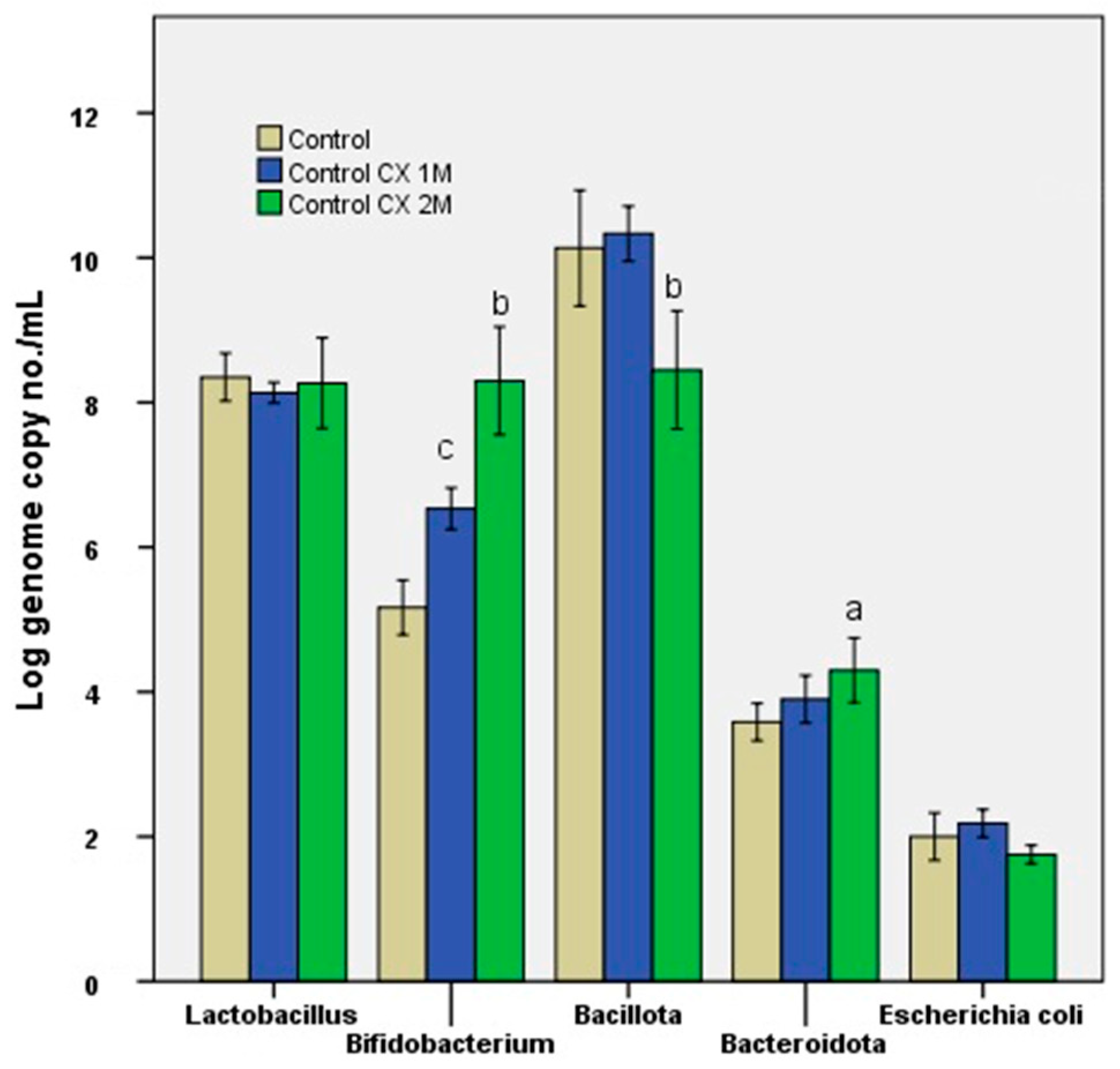
Figure 1. The effect of ColonX administration (60 days) on the microbiota pattern. Different letters mean statistical differences vs. control, p < 0.0001, n = 3. Control was represented by the untreated microbiota.
Nutraceuticals 06 00054 g001
Figure 2. The effect of acacia gum, HPMC șand resistant dextrin administration (60 days) on the microbiota pattern. Different letters mean statistical differences vs. control, p < 0.0001, n = 3. Control was represented by the untreated microbiota.
Nutraceuticals 06 00054 g002
Figure 3. The effect of ColonX administration (60 days) on the metabolomic pattern (SCFAs). Different letters mean statistical differences vs. control, p < 0.0001, n = 3. Control was represented by the untreated microbiota.
Nutraceuticals 06 00054 g003
Figure 4. The effect of acacia gum, HPMC and resistant dextrin administration (60 days) on the metabolomic pattern (SCFAs). Different letters mean statistical differences vs. control, p < 0.0001, n = 3. Control was represented by the untreated microbiota.
Nutraceuticals 06 00054 g004
Figure 5. Principal component analysis (PCA) of the organic acid profiles in the control, ColonX 1M, and ColonX 2M groups.
Nutraceuticals 06 00054 g005
Table 1. Ratios between bacterial groups relevant for characterizing the modulatory effect on the microbiota.
ReportControlColonX 1MColonX 2MAcacia GumHPMCResistant Dextrin
Lactobacillaceae/
Enterobacteriaceae
3.403.734.614.504.335.59
Bifidobacterium/
Enterobacteriaceae
2.603.004.613.553.063.76
Strict anaerobes/
facultative anaerobes
6.906.507.116.187.567.88
Table 2. Content of total glucans, alpha-glucans, and beta-glucans in the analyzed samples.
No.SampleContent of Total Glucans
(%)
Content of Alpha-Glucans (%)Content of Beta-Glucans (%)
1ColonX40.772.3438.43
2ColonX without barley41.702.6739.03
3ColonX with barley35.971.5834.39
Disclaimer/Publisher’s Note: The statements, opinions and data contained in all publications are solely those of the individual author(s) and contributor(s) and not of MDPI and/or the editor(s). MDPI and/or the editor(s) disclaim responsibility for any injury to people or property resulting from any ideas, methods, instructions or products referred to in the content.