Article

Anti-Obesity Effects of Carnosine Supplementation and Exercise in Mice Fed a High-Fat Diet with Concomitant UCP1 Expression in White Adipocytes

1
Graduate School of Sports and Health Science, Daito Bunka University, Saitama 355-8501, Japan
2
Division of Nutrition, Department of Health Science, Daito Bunka University, Saitama 355-8501, Japan
*
Author to whom correspondence should be addressed.
Nutraceuticals 2025, 5(4), 39; https://doi.org/10.3390/nutraceuticals5040039
Submission received: 19 August 2025 / Revised: 1 November 2025 / Accepted: 13 November 2025 / Published: 21 November 2025

Abstract

Obesity and obesity-related diseases represent increasingly serious global health challenges, and effective preventive strategies are urgently needed. This study investigated the anti-obesity effects of carnosine and anserine, representative imidazole dipeptides known for their antioxidant and metabolic regulatory properties, using a mouse model of high-fat (HF) diet-induced obesity. Thirty 6-week-old male C57BL/6n mice were fed an HF diet (56% fat) for eight weeks. Carnosine or anserine was administered in drinking water ad libitum (4 mM). After one week of dietary acclimation, the mice were divided into sedentary and exercise groups (n = 5 per group). The exercise protocol consisted of treadmill running for 30 min/day at 9 m/min, five days per week, for seven consecutive weeks. The results demonstrated that only carnosine supplementation, and not anserine, significantly suppressed body weight gain, visceral white adipose tissue accumulation, and adipocyte hypertrophy induced by the HF diet. Moreover, carnosine supplementation enhanced uncoupling protein 1 (UCP1) expression in epididymal adipocytes and improved serum blood glucose levels. These findings indicate that carnosine exerts anti-obesity effects, potentially through the enhancement of thermogenic and metabolic pathways, and may have therapeutic potential as a dietary intervention for the prevention of obesity-related metabolic disorders.

1. Introduction

Obesity is a growing global health issue linked to a wide range of chronic diseases, including diabetes, cardiovascular disease, and certain cancers [1,2,3,4]. The increasing prevalence of obesity worldwide has become a major public health concern, and effective strategies for prevention and treatment are urgently needed.
Adipose tissue is currently recognized as an endocrine organ that plays a regulatory role in metabolism-related diseases [5]. Among the various mechanisms underlying obesity-related pathologies, dysfunction of visceral white adipocytes has been identified as a key mediator of disease onset and progression [5]. Adipocytes are classified into two distinct categories: white adipocytes and brown adipocytes. These cells constitute white adipose tissue (WAT) and brown adipose tissue (BAT) [6,7]. WAT is predominantly located in the subcutaneous and visceral regions of the body. It plays a pivotal role in storing energy and secreting various adipocytokines, including adiponectin, leptin, TNF-α, and IL-6 [2,5,6,7]. These adipocytokines are implicated in the regulation of insulin sensitivity, inflammation, and lipid metabolism. Excessive accumulation of WAT, particularly in visceral depots, contributes to metabolic dysfunction and chronic inflammation, which are key mediators in the onset and progression of obesity-related diseases [1,2,6]. BAT, in contrast, is specialized for thermogenesis through the expression of uncoupling protein 1 (UCP1) [8]. The presence of BAT in human adults has been confirmed [7,8,9], and it has become a focus of active research in the fields of obesity prevention and treatment. Recent studies have shown that certain stimuli, such as cold exposure and exercise, can induce the expression of UCP1 in WAT, a process known as “browning.” Browning of WAT is associated with increased energy expenditure and improved metabolic profiles, suggesting a promising target for obesity intervention [10,11,12,13].
Carnosine and anserine are well-known imidazole dipeptides with distinct structural and functional properties [14]. Carnosine (β-alanyl-L-histidine) and anserine (β-alanyl-1-methylhistidine) differ in methylation, which affects their antioxidant capacity, tissue distribution, and metabolic fate [15]. Carnosine is a naturally occurring dipeptide composed of β-alanine and histidine, found in high concentrations in skeletal muscle and brain tissue [15]. It has been reported to possess antioxidant, anti-inflammatory, and anti-glycation properties [16,17,18,19,20]. Anserine, a methylated analog of carnosine, shares similar biochemical characteristics but may differ in tissue distribution and biological activity. These compounds have attracted attention as potential nutraceuticals for the prevention of lifestyle-related diseases.
Furthermore, exercise is well known to improve metabolic health and promote the browning of white adipose tissue (WAT), leading to enhanced thermogenic capacity and energy expenditure [21]. Carnosine and Anserine have been recognized as an ergogenic aid that enhances exercise endurance and reduces fatigue through its buffering and antioxidant effects [22,23]. Recent studies also suggest synergistic metabolic benefits when nutritional supplementation is combined with physical activity [24]. Among imidazole dipeptides, increased muscle carnosine—typically achieved through β-alanine supplementation—has been recognized as an ergogenic mechanism that enhances high-intensity exercise performance [25]. Despite the growing interest in imidazole dipeptides, the comparative effects of carnosine and anserine supplementation, particularly in combination with exercise, on adipose tissue morphology and uncoupling protein 1 (UCP1) expression remain poorly understood. Further elucidation of these interactions may provide new insights into strategies for prevention of obesity and metabolic health improvement.
Therefore, in the present study, we hypothesized that carnosine and anserine supplementation would attenuate obesity-related phenotypes by modulating UCP1 expression in white adipocytes. The objective of this study was to examine the anti-obesity effects of these imidazole dipeptides, both individually and in combination with exercise, using a high-fat diet-induced obese mouse model.

2. Materials and Methods

2.1. Experimental Animals and Diet Groups

Six-week-old C57BL/6n male mice (n = 30; Sankyo Laboratory Inc., Tokyo, Japan) were used as an experimental model of diet-induced obesity. C57BL/6 mice were selected because they are one of the most widely used strains in obesity research, and when fed a high-fat diet, they reliably develop obesity characterized by weight gain, adipose tissue accumulation, and metabolic abnormalities such as impaired glucose tolerance, hyperinsulinemia, hyperlipidemia, hyperleptinemia, and fatty liver [3,6,25]. The composition of the experimental high-fat (HF) diet (56% fat), which was modified based on the American Institute of Nutrition (AIN)-93G formulation (16% fat; Research Diets Inc., New Brunswick, NJ, USA), is shown in Table 1. After one week of acclimatization, thirty mice were randomly divided into six groups (five mice per group) as follows: (1) HF group, fed the HF diet; (2) HFEx group, fed the HF diet with exercise; (3) HFC group, fed the HF diet supplemented with carnosine; (4) HFCEx group, fed the HF diet supplemented with carnosine and subjected to exercise; (5) HFA group, fed the HF diet supplemented with anserine without exercise; and (6) HFAEx group, fed the HF diet supplemented with anserine and subjected to exercise. Mice were allowed free access to food and drinking water throughout the experimental periods for 8 weeks. Environmental housing conditions were maintained at 22 ± 2 °C with 55 ± 10% humidity under a 12 h light/dark cycle. Cage density was 5 mice per cage. All experimental procedures were approved by and conducted in accordance with the institutional animal care and use guidelines. Carnosine or anserine (Tokai Bussan Co., Ltd., Tokyo, Japan) was dissolved in drinking water ad libitum at a concentration of 4 mM. This concentration was selected based on previous studies demonstrating physiological efficacy in murine models [26]. Randomization was performed by simple random selection. Blinding was implemented by coding groups (A–F) by a third party, and cages were labeled accordingly.
The number of mice in each group (n = 5) was chosen as the minimum required for statistical analysis, based on previous studies using similar mouse models and in accordance with institutional guidelines. Although a formal power analysis was not performed, this limitation is acknowledged in the Section 4.
This study was approved by the Daito Bunka University Animal Experiment Committee (approval no. ASH22-005, approved on 1 February 2023) and complied with the guidelines of the Japanese Council on Animal Research at Daito Bunka University in Saitama, Japan. Efforts were made to minimize animal suffering and to reduce the number of animals used.

2.2. Measurement of Diet and Water Intake, and Body Weight

The intake of the experimental diet and drinking water (plain water or water containing carnosine or anserine) was measured once per week. Body weight was also recorded weekly throughout the experimental period.

2.3. Sample Collection and Tissue Preparation

At the end of the 8-week feeding period, the mice were fasted for 5 h, and blood samples were collected from the orbital sinus under isoflurane anesthesia. Serum was obtained by centrifugation. Subsequently, the mice were sacrificed by cervical dislocation under anesthesia, and the liver, epididymal adipose tissue, perirenal adipose tissue, and skeletal muscles (gastrocnemius and soleus) were immediately excised and weighed. After weighing, the epididymal adipose tissue was fixed in formalin and used for histological analysis.

2.4. Exercise Loading Protocol

After one week of feeding on the experimental diets, the mice were divided into exercise and sedentary groups. All animals in the exercise groups (HFEx, HFCEx, and HFAEx) were adapted to treadmill running for 30 min per day at a speed corresponding to moderate exercise intensity, using a TMC-200 treadmill (Melquest, Toyama, Japan). During the first week, the running speed was maintained at 9 m/min, followed by 11 m/min for the subsequent six weeks, based on previously published studies that confirmed these speeds as moderate-intensity exercise for obese C57BL/6 mice. The classification of “moderate” exercise intensity was further supported by preliminary measurements of serum lactate levels, which remained within the range of 2–4 mmol/L, consistent with moderate aerobic activity in C57BL/6 mice [25]. The exercise regimen was conducted five days per week for a total of seven weeks and performed during the dark cycle period. All sedentary mice were fasted during the running sessions of the exercise groups. All sedentary mice were fasted during the running sessions of the exercise groups.

2.5. Morphological Observations in Epididymal Adipose Tissue

Epididymal adipose tissue was weighed, fixed in formalin, embedded in paraffin, and sectioned into 3-μm-thick slices. The sections were stained with hematoxylin and eosin (HE stain). Microscopic observation and image acquisition were performed using ImageJ software (version 1.51; National Institutes of Health, Bethesda, MD, USA). The cross-sectional areas of 30 individual adipocytes per sample were measured for quantitative analysis.

2.6. UCP1 Expression in Epididymal Adipose Tissue by Immunostaining

The expression of UCP1 in epididymal adipose tissue was evaluated by immunohistochemical staining of paraffin-embedded sections (3 μm thick). Sections were deparaffinized, rehydrated, treated with methanol for antigen activation, and blocked using Block Ace (DS Pharma Biomedical, Osaka, Japan). An anti-mouse UCP1 antibody (23673-1-AP; Proteintech, Rosemont, IL, USA) was used as the primary antibody at a 1:200 dilution. UCP1 expression was visualized using 3,3′-diaminobenzidine (DAB) staining, and the UCP1-positive area per 100 μm2 was quantified from microscopic images.

2.7. UCP1 Gene Expression in Epididymal Adipose Tissue by RT-PCR

Total RNA was extracted from epididymal adipose tissue using Isogen II (Nippon Gene Co., Ltd., Toyama, Japan) according to the manufacturer’s instructions. During the initial extraction step, the fat layer on the surface was carefully removed after centrifugation. One microgram of total RNA was reverse transcribed into cDNA using the PrimeScript RT Reagent Kit (Takara Bio Inc., Shiga, Japan). Quantitative real-time PCR (RT-PCR) was performed using SYBR Green Master Mix (Takara Bio Inc.) and gene-specific primers (Applied Biosystems, Foster City, CA, USA). Relative UCP1 mRNA expression levels were normalized to 18S rRNA as the internal control. The primer sequences used were as follows: Ucp1 (forward: 5′-GTACACCAAGGAAGGACCGA-3′; reverse: 5′-TTTATTCGTGGTCTCCCAGC-3′), 18S rRNA (forward: 5′-CTTAGAGGGACAAGTGGCG-3′; reverse: 5′-ACGCTGAGCCAGTCAGTGTA-3′).

2.8. Serum Biochemical Marker

Blood samples collected from the mice were centrifuged at 1200 rpm for 10 min, and the separated serum from each sample was used for biochemical analyses. The biochemical measurements were conducted at the Nagahama Life Science Laboratory (Oriental Yeast Co., Ltd., Tokyo, Japan). Serum glucose levels were determined using the hexokinase/glucose-6-phosphate dehydrogenase (HK/G6PDH) method. Total protein was measured by the Biuret method, albumin by the bromocresol green (BCG) method, serum lipids by enzymatic colorimetry, and aspartate aminotransferase (AST) and alanine aminotransferase (ALT) activities by Japan Society of Clinical Chemistry (JSCC)-standardized methods.

2.9. Statistical Analysis

The results are presented as means ± standard errors (SE). Prior to conducting analysis, the assumptions of normality and homogeneity of variance were evaluated using the Shapiro–Wilk and Levene’s tests, respectively. Dunnett’s test was applied to compare body weight, organ weight, and adipose tissue mass among groups relative to the HF group. Two-way analysis of variance (ANOVA) was used to assess the main and interactive effects of carnosine or anserine supplementation and exercise as independent factors. When significant interactions were detected, Tukey’s post hoc test was performed for multiple comparisons. All statistical analyses were conducted using SPSS Statistics software, version 22 (IBM Corp., Armonk, NY, USA). A p-value of less than 0.05 was considered statistically significant.

3. Results

3.1. Body Weight Changes and the HF Diet Intake

The changes in body weight (%) of the mice over the 8-week period following the initiation of the HF diet are shown in Figure 1. After 8 weeks, all groups exhibited an increase in body weight ranging from 133% to 173% as a result of HF diet consumption. The HF group showed the greatest increase, with a 173% gain in body weight at week 8. Body weight gain induced by the HF diet was attenuated in all exercise-loaded groups (HFEx, HFCEx, and HFAEx) by the end of the experimental period. However, the onset of the exercise-induced weight-suppressive effect differed among these groups: it was observed from week 4 in the HFEx group, week 5 in the HFCEx group, and week 7 in the HFAEx group. In addition, the HFC group, which received carnosine supplementation, showed a significant suppression of body weight gain compared with the HF group from week 6 onward. Conversely, the HFA group, supplemented with anserine, did not show a statistically significant difference in body weight change compared with the HF group.
The amount of dietary intake and carnosine or anserine supplementation per mouse was calculated by dividing the total amount consumed per cage by the number of mice. Although exercise loading tended to reduce total energy intake, no statistically significant differences in food or water intake were observed among the groups, indicating that the observed effects were not attributable to differences in consumption volume (Table 2). During the experimental period, the average carnosine supplementation was 1.1 mmol/day in the HFC group and 1.2 mmol/day in the HFCEx group. The average anserine supplementation was 1.1 mmol/day in both the HFA and HFAEx groups, with no significant differences between the supplementation-only and exercise-loaded groups.

3.2. Weight of Visceral Adipose Tissue, Liver and Muscle

Weights of epididymal adipose tissue, perirenal adipose tissue, liver, and muscles (gastrocnemius and soleus) are shown in Table 2. All these weights were analyzed per 100 g of body weight. The weights of epididymal adipose tissue and perirenal adipose tissue were notably lower in the HFEx, HFC, and HFCEx groups than in the HF group. Weights of gastrocnemius and soleus muscles tended to slightly increase owing to the exercise group. The carnosine and anserine supplementation did not influence the weight of the skeletal muscles. Therefore, subsequent analyses focused on the carnosine-supplemented groups, which demonstrated significant suppression of weight gain and visceral adiposity, to further investigate morphological changes and biochemical markers associated with obesity inhibition.

3.3. Morphological Observation of Epididymal Adipose Tissue

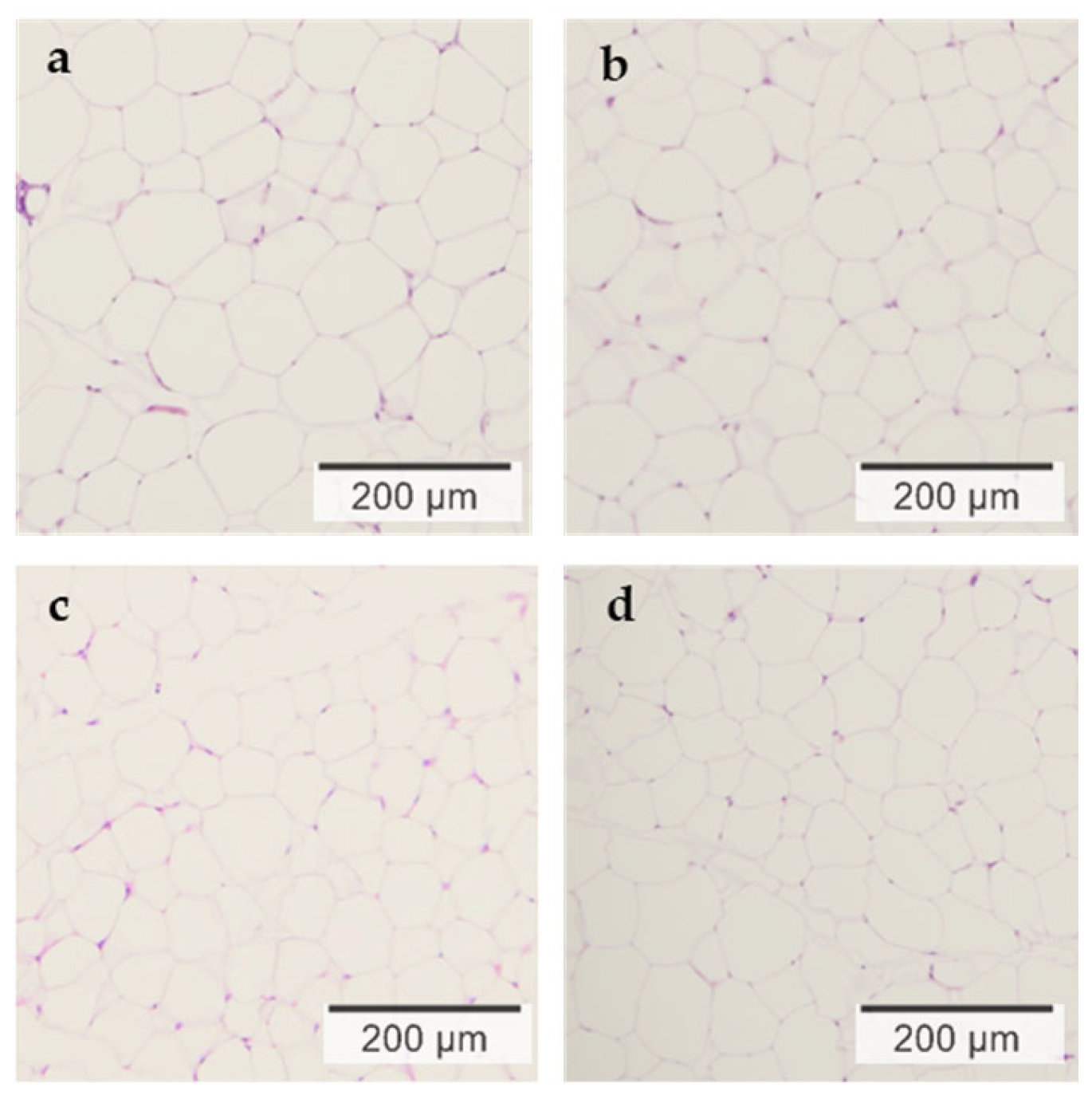
Microscopic images of epididymal adipose tissue stained with hematoxylin and eosin (HE) and the corresponding measurements of adipocyte cross-sectional area are presented in Figure 2 and Table 3, respectively. A two-way ANOVA revealed a significant interaction between exercise and carnosine supplementation (p = 0.034; Table 3). The cross-sectional area of white adipocytes was smaller in the HFEx, HFC, and HFCEx groups compared with the HF group, indicating that both exercise and carnosine supplementation effectively suppressed adipocyte hypertrophy in epididymal adipose tissue.

3.4. Histological Expression of UCP1 in Epididymal Adipose Tissue by Immunostaining

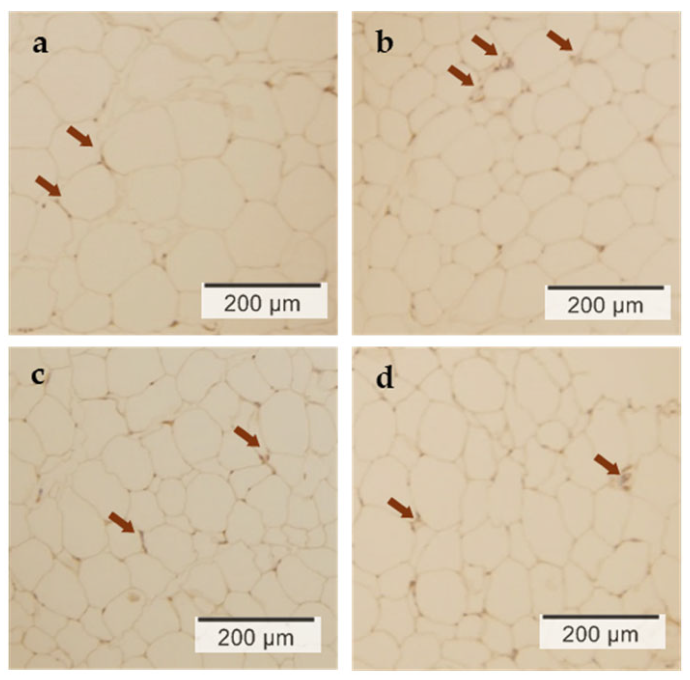
The expression levels of UCP1 in epididymal adipose tissue determined by immunostaining are presented in Figure 3 and Table 3, respectively. The HF group showed the lowest UCP1 expression (8.1 ± 2.4 counts/100 μm2). A two-way ANOVA revealed a significant main effect of carnosine supplementation on UCP1 expression (p = 0.039), whereas the main effect of exercise was not significant (Table 3). There was also no significant interaction between exercise and carnosine supplementation (p = 0.058; Table 3). The HFEx group exhibited nearly double the UCP1 expression (15.7 ± 6.2 counts/100 μm2) compared with the HF group. Although individual variability was observed, UCP1 expression in both the HFC and HFCEx groups was higher than that in the HF group (17.9 ± 10.9 counts/100 μm2 and 19.5 ± 11.0 counts/100 μm2, respectively).

3.5. Gene Expression of UCP1 in Epididymal Adipose Tissue

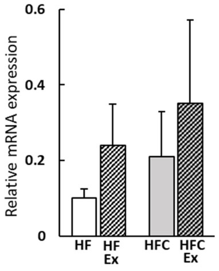
The relative gene expression levels of UCP1 in epididymal adipose tissue are presented in Figure 4. Both carnosine supplementation (p = 0.044) and exercise (p = 0.031) significantly upregulated UCP1 gene expression, whereas no significant interaction was observed between the two factors (p = 0.317).

3.6. Serum Biochemical Parameter

The results of serum biochemical analyses are summarized in Table 4. Serum glucose levels were significantly lower in the HFEx, HFC, and HFCEx groups compared with the HF group (p < 0.05). Triglyceride (TG) levels also tended to decrease in these groups, although the differences did not reach statistical significance. High-density lipoprotein cholesterol (HDL-C) levels were significantly higher in the HFC and HFCEx groups, which received carnosine supplementation, compared with both the HF and HFEx groups. Low-density lipoprotein cholesterol (LDL-C) levels were significantly reduced by carnosine supplementation, as revealed by two-way ANOVA (p = 0.028). Total cholesterol (T-CHO) and free cholesterol (F-CHO) levels tended to increase with carnosine supplementation; however, these changes were not statistically significant. No significant differences were observed among the groups in the levels of aspartate aminotransferase (AST) and alanine aminotransferase (ALT).

4. Discussion

In the present study, carnosine supplementation, either alone or in combination with exercise, effectively suppressed body weight gain, reduced visceral adipose tissue mass, and prevented hypertrophy of white adipocytes induced by a high-fat diet. These effects were accompanied by increased UCP1 expression in epididymal adipocytes and improved glucose tolerance. In contrast, anserine supplementation did not produce comparable effects, suggesting that structural or metabolic differences between these two imidazole dipeptides may account for their divergent physiological outcomes.
Carnosine and anserine are abundant in skeletal and cardiac muscles, as well as in the brain and blood of animals including humans [14,15,16]. Carnosine differs from anserine by a single methyl group, yet this structural difference alters tissue distribution and antioxidant potential. Carnosine is more abundant in skeletal muscle and exhibits stronger metal ion chelating and antiglycation properties than anserine [15,16,17]. These characteristics may enhance mitochondrial activity and lipid oxidation, thereby contributing to UCP1 upregulation and reduced adiposity. This study aimed to investigate the effects of obesity and focused on analyzing the effects of carnosine supplementation, including analysis of visceral white adipocytes. Furthermore, carnosine supplementation demonstrated a similar suppression of white adipose tissue mass as exercise (Figure 1). These results suggest that carnosine supplementation alone may be able to suppress weight gain and adipose tissue loss, similar to exercise. Carnosine and its precursor, β-alanine, have been studied to examine the effects of supplementation on anti-inflammatory, antioxidant, anti-glycation, chelating, skeletal muscle buffering, and improved glucose tolerance [17,18,19,20,25,26,27].
However, the mechanism by which carnosine regulates UCP1 expression in white adipocytes remains unclear. Previous studies have shown that certain functional food components can induce the browning of white adipocytes, accompanied by increased UCP1 expression, reduced secretion of inflammatory adipocytokines such as TNF-α, and improved glycemic control via AMP-activated protein kinase (AMPK) activation [28,29,30]. This suggests that energy expenditure in adipocytes is enhanced. Furthermore, in the present study, carnosine reduced blood glucose levels to a degree similar to that achieved by exercise (p < 0.05). This finding is consistent with previous reports, suggesting that carnosine supplementation in combination with exercise may improve glucose tolerance and has the potential to serve as a preventive intervention for lifestyle-related diseases, including diabetes, in humans [29,31]. In addition, carnosine supplementation increased high-density lipoprotein cholesterol (HDL-C), which may indicate beneficial effects on lipid metabolism [30,31]. A slight increase in serum total cholesterol (T-CHO) levels and a slight increase in low-density lipoprotein cholesterol (LDL-C) may relate to altered hepatic cholesterol transport or lipoprotein remodeling. However, the concurrent rise in HDL-C levels suggests a compensatory mechanism to maintain lipid homeostasis and possibly confer a protective metabolic effect. Further studies measuring hepatic enzymes and cholesterol efflux transporters are warranted to clarify this phenomenon.
Furthermore, given that carnosine concentrations in human skeletal muscle decrease with age [32,33,34], the effects observed in this study may contribute to counteracting age-related metabolic suppression. Previous studies have demonstrated that carnosine regulates blood glucose levels by modulating the autonomic nervous system and improving insulin resistance through the activation of AMP-activated protein kinase (AMPK) [17]. These mechanisms are similar to those involved in UCP1 expression in white adipocytes. Exercise and cold exposure increase UCP1 expression and activation, thereby promoting fat oxidation and suppressing obesity [10,29,35,36]. Moreover, functional food ingredients with antioxidant properties—such as resveratrol from grape skins [37], curcumin from turmeric [38], and berberine from various medicinal herbs [13]—have been reported to promote lipid metabolism through AMPK activation and upregulation of UCP1 expression in white adipose tissue [37,38]. Therefore, we speculate that the anti-obesity effects of carnosine observed in this study are mediated, at least in part, through the enhancement of UCP1 expression in white adipocytes via autonomic nervous system regulation and antioxidant activity. Therefore, we speculate that the anti-obesity effects of carnosine observed in this study are mediated, at least in part, through the enhancement of UCP1 expression in white adipocytes via autonomic nervous system regulation and antioxidant activity.
This study had some limitations. The first limitation is the inability to measure adipocytokines, an indicator of adipocyte function, such as adiponectin, leptin, tumor necrosis factor-α, and other indices that would clarify the mechanism of changes in glucose and lipid metabolism. These factors are critical in linking adipose tissue metabolism to systemic glucose and lipid regulation [6]. Future studies incorporating these markers will provide deeper mechanistic insights into carnosine’s anti-obesity effects. Second, the sample size was n = 5 for each group, based on previous murine studies and institutional ethical guidelines. We acknowledge that this small sample size limits generalizability and statistical power, and future studies with more and increase analyses power are needed. Third, the dependency on carnosine concentration or exercise intensity could not be confirmed. The hypothesis that the simultaneous ingestion of carnosine and exercise would result in a synergistic effect was not supported by the findings of this study. Because carnosine predominantly accumulates in fast-twitch muscle fibers [16,39,40], the moderate exercise intensity used here (9 m/min) might not have fully activated carnosine’s muscular benefits. Additional limitations include the short experimental duration (8 weeks), male-only animals, and the absence of a standard diet control. These constraints should be addressed in future work to strengthen translational validity. It is hypothesized that further investigation is required into the relationship between the supplementation value of carnosine intensity and duration of exercise and the effect of carnosine supplementation on obesity control. Finally, caution is advised when extrapolating these findings to humans. The average carnosine intake of mice in this study corresponds to approximately 0.3~0.5 g/day for a 60 kg adult, an amount that humans can ingest through their diet. However, dose-finding and bioavailability studies in humans are needed to assess the feasibility and safety of daily supplementation. Together, these findings suggest that carnosine may modulate energy metabolism through UCP1 upregulation, antioxidant activity, and improved glucose regulation. However, the modest sample size and lack of mechanistic biomarkers warrant cautious interpretation.

5. Conclusions

Carnosine supplementation effectively suppressed weight gain, reduced visceral adiposity, inhibited adipocyte hypertrophy, and enhanced UCP1 expression in a mouse model of high-fat diet-induced obesity. This study provides new evidence supporting the potential of carnosine as a bioactive compound for metabolic regulation, including improved glucose tolerance and enhanced energy metabolism in adipose tissue. However, as this was an exploratory study with a limited sample size, the findings should be interpreted with caution. Future studies should confirm these effects in larger models and clarify the underlying molecular mechanisms. Overall, these results suggest that carnosine may serve as a promising dietary component for improving metabolic resilience and preventing obesity.

Author Contributions

Conceptualization, T.K.; methodology and format analysis T.K., Data curation X.S.; investigation, X.S., M.H., S.M. and T.K.; writing—original draft preparation, X.S.; writing—review and editing, T.K.; funding acquisition, T.K. All authors have read and agreed to the published version of the manuscript.

Funding

This research received no external funding.

Institutional Review Board Statement

This study was approved by the Daito Bunka University Animal Experiment Committee (ASH22-005, approved on 1 February 2023) and complied with the guidelines of the Japanese Council on Animal Research at Daito Bunka University in Saitama, Japan.

Data Availability Statement

The data that support the findings of this study are available from the corresponding author upon reasonable request.

Acknowledgments

We would like to thank Tokai Bussan Co., Ltd. for providing the carnosine and anserine used in this research. We are especially grateful to Kenichiro Sato for his kind assistance in facilitating the sample provision.

Conflicts of Interest

The authors declare no conflicts of interest.

Abbreviations

The following abbreviations are used in this manuscript:
HFhigh fat
WATwhite adipose tissue
BATbrown adipose tissue
UCP1uncoupling protein-1
AINAmerican Institute of Nutrition
HFExHF diet, with exercise
HFCHF diet supplemented carnosine
HFAHF diet supplemented anserine
HFCExHF diet supplemented carnosine, with exercise
HFAExHF diet supplemented anserine, with exercise
ANOVAanalyses of variance
SEstandard errors
T-CHOtotal cholesterol
F-CHOfree cholesterol
TGtriglycerides
HDL-Chigh-density lipoprotein cholesterol
LDL-Clow-density lipoprotein cholesterol
ASTaspartate aminotransferase
ALTalanine aminotransferase
AMPKAMP-activated protein kinase.

References

  1. World Health Organization. Obesity and Overweight. 2021. Available online: https://www.who.int/news-room/fact-sheets/detail/obesity-and-overweight (accessed on 31 July 2025).
  2. Haslam, D.W.; James, W.P.T. Obesity. Lancet 2005, 366, 1197–1209. [Google Scholar] [CrossRef]
  3. Grundy, S.M. Obesity, Metabolic Syndrome, and Cardiovascular Disease. J. Clin. Endocrinol. Metab. 2004, 89, 2595–2600. [Google Scholar] [CrossRef]
  4. Lissner, L.; Heitmann, B.L. Dietary Fat and Obesity: Evidence from Epidemiology. Eur. J. Clin. Nutr. 1995, 49, 79–90. [Google Scholar]
  5. Cao, H. Adipocytokines in Obesity and Metabolic Disease. J. Endocrinol. 2014, 220, T47–T59. [Google Scholar] [CrossRef]
  6. Morigny, P.; Boucher, J.; Arner, P.; Langin, D. Lipid and Glucose Metabolism in White Adipocytes: Pathways, Dysfunction and Therapeutics. Nat. Rev. Endocrinol. 2021, 17, 276–295. [Google Scholar] [CrossRef]
  7. Singh, R.; Barrios, A.; Dirakvand, G.; Pervin, S. Human Brown Adipose Tissue and Metabolic Health: Potential for Therapeutic Avenues. Cells 2021, 10, 3030. [Google Scholar] [CrossRef]
  8. Ballinger, M.A.; Andrews, M.T. Nature’s Fat-Burning Machine: Brown Adipose Tissue in a Hibernating Mammal. J. Exp. Biol. 2018, 221, jeb162586. [Google Scholar] [CrossRef]
  9. Nedergaard, J.; Bengtsson, T.; Cannon, B. Unexpected Evidence for Active Brown Adipose Tissue in Adult Humans. Am. J. Physiol. Endocrinol. Metab. 2007, 293, E444–E452. [Google Scholar] [CrossRef] [PubMed]
  10. Saito, M.; Okamatsu-Ogura, Y.; Matsushita, M.; Watanabe, K.; Yoneshiro, T.; Nio-Kobayashi, J.; Iwanaga, T.; Miyagawa, M.; Kameya, T.; Nakada, K.; et al. High Incidence of Metabolically Active Brown Adipose Tissue in Healthy Adult Humans: Effects of Cold Exposure and Adiposity. Diabetes 2019, 58, 1526–1531. [Google Scholar] [CrossRef] [PubMed]
  11. Samuelson, I.; Vidal-Puig, A. Studying Brown Adipose Tissue in a Human in vitro Context. Front. Endocrinol. 2020, 11, 629. [Google Scholar] [CrossRef] [PubMed]
  12. Osuna-Prieto, F.J.; Martinez-Tellez, B.; Sanchez-Delgado, G.; Aguilera, C.M.; Lozano-Sánchez, J.; Arráez-Román, D.; Segura-Carretero, A.; Ruiz, J.R. Activation of Human Brown Adipose Tissue by Capsinoids, Catechins, Ephedrine, and Other Dietary Components: A Systematic Review. Adv. Nutr. 2019, 10, 291–302. [Google Scholar] [CrossRef] [PubMed]
  13. Zhang, Z.; Zhang, H.; Li, B.; Meng, X.; Wang, J.; Zhang, Y.; Yao, S.; Ma, Q.; Jin, L.; Yang, J.; et al. Berberine Activates Thermogenesis in White and Brown Adipose Tissue. Nat. Commun. 2014, 5, 5493. [Google Scholar] [CrossRef] [PubMed]
  14. Mannion, A.F.; Jakeman, P.M.; Dunnett, M.; Harris, R.C.; Willan, P.L.T. Carnosine and Anserine Concentrations in the Quadriceps Femoris Muscle of Healthy Humans. Eur. J. Appl. Physiol. Occup. Physiol. 1992, 64, 47–50. [Google Scholar] [CrossRef] [PubMed]
  15. Boldyrev, A.A.; Aldini, G.; Derave, W. Physiology and Pathophysiology of Carnosine. Physiol. Rev. 2013, 93, 1803–1845. [Google Scholar] [CrossRef]
  16. Aldini, G.; de Courten, B.; Regazzoni, L.; Gilardoni, E.; Ferrario, G.; Baron, G.; Altomare, A.; D’amato, A.; Vistoli, G.; Carini, M. Understanding the antioxidant and carbonyl sequestering activity of carnosine: Direct and indirect mechanisms. Free Radic. Res. 2020, 55, 321–330. [Google Scholar] [CrossRef]
  17. Nagai, K.; Niijima, A.; Yamano, T.; Otani, H.; Okumura, N.; Tsuruoka, N.; Nakai, M.; Kiso, Y. Possible Role of L-Carnosine in the Regulation of Blood Glucose Through Controlling Autonomic Nerves. Exp. Biol. Med. 2003, 228, 1138–1145. [Google Scholar] [CrossRef]
  18. Caruso, G.; Caraci, F.; Jolivet, R.B. Pivotal Role of Carnosine in the Modulation of Brain Cells Activity: Multimodal Mechanism of Action and Therapeutic Potential in Neurodegenerative Disorders. Prog. Neurobiol. 2019, 175, 35–53. [Google Scholar] [CrossRef]
  19. Aydın, A.F.; Küçükgergin, C.; Bingül, İ.; Doğan-Ekici, I.; Doğru-Abbasoğlu, S.; Uysal, M. Effect of Carnosine on Renal Function, Oxidation and Glycation Products in the Kidneys of High-Fat Diet/Streptozotocin-Induced Diabetic Rats. Exp. Clin. Endocrinol. Diabetes 2017, 125, 282–289. [Google Scholar] [CrossRef]
  20. Albrecht, T.; Schilperoort, M.; Zhang, S.; Braun, J.D.; Qiu, J.; Rodriguez, A.; Pastene, D.O.; Kramer, B.K.; Köppel, H.; Baelde, H.; et al. Carnosine Attenuates the Development of Both Type 2 Diabetes and Diabetic Nephropathy in BTBR ob/ob Mice. Sci. Rep. 2017, 7, 44492. [Google Scholar] [CrossRef]
  21. Stanford, K.I.; Middelbeek, R.J.; Goodyear, L.J. Exercise Effects on White Adipose Tissue: Beiging and Metabolic Adaptations. Diabetes 2015, 64, 3334–3342. [Google Scholar] [CrossRef]
  22. de Jager, S.; Blancquaert, L.; Van der Stede, T.; Lievens, E.; De Baere, S.; Croubels, S.; Gilardoni, E.; Regazzoni, L.G.; Aldini, G.; Bourgois, J.G.; et al. The ergogenic effect of acute carnosine and anserine supplementation: Dosing, timing, and underlying mechanism. J. Int. Soc. Sports Nutr. 2022, 19, 70–91. [Google Scholar] [CrossRef] [PubMed]
  23. Maemura, H.; Goto, K.; Yoshioka, T.; Sato, M.; Takahata, Y.; Morimatsu, F.; Takamatsu, K. Effects of Carnosine and Anserine Supplementation on Relatively High Intensity Endurance Performance. Int. J. Sport Health Sci. 2006, 4, 86–94. [Google Scholar] [CrossRef]
  24. Shibuya, T.; Kaburagi, T.; Nagai, R.; Oshiro, S. The Effects of Moderate Exercise on Secretory IgA Production in Mice Depends on Dietary Carbohydrate Intake. J. Clin. Biochem. Nutr. 2015, 57, 44–49. [Google Scholar] [CrossRef] [PubMed]
  25. Saunders, B.; Elliott-Sale, K.; Artioli, G.G.; Swinton, P.A.; Dolan, E.; Roschel, H.; Sale, C.; Gualano, B. β-Alanine Supplementation to Improve Exercise Capacity and Performance: A Systematic Review and Meta-Analysis. Br. J. Sports Med. 2017, 51, 658–669. [Google Scholar] [CrossRef]
  26. Matthews, J.J.; Dolan, E.; Swinton, P.A.; Santos, L.; Artioli, G.G.; Turner, M.D.; Elliott-Sale, K.J.; Sale, C. Effect of Carnosine or β-Alanine Supplementation on Markers of Glycemic Control and Insulin Resistance in Humans and Animals: A Systematic Review and Meta-analysis. Adv. Nutr. 2021, 12, 343–362. [Google Scholar] [CrossRef]
  27. Jin, S.Y.; Lee, J.H.; Kim, H.J.; Park, S.J.; Cho, Y.S. Effects of 4 Weeks of Beta-Alanine Intake on Inflammatory Cytokines after Long-Distance Running Exercise. Exerc. Sci. 2022, 31, 188–196. [Google Scholar] [CrossRef]
  28. Machado, S.A.; Pasquarelli-do-Nascimento, G.; Da Silva, D.S.; Farias, G.R.; de Oliveira Santos, I.; Baptista, L.B.; Magalhães, K.G. Browning of the White Adipose Tissue Regulation: New Insights into Nutritional and Metabolic Relevance in Health and Diseases. Nutr. Metab. 2022, 19, 61. [Google Scholar] [CrossRef]
  29. Schirinzi, V.; Poli, C.; Berteotti, C.; Leone, A. Browning of Adipocytes: A Potential Therapeutic Approach. Nutrients 2023, 15, 2229. [Google Scholar] [CrossRef]
  30. Bellia, F.; Vecchio, G.; Rizzarelli, E. Carnosine derivatives: New multifunctional drug-like molecules. Amino Acids 2012, 43, 153–163. [Google Scholar] [CrossRef]
  31. Peng, W.; Mao, P.; Liu, L.; Chen, K.; Zhong, Y.; Xia, W.; Guo, Q.; Tan, S.C.; Rahmani, J.; Varkaneh, H.K.; et al. Effect of carnosine supplementation on lipid profile, fasting blood glucose, HbA1C and insulin resistance: A systematic review and meta-analysis of long-term randomized controlled trials. Complement. Ther. Med. 2020, 48, 102241. [Google Scholar] [CrossRef]
  32. Everaert, I.; Mooyaart, A.; Baguet, A.; Zutinic, A.; Baelde, H.; Achten, E.; Taes, Y.; De Heer, E.; Derave, W. Vegetarianism, Female Gender and Increasing Age, but Not CNDP1 Genotype, Are Associated with Reduced Muscle Carnosine Levels in Humans. Amino Acids 2011, 40, 1221–1229. [Google Scholar] [CrossRef]
  33. Baguet, A.; Everaert, I.; Achten, E.; Thomis, M.; Derave, W. The influence of sex, age and heritability on human skeletal muscle carnosine content. Amino Acids 2011, 43, 13–20. [Google Scholar] [CrossRef] [PubMed]
  34. Szcześniak, D.; Budzeń, S.; Kopeć, W.; Rymaszewska, J. Anserine and Carnosine Supplementation in the Elderly: Effects on Cognitive Functioning and Physical Capacity. Arch. Gerontol. Geriatr. 2014, 59, 485–490. [Google Scholar] [CrossRef] [PubMed]
  35. Wu, J.; Cohen, P.; Spiegelman, B.M. Adaptive Thermogenesis in Adipocytes: Is Beige the New Brown? Genes Dev. 2013, 27, 234–250. [Google Scholar] [CrossRef] [PubMed]
  36. De Matteis, R.; Lucertini, F.; Guescini, M.; Polidori, E.; Zeppa, S.; Stocchi, V.; Cinti, S. Exercise as a New Physiological Stimulus for Brown Adipose Tissue Activity. Nutr. Metab. Cardiovasc. Dis. 2013, 23, 582–590. [Google Scholar] [CrossRef]
  37. Terzo, M.; Iantomasi, M.; Tsiani, E. Effects of Resveratrol on Adipocytes: Evidence from In Vitro and In Vivo Studies. Molecules 2024, 29, 5359. [Google Scholar] [CrossRef]
  38. Lone, J.; Choi, J.H.; Kim, S.W.; Yun, J.W. Curcumin induces brown fat-like phenotype in 3T3-L1 and primary white adipocytes. J. Nutr. Biochem. 2016, 27, 193–202. [Google Scholar] [CrossRef]
  39. Everaert, I.; Derave, W. Human Skeletal Muscle Carnosine and Its Function: A Focus on Homeostasis, Muscle Contractility and pH. In Imidazole Dipeptides: Chemistry, Analysis, Function and Effects; Royal Society of Chemistry: London, UK, 2015; pp. 295–312. [Google Scholar]
  40. Dutka, T.L.; Lamboley, C.R.; McKenna, M.J.; Murphy, R.M.; Lamb, G.D. Effects of carnosine on contractile apparatus Ca2+ sensitivity and sarcoplasmic reticulum Ca2+ release in human skeletal muscle fibers. J. Appl. Physiol. 2012, 112, 728–736. [Google Scholar] [CrossRef]
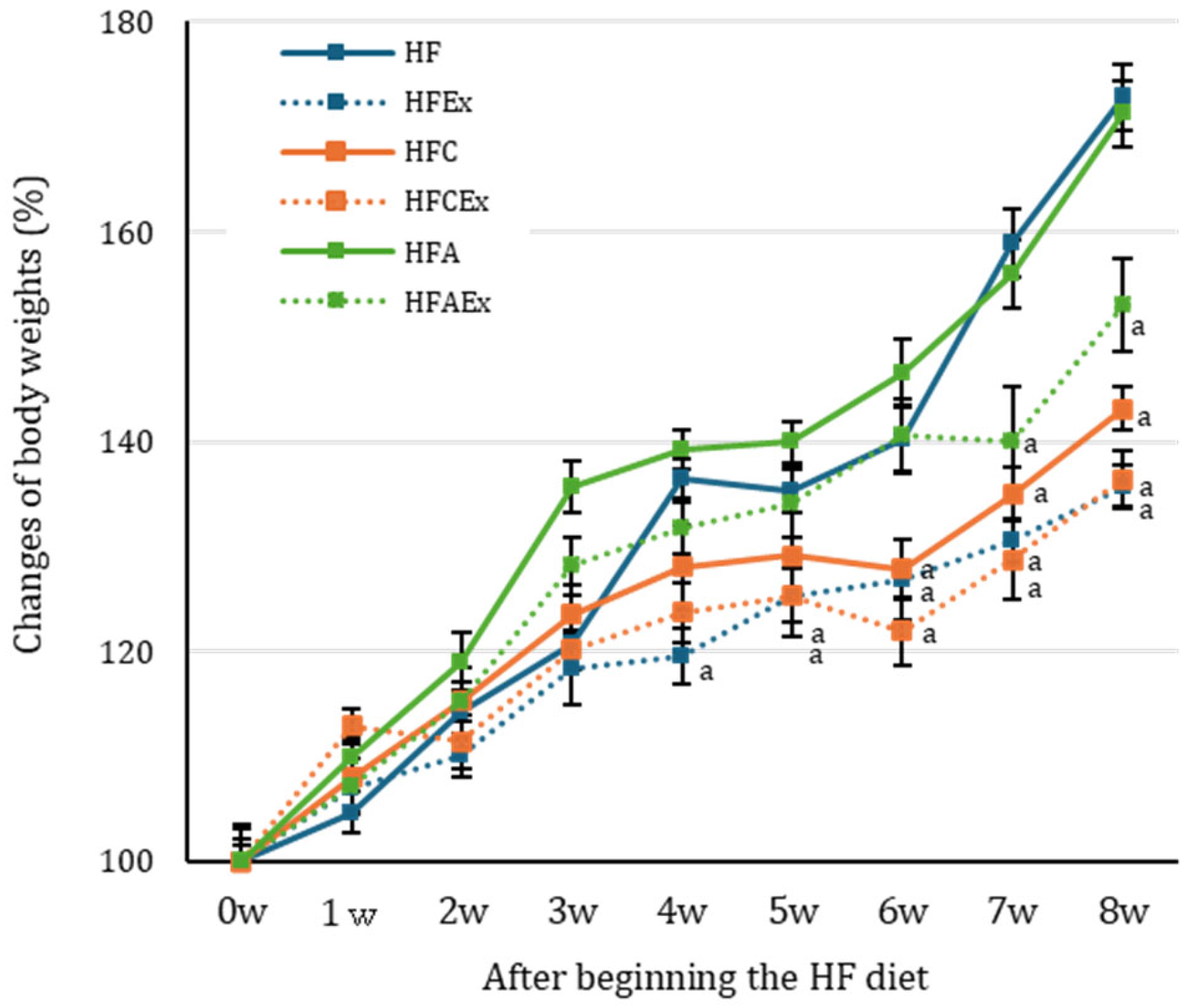
Figure 1. Changes in body weight in the high-fat (HF) diet–induced obese mouse model. Percentage changes in body weight were calculated by dividing the body weight at each time point by the initial body weight (day 0) and multiplying by 100. a p < 0.05 vs. HF group by Dunnett’s test. HF: high-fat diet; HFEx: HF diet with exercise; HFC: HF diet supplemented with carnosine; HFCEx: HF diet supplemented with carnosine and exercise; HFA: HF diet supplemented with anserine; HFAEx: HF diet supplemented with anserine and exercise. Values are expressed as mean ± SE (n = 5).
Nutraceuticals 05 00039 g001
Figure 2. Morphological images of epididymal adipose tissue stained with hematoxylin and eosin (HE). (a) HF group; (b) HFEx group; (c) HFC group; (d) HFCEx group. HF: high-fat diet; HFEx: HF diet with exercise; HFC: HF diet supplemented with carnosine; HFCEx: HF diet supplemented with carnosine and exercise; HFA: HF diet supplemented with anserine; HFAEx: HF diet supplemented with anserine and exercise. Scale bar = 100 μm.
Nutraceuticals 05 00039 g002
Figure 3. Immunostaining of UCP1 in epididymal adipose tissue. UCP1 expression was examined under a light microscope in epididymal adipose tissue. (a) HF group; (b) HFEx group; (c) HFC group; (d) HFCEx group. The arrows indicate some of the sites of UCP1 expression. HF: HF diet with no exercise; HFEx HF diet with exercise; HFC: HF diet with carnosine supplementation and with no exercise; HFCEx: HF diet with carnosine supplementation and with exercise.
Nutraceuticals 05 00039 g003
Figure 4. Gene expression of UCP1 in epididymal adipose tissue. mRNA expression normalized by s17 rRNA as a loading control. Values are mean ± SE (n = 5). HF: HF diet with no exercise; HFEx HF diet with exercise; HFC: HF diet with carnosine supplementation and with no exercise; HFCEx: HF diet with carnosine supplementation and with exercise. The results of the two-way ANOVA were as follows: carnosine: p = 0.044, exercise: p = 0.031, carnosine × exercise interaction: p = 0.317.
Nutraceuticals 05 00039 g004
Table 1. Composition of the high-fat (HF) diet.
Contents
 PFC ratios (kcal%)
    Protein20 
    Fat56 
    Carbohydrate24 
    Total100 
    kcal/g5.1 
 Ingredients (g)
    Casein200.0 
    L-Cystine3.0 
    Corn Starch- 
    Maltodextrin 10132.0 
    Sucrose100.0 
    Cellulose,50.0 
    Soybean Oil30.0 
    Lard216.7 
    t-Butylhydroquinone0.014 
    Mineral Mix S10022G 135.0 
    Vitamin Mix V10037 110.0 
    Choline Bitartrate2.5 
The HF diet (56% fat) was prepared by Research Diets Inc., based on the AIN-93G diet (16% fat). 1 Research Diets Inc.
Table 2. Body weight and weight of visceral fat, liver, and muscle.
HF HFExHFC HFCExHFA HFAEx
initial body weight (g) 21.12
±0.47
22.31
±0.26
21.08
±0.26
21.74
±0.46
22.03
±0.48
22.12
±0.56
final body weight (g)37.80
±1.21
29.83
±0.48 a
30.14 a
±0.16
29.78 a
±0.16
37.23
±1.00
34.48
±0.16
Total intake of diet (kcal)577.63543.08581.42557.36582.63561.04
(mg/100 g body weight)
Liver 2.54
±0.12
2.39
±0.18
2.75
±0.12
2.50
±0.11
2.64
±0.21
2.47
±0.12
muscles (1)0.72
±0.09
0.99
±0.15
0.74
±0.08
0.99
±0.14
0.79
±0.08
088
±0.09
epidydimal adipose tissue5.85
±0.48
3.06
±0.52 a
3.66 a
±0.80
3.46 a
±0.45
5.54
±0.53
4.38
±0.93
perirenal adipose tissue2.20
±0.23
1.31
±0.26 a
1.34 a
±0.13
1.33 a
±0.14
2.11
±0.17
1.91
±0.43
Values are shown as mean ± SE (n = 5). HF: high-fat diet; HFEx: HF diet with exercise; HFC: HF diet supplemented with carnosine; HFCEx: HF diet supplemented with carnosine and exercise; HFA: HF diet supplemented with anserine; HFAEx: HF diet supplemented with anserine and exercise. (1) Combined value of gastrocnemius and soleus muscles. a p < 0.05 vs. HF group by Dunnett’s tests.
Table 3. Areas of adipocytes and UCP1 expression in epididymal adipose tissue.
2-Way ANOVA (1)
HFHFExHFCHFCExCarnosineExerciseInteraction
Area of
white adipocytes (μm2)
8637
±1050
4375 *
±038
3859 *
±1185
4088 *
±1926
0.1190.7550.034
UCP1 expression (counts/
100 μm2 (1)
8.1
±2.4
15.7
±6.2
17.9
±10.9
19.5
±11.0
0.0390.3980.058
Values are shown as mean ± SE (n = 5). HF: HF diet with no exercise; HFEx HF diet with exercise; HFC: HF diet with carnosine supplementation and with no exercise; HFCEx: HF diet with carnosine supplementation and with exercise. * p < 0.05 (vs. HF) by Tukey’s post hoc test. (1) The results of the two-way ANOVA.
Table 4. Serum biochemical parameters.
2-Way ANOVA (1)
HFHFExHFCHFCExCarnosineExerciseInteraction
glucose
(mg/dL)
171.3
±37.0
113.3 *
±6.0
127.3 *
±10.4
124.7 *
±14.5
0.2110.3500.049
T-CHO
(mg/dL)
145.7
±16.8
78.3
±10.1
163.0
±19.0
176.3
±14.9
0.0570.2390.075
F-CHO
(mg/dL)
27.0
±2.6
22.0
±2.0
32.0
±3.0
32.0
±2.0
0.1100.1150.115
TG
(mg/dL)
45.0
±8.9
25.7
±5.9 *
30.0
±2.6
32.3
±9.5
0.3490.0770.320
HDL-C
(mg/dL)
37.3
±13.1
69.7
±9.3
81.0 *#
±14.5
91.3 *#
±3.8
0.0050.2270.035
LDL-C
(mg/dL)
57.0
±19.0
35.7
±2.0
75.7
±27.8
78.7
±13.0
0.0280.3990.290
AST
(IU/L)
137.3
±19.6
156.0
±70.1
149.0
±54.0
110.3
±10.7
0.1870.4150.129
ALT
(IU/L)
28.3
±7.8
40.0
±29.0
33.7
±12.5
24.3
±1.5
0.4130.5430.277
Values are presented as mean ± SE (n = 5). HF: high-fat diet with no exercise; HFEx: high-fat diet with exercise; HFC: high-fat diet supplemented with carnosine and no exercise; HFCEx: high-fat diet supplemented with carnosine and exercise. * p < 0.05 (vs. HF), # p < 0.05 (vs. HFEx) by Turkeys post hoc test. (1) Results of the two-way ANOVA.
Disclaimer/Publisher’s Note: The statements, opinions and data contained in all publications are solely those of the individual author(s) and contributor(s) and not of MDPI and/or the editor(s). MDPI and/or the editor(s) disclaim responsibility for any injury to people or property resulting from any ideas, methods, instructions or products referred to in the content.