Article

Mechanism of Action and Antimicrobial Potential of Weissellicin LM85 from Weissella confusa

by
Manoj Kumar Yadav
1,2 and
Santosh Kumar Tiwari
1,*
1
Department of Genetics, Maharshi Dayanand University, Rohtak 124001, Haryana, India
2
Department of Animal Biotechnology, Dankook University, 119 Dandae-ro, Cheonan 31116, Republic of Korea
*
Author to whom correspondence should be addressed.
Nutraceuticals 2025, 5(4), 33; https://doi.org/10.3390/nutraceuticals5040033
Submission received: 30 August 2025 / Revised: 9 October 2025 / Accepted: 13 October 2025 / Published: 16 October 2025

Abstract

Bacteriocins from lactic acid bacteria have attracted considerable attention as natural alternatives to conventional antimicrobial agents. Weissellicin LM85, a bacteriocin purified from Weissella confusa LM85, has been less extensively studied in terms of its mechanism of action and potential applications. In this study, purified weissellicin LM85 exhibited potent inhibitory effects against Gram-positive bacteria, with minimum inhibitory and bactericidal concentrations determined against Micrococcus luteus MTCC106. Time-kill assays and fluorescence staining indicated a concentration-dependent reduction in cell viability, accompanied by membrane disruption. Further analyses revealed potassium ion efflux, dissipation of membrane potential (Δψ) and pH gradient (ΔpH), genomic DNA fragmentation, and pronounced morphological alterations in target cells. These findings are strongly suggestive of membrane-targeted bactericidal activity, likely involving pore-forming effects. In addition, weissellicin LM85 inhibited both growth and biofilm formation of Salmonella enterica subsp. enterica serovar Typhimurium ATCC13311 and Staphylococcus aureus subsp. aureus ATCC25923. Mechanistic analyses revealed the disruption of cell membrane integrity, leakage of potassium ions, cytoplasmic contents, and non-specific DNA degradation, indicating a multifaceted antibacterial mode of action. These findings highlight weissellicin LM85 as a promising natural antimicrobial with potential applications in food preservation and the control of foodborne pathogens and biofilm-associated infections. Further studies on cytotoxicity and in vivo efficacy are required to advance its practical application.

1. Introduction

Antimicrobial peptides (AMPs) are naturally occurring antibiotics produced by microorganisms, plants, insects, and mammalian cells [1]. They have recently gained attention as potential antimicrobial agents against antibiotic-resistant microbes and may serve as templates for novel drug design [2]. Among these, bacteriocins are ribosomally synthesized AMPs produced by bacteria increasingly regarded as promising alternatives to conventional antibiotics [3]. Many lactic acid bacteria (LAB) are well known for producing diverse bacteriocins and other antimicrobial compounds [1,4]. Bacteriocins are relatively simple to produce, considered safe, and act through targeted mechanisms, making them suitable antimicrobial agents [5]. Bacteriocins from LAB are typically small cationic peptides or proteins that display bacteriostatic or bactericidal activity against related or non-related bacteria [2,6]. Their antimicrobial effects involve binding to cell surface receptors, forming pores that collapse the proton motive force (PMF), inhibiting cell wall or protein synthesis, and in some cases degrading nucleic acids [2]. The PMF composed of membrane potential (Δψ) and pH gradient (ΔpH) is essential for ATP synthesis, ion transport, and phosphorylation [7]. Because of these activities, bacteriocins are widely applied in food safety and pharmaceuticals as safe alternatives to chemical preservatives and antibiotics [6,8]. Currently, nisin and pediocin PA-1 are the main bacteriocins available commercially [8].
Although extensive research has been conducted on bacteriocins produced by Lactobacillus, Leuconostoc, and Enterococcus species, bacteriocins from Weissella species remain poorly characterized, highlighting the need for additional investigation into their potential uses [2]. The genus Weissella is a member of the family Leuconostocaceae, which includes Gram-positive, heterofermentative LAB that are considered probiotic candidates. Members of this genus exhibit spherical to irregular rod-shaped morphology, and to date 22 species have been described [9,10]. Weissella are known to produce various bacteriocins, called weissellicins, which have potential applications in biotechnology for pathogen control or as natural bio-preservatives [10]. Fhoula et al. [11] reported 54 environmental isolates of Weissella spp., confirming their antibacterial activity and safety for potential application as probiotics in animal feed. In 2007, Srionnual et al. [12] first identified and characterized weissellicin 110, a novel bacteriocin with a molecular mass of 3490.8 Da and 27 amino acids sequence, isolated from W. cibaria 110, a strain obtained from plaa-som. Previous studies have reported the purification and characterization of weissellicins 110, L, D, A, Y, and M from W. cibaria 110, W. hellenica 4-7, W. hellenica D1501, W. paramesenteroides DX, and W. hellenica QU13, demonstrating antimicrobial activity against foodborne pathogenic and spoilage bacteria [12,13,14,15,16]. In another study, bacteriocins 7293A and 7293B produced by W. hellenica BCC7293 likely target the cytoplasmic membrane of cells, developing hydrophilic regions that allow leakage of essential molecules and ultimately lead to cell death [17]. To the best of our knowledge, Goh and Philip [1] and Tenea and Lara [18] investigated the mechanism of bactericidal action of a weissellicin purified from W. confusa A3 and W. confusa Cys2-2, demonstrating membrane disruption, intracellular penetration, and DNA binding. More recently, Sreelakshmi et al. [19] reported the antimicrobial, hemolytic, antioxidant, and anticancer activities of the cell-free supernatant from W. confusa. However, comprehensive studies elucidating the mechanism of action of W. confusa-derived weissellicins are still lacking. Therefore, further detailed studies are required to elucidate the mechanism of action of W. confusa-derived weissellicins.
The LAB strain W. confusa LM85 was originally isolated from the rhizosphere of mulberry plants; it was molecularly identified, and its bacteriocin-producing potential was analyzed by Kaur and Tiwari [20]. In our previous study, weissellicin LM85 was purified from W. confusa LM85 using ultrafiltration, cation-exchange, and gel-filtration chromatography, which exhibited broad-spectrum antimicrobial activity (zone of growth inhibition) against L. acidophilus NRRLB4495, L. plantarum NRRLB4496, L. plantarum LD4, E. faecalis ATCC29212, E. faecium NRRLB2354, E. hirae LD3, Pediococcus pentosaceus LB44, Micrococcus luteus MTCC106, Shigella flexneri, S. aureus ATCC25923, Escherichia coli ATCC25922, S. Typhimurium ATCC13311, Vibrio sp., and Pseudomonas aeruginosa ATCC27853 [7]. The current study focused on the detailed mechanism of action of weissellicin LM85 against M. luteus MTCC106 (Gram-positive) and evaluated its potential applications by assessing in vitro antibiofilm activity against the pathogenic bacteria S. Typhimurium ATCC13311 (Gram-negative) and S. aureus ATCC25923 (Gram-positive).

2. Materials and Methods

2.1. Bacterial Strains and Growth Conditions

W. confusa LM85 was originally isolated from the rhizosphere of mulberry plant and is maintained in our culture collection [20,21]. W. confusa LM85 was cultured in de Man, Rogosa, and Sharpe (MRS) medium for growth and production of weissellicin LM85 at 37 °C for 18 h [7]. The indicator strains, M. luteus MTCC106, S. Typhimurium ATCC13311, and S. aureus ATCC25923, were grown in Nutrient Broth (NB) medium at 37 °C for 18 h with agitation at 200 rpm [7].

2.2. Determination of Minimum Inhibitory Concentration (MIC) and Minimum Bactericidal Concentration (MBC)

Weissellicin LM85 was previously purified from the cell-free supernatant of W. confusa LM85 [7]. The MIC of weissellicin LM85 was evaluated using microdilution assay following the method of Goh and Philip [1]. Serial two-fold dilutions of weissellicin LM85 (2.16, 4.32, 8.64, 17.29, 34.57, 69.15, 138.3, 276.6, and 553.2 µg/mL) were prepared in 10 mM sodium acetate buffer (pH 4.5), and each dilution (100 µL) was filled into the wells of microtiter already containing 200 µL of culture of M. luteus MTCC106 (OD600 0.02, 106 CFU/mL). The same buffer (100 µL) was used in the control set at the place of weissellicin LM85. The assay plate was incubated at 37 °C for 18 h with agitation (200 rpm). After the incubation, the growth was assessed by calculating the difference between the final and initial OD using a microplate reader. The MIC was considered the lowest concentration that inhibited turbidity and maintained OD600 < 0.1.
Serial tenfold dilutions of each well were prepared in 0.8% normal saline (NS), and each dilution was spread onto nutrient agar plates. The assay plates were incubated at 37 °C for 18 h. After incubation, the visible colonies grown on the agar plate were manually counted and expressed colony-forming units per milliliter (CFU/mL). The MBC was defined as the lowest concentration causing complete loss of viability.

2.3. Determination of Kill Kinetics and Estimation of Live/Dead Cells

The bactericidal activity of weissellicin LM85 against M. luteus MTCC106 was determined at different time intervals. The M. luteus MTCC106 was inoculated into 10 mL of fresh NB medium (OD600 0.02) and incubated with agitation (200 rpm) at 37 °C. The cell pellets were collected from mid-log phase (OD600 0.5) through centrifugation at 10,000 rpm for 15 min at 4 °C, washed, and reconstituted in 0.8% NS to a final concentration of ~106 CFU/mL. The cell suspensions were treated with ½ MIC (34.57 µg/mL), MIC (69.15 µg/mL), and 2 MIC (138.3 µg/mL) of weissellicin LM85 and incubated with agitation (200 rpm) at 37 °C for up to 10 h. In the control set, 10 mM sodium acetate buffer (pH 4.5) served as the diluent of weissellicin LM85. Samples of 100 µL were taken at 2 h intervals for a total duration of 10 h, and the CFU/mL was calculated as mentioned above. The percentage (%) of cell viability was measured and compared with that of the untreated (control) [22].
Fluorescence microscopy with DAPI and PI staining was used to qualitatively distinguish between live and dead cell populations, respectively, as well as to evaluate membrane integrity. Cells from all treatments were collected from 10 h of incubation (as mentioned above), were harvested, washed twice, and reconstituted in 0.8% NS. The untreated cells (~106 CFU/mL) were used as a negative (live) control. The cells treated with 70% ethanol, incubated at 60 °C for 10 min, and washed twice with NS were used as a positive (dead) control. For fluorescence staining, 10 µL of DAPI and PI stock solutions (1 mg/mL) were added to the treated and untreated cell suspensions (1 mL), following the method of Sheoran et al. [23]. All samples were incubated at room temperature (RT) for 10 min, then placed on a glass slide and examined under a fluorescence microscope at 40× magnification using an excitation wavelength of 330–380 nm. Representative images were captured from multiple randomly selected microscopic fields for each treatment. The observations were used for visual comparison of live (blue-stained) and dead (red-stained) cells, but no manual or automated quantitative counting was performed.

2.4. Determination of Efflux of Potassium (K+) Ions

M. luteus MTCC106 was cultured in NB medium (10 mL) until reaching mid-log phase (OD600 0.5), then the cell pellets were harvested by centrifugation and washed thrice with 10 mM tris-acetate buffer (pH 7.4) supplemented with NaCl (100 mM) to remove residual medium. The cell pellets were reconstituted in the same buffer and treated with different concentrations of weissellicin LM85 (as above, Section 2.3). For positive control, nisin (250 µg/mL) was used, whereas the respective diluents (0.02 N HCl for nisin and sodium acetate buffer for weissellicin LM85) were used as a negative control. The efflux of K+ ions was monitored every minute for a duration of 5 min following treatment employing a potassium-selective electrode, calibrated with 20 and 100 ppm KCl solutions and connected with a digital flame photometer [24,25].

2.5. Determination of Dissipation of Membrane Potential (∆ψ)

Mid-log phase cells of M. luteus MTCC106 (10 mL, OD600 0.5) were harvested, washed twice with HEPES buffer (50 mM, pH 7), reconstituted using the same buffer (100 µL), kept at 4 °C, and utilized within 30 min. The changes in ∆ψ were measured with fluorescent probe di-S-C3-(3) [22,26]. An aliquot of 5 µL of the 2 mM probe di-S-C3-(3) was mixed with 2 mL of same buffer in a fluorescent cuvette, and the fluorescence spectra were monitored for 1500 s at 575 nm (excitation and emission wavelengths set at 532 and 575 nm, respectively) by a modular spectrofluorometer. Once the baseline signal was stabilized, 10 µL of concentrated M. luteus MTCC106 cells were rapidly mixed with the mixture, followed by 20 µL glucose (20%). Once the signal stabilized, 2 µL nigericin (5 µM) was added to the mixture. Subsequently, different concentrations of weissellicin LM85 (as above, Section 2.3) and nisin (250 µg/mL) were mixed into the mixture after signal stabilization. The respective bacteriocin diluents were used as a negative control, while nisin was used as a positive control, as mentioned in the above section. Finally, valinomycin (2 µL, 2 µM) was mixed following signal stabilization.

2.6. Determination of Dissipation of Transmembrane pH Gradient (∆pH)

Mid-log phase cells of M. luteus MTCC106 (OD600 0.5) grown in NB medium (50 mL) were harvested by centrifugation, washed twice with 5 mM HEPES buffer (pH 7), reconstituted in the same buffer (5 mL), and incubated for 1 h on ice. The cells were then loaded with the fluorescent probe BCECF,AM by adding 1 µL of probe (2 mM) to the cell suspension and incubated in the dark for 1 h at 37 °C [7,26]. The 2 mL probe-loaded cells were transferred into a fluorescent cuvette, and spectra were recorded for 600 s at 535 nm (excitation and emission wavelengths set at 488 and 535 nm, respectively) by a modular spectrofluorometer. After baseline stabilization, glucose (20 µL, 20%) was added, followed by valinomycin (2 µL, 5 µM). Once the fluorescence signal stabilized, different concentrations of weissellicin LM85 (as above, Section 2.3) and nisin (250 µg/mL) were mixed. The respective bacteriocin diluents were used as a negative control, while nisin was used as a positive control, as mentioned above. Finally, nigericin (2 µL, 2 µM) was added after signal stabilization.

2.7. Effects of Weissellicin LM85 on Genomic DNA

The M. luteus MTCC106 cells were harvested from mid-log phase (OD600 0.5) culture by centrifugation, washed twice, and reconstituted in 0.8% NS. The cell suspension was treated with MIC (69.15 µg/mL), 2 MIC (138.3 µg/mL), and 4 MIC (276.6 µg/mL) of weissellicin LM85, while treated with diluent of weissellicin LM85 used as a control [24,27]. The treated and untreated cells were incubated with shaking (200 rpm) at 37 °C for 18 h. Genomic DNA was isolated using a genomic DNA isolation kit. DNA integrity was evaluated using standard agarose gel electrophoresis protocol, and fragmentation was visualized under UV trans-illuminator and compared with the control.
For direct interaction, genomic DNA was extracted from an overnight-grown M. luteus MTCC106 culture (OD600 1.12) using the same kit. The purified DNA (0.8 µg) was incubated with different concentrations of weissellicin LM85 (as mentioned above) at RT for 8 h under static conditions [28,29]. DNA incubated without bacteriocin was used as a control. Following incubation, DNA fragmentation was evaluated by a standard agarose gel electrophoresis protocol.

2.8. Morphological Analysis

The cell morphology was examined using a scanning electron microscope (SEM) and transmission electron microscope (TEM) following the method of Goh and Philip [1] and Du et al. [27]. Briefly, overnight-grown culture of M. luteus MTCC106 (~106 CFU/mL) was inoculated into fresh NB medium (10 mL) and incubated at 37 °C with shaking at 200 rpm for 4 h to obtain mid-log phase cells (OD600 0.5). Cells were harvested, washed two times with sodium phosphate buffer (0.1 M, pH 7.4), and treated with 2 MIC (138.3 µg/mL) of weissellicin LM85 and 250 µg/mL of nisin. The untreated cells were used as a control. The treated and untreated cells were incubated with shaking (200 rpm) at 37 °C for 18 h. After that cell pellets were washed with the same buffer and fixed in 2.5% glutaraldehyde and 2% paraformaldehyde at 4 °C for 12 h. After incubation, cells were post-fixed with 1% osmium tetroxide, dehydrated with series of ethanol, and dried with hexamethyldisilazane. For SEM analysis, cells were mounted and coated with gold/palladium (6:4) and visualized at 10.25 Kx magnification. For TEM analysis, dehydrated and dried cells were embedded, sectioned, mounted onto grids, stained with alkaline lead citrate and uranyl acetate, and examined under TEM at 14 Kx magnification.

2.9. In Vitro Applications

The applications of weissellicin LM85 were evaluated by assessing its in vitro antimicrobial and antibiofilm activity against the pathogenic bacteria S. aureus ATCC25923 and S. Typhimurium ATCC13311, as described by Leslie et al. [25] and Shastry et al. [30]. Overnight-grown culture (~106 CFU/mL) of each strain (150 µL) was added to microtiter plate wells containing various concentrations of weissellicin LM85 (4.94–316.66 µg/mL) and incubated at 37 °C for 18 h. The diluent was used as a negative control. Bacterial growth inhibition was measured at 600 nm, while biofilm inhibition was assessed using the crystal violet (CV) assay. For inhibition of biofilm formation, wells were washed twice with phosphate-buffered saline (1X PBS, pH 7.4) to remove the unbound cells, and biofilm was fixed with 99% methanol for 20 min at RT and air-dried. Biofilm was stained by adding 200 µL of 0.1% CV at RT for 30 min and washed with sterilized ddH2O to remove excess dye. The bound dye was solubilized with 150 µL acetic acid (33%) at RT for 15 min. The absorbance of the solubilized CV was determined at 585 nm to observe biofilm formation.

2.10. Statistical Analysis

All experiments were performed in triplicate, and data were expressed as the mean ± standard deviation (SD) using SigmaPlot 11.0. Statistically analyses were performed using Graphpad Prism. Two-way ANOVA was applied to access the effect of treatment concentration and incubation time followed by Tukey’s multiple comparison tests. Significance was considered at p < 0.05.

3. Results

3.1. MIC and MBC

Weissellicin LM85 showed strong inhibitory and bactericidal activity against M. luteus MTCC106. The growth inhibition was not observed in the untreated set. In contrast, treatment with increasing concentrations of weissellicin LM85 caused a dose-dependent increase in the percentage of growth inhibition and inhibition of cell viability. The percentage of growth inhibition at different concentrations is shown in Supplementary Table S1. Almost complete growth inhibition (98.77 ± 0.62%) was observed at 69.15 µg/mL and considered the MIC (Figure 1). The untreated M. luteus MTCC106 did not show inhibition of cell viability. Treatment with weissellicin LM85 led to a concentration-dependent reduction in cell viability, as shown in Supplementary Table S1. Weissellicin LM85 completely (100%) inhibited the cell viability of M. luteus MTCC106 at 276.6 µg/mL and was considered the MBC (Figure 1).

3.2. Kill Kinetics and Estimation of Live/Dead Cells

The weissellicin LM85 exhibited a clear time- and dose-dependent reduction in the cell viability. The viability of untreated M. luteus MTCC106 was considered 100% and almost remained stable throughout the 10 h incubation period. In contrast, treatment with weissellicin LM85 caused a time- and dose-dependent increase in the inhibition of cell viability. The percentage of cell viability at different time intervals after treatment with different concentrations is shown in Supplementary Table S2. The presence of lower concentrations (½ MIC, 34.57 µg/mL) of weissellicin LM85 caused a gradual increase in the inhibition of cell viability (59.87 ± 1.95% at 10 h), whereas higher concentrations, MIC (69.15 µg/mL) and 2 MIC (138.3 µg/mL), caused the maximum loss of cell viability, 22.16 ± 0.05% and 2.75 ± 0.01% at 10 h, respectively (Figure 2A), confirming its bactericidal nature. These results demonstrate that weissellicin LM85 exerts rapid bactericidal activity against M. luteus MTCC106.
Fluorescence staining further confirmed this effect. The untreated cells fluoresced blue (live), whereas bacteriocin-treated cells exhibited pink/red fluorescence (dead), indicating disruption of membrane integrity. The untreated M. luteus MTCC106 cells appeared blue with DAPI staining, indicating live cells (Figure 2B), while ethanol-treated cells fluoresced red with PI staining, representing dead cells (Figure 2C). The cells showed a mixture of blue and reddish-pink after being treated with weissellicin LM85 (34.57 µg/mL) (Figure 2D), whereas 69.15 µg/mL resulted in a predominance of light-pink cells (Figure 2E). A greater proportion of red cells was observed after treatment with 138.3 µg/mL, indicating the concentration-dependent bactericidal activity of weissellicin LM85 (Figure 2F).

3.3. Efflux of Potassium (K+) Ions

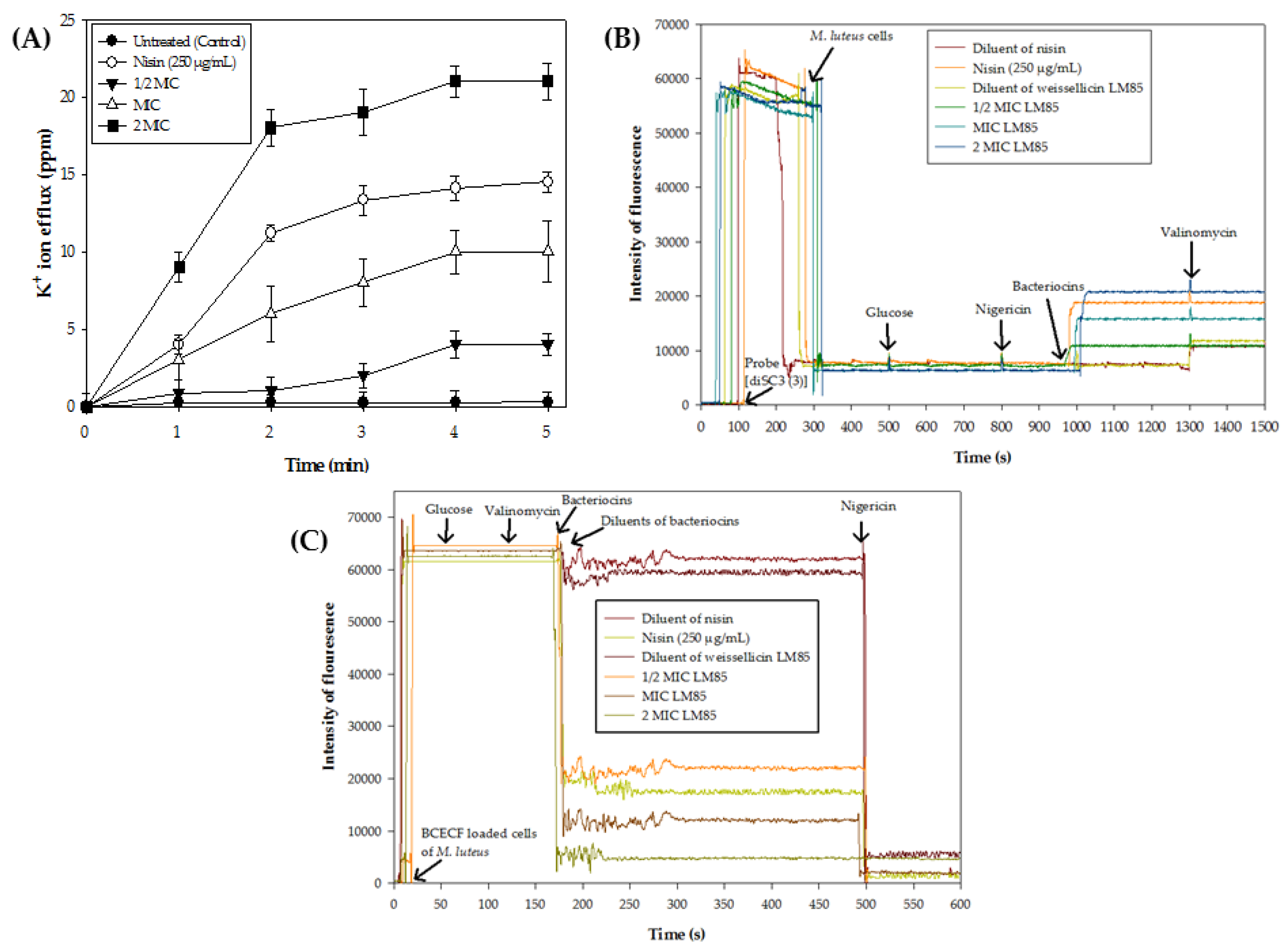
The cells of M. luteus MTCC106 caused quick efflux of K+ ions, and the efflux was found higher at high concentrations of weissellicin LM85. The untreated M. luteus MTCC106 cells showed negligible K+ ion release, whereas 14.62 ± 0.10 ppm was found after treatment with nisin (250 µg/mL) at 5 min. The weissellicin LM85 caused a rapid and concentration-dependent efflux of intracellular K+ ions, as shown in Supplementary Table S3. The efflux of K+ ions reached 4.33 ± 0.31 ppm at 5 min after treatment with 34.57 µg/mL, whereas treatment with 69.15 µg/mL and 138.3 µg/mL of weissellicin LM85 caused 10.1 ± 0.1 and 21.07 ± 0.06 ppm efflux of K+ ions, respectively. The efflux increased sharply during the first 3 min and then stabilized, indicating that most K+ ion efflux occurred rapidly after exposure (Figure 3A). These results demonstrate that weissellicin LM85 disrupts membrane integrity, leading to loss of intracellular ions and contributing to cell death.

3.4. Dissipation of Membrane Potential (∆ψ)

Weissellicin LM85 treatment caused complete dissipation of Δψ in M. luteus MTCC106, as shown by a marked increase in fluorescence intensity. M. luteus MTCC106 cells caused a rapid reduction in fluorescence signal just after the addition of cells with the probe di-S-C3-(3), which then stabilized. After the addition of nigericin, no change in fluorescence was detected in all sets. In contrast, nisin treatment increased the fluorescence signal, showing dissipation of ∆ψ. Similarly, treatment with weissellicin LM85 (34.57, 69.15, and 138.3 µg/mL) also led to a concentration-dependent increase in fluorescence, suggesting the dissipation of ∆ψ, after which the signal was stabilized. The cells treated with bacteriocin diluents did not show fluorescence increase. With the addition of the ionophore valinomycin, the fluorescence signal was stabilized in treated cells, whereas there was an increase in untreated cells, showing dissipation of ∆ψ. These findings suggest that bacteriocins, including weissellicin LM85, induce complete dissipation of ∆ψ in M. luteus MTCC106 (Figure 3B).

3.5. Transmembrane pH Gradient (∆pH)

Weissellicin LM85 also dissipated the ΔpH in M. luteus MTCC106 cells. The fluorescence signal was detected in the BCECF,AM probe-loaded cells of M. luteus MTCC106. The addition of valinomycin showed no reduction in fluorescence in any treatment sets. The cells treated with diluents of bacteriocins also showed no decrease in fluorescence, whereas nigericin caused a reduction in fluorescence signal, suggesting the dissipation of ∆pH. The fluorescence signal was reduced just after mixing with nisin in BCECF,AM probe-loaded cells, and some reduction was also found after adding nigericin; after that, it was stabilized. A concentration-dependent reduction in the fluorescence was observed in BCECF,AM-loaded cells after treatment with weissellicin LM85 (34.57, 69.15, and 138.3 µg/mL), suggesting dissipation of ∆pH (Figure 3C). These findings indicate that weissellicin LM85 disrupts ∆pH, supporting its pore-forming mode of action against M. luteus MTCC106.

3.6. Effects on Genomic DNA

Weissellicin LM85 affected genomic DNA integrity. Genomic DNA from untreated cells appeared as a sharp band on agarose gel, whereas DNA from treated cells with 69.15 µg/mL showed a smeared pattern. Treatment with higher concentrations (138.3 and 276.6 µg/mL) resulted in more smearing, with DNA migrating faster on agarose gel (Figure 4A). In vitro assay further confirmed the direct interaction, as purified DNA incubated with weissellicin LM85 migrated more slowly during electrophoresis. Genomic DNA in the absence of weissellicin LM85 migrated normally on agarose gel, whereas DNA in the presence of MIC (69.15 µg/mL), 2 MIC (138.3 µg/mL), and 4 MIC (276.6 µg/mL) of weissellicin LM85 showed progressively slower migration in a concentration-dependent manner (Figure 4B).

3.7. Morphological Analysis

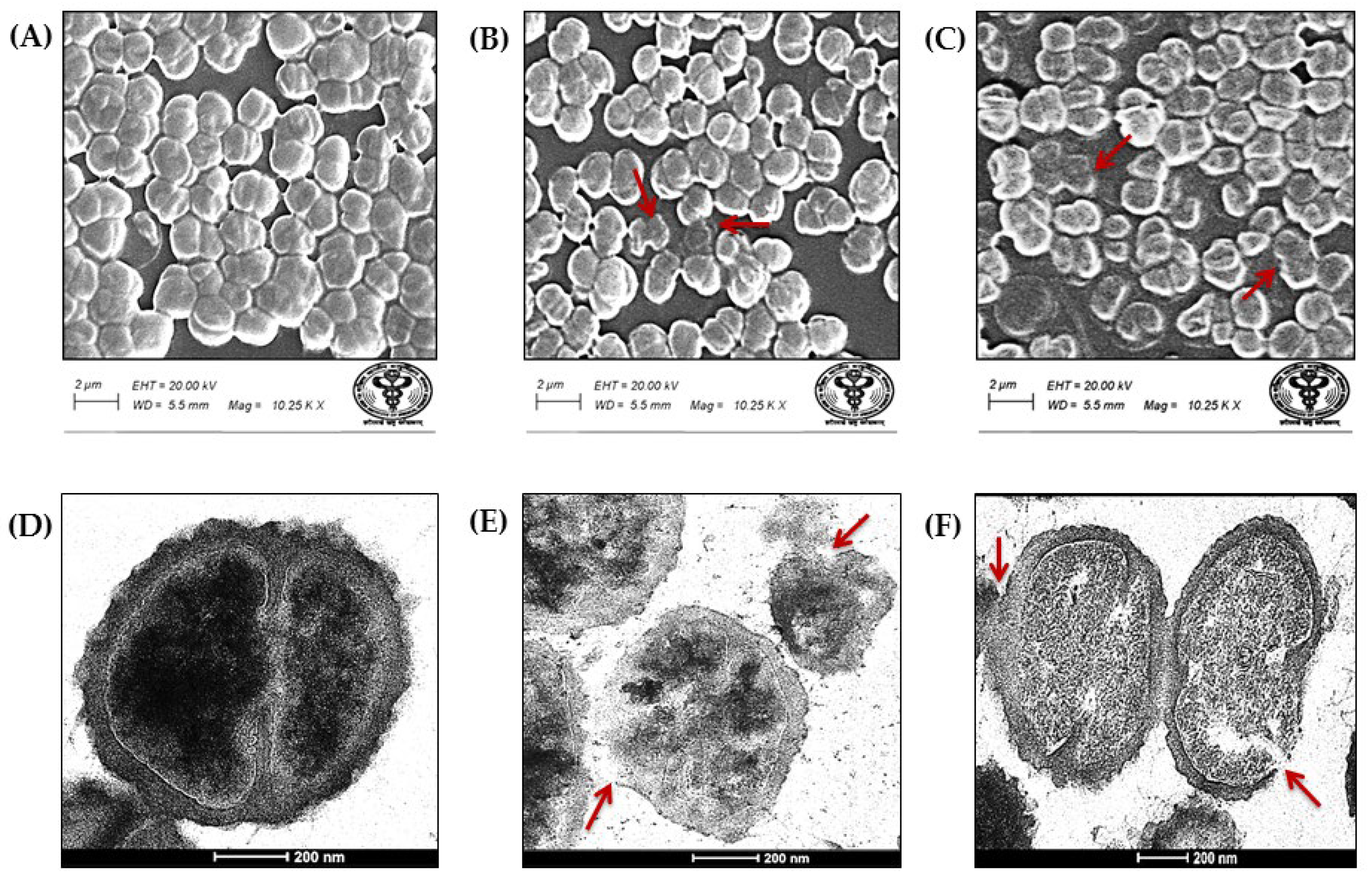
Microscopy confirmed that weissellicin LM85 disrupted the cell structure of M. luteus MTCC106. The untreated cells of M. luteus MTCC106 used as a control display a typical cocci shape with intact and well-defined cell boundaries in SEM analysis (Figure 5A). In contrast, cells treated with nisin showed ruptured boundaries and appeared smaller in size (Figure 5B), with severe cellular damage, including swollen, broken, and rough cell boundaries, with cells also appearing smaller after treatment with 138.3 µg/mL (Figure 5C).
The untreated cells showed normal morphology with an intact cytoplasmic membrane and complete cell boundaries in TEM analysis (Figure 5D). In treated cells, bursting of cells, membrane rupture, and separation between the cytoplasmic and outer membrane were clearly observed (Figure 5E). Additional signs of damage, such as intracellular contents leakage, cell lysis, membrane deformation, and surface cuts, were evident in cells exposed to weissellicin LM85 (Figure 5F). These observations are strongly suggestive that bacteriocins, including weissellicin LM85, disrupt both the cytoplasmic membrane and damage the cell wall of M. luteus MTCC106, likely through pore-forming activity that results in leakage of intracellular components into the surrounding environment, ultimately leading to a strong bactericidal effect.

3.8. Inhibition of Growth and Biofilm Formation of S. Typhimurium ATCC13311 and S. aureus ATCC 25923

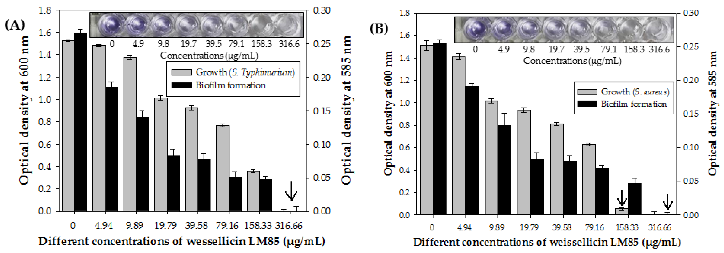
Weissellicin LM85 strongly inhibited both growth and biofilm formation of pathogenic bacteria. The untreated S. Typhimurium ATCC13311 exhibited growth up to OD600 1.52 ± 0.05. In contrast, treatment with weissellicin LM85 led to a dose-dependent decrease in OD600, suggesting the inhibition of growth, as shown in Supplementary Table S4. The complete growth was inhibited after treatment with 316.66 µg/mL and was considered as the MIC against S. Typhimurium ATCC13311 (Figure 6A). A concentration-dependent inhibition of biofilm formation was observed. In the control, cells produced a dark purple color with OD585 0.24 ± 0.002, indicating strong biofilm formation. However, treatment with different concentrations of weissellicin LM85 reduced the intensity of purple color and OD585, suggesting the reduction of biofilm formation, as shown in Supplementary Table S4. The transparent color was observed with nil OD585 at 316.66 µg/mL, indicating the complete inhibition of biofilm formation (Figure 6A).
The untreated S. aureus ATCC25923 reached an OD600 of 1.51 ± 0.043. The different concentrations of weissellicin LM85 led to a progressive decrease in OD600, suggesting growth inhibition, as shown in Supplementary Table S4. The complete inhibition of growth was observed at 158.33 µg/mL and was considered as the MIC against S. aureus ATCC25923 (Figure 6B). The untreated cells produced a purple color with OD585 0.23 ± 0.003, indicating strong biofilm formation. Different concentrations of weissellicin LM85 reduced purple color intensity and OD585, indicating the dose-dependent reduction in biofilm formation, as shown in Supplementary Table S4. The purple color completely disappeared when treated with 158.33 µg/mL and higher concentrations. Hence, 316.66 µg/mL of weissellicin LM85 completely prevented the biofilm formation of S. aureus ATCC25923 (Figure 6B).

4. Discussion

Bacteriocins inhibit bacteria mainly through a membrane interaction that causes pore-formation [31]. Electrostatic interactions between bacteriocins and negatively charged membrane components, such as phospholipids and teichoic acids in Gram-positive bacteria and lipopolysaccharides in Gram-negative bacteria, facilitate their binding [32]. Following this interaction, bacteriocins are proposed to integrate into the cytoplasmic membrane, causing the formation of ion-permeable pores. This results in leakage of intracellular metabolites, dissipation of energy gradients, and loss of cell viability [8,33]. Additionally, cell lysis can occur through the stimulation of autolytic enzymes linked with lipoteichoic, teichoic, and teichuronic acids, which influence the bacteriocin interaction with the bacterial cell wall [8,31,32]. There has been increasing interest in researching and developing new bacteriocins from different bacteria.
This study provides a detailed killing mechanism of purified weissellicin LM85 against M. luteus MTCC106, demonstrating a bactericidal effect through K+ ion efflux, disruption of Δψ and ΔpH, and genomic DNA degradation. The antimicrobial potency of weissellicin LM85 was quantitatively confirmed by effective inhibitory and bactericidal concentrations for M. luteus MTCC106. In comparison, Goh and Philip [1] reported that bacteriocin A3 from W. confusa A3 displayed considerably lower MIC and MBC values against M. luteus ATCC10240. In another study, weissellicin A purified from W. paramesenteroides DX showed similar inhibitory and bactericidal effects on M. flavus ATCC400 [15]. In contrast, plantaricin Pln1 required a higher concentration to inhibit the growth of M. luteus CMCC63202 [34]. Several methods are available to study bacteriocin mechanisms, and colony-forming unit (CFU) enumeration remains a standard for assessing bacterial viability. The kill-kinetics assay further confirmed that weissellicin LM85 reduced cell viability of M. luteus MTCC106 in a concentration- and time-dependent manner. At sub-MICs, only a gradual decline in CFU counts was observed, while MIC and higher doses produced more rapid reductions. Similarly, peptide UTNGt21O from W. cibaria UTNGt21O showed the loss in cell viability in E. coli ATCC25922 in a dose- and time-dependent way [35].
The fluorescent staining with DAPI and PI was used to determine the live and dead cells, respectively [36]. The intact cell membranes prevent the entry of dyes that normally penetrate compromised membranes of dead cells. In contrast, PI is excluded by intact membranes but readily enters damaged cells, where it binds to cellular DNA [37,38]. It is considered a high-affinity nuclear staining reagent [8]. The untreated M. luteus MTCC106 cells displayed blue fluorescence due to DAPI binding, indicative of intact membranes and viability. The cells treated with weissellicin LM85 exhibited both blue (DAPI) and red (PI) fluorescence, suggesting a mixture of live and dead populations due to membrane disruption. Similarly, a mixture of blue (live) and red (dead) populations of M. luteus MTCC106 was observed after treatment with plantaricin LD1 [39]. These fluorescence microscopy observations are consistent with membrane disruption in M. luteus MTCC106 following exposure to weissellicin LM85. Together, the results strongly suggest a bactericidal mode of action of weissellicin LM85 against M. luteus MTCC106.
Bacteriocins are widely recognized to exert their effects by inducing pore-formation in the membranes of indicator strains [40]. Bacteriocins integrate into the membrane through potential-dependent alignment, leading to the leakage of intracellular components such as K+ ions, DNA, and RNA. This process results in ATP hydrolysis, disruption of ∆ψ, and finally cell death [41,42]. The K+ ion efflux from cells of the indicator strain is a standard method used to evaluate membrane disruption initiated by bacteriocins [24,43]. Weissellicin LM85 induced K+ ion efflux from M. luteus MTCC106 cells, with higher bacteriocin concentrations, causing higher release of K+ ions. Similarly, the other membrane-targeted peptides, such as plantaricin MG, NC8α, and NC8β, also induce the release of K+ ions in M. luteus CGMCC1.193, which causes cell death [44]. These findings also prompted us to examine the dissipation of ∆ψ in the indicator strains. The observed K+ ion efflux in M. luteus MTCC106 cells provides a strong indication that weissellicin LM85 disrupts membrane integrity, consistent with previous reports [24,29].
The involvement of membrane potential in the bactericidal effect of weissellicin LM85 was examined. Bacterial transmembrane potential plays an essential role in energy-dependent processes, including ion transport, ATP synthesis, and movement [45]. In this study, membrane permeabilization in cells of M. luteus MTCC106 was assessed by monitoring the dissipation of both ∆ψ and ∆pH. After treatment with weissellicin LM85 (138.3 µg/mL), this resulted in total dissipation of both ∆ψ and ∆pH in the cells. In contrast, cells treated with bacteriocin diluents showed no dissipation of both components, which indicates the pH of the intracellular space remained similar to that of the buffer [26]. The control treatment with valinomycin led to full cellular depolarization. Furthermore, the absence of additional fluorescence changes upon valinomycin addition confirmed that weissellicin LM85 alone was sufficient to completely dissipate ∆ψ. These results suggest that purified weissellicin LM85 can disrupt both ∆ψ and ∆pH in M. luteus MTCC106, effects that are strongly indicative of membrane-targeting bactericidal activity. Similarly, plantaricin NC8 caused dissipation of Δψ and ΔpH in M. luteus CGMCC1.193 through membrane permeabilization [44]. These pores caused efflux of ions (K+/Na+), ATP, nucleic acids, and other intracellular components, thereby disrupting PMF and confirming bactericidal activity [26,46]. Overall, weissellicin LM85 exerts its antimicrobial effect through membrane-acting by pore-formation, resulting in dissipation of ∆ψ and ion leakage, ultimately resulting in cell death in M. luteus MTCC106.
Antimicrobial peptides had the ability to inhibit the synthesis and functions of intracellular biopolymers, particularly those that exhibited specific inhibition of DNA synthesis by binding directly to bacterial DNA [24]. Similarly, weissellicin LM85 was found to penetrate M. luteus MTCC106 cells and interact with their DNA. Genomic DNA isolated from untreated M. luteus MTCC106 cells appeared as a sharp intact band, whereas DNA from weissellicin LM85-treated cells displayed a progressively smeared pattern with increasing concentrations. Instead, the observed smear reflects nonspecific DNA damage, including random fragmentation and altered migration, likely caused by direct interaction of weissellicin LM85 with genomic DNA and/or secondary effects of membrane disruption leading to leakage and subsequent DNA breakdown. Du et al. [27] also demonstrated that treatment with plantaricin GZ1-27 blocked the DNA repair pathway, resulting in genomic instability of the indicator stain and ultimately causing cell damage or death. The in vitro study further confirmed the interaction between weissellicin LM85 and the genomic DNA of M. luteus MTCC106. The isolated genomic DNA treated with weissellicin LM85 showed reduced migration during electrophoresis, indicating direct interaction, and this effect was concentration-dependent. Similarly, Miao et al. [24] reported that peptide F1 interacted with the genomic DNA of E. coli ATCC25922 in vitro in a concentration-dependent manner. In another study, bacteriocin from W. confusa A3 showed bactericidal action through DNA binding [1]. Zhao et al. [29] also demonstrated that plantaricin 827 interacts with genomic DNA of S. aureus ATCC25923 in vitro and in silico binding to AT-rich regions of the DNA minor groove. Such DNA-binding capacity of bacteriocins is a distinctive feature, suggesting that interference with DNA integrity and function contributes to growth inhibition and cell death. Future work using DNase protection assays or oxidative stress inhibition studies will be essential to clarify whether the observed DNA degradation arises from direct nucleic acid interaction or secondary effects of cellular stress.
Microscopic analysis of weissellicin LM85-treated cells confirmed its activity against M. luteus MTCC106. This structural damage indicates the membrane-acting nature of weissellicin LM85. These observations are consistent with a bactericidal effect of weissellicin LM85 on M. luteus MTCC106, likely mediated by membrane permeabilization and associated leakage of cellular contents. Such membrane disruption is a well-recognized mechanism of bacteriocins, as also reported by Zhang et al. [47]. TEM analysis further confirmed the damage in weissellicin LM85-treated cells. Many cells showed rupture and cytoplasmic leakage, strongly suggestive that weissellicin LM85 disrupts the cytoplasmic membrane and cell wall. Membrane penetrations and increased intracellular leakage were also observed, providing strong evidence of the bactericidal effects of weissellicin LM85.
Consumption of contaminated raw vegetables and fruit juices often leads to salmonellosis, a food-borne infection commonly caused by Salmonella, manifested as fever, diarrhea, vomiting, etc. [22]. Skin and soft tissue infections, potentially life-threatening, are caused by several Staphylococcus species [48]. Bacteriocins from LAB are widely studied for their application in food safety and therapeutics. In this study, weissellicin LM85 inhibited the growth and biofilm formation of pathogenic bacteria S. Typhimurium ATCC13311 and S. aureus ATCC25923 in a dose-dependent manner. Similarly, growth of S. UTNSm2, S. enterica ATCC51741, and S. Abaetetuba ATCC35640 was strongly inhibited by bacteriocin Cys2-2 isolated from W. confusa Cys2-2 [18]. In another study, growth of S. typhi DMST22842 was suppressed by bacteriocin-containing CFS of W. confusa WM36 and W. viridescens WM33 [49]. Tenea et al. [35] also reported that peptide UTNGt21O from W. cibaria UTNGt21O inhibited the growth of S. enterica ATCC 51741. However, weissellicin Y and M from W. hellenica QU13 and bacteriocin A3 from W. confusa A3 did not inhibit the growth of S. aureus ATCC12600 and S. aureus RF122, respectively [1,16].
In the present study, weissellicin LM85 was purified using multi-step chromatography and characterized for its bactericidal activity, which involved intracellular ion efflux and dissipation of Δψ and ΔpH in M. luteus MTCC106. Furthermore, its ability to inhibit both growth and biofilm formation of pathogenic bacteria highlights the potential of weissellicin LM85 as a candidate for further evaluation in food safety and therapeutic applications. This study highlights the novel dual mechanism of weissellicin LM85, targeting both the cell membrane and genomic DNA. It is limited to in vitro experiments and lacks cytotoxicity assays or in vivo/food model evaluations. Future studies should address these limitations by evaluating cytotoxicity, conducting in vivo testing, and exploring potential synergy with antibiotics to fully assess their therapeutic and industrial potential.

5. Conclusions

The present study demonstrates that weissellicin LM85 exerts strong antimicrobial activity against M. luteus MTCC106 through multiple mechanisms and in vitro application against pathogenic bacteria S. Typhimurium ATCC13311 and S. aureus ATCC25923. Weissellicin LM85 exhibits a multifaceted antibacterial mode of action, suggestive of pore-forming activity that disrupts cell membrane integrity and induces DNA degradation, ultimately ensuring strong bactericidal and antibiofilm effects. These findings highlight its potential as a natural antimicrobial candidate for food and therapeutic applications. While weissellicin LM85 shows promising antimicrobial and antibiofilm activity, future work will need to evaluate its safety, cytotoxicity, and compatibility with host cells (e.g., Caco-2, Vero, or other mammalian models) to support its therapeutic application.

Supplementary Materials

The following supporting information can be downloaded at https://www.mdpi.com/article/10.3390/nutraceuticals5040033/s1: Table S1: The percentage (%) of inhibition of growth and cell viability of Micrococcus luteus MTCC106 after treatment with different concentrations of weissellicin LM85; Table S2: Percentage (%) of cell viability of Micrococcus luteus MTCC106 at different time interval after treatment with different concentrations of weissellicin LM85; Table S3: Efflux of potassium (K+) ions from cells of Micrococcus luteus MTCC106 at different time interval after treatment with different concentrations of weissellicin LM85 and nisin (positive control); Table S4: Inhibition of growth and biofilm formation of Salmonella enterica subsp. enterica serovar Typhimurium ATCC13311 and Staphylococcus aureus subsp. aureus ATCC25923 after treatment with different concentrations of weissellicin LM85.

Author Contributions

The experiments were carried out by M.K.Y., who also prepared the original draft of the manuscript; S.K.T. designed the study and supplied all laboratory resources. All authors have read and agreed to the published version of the manuscript.

Funding

This work was financially supported by the Indian Council of Medical Research (5/9/1117/2013-NUT) and the Indian National Science Academy (IA/INDO-AUST/F-4/2017/1872), New Delhi, India.

Institutional Review Board Statement

Not applicable.

Informed Consent Statement

Not applicable.

Data Availability Statement

All data supporting the findings of this study are presented in the manuscript and Supplementary Materials; no additional data are available.

Acknowledgments

We greatly acknowledge the Indian Council of Medical Research, New Delhi, India, and the University Research scholarship from the Department of Genetics, M.D. University Rohtak, Haryana, India, for supporting M.K.Y. with fellowships.

Conflicts of Interest

The authors confirm that there are no conflicts of interest.

Abbreviations

The following abbreviations are used in this manuscript:
AMPsAntimicrobial peptides
MICMinimum inhibitory concentration
MBCMinimum bactericidal concentration
K+Potassium ion
∆ψMembrane potential
∆pHTransmembrane pH gradient
MRSDe Man, Rogosa, and Sharpe
LABLactic acid bacteria
PMFProton motive force
NBNutrient broth
DAPI4′,6-Diamidino-2-Phenylindole
PIPropidium iodide
RTRoom temperature
NaClSodium chloride
KClPotassium chloride
HEPES4-(2-hydroxyethyl)-1-piperazineethanesulfonic acid
di-S-C3-(3)3,3′-dipropylthicarbocyanine iodide
BCECF, AM2′,7′-Bis-(2-Carboxyethyl)-5-(and-6)-Carboxyfluorescein, Acetoxymethyl Ester
SEMScanning electron microscope
TEMTransmission electron microscope
ddH2ODouble distilled water
HMDSHexamethyldisilazane
CVCrystal violet
PBSPhosphate buffer saline
CFU/mLColony-forming units per milliliter

References

  1. Goh, H.F.; Philip, K. Purification and characterization of bacteriocin produced by Weissella confusa A3 of dairy origin. PLoS ONE 2015, 10, e0140434. [Google Scholar] [CrossRef]
  2. Singh, J.K.; Devi, P.B.; Reddy, G.B.; Jaiswal, A.K.; Kavitake, D.; Shetty, P.H. Biosynthesis, classification, properties, and applications of Weissella bacteriocins. Front. Microbiol. 2024, 15, 1406904. [Google Scholar] [CrossRef]
  3. Allen, H.K.; Trachsel, J.; Looft, T.; Casey, T.A. Finding alternatives to antibiotics. Ann. N. Y. Acad. Sci. 2014, 1323, 91–100. [Google Scholar] [CrossRef] [PubMed]
  4. Cleveland, J.; Montville, T.J.; Nes, I.F.; Chikindas, M.L. Bacteriocins: Safe, natural antimicrobials for food preservation. Int. J. Food Microbiol. 2001, 71, 1–20. [Google Scholar] [CrossRef] [PubMed]
  5. Todorov, S.D.; Popov, I.; Weeks, R.; Chikindas, M.L. Use of bacteriocins and bacteriocinogenic beneficial organisms in food products: Benefits, challenges, concerns. Foods 2022, 11, 3145. [Google Scholar] [CrossRef] [PubMed]
  6. He, W.; Zeng, Y.; Shen, J.; Li, K.; Zhou, Y.; Zeng, X.; Pan, D. Enhanced bacteriocin production by lactic acid bacteria by co-culture of Lactiplantibacillus plantarum and Limosilactobacillus fermentum. Food Biosci. 2025, 68, 106358. [Google Scholar] [CrossRef]
  7. Yadav, M.K.; Tiwari, S.K. Weissellicin LM85 purified from Weissella confusa LM85 effluxes potassium ions and depletes proton motive force in Escherichia coli ATCC 25922. Int. J. Pept. Res. Ther. 2024, 30, 44. [Google Scholar] [CrossRef]
  8. Cui, Y.; Luo, L.; Wang, X.; Lu, Y.; Yi, Y.; Shan, Y.; Liu, B.; Zhou, Y.; Lü, X. Mining, heterologous expression, purification, antibactericidal mechanism, and application of bacteriocins: A review. Compr. Rev. Food Sci. Food Saf. 2021, 20, 863–899. [Google Scholar] [CrossRef]
  9. Manberger, A.; Verbrugghe, P.; Guðmundsdóttir, E.E.; Santesson, S.; Nilsson, A.; Hreggviðsson, G.Ó.; Linares-Pastén, J.A.; Nordberg Karlsson, E. Taxogenomic assessment and genomic characterisation of Weissella cibaria strain 92 able to metabolise oligosaccharides derived from dietary fibres. Sci. Rep. 2020, 10, 5853. [Google Scholar] [CrossRef]
  10. Kavitake, D.; Devi, P.B.; Shetty, P.H. Overview of exopolysaccharides produced by Weissella genus—A review. Int. J. Biol. Macromol. 2020, 164, 2964–2973. [Google Scholar] [CrossRef]
  11. Fhoula, I.; Boumaiza, M.; Tayh, G.; Rehaiem, A.; Klibi, N.; Ouzari, I.H. Antimicrobial activity and safety features assessment of Weissella spp. from environmental sources. Food Sci. Nutr. 2022, 10, 2896–2910. [Google Scholar] [CrossRef]
  12. Srionnual, S.; Yanagida, F.; Lin, L.H.; Hsiao, K.N.; Chen, Y.S. Weissellicin 110, a newly discovered bacteriocin from Weissella cibaria 110, isolated from plaa-som, a fermented fish product from Thailand. Appl. Environ. Microbiol. 2007, 73, 2247–2250. [Google Scholar] [CrossRef]
  13. Leong, K.H.; Chen, Y.S.; Lin, Y.H.; Pan, S.F.; Yu, B.; Wu, H.C.; Yanagida, F. Weissellicin L, a novel bacteriocin from sian-sianzih-isolated Weissella hellenica 4-7. J. Appl. Microbiol. 2013, 115, 70–76. [Google Scholar] [CrossRef] [PubMed]
  14. Chen, C.; Chen, X.; Jiang, M.; Rui, X.; Li, W.; Dong, M. A newly discovered bacteriocin from Weissella hellenica D1501 associated with Chinese Dong fermented meat (Nanx Wudl). Food Control 2014, 42, 116–124. [Google Scholar] [CrossRef]
  15. Papagianni, M.; Papamichael, E.M. Purification, amino acid sequence and characterization of the class IIa bacteriocin weissellin A, produced by Weissella paramesenteroides DX. Bioresour. Technol. 2011, 102, 6730–6734. [Google Scholar] [CrossRef] [PubMed]
  16. Masuda, Y.; Zendo, T.; Sawa, N.; Perez, R.H.; Nakayama, J.; Sonomoto, K. Characterization and identification of weissellicin Y and weissellicin M, novel bacteriocins produced by Weissella hellenica QU 13. J. Appl. Microbiol. 2012, 112, 99–108. [Google Scholar] [CrossRef]
  17. Woraprayote, W.; Pumpuang, L.; Tosukhowong, A.; Roytrakul, S.; Perez, R.H.; Zendo, T.; Sonomoto, K.; Benjakul, S.; Visessanguan, W. Two putatively novel bacteriocins active against Gram-negative food borne pathogens produced by Weissella hellenica BCC 7293. Food Control 2015, 55, 176–184. [Google Scholar] [CrossRef]
  18. Tenea, G.N.; Lara, M.I. Antimicrobial compounds produced by Weissella confusa Cys2-2 strain inhibit Gram-negative bacteria growth. CyTA-J. Food 2019, 17, 105–111. [Google Scholar] [CrossRef]
  19. Sreelakshmi, K.; Kadirvelu, K.; Ramasubramanian, V. Probiotic efficacy, antioxidant, antimicrobial and anticancer activities of cell free supernatant derived from Weissella confusa isolated from Oreochromis niloticus—An in vitro study. The Microbe 2025, 6, 100291. [Google Scholar] [CrossRef]
  20. Kaur, R.; Tiwari, S.K. Isolation, identification and characterization of Pediococcus pentosaceus LB44 and Weissella confusa LM85 for the presence of bacteriocin-like inhibitory substances (BLIS). Microbiology 2016, 85, 540–547. [Google Scholar] [CrossRef]
  21. Kaur, R.; Tiwari, S.K. Optimization of culture conditions for bacteriocin production by soil isolates Pediococcus pentosaceus LB44 and Weissella confusa LM85. Int. J. Infect. 2017, 4, e15842. [Google Scholar] [CrossRef]
  22. Gao, Y.; Li, D. Antibacterial mode of action of garviecin LG34 against Gram-negative bacterium Salmonella typhimurium. FEMS Microbiol. Lett. 2024, 371, fnae066. [Google Scholar] [CrossRef] [PubMed]
  23. Sheoran, P.; Yadav, M.K.; Kumari, I.; Tiwari, S.K. Enterocin LD3 from Enterococcus hirae LD3 Inhibits the Growth of Salmonella enterica subsp. enterica serovar Typhimurium ATCC13311 in Fruit Juice. Probiotics Antimicrob. Proteins 2024, 16, 1205–1213. [Google Scholar] [CrossRef] [PubMed]
  24. Miao, J.; Liu, G.; Ke, C.; Fan, W.; Li, C.; Chen, Y.; Dixon, W.; Song, M.; Cao, Y.; Xiao, H. Inhibitory effects of a novel antimicrobial peptide from kefir against Escherichia coli. Food Control 2016, 65, 63–72. [Google Scholar] [CrossRef]
  25. Leslie, V.A.; Alarjani, K.M.; Malaisamy, A.; Balasubramanian, B. Bacteriocin producing microbes with bactericidal activity against multidrug resistant pathogens. J. Infect. Public Health 2021, 14, 1802–1809. [Google Scholar] [CrossRef]
  26. Shi, F.; Wang, Y.; Li, Y.; Wang, X. Mode of action of leucocin K7 produced by Leuconostoc mesenteroides K7 against Listeria monocytogenes and its potential in milk preservation. Biotechnol. Lett. 2016, 38, 1551–1557. [Google Scholar] [CrossRef]
  27. Du, H.; Zhou, L.; Lu, Z.; Bie, X.; Zhao, H.; Niu, Y.D.; Lu, F. Transcriptomic and proteomic profiling response of methicillin-resistant Staphylococcus aureus (MRSA) to a novel bacteriocin, plantaricin GZ1-27 and its inhibition of biofilm formation. Appl. Microbiol. Biotechnol. 2020, 104, 7957–7970. [Google Scholar] [CrossRef]
  28. Zhang, J.; Gu, S.; Zhang, T.; Wu, Y.; Ma, J.; Zhao, L.; Li, X.; Zhang, J. Characterization and antibacterial modes of action of bacteriocins from Bacillus coagulans CGMCC 9951 against Listeria monocytogenes. LWT 2022, 160, 113272. [Google Scholar] [CrossRef]
  29. Zhao, D.; Wang, Q.; Lu, F.; Bie, X.; Zhao, H.; Lu, Z.; Lu, Y. A novel plantaricin 827 effectively inhibits Staphylococcus aureus and extends shelf life of skim milk. LWT 2022, 154, 112849. [Google Scholar] [CrossRef]
  30. Shastry, R.P.; Arunrenganathan, R.R.; Rai, V.R. Characterization of probiotic Enterococcus lactis RS5 and purification of antibiofilm enterocin. Biocatal. Agric. Biotechnol. 2021, 31, 101897. [Google Scholar] [CrossRef]
  31. Yap, P.G.; Lai, Z.W.; Tan, J.S. Bacteriocins from lactic acid bacteria: Purification strategies and applications in food and medical industries: A review. Beni-Suef Univ. J. Basic Appl. Sci. 2022, 11, 51. [Google Scholar] [CrossRef]
  32. Soltani, S.; Hammami, R.; Cotter, P.D.; Rebuffat, S.; Said, L.B.; Gaudreau, H.; Bédard, F.; Biron, E.; Drider, D.; Fliss, I. Bacteriocins as a new generation of antimicrobials: Toxicity aspects and regulations. FEMS Microbiol. Rev. 2021, 45, fuaa039. [Google Scholar] [CrossRef]
  33. Deng, Z.; Hou, K.; Zhao, J.; Wang, H. The probiotic properties of lactic acid bacteria and their applications in animal husbandry. Curr. Microbiol. 2022, 79, 22. [Google Scholar] [CrossRef]
  34. Meng, F.; Zhao, H.; Zhang, C.; Lu, F.; Bie, X.; Lu, Z. Expression of a novel bacteriocin the plantaricin Pln1-in Escherichia coli and its functional analysis. Protein Expr. Purif. 2016, 119, 85–93. [Google Scholar] [CrossRef] [PubMed]
  35. Tenea, G.N.; Hurtado, P.; Ortega, C. A novel Weissella cibaria strain UTNGt21O isolated from wild Solanum quitoense fruit: Genome sequence and characterization of a peptide with highly inhibitory potential toward Gram-negative bacteria. Foods 2020, 9, 1242. [Google Scholar] [CrossRef] [PubMed]
  36. Duan, F.; Xin, G.; Niu, H.; Huang, W. Chlorinated emodin as a natural antibacterial agent against drug-resistant bacteria through dual influence on bacterial cell membranes and DNA. Sci. Rep. 2017, 7, 12721. [Google Scholar] [CrossRef] [PubMed]
  37. Johnson, M.B.; Criss, A.K. Fluorescence microscopy methods for determining the viability of bacteria in association with mammalian cells. J. Vis. Exp. 2013, 79, e50729. [Google Scholar] [CrossRef]
  38. Jiang, Y.; Mei, C.; Huang, X.; Gu, Q.; Song, D. Antibacterial activity and mechanism of a bacteriocin derived from the valine-cecropin a (1-8)-plantaricin zj5 (1-18) hybrid peptide against Escherichia coli O104. Food Biophys. 2020, 15, 442–451. [Google Scholar] [CrossRef]
  39. Yadav, M.; Tiwari, S.K. Mechanism of cell-killing activity of plantaricin LD1 against Escherichia coli ATCC 25922. Appl. Biochem. Biotech. 2024, 196, 7570–7587. [Google Scholar] [CrossRef]
  40. Perez-Ramos, A.; Madi-Moussa, D.; Coucheney, F.; Drider, D. Current knowledge of the mode of action and immunity mechanisms of LAB-bacteriocins. Microorganisms 2021, 9, 2107. [Google Scholar] [CrossRef]
  41. Elsilk, S.E.; Azab, E.A.; Tahwash, A.M. Bacteriocins-like substances produced by Enterococcus sanguinicola isolated from traditional Egyptain food Sires (Chicorium pumilum). JSM Microbiol. 2015, 3, 1018. [Google Scholar]
  42. Tenea, G.N.; Pozo, T.D. Antimicrobial peptides from Lactobacillus plantarum UTNGt2 prevent harmful bacteria growth on fresh tomatoes. CyTA-J. Food 2019, 17, 105–111. [Google Scholar] [CrossRef]
  43. Sheoran, P.; Tiwari, S.K. Enterocin LD3 from Enterococcus hirae LD3 causing efflux of intracellular ions and UV-absorbing materials in Gram-negative bacteria. J. Appl. Microbiol. 2019, 126, 1059–1069. [Google Scholar] [CrossRef] [PubMed]
  44. Jiang, H.; Tang, X.; Zhou, Q.; Zou, J.; Li, P.; Breukink, E.; Gu, Q. Plantaricin NC8 from Lactobacillus plantarum causes cell membrane disruption to Micrococcus luteus without targeting lipid II. Appl. Microbiol. Biotechnol. 2018, 102, 7465–7473. [Google Scholar] [CrossRef]
  45. Buttress, J.A.; Halte, M.; Te Winkel, J.D.; Erhardt, M.; Popp, P.F.; Strahl, H. A guide for membrane potential measurements in Gram-negative bacteria using voltage-sensitive dyes. Microbiology 2022, 168, 001227. [Google Scholar] [CrossRef]
  46. Gong, H.S.; Meng, X.C.; Wang, H. Mode of action of plantaricin MG, a bacteriocin active against Salmonella typhimurium. J. Basic Microbiol. 2010, 50, S37–S45. [Google Scholar] [CrossRef]
  47. Zhang, X.; Wang, Y.; Liu, L.; Wei, Y.; Shang, N.; Zhang, X.; Li, P. Two-peptide bacteriocin PlnEF causes cell membrane damage to Lactobacillus plantarum. Biochim. Biophys. Acta Biomembr. 2016, 1858, 274–280. [Google Scholar] [CrossRef]
  48. Yadav, M.K.; Baldia, A.; Tiwari, S.K. Plantaricin LD1 inhibits the growth and biofilm formation of Staphylococcus aureus in milk. J. Explor. Res. Pharmacol. 2024, 9, 1–7. [Google Scholar] [CrossRef]
  49. Pelyuntha, W.; Chaiyasut, C.; Kantachote, D.; Sirilun, S. Cell-free supernatants from cultures of lactic acid bacteria isolated from fermented grape as biocontrol against Salmonella typhi and Salmonella typhimurium virulence via autoinducer-2 and biofilm interference. PeerJ 2019, 7, e7555. [Google Scholar] [CrossRef]
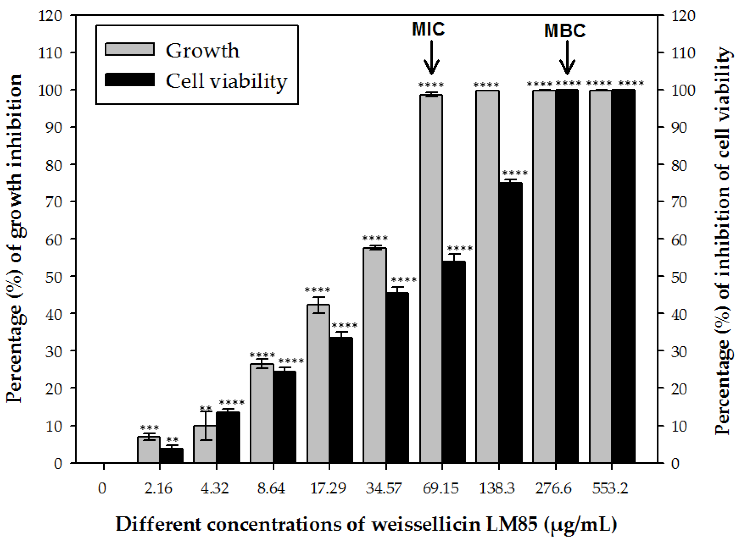
Figure 1. Determination of the minimum inhibitory concentration (MIC) and minimum bactericidal concentration (MBC) of weissellicin LM85 against Micrococcus luteus MTCC106 (arrows indicate MIC and MBC values). The significance levels are shown as ** p < 0.01, *** p < 0.001, and **** p < 0.0001 compared to the data of control.
Nutraceuticals 05 00033 g001
Figure 2. Time- and dose-dependent effect of weissellicin LM85 on the cell viability of Micrococcus luteus MTCC106 (A). Two-way ANOVA showed a significant time × concentration (F (15, 48) = 2825, p < 0.0001). Fluorescence microscopy images of M. luteus MTCC106 cells: untreated (B), ethanol-treated (C), and treated with ½ MIC (34.57 µg/mL) (D), MIC (69.15 µg/mL) (E), and 2 MIC (138.3 µg/mL) (F) of weissellicin LM85, observed at 40× magnification. The images represent qualitative observations captured from multiple randomly selected fields under each condition.
Nutraceuticals 05 00033 g002
Figure 3. Effects of weissellicin LM85 at different concentrations on (A) K+ ion efflux (significant time × concentration F (20, 60) = 2750, p < 0.0001), (B) membrane potential (∆ψ), and (C) transmembrane pH gradient (∆pH) in Micrococcus luteus MTCC106. The diluents of bacteriocins were used as the negative control, whereas nisin (250 µg/mL) was used a positive control.
Nutraceuticals 05 00033 g003
Figure 4. Effects of weissellicin LM85 on genomic DNA (A) in vivo and (B) in vitro of Micrococcus luteus MTCC106 after treatment of cells with MIC (69.15 µg/mL) (well 2), 2 MIC (138.3 µg/mL) (well 3), and 4 MIC (276.6 µg/mL) (well 4). The genomic DNA of untreated cells (well 1) showed a sharp band on 0.8% agarose gel and was used as a control.
Nutraceuticals 05 00033 g004
Figure 5. Scanning electron microscopic (SEM) and transmission electron microscopic (TEM) analysis at 10.25 Kx and 14 Kx magnification, respectively, of untreated Micrococcus luteus MTCC106 cells (A,D) treated with weissellicin LM85 (B,E) and nisin (C,F). The arrows indicate smaller and broken cells, after treatment with the weissellicin LM85.
Nutraceuticals 05 00033 g005
Figure 6. Effects of various concentrations of weissellicin LM85 on inhibition of growth and biofilm formation of (A) Salmonella enterica subsp. enterica serovar Typhimurium ATCC13311 and (B) Staphylococcus aureus subsp. aureus ATCC25923. The concentrations marked by the arrow correspond to those inhibiting growth and biofilm formation. Inset: the decrease in the color intensity indicates the inhibition of biofilm formation.
Nutraceuticals 05 00033 g006
Disclaimer/Publisher’s Note: The statements, opinions and data contained in all publications are solely those of the individual author(s) and contributor(s) and not of MDPI and/or the editor(s). MDPI and/or the editor(s) disclaim responsibility for any injury to people or property resulting from any ideas, methods, instructions or products referred to in the content.