Article

In Vitro Reversal of Escherichia coli Quiescence by Catechol-Containing Polyphenols and Phenolic Acids Across Multiple Strains

by
Terra Marie M. Jouaneh
1,†,
Josiah J. Morrison
2,†,
Abigail C. Luthern
3,
Riley D. Kirk
4,
Jodi L. Camberg
2 and
Matthew J. Bertin
3,*
1
School of Medicine, Johns Hopkins University, Baltimore, MD 21287, USA
2
Department of Cell and Molecular Biology, University of Rhode Island, Kingston, RI 02881, USA
3
Department of Chemistry, Case Western Reserve University, Cleveland, OH 44106, USA
4
Department of Biomedical and Pharmaceutical Sciences, College of Pharmacy, University of Rhode Island, Kingston, RI 02881, USA
*
Author to whom correspondence should be addressed.
These authors contributed equally to this work.
Nutraceuticals 2025, 5(4), 29; https://doi.org/10.3390/nutraceuticals5040029
Submission received: 20 August 2025 / Revised: 17 September 2025 / Accepted: 28 September 2025 / Published: 9 October 2025

Abstract

Urinary tract infections (UTIs) are common and create significant clinical challenges. Most UTIs are caused by uropathogenic Escherichia coli (UPEC) and affect 50 to 70% of women at some point in their lives. Of this population, 25% will have a recurrent urinary tract infection (rUTI) within 3 to 12 months of the first episode. High rates of rUTIs may occur because UPEC can enter a non-proliferative or quiescent state within the urothelium of the bladder. This state allows UPEC to evade the host’s immune response and antibiotic treatment. We utilized a library of plant extracts derived from the URI Heber W. Youngken Jr. Medicinal Garden to determine if they reversed UPEC quiescence with a novel in vitro quiescence assay using the classic UPEC endemic lineage ST73 strain CFT073. We found an overall active extract hit rate of 69% (79/114 active) and that active extracts contained high levels of polyphenolic compounds. Further testing showed that polyphenols with adjacent hydroxyl groups on a benzene ring (catechol moiety) were the most effective and potent in reversing quiescence. The ability to reverse quiescence was also linked to the compound’s ability to bind iron (e.g., epigallocatechin gallate and rosmarinic acid were the most potent reversing agents—0.1 mM—and they both had the strongest iron-binding activity as determined via CAS assay). These findings reveal a new class of metabolites that can reverse quiescence in UPEC strains.

Graphical Abstract

1. Introduction

Bacterial resistance to antibiotics continues to be a major public health issue for the entire world [1]. However, beyond developing resistance to antibiotics, some bacteria develop alternative ways to persist despite the introduction of drugs designed to eradicate them. One persistent pathogen is uropathogenic Escherichia coli (UPEC). UPEC infections are the most common type of bacterial infection and are responsible for 65–90% of urinary tract infections (UTIs) [2]. In the United States alone, this accounts for approximately $3.5 billion dollars in healthcare costs annually [3]. Even after the UTI is initially treated, recurrent infections are a pervasive public health issue, and in 77% of rUTIs, it is the original bacterial strain that is responsible for the recurrence [2,4,5]. This is possibly due to the E. coli developing a biofilm-like intracellular bacterial community (IBC) and quiescent intracellular reservoirs [6]. These adaptations allow E. coli to enter a quiescent state where the bacteria hide in the epithelium of the bladder, evade antibiotic molecules, and then periodically re-emerge and re-colonize to facilitate a rUTI [7]. Antibiotic drugs typically target components of actively dividing bacteria, allowing the E. coli in the quiescent state to avoid eradication [8]. Some natural products such as components in cranberry (Vaccinium macrocarpon) have shown efficacy in interfering with bacterial adherence to bladder cells, which can help reduce E. coli populations in cells [2]. Another approach is to use small molecule proliferants, which have emerged as a promising approach in reversing bacterial quiescence by reactivating dormant cells. These compounds work by targeting specific metabolic pathways and cellular processes that are downregulated during quiescence, thereby restoring bacterial growth and division. By reawakening quiescent bacteria, small molecule proliferants can enhance the efficacy of antibiotics, as actively growing bacteria are more susceptible to antibiotic action. Thus, the identification and application of new small molecule proliferants represents an important strategy in the fight against chronic bacterial infections and antibiotic resistance. A novel in vitro quiescence assay developed at the University of Rhode Island utilizes glucose minimal medium to culture E. coli CFT073 on agar plates at low cell density (i.e., below 106 cells per milliliter), which elicits quiescence in bacteria. Once established, the quiescent state can be reversed by supplementation with small molecule cues (proliferants: e.g., succinate, l-lysine and l-methionine, pectic oligosaccharides from cranberry, and d-amino acid containing peptidoglycan fragments). Resuscitation is observable as colonies growing in a zone on the surface of the agar surrounding the location of the cue addition [2,9].
Plant metabolites offer promise in providing new molecular scaffolds to use as antibiotics and quiescence-reversing agents either alone or in combination with previously discovered antibiotics to increase their efficacy [10,11]. The same metabolites that the plant uses to fight microbes in the environment can be harnessed to combat human pathogens. The University of Rhode Island houses a medicinal garden comprised of over 200 specimens. Previously, we developed a new plant extract library comprised of aqueous and organic extracts of aerial and root portions of each specimen from the garden [12]. In the current report, the extract library was utilized in a quiescence-reversing assay using the uropathogenic E. coli strain CFT073.
Herein, plant extracts were tested against E. coli CFT073 to assess the extracts’ ability to reverse quiescence. Following identification of active extracts, the extracts were subjected to bioassay-guided fractionation and isolation, which led to the identification of high concentrations of plant polyphenols in active samples. Panels of pure polyphenols, phenolic acids, and other plant metabolites were tested in pure form in reversal assays, and a clear structure–activity relationship was observed between compounds containing catechol moieties or multiple adjacent hydroxylations and reversal activity.

2. Materials and Methods

2.1. Chemicals

The compounds tested in this study (including (-)-epigallocatechin gallate (EGCG); (+)-alpha-tocopherol; 3-methoxybenzoic acid; 3,4-(methylenedioxy)cinnamic acid; 3,4-dimethoxycinnamic acid; 3,4,5-trimethoxycinnamic acid; adenosine; allantoin; aloin; baicalein; caffeic acid; caffeine; chromone; coumarin; cyanidin chloride; dopamine; enterodiol; flavone; genistein; gentisic acid; hydrocaffeic acid; l-ascorbic acid; levodopa; p-coumaric acid; quercetin; resveratrol; rosmarinic acid; sesamolin; sinapic acid; stigmasterol; trans-cinnamic acid; and trans-ferulic acid) and the solvents used (including dimethyl sulfoxide and methanol) were purchased from Sigma-Aldrich Chemical Co. (St. Louis, MO, USA). Additional compounds (including ellagic acid and syringic acid) were purchased from Acros Organics (Geel, Antwerp, Belgium). Vanillin and 3-aminoquinoline were purchased from Fisher-Scientific (Waltham, MA, USA).

2.2. General Experimental Procedures

Liquid chromatography–tandem mass spectrometry (LC-MS/MS) was performed using a ThermoFisher LTQ XL mass spectrometer (Thermo Fisher Scientific, Inc., Waltham, MA, USA) with an electrospray ionization (ESI) source equipped with a Dionex Ultimate 3000 high-performance liquid chromatography (HPLC) system (Thermo Fisher Scientific, Inc., Waltham, MA, USA). Analytical and semi-preparative HPLC was carried out using a Dionex UltiMate 3000 HPLC system equipped with a micro vacuum degasser, an autosampler, and a diode-array detector (Thermo Fisher Scientific, Inc., Waltham, MA, USA).

2.3. Preparation of Plant Extracts

Plant specimens were harvested with assistance from the Medicinal Garden Coordinator, Elizabeth Leibovitz, from the Heber W. Youngken Jr. Medicinal Garden in the College of Pharmacy at the University of Rhode Island (URI). Specimens were identified by the Garden Coordinator and voucher specimens are kept in the URI College of Pharmacy Herbarium. Plant extracts were prepared as previously described [12]. Briefly, plants were separated into aerial and underground portions, then further divided for extraction in both aqueous (water) and organic (methanol) solvents. Aerial portions included bark, leaves, seeds, flowers, and stems. Following 24 to 48 h of extraction, each extract was vacuum filtered to remove large particulates, then either lyophilized or evaporated under reduced pressure to remove any remaining solvent. The dried product was sub-aliquoted into small vials, reconstituted at 10 mg/mL in (CH3)2SO, and stored at −20 °C until the assays were performed. The plant extracts tested are listed in Table S1.

2.4. Bacterial Strains and Cultivation

The E. coli strains CFT073, JJ1186, and Nissle 1917 were obtained from cryogenically preserved frozen stocks in the Camberg lab (University of Rhode Island) consisting of a 1:1 combination of Luria-Bertani (LB broth) and 50% glycerol. For E. coli cultivation and handling, both LB broth and LB agar were routinely used. Liquid M9 medium containing glucose (0.2%), and supplemented with noble agar (1.5%) for solid medium plates where indicated, was used to culture cells for the quiescence assay as described previously [5].

2.5. Molecular Network Analysis of Active Plant Extracts

Prioritized organic active extracts were further fractionated using a C18 SPE cartridge and an elution scheme (CH3OH/H2O) from 20:80 to 100:0, resulting in five fractions. The fractions were reconstituted at 0.6 mg/mL in methanol and were subjected to LC-MS/MS analysis on an LTQ XL mass spectrometer. The HPLC method used a Thermo Scientific Acclaim™ 3 µm C18 column (150 mm × 3 mm) and a 70 min gradient beginning with 15% CH3CN for 10 min, then increasing the concentration of CH3CN in a linear manner to 45 min, with a 20 min hold at 100% CH3CN, followed by a return to starting conditions for 5 min. The flow rate was held constant at 0.4 mL/min. The MS spray voltage was 3.5 kV with a capillary temperature of 325 °C. For the MS/MS component, the collision-induced dissociation (CID) isolation width was 1.0 and the collision energy was 35.0 eV. Following acquisition, the raw data files were converted to .mgf format using MSConvert from the ProteoWizard suite. Following conversion, the files were uploaded to the Global Natural Products Social Molecular Network (GNPS at http://gnps.ucsd.edu, accessed on 11 May 2022) website for known metabolite identification. Networks were created where edges were filtered to have a cosine score above 0.7 and 6 matched peaks. The networks were visualized using the Browser Network Visualizer tool available on the platform. Known library hits were accessed using the default Molecular Networking Results Views.

2.6. In Vitro Quiescence Inhibition Assay

The procedure for this assay closely followed the established quiescence inhibition assay protocol that has been used and validated previously [2,5,9]. Briefly, cultures of E. coli CFT073 were grown overnight in 0.4% glucose with M9 minimal medium and quiescence assays were performed as described [5]. Bacteria were diluted to 104 CFU per mL from an overnight culture and added to 3 mL of a liquid agar overlay containing glucose minimal medium with 0.75% noble agar on a prewarmed plate containing the solid culture medium (1.5% noble agar). Overlays containing cells were allowed to solidify for 1 h, cues were added where indicated (5 μL to 20 μL), and plates were incubated at 37 °C for 24 to 48 h. Where indicated, assays were performed similarly for Nissle 1917 (104 CFU per mL) and JJ1886 (103 CFU per mL). The lower cell density for the JJ1886 strain was discovered through assay optimization in previous work [9].
Plant extracts reconstituted at 10 mg/mL were spotted onto the CFT073 plates with 10 μL of each test solution (100 µg/mL). The initial plant extract test concentrations were chosen based on previous research with the assay testing cranberry components [2]. Pure test compounds were initially reconstituted at 10 mg/mL (ranging from 20 mM–90 mM), and 5 μL of the solutions was spotted on the CFT073 plates. The pure test compounds identified as active were titrated (0.1 mM, 1 mM, 10 mM, and 100 mM), and 5 μL was spotted on the CFT073 plates. Five of the active pure test compounds (EGCG, caffeic acid, quercetin, levodopa, and dopamine) were titrated and spotted on the JJ1886 and Nissle 1917 plates.
All quiescence assays were performed in triplicate. E. coli was deemed to be quiescent if the growth was able to be induced by the positive control consisting of 5 μL co-spotted l-lysine and l-methionine (1 mM). The negative control consisted of the vehicle carrier (DMSO), which showed no reversal activity. A positive result was defined in this assay as visually observable bacteria growth at the spot of testing following incubation, which we termed a “zone of activation” (approximately 24 h at 37 °C and an additional 20 h at 23 °C for CFT073 and 24 h at 37 °C for JJ1886 and Nissle 1917). Negative results were defined as no observable growth at the test site following the outline period of incubation. Images of each agar plate were taken using a Molecular Imager Gel Doc XR+ (Bio-rad, Hercules, CA, USA) system with Image Lab Software 6.1.

2.7. Chrome Azurol S (CAS) Assay

The CAS solution was prepared as previously described [13]. In a glass container, 60.5 mg of CAS (Sigma Aldrich, Inc., St. Louis, MO, USA) was dissolved in 50 mL deionized water. Iron(III) chloride hexahydrate (ThermoFisher Scientific) was added to an HCl solution to create 1 mg/mL FeCl3·6H2O in 10 mL 10 mM HCl. The CAS solution was combined and stirred with the iron(III) solution, and a solution of 72.9 mg of hexadecyltrimethylammonium bromide (HDTMA) (Sigma Aldrich, Inc.) in 40 mL deionized water was slowly added. The total volume was adjusted to 100 mL with deionized water.
The EGCG, rosmarinic acid, levodopa, and tyrosine samples were dissolved in deionized water to create 10 mM stock solutions. The stock solutions were then diluted to additional concentrations of 0.1 mM and 1 mM. In a 96-well plate, 50 μL of the CAS reagent was combined with 50 μL of the polyphenols at their varying concentrations, testing in triplicate. These mixtures were left to sit for 1 h. The absorbance was measured at 630 nm using a Tecan Spark multimode plate reader. The siderophore activity was expressed as a percent using the following equation:
Siderophore   units   ( % )   =   [ A r A s A r ] × 100
where Ar is the absorbance of the reference (CAS reagent with water) and As is the absorbance of the sample.

3. Results and Discussion

3.1. Initial Extract Screening: Aqueous and Organic Medicinal Plant Extracts Exhibited Quiescence-Reversal Activity in CFT073 Assay

The uropathogenic E. coli quiescence-reversing activity of 114 plant extracts from the PRISM library was evaluated against E. coli CFT073. After the initial testing of the crude extracts, 79 of the 114 (69%) showed some observable activity (Figure 1 and Figure S1, Table S1), and the remaining 35 extracts (31%) showed no observable activity. Out of the 79 extracts that were active, we prioritized pursuing the potential active constituents of the extracts which had the largest zone of activation, were organic extracts, and showed increased proliferative activity over the aqueous extract. The reason we initially prioritized organic extracts stemmed from previous research with quiescence-reversing molecules. We knew that amino acids such as lysine have shown activity as well as sugar molecules like those in the cranberry plant, which would be extracted in an aqueous solvent. Therefore, putative, new proliferative small molecules had the highest likelihood of being identified in these organic extracts. Active extracts were fractionated using reversed-phase chromatography, retested for proliferative activity (Figure S2), and analyzed by HPLC and LC-MS/MS.

3.2. GNPS Networking Identified Polyphenols and Phenolic Acids as Abundant in the Aerial Extracts

Active chromatography fractions were subjected to LC-MS/MS analysis following by molecular networking using the Global Natural Products Social Molecular Networking site (GNPS) [14]. Networks from the active chromatography fractions were visualized using the online tools at GNPS and annotated using the library search function. Several analytes were annotated as polyphenol compounds from multiple active extracts and chromatography fractions (Figure S3). To abrogate the time-consuming process of isolation and characterization of polyphenols from organic extracts, we shifted our approach from a “top-down” discovery approach using bioassay-guided isolation to a “bottom up” approach utilizing a panel of commercially available pure polyphenol compounds.

3.3. Structure Activity Relationships: Pure Compound Screen Identifies Hydroxylated Compounds as Active on the CFT073 Assay

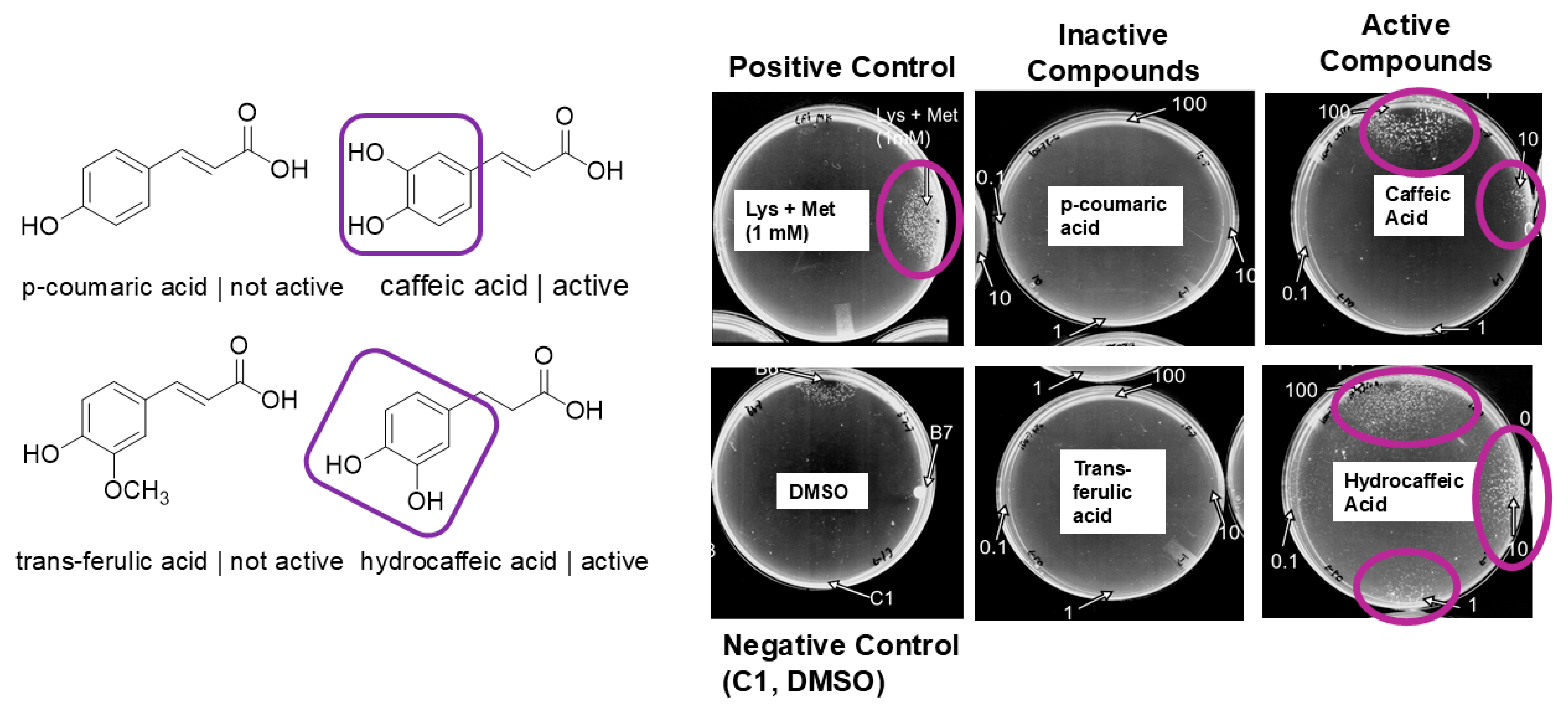
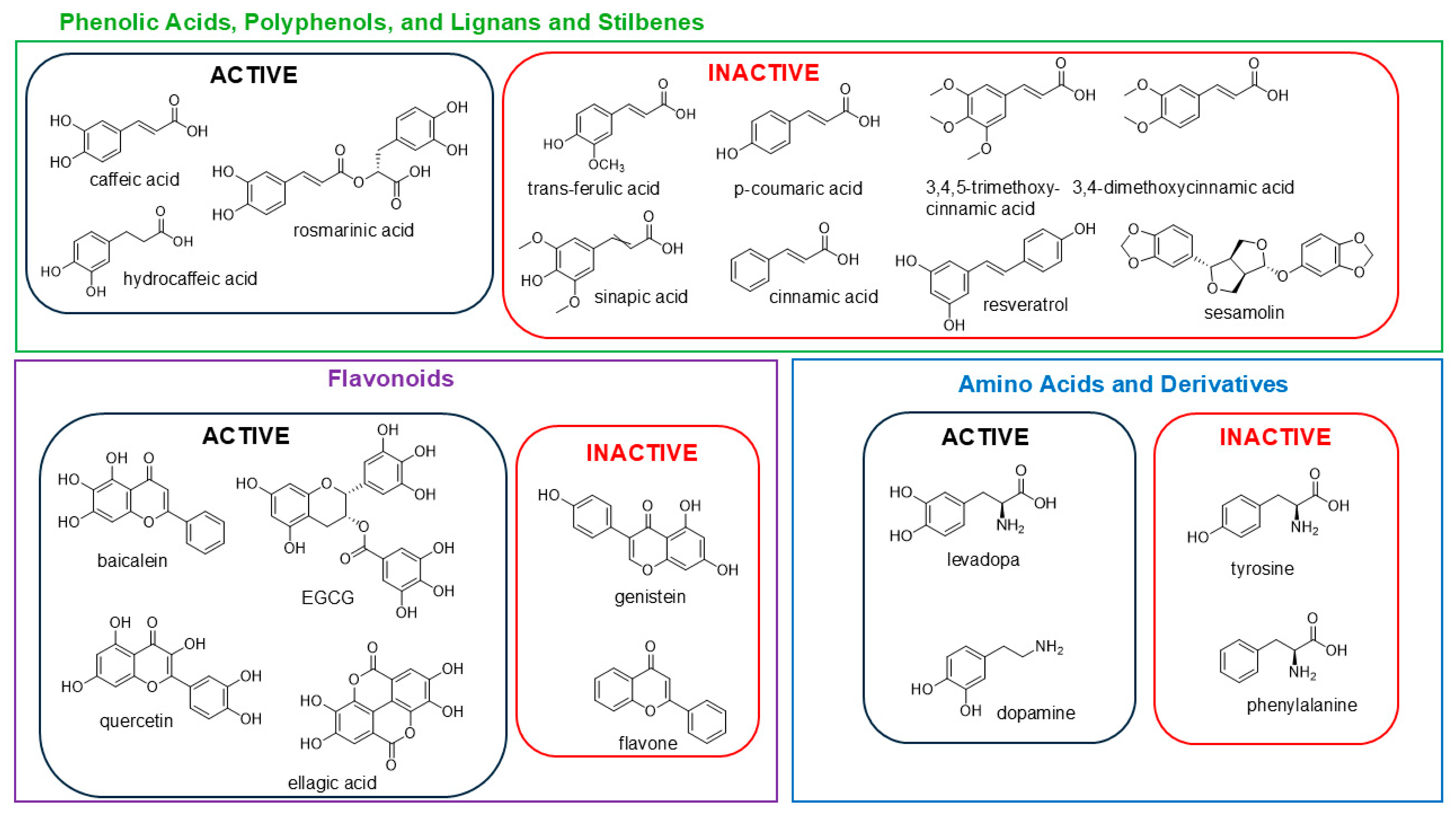
We accumulated a pure compound library including plant-derived phytochemicals and/or well-known polyphenols, phenolic acids, and antioxidants, which were screened in a quiescence-reversal assay to assess reversal activity. Initial screening of the test compounds (chosen to represent polyphenol classes: e.g., anthocyanins, flavonoids, etc.) was carried out at 10 mg/mL concentrations, which translated to a range of 20 to 90 mM for the panel of compounds. In total, the polyphenols tested included nearly all categories of flavonoids (anthocyanins, flavanones, isoflavones, flavanols, flavan-3-ols), phenolic acids (benzoic acid derivatives and cinnamic acid derivatives), coumarins, stilbenes, and lignans as well as other common plant metabolites. The compounds showing activity were hydroxylated compounds, and no activity was identified in the tested steroid molecules, alkaloids, coumarins, stilbenes, and lignans. However, certain inactive compounds were hydroxylated, but the hydroxyl groups were not adjacent (e.g., resveratrol), were not on a six-membered ring (e.g., l-ascorbic acid), or were sterically hindered (alpha-tocopherol). A comparison of genistein (not active), quercetin (active), and flavone (not active) helped to support this structure–activity relationship with respect to hydroxylation pattern (Table S2. There were several direct comparisons that unequivocally showed the SAR between adjacent hydroxylation and reversal. A comparison of caffeic acid (active) and p-coumaric acid (inactive), which differed only in the addition of a hydroxy group to form a catechol moiety in caffeic acid, shed light on the potential active functionality of the catechol; a hydroxy group in the catechol acid (i.e., trans-ferulic acid) rendered the compound inactive; and reducing the double bond on caffeic acid (hydrocaffeic acid) still retained its activity (Figure 2 and Figure 3).
The inactivity of p-coumaric acid and cinnamic acid was initially attributed to the lack of hydroxylation, and that hydrogen bonding or sterics could be contributors. While we found that the removal of one or more hydroxyl groups resulted in the loss of proliferative activity, we hypothesized that protection (alkylation) of one or more hydroxyl groups would also result in the loss of proliferative activity. Therefore, we tested derivatives of caffeic acid with one or more of the hydroxyl groups protected. 3,4-(methylenedioxy)cinnamic acid and 3,4-dimethoxycinnamic acid have both hydroxyl groups protected, and both are inactive (Table S2 and Figure 3). As mentioned above, ferulic acid has the hydroxyl group at the C3 position O-methylated, but the C4 hydroxyl is deprotected (like p-coumaric acid). It is also inactive. Furthermore, neither sinapic acid, which has the unmasked C4 hydroxyl but protected C3 and C5 hydroxyls, nor 3,4,5-trimethoxycinnamic acid, which has protected hydroxyls as positions C3, C4, and C5, exhibit reversal activity (Table S2 and Figure 3).
Amino acids have been found to exhibit reversal activity and a cocktail of two were used as positive controls (1 mM of lysine and methionine or lysine and tyrosine in combination). Previous studies found that seventeen of the essential amino acids exhibited no proliferative activity on their own [9]. Phenylalanine and tyrosine were selected for further examination. Structurally, phenylalanine and tyrosine only differ with a hydroxyl substituent at the C4 position of the ring. Levodopa, a neurotransmitter amino acid, is the result of the oxidation of the C3 on tyrosine. While phenylalanine and tyrosine exhibited no proliferative activity on the assay, both levodopa and dopamine (a decarboxylated derivative of levodopa) were highly proliferative on the assay (Figure S4). The direct comparison of these four compounds further points to the catechol moiety being the pharmacophore driving this proliferative process. A general structure–activity relationship of the different chemical classes tested is shown in Figure 3.
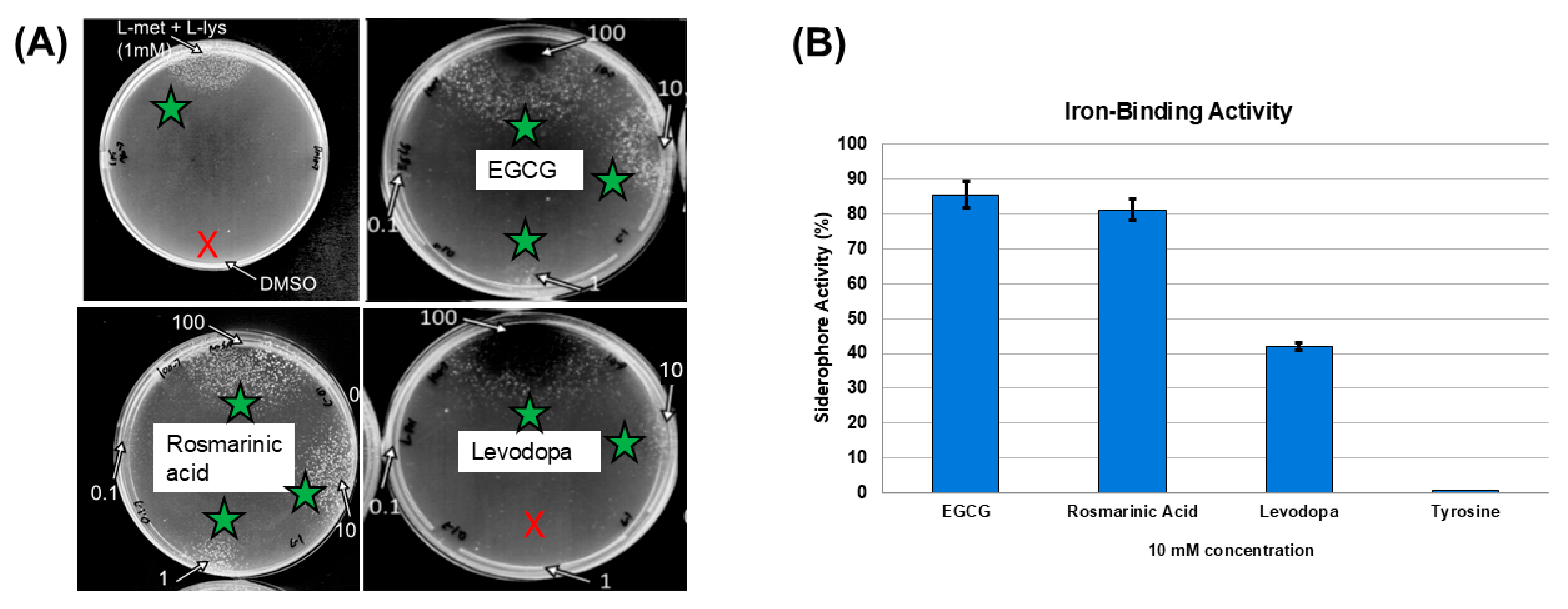
When assessing the entire panel of compounds, epigallocatechin (EGCG) and rosmarinic acid proved to be amongst the most potent, as they were the only compounds to exhibit reversal activity at 0.1 mM (Figure 4A). In total, these results point to the catechol moiety, or three neighboring hydroxyl groups attached to a benzene ring, driving the quiescence-reversal activity.

3.4. Quiescence-Reversal Assays in Additional Strains

While CFT073 is a predominant, prototypical uropathogenic strain studied in the laboratory, five of the most proliferative compounds (EGCG, quercetin, dopamine, levodopa, and caffeic acid) were selected for testing against two additional strains. UPEC JJ1886 is a clinically relevant and antibiotic-multidrug-resistant strain and Nissle 1917 is a probiotic strain but closely related to CFT073. The trend observed with CFT073 generally applied to these two strains, showing that the observed results are not strain-specific effects (Figure S5).

3.5. Iron-Binding Activities of Test Compounds

We speculated that the iron-binding ability of catechol-containing polyphenols could play a role in their ability to reverse quiescence. Previous studies have shown that catechols have ferric-iron-binding capabilities [15], and catechols produced by bacteria are key virulence factors as they bind ferric iron and contribute to the abrogation of bacteriostasis [16]. Quercetin and EGCG have also been shown as iron chelators [13,17]. We did observe that the strongest quiescence-reversing agents also showed the strongest iron-binding ability in CAS assays (Figure 4B, Figure S6). This result was intriguing, and further work will explore the mechanism for this phenomenon. Certain possibilities may be that the sequestering of iron and limiting it from the bacteria instigates the quiescence reversal, or perhaps the bacteria import the iron-bound polyphenols for their own metabolism with quiescence-reversal effects.

4. Conclusions

Plant extracts and metabolites continue to show therapeutic value against UTIs [18]. The results from this study shed light on new biological activities from polyphenol natural products and provoke intriguing questions as to the future development of diet-derived polyphenols to mitigate UTIs and rUTIs [19]. However, there are several limitations with respect to the therapeutic value of plant polyphenols and their ability to reverse quiescence in UPEC. The in vitro nature of the assay itself does not directly reflect the in vivo environment of the bladder epithelium, where UPEC quiescence has been observed. The bacteria form quiescent communities intracellularly within the urothelium of the bladder [8]. If the in vivo response of UPEC to these active compounds is similar to that of its in vitro response observed in this study, only then could a potential dual antibiotic therapy be proposed. We did not validate the quiescent reversal phenomenon in cellular models or animal models, which will be necessary for future therapeutic development. We will need to determine if polyphenol administration results in bioavailable polyphenols at effective concentrations in the urothelium of the bladder. As polyphenols generally have low bioavailability in blood and urine (1–100 µmol) [20], this may be a challenge requiring medicinal chemistry optimization. Additionally, we did not test the toxicity of these polyphenols to cells or animals at the concentrations tested. However, previous work showed that concentrations of polyphenols such as EGCG near or in our test range showed cytoprotective effects in human bladder cells [21,22]. Future work is necessary to determine the potential pharmacophore for quiescence reversal among plant metabolites and the overall molecular mechanism of quiescence reversal in UPEC and the role of iron chelation and siderophores in the mechanism. It may be that in the bacterial assay the exogenous application of the catechol-containing polyphenols is interfering with bacterial iron acquisition and disrupting quiescence. Interestingly, previous research has shown that certain bacterial strains can utilize catechol-containing plant polyphenols as an iron source by transporting them into their cells using TonB transporters [23]. To date, genes reported to be essential for establishing the quiescent state in UPEC include metabolism (sdhA, pykF, zwf, gnd, gdhA), division (zapE), and transcriptional regulation (ihfB) [5,24]. Gene expression of this suite of genes can be probed following iron manipulation and exogenous application of siderophores including plant polyphenols. This is planned for future studies. In this work, we provide evidence that the quiescence-reversal activity of these catechol-containing metabolites is likely related to iron-binding ability, and we demonstrate a new intriguing activity (quiescence-reversal) for plant-derived polyphenols. However, as demonstrated by the positive controls used in this study (lysine with methionine), which presumably do not bind iron, and the role of central carbon metabolism, there are likely additional molecular mechanisms involved in quiescence reversal related to transcriptional regulation, bacterial quorum signaling, and growth transitions [9].

Supplementary Materials

The following supporting information can be downloaded at: https://www.mdpi.com/article/10.3390/nutraceuticals5040029/s1, Figure S1: Quiescence reversal assays in which the PRISM Library extracts and fraction samples were tested against UPEC strain CFT073; Figure S2: Maiz morado organic husk and leaves five chromatography fractions (W = water; M = CH3OH) tested against quiescence-reversal assay; Figure S3: Molecular networks created with mass spectrometry data from (A) the Liriodendron tulipifera organic extract and (B) all 5 Maiz morado chromatography fractions. Select nodes (stars) matched with respect to MS/MS spectra to known library molecules in the GNPS database, which were identified as polyphenols. The side of the node is relative to the amount of ion detected; Figure S4: Comparison of E. coli CFT073 quiescence reversal. Levadopa and dopamine showed quiescence reversal at 100 mM and 10 mM concentrations while phenylalanine and tyrosine were not active; Figure S5: Quiescence-reversal activity of caffeic acid, EGCG, quercetin, levodopa, and dopamine against E. coli strains J1886 and Nissle 1917; Figure S6: CAS assay with EGCG, rosmarinic acid, levodopa, and tyrosine; Table S1: Quiescence reversal activity of extracts from the PRISM Library; Table S2: Compounds tested in this study, their chemical class and ability to reverse quiescence.

Author Contributions

T.M.M.J.: formal analysis, methodology, data curation, and writing original draft; J.J.M.: methodology and formal analysis; A.C.L.: methodology and formal analysis; R.D.K.: conceptualization and formal analysis; J.L.C.: methodology, resources, validation, and supervision; M.J.B.: conceptualization, formal analysis, validation, supervision, and writing original draft. All authors were involved in reviewing and editing the final version of this work. All authors have read and agreed to the published version of the manuscript.

Funding

Research reported in this publication was supported in part by the National Institutes of Health under Award Number 1R21AI156574 to J.L.C. The content is solely the responsibility of the authors and does not necessarily represent the official views of the National Institutes of Health or the authors’ respective institutions.

Institutional Review Board Statement

Not applicable.

Informed Consent Statement

Not applicable.

Data Availability Statement

Most data are available in the Supplementary Materials and the manuscript itself. Any other data will be made available upon request. The Supplementary Materials are available free of charge with the online version of this article and include quiescence assays of plant extracts and a table detailing reversal activity, a table of active and inactive pure compounds, LC-MS/MS networking information, and biological assay results.

Acknowledgments

We gratefully acknowledge all of the researchers who were involved in the generation of the PRISM library with special thanks to Garden Coordinator Elizabeth Leibovitz.

Conflicts of Interest

The authors declare no competing interests.

References

  1. Ventola, C.L. The antibiotic resistance crisis Part 1. J. Clin. Pharm. Ther. 2015, 40, 277–283. [Google Scholar]
  2. Sun, J.; Deering, R.W.; Peng, Z.; Najia, L.; Khoo, C.; Cohen, P.S.; Seeram, N.P.; Rowley, D.C. Pectic oligosaccharides from cranberry prevent quiescence and persistence in the uropathogenic Escherichia coli CFT073. Sci. Rep. 2019, 9, 19590. [Google Scholar] [CrossRef]
  3. Flores-Mireles, A.L.; Walker, J.N.; Caparon, M.; Hultgren, S.J. Urinary tract infections: Epidemiology, mechanisms of infection and treatment options. Nat. Rev. Microbiol. 2015, 13, 269–284. [Google Scholar] [CrossRef]
  4. Nuutinen, M.; Uhari, M. Recurrence and follow-up after urinary tract infection under the age of 1 year old. Pediatr. Nephrol. 2001, 16, 69–72. [Google Scholar] [CrossRef] [PubMed]
  5. Leatham-Jensen, M.P.; Mokszycki, M.E.; Rowley, D.C.; Deering, R.; Camberg, J.L.; Sokurenko, E.V.; Tchesnokova, V.L.; Frimodt-Møller, J.; Krogfelt, K.A.; Leth Nielsen, K.; et al. Uropathogenic Escherichia coli metabolite-dependent quiescence and persistence may explain antibiotic tolerance during urinary tract infection. mSphere 2016, 1, e00055-15. [Google Scholar] [CrossRef]
  6. Mysorekar, I.U.; Hultgren, S.J. Mechanisms of uropathogenic Escherichia coli persistence and eradication from the urinary tract. Proc. Natl. Acad. Sci. USA 2006, 103, 14170–14188. [Google Scholar] [CrossRef]
  7. Silverman, J.A.; Schreiber, H.L.; Hooton, T.M.; Hultgren, S.J. From physiology to pharmacy: Developments in the pathogenesis and treatment of recurrent urinary tract infections. Curr. Urol. Rep. 2013, 14, 448–456. [Google Scholar] [CrossRef]
  8. Rosen, D.A.; Hooton, T.M.; Stamm, W.E.; Humphrey, P.A.; Hultgren, S.J. Detection of intracellular bacterial communities in human urinary tract infection. PLoS Med. 2007, 4, e329. [Google Scholar] [CrossRef]
  9. DiBiasio, E.C.; Ranson, H.J.; Johnson, J.R.; Rowley, D.C.; Cohen, P.S.; Camberg, J.L. Peptidoglycan sensing prevents quiescence and promotes quorum-independent colony growth of uropathogenic Escherichia coli. J. Bacteriol. 2020, 202, e00157-20. [Google Scholar] [CrossRef]
  10. Rossiter, S.E.; Fletcher, M.H.; Wuest, W.M. Natural products as platforms to overcome antibiotic resistance. Chem. Rev. 2017, 117, 12415–12474. [Google Scholar] [CrossRef] [PubMed]
  11. Fazly, B.S.; Khameneh, B.; Zahedian, M.R.; Hosseinzadeh, H. In vitro evaluation of antibacterial activity of verbascoside, lemon verbena extract and caffeine in combination with gentamicin against drug-resistant Staphylococcus aureus and Escherichia coli clinical isolates. Avicenna J. Phytomed. 2018, 8, 246–253. [Google Scholar]
  12. Kirk, R.D.; Carro, M.A.; Wu, C.; Jamal, M.; Wharton, A.M.; Goldstein, D.G.; Rosario, M.E.; Gallucci, G.M.; Zhao, Y.; Leibovitz, E.; et al. Integrating natural product chemistry workflows into medicinal chemistry laboratory training: Building the PRISM library and cultivating independent research. J. Chem. Educ. 2020, 98, 410–415. [Google Scholar] [CrossRef]
  13. Alhafez, M.; Kheder, F.; Alijoubbeh, M. Synthesis, antioxidant activity and antibacterial study of EGCG complexes with iron(III) ions. Results Chem. 2022, 4, 100638. [Google Scholar] [CrossRef]
  14. Wang, M.; Carver, J.J.; Phelan, V.V.; Sanchez, L.M.; Garg, N.; Peng, Y.; Nguyen, D.D.; Watrous, J.; Kapono, C.A.; Luzzatto-Knaan, T.; et al. Sharing and community curation of mass spectrometry data with Global Natural Products Social Molecular Networking. Nat. Biotechnol. 2016, 34, 828–837. [Google Scholar] [CrossRef]
  15. Bijlsma, J.; de Bruijn, W.J.C.; Hageman, J.A.; Goos, P.; Velikov, K.P.; Vincken, J.-P. Revealing the main factors and two-way interactions contributing to food discolouration caused by iron-catechol complexation. Sci. Rep. 2020, 10, 8288. [Google Scholar] [CrossRef] [PubMed]
  16. Rogers, H.J. Iron-binding catechols and virulence in Escherichia coli. Infect. Immun. 1973, 7, 445–456. [Google Scholar] [CrossRef]
  17. Regner, A.; Szepannek, N.; Wiederstein, M.; Fakhimahmadi, A.; Paciosis, L.F.; Blokhuis, B.R.; Redegeld, F.A.; Hofstetter, G.; Dvorak, Z.; Jensen-Jarolim, E.; et al. Binding to iron quercetin complexes increases the antioxidant capacity of the major birch pollen allergen Bet v 1 and reduces its allergenicity. Antioxidants 2020, 12, 42. [Google Scholar] [CrossRef] [PubMed]
  18. Zhou, Y.; Zhou, Z.; Jin, L.; Peng, Y.; Tang, J.; Wang, A.; Zhou, M.; Li, Y.; Zheng, L.; Huang, Y. The effects and mechanisms of aqueous Persicaria capitata extract on uropathogenic Escherichia coli adhesion. Phytomedicine 2025, 139, 156515. [Google Scholar] [CrossRef]
  19. Maisto, M.; Iannuzzo, F.; Novellino, E.; Schiano, E.; Piccolo, V.; Tenore, G.C. Natural polyphenols for prevention and treatment of urinary tract infections. Int. J. Mol. Sci. 2023, 24, 3277. [Google Scholar] [CrossRef]
  20. van Duynhoven, J.; Vaughan, E.E.; Jacobs, D.M.A.; Kemperman, R.; van Velzen, E.J.J.; Gross, G.; Roger, L.C.; Possemiers, S.; Smilde, A.K.; Doré, J.; et al. Metabolic fate of polyphenols in the human superorganism. Proc. Natl. Acad. Sci. USA 2011, 108 (Suppl. 1), 4531–4538. [Google Scholar] [CrossRef]
  21. Coyle, C.H.; Philips, B.J.; Morrisroe, S.N.; Chancellor, M.B.; Yoshimura, N. Antioxidant effects of green tea and its polyphenols on bladder cells. Life Sci. 2008, 83, 12–18. [Google Scholar] [CrossRef]
  22. González de Llano, D.; Roldán, M.; Taladrid, D.; Relaño de la Guía, E.; Moreno-Arribas, M.V.; Bartolomé, B. Cranberry polyphenols and prevention against urinary tract infections: New findings related to the integrity and functionality of intestinal and urinary barriers. J. Agric. Food Chem. 2024, 72, 10328–10338. [Google Scholar] [CrossRef] [PubMed]
  23. Luscher, A.; Moynié, L.; Auguste, P.S.; Bumann, D.; Mazza, A.; Abergel, R.J.; Schalk, I.J. Plant-derived catechols are substrates of TonB-dependent transporters and sensitize Pseudomonas aeruginosa to siderophore-drug conjugates. mBio 2022, 13, e01498-22. [Google Scholar] [CrossRef] [PubMed]
  24. Morrison, J.J.; Madden, E.K.; Banas, D.A.; DiBiasio, E.C.; Hansen, M.; Krogfelt, K.A.; Rowley, D.C.; Cohen, P.S.; Camberg, J.L. Metabolic flux regulates growth transitions and antibiotic tolerance in uropathogenic Escherichia coli. J. Bacteriol. 2024, 206, e00162-24. [Google Scholar] [CrossRef] [PubMed]
Figure 1. E. coli CFT073 quiescence assays. Control, lysine + methionine 1 mM (left panel), Maiz morado organic husk and leaves extract, 64 (middle panel), and Amsonia tabernaemontana organic aerial extract, 30 (right panel) showed strong quiescence-reversing activity (circled), while, e.g., extract 29 (Amsonia tabernaemontana aqueous root extract) did not show activity. The negative control was DMSO. Concentrations for extracts were spotted at 100 µg/mL. Plate diameter = 9 cm. Arrows illustrate plant extract numbers and zones of activation or no activation associated with extracts.
Nutraceuticals 05 00029 g001
Figure 2. Comparison of E. coli CFT073 quiescence reversal. Caffeic acid and hydrocaffeic acid showed quiescence reversal at 100 mM and 10 mM concentrations with hydrocaffeic acid showing reversal activity at 1 mM (purple circles), while p-coumaric acid and trans-ferulic acid were not active (note absence of colony growth on those plates). Concentration values (mM) for proliferants are labeled near spots with arrows. The positive control was 1 mM of a lysine + methionine, while the negative control was DMSO (labeled as C1). Catechol moieties circled in purple. Plate diameter = 9 cm.
Nutraceuticals 05 00029 g002
Figure 3. Summary of active and inactive quiescence-reversing agents grouped into chemical classes, which were tested in this study.
Nutraceuticals 05 00029 g003
Figure 4. Evaluation of the potency of test compounds in E. coli CFT073. (A) Representative assay plates are shown at left. The positive control is Lys + Met (1 mM) and the negative control is DMSO. The two most potent reversing agents were epigallocatechin gallate and rosmarinic acid (reversal shown with green stars), while levodopa showed more moderate potency (lack of reversal shown with red X). Potency was defined as the lowest concentration that was able to reverse quiescence. Concentration values (mM) for proliferants are labeled near spots with arrows. (B) Siderophore activity of select compounds (10 mM concentration) determined using the CAS assay. Plate diameter = 9 cm.
Nutraceuticals 05 00029 g004
Disclaimer/Publisher’s Note: The statements, opinions and data contained in all publications are solely those of the individual author(s) and contributor(s) and not of MDPI and/or the editor(s). MDPI and/or the editor(s) disclaim responsibility for any injury to people or property resulting from any ideas, methods, instructions or products referred to in the content.