Article

Modeling the Interactions Between Chemicals and Proteins to Predict the Health Consequences of Air Pollution

1
Department of Biochemistry and Molecular Biology, Gono Bishwabidyalay, Dhaka 1344, Bangladesh
2
Department of Disaster Science and Climate Resilience, University of Dhaka, Dhaka 1000, Bangladesh
3
Department of Soil and Environmental Sciences, University of Barishal, Barishal 8254, Bangladesh
4
Department of Crop Physiology and Ecology, Hajee Mohammad Danesh Science and Technology University, Dinajpur 5200, Bangladesh
5
Institute of Food and Radiation Biology, Bangladesh Atomic Energy Commission, Dhaka 1000, Bangladesh
6
Center for Personalized Nanomedicine, Australian Institute for Bioengineering & Nanotechnology (AIBN), The University of Queensland, Brisbane, QLD 4072, Australia
*
Author to whom correspondence should be addressed.
Int. J. Environ. Res. Public Health 2025, 22(3), 418; https://doi.org/10.3390/ijerph22030418
Submission received: 7 January 2025 / Revised: 4 March 2025 / Accepted: 8 March 2025 / Published: 13 March 2025
(This article belongs to the Collection Environmental Risk Assessment)

Abstract

The impacts of air pollution on human health have become a major concern, especially with rising greenhouse gas emissions and urban development. This study investigates the molecular mechanisms using the STITCH 4.0 and STRING 9.0 databases to analyze the interaction networks (PCI and PPI) associated with two air pollutants: carbon monoxide and hydrogen sulfide. The functional and pathway analysis related to these pollutants were performed by OmicsBox v.3.0. Additionally, critical proteins and their essential pathways were also identified by the Cytoscape networking tool v.3.10.3. AutoDock vina was employed to hypothetically determine the direct interactions of CO and H2S with the proteins that were found by STITCH. This study revealed that CO and H2S interacted with the different biological processes related to human health, including erythropoiesis, oxidative stress, energy production, amino acids metabolism, and multiple signaling pathways associated with respiratory, cardiovascular, and neurological functions. Six essential proteins were identified based on their degree of centrality, namely, FECH, HMOX1, ALB, CTH, CBS, and CBSL, which regulate various Reactome and KEGG pathways. Molecular docking analysis revealed that CO exhibited a strong interaction with ADI1, demonstrating a binding affinity of −1.9 kcal/mL. Alternately, the binding energy associated with the H2S interaction was notably weak (below −0.9 kcal/mL). This present research highlights the necessity for ongoing investigation into the molecular effects of air pollution to guide public health policies and interventions.

1. Introduction

Air pollution is a pervasive environmental issue that affects millions of people worldwide, contributing to a range of health problems [1]. The increased amount of greenhouse emissions has caused a significant deterioration of air quality, which is recognized as a growing concern, specifically due to its far-reaching implications for human health [2]. Due to industrialization and rapid urbanization, populations have faced multiple health consequences, such as respiratory, cardiovascular, and neurological diseases related to the increased prevalence of atmospheric pollutants [3]. These pollutants include, but are not limited to, particulate matter (PM), nitrogen dioxide (NO2), sulfur dioxide (SO₂), carbon monoxide (CO), ozone (O₃), and volatile organic compounds (VOCs) [4]. However, extensive epidemiological research concerning these health consequences and their biomolecular basis has remained insufficiently understood [5]. Therefore, there is an urgent need for studies that examine the molecular and cellular mechanisms that lead to the negative effects of air pollution on health [6]. In this regard, one of the promising strategies for understanding these mechanisms is the chemical–protein networks, which act as an integrated model of protein–protein interactions (PPIs) and reveal how pollutants can change the biological processes at the molecular level [7].
Integrating chemical–protein and protein–protein interaction networks into air pollution-related studies will enable the harmonic exploration of complex biochemical paths disrupted by pollutants. Interactions of human chemicals and proteins are known as chemical–protein interaction networks [8]. These networks give information on how pollutants can attach to proteins, altering their behavior, status, and metabolism and initiating pathological conditions. Particulate matter and heavy metals can induce oxidative stress by interacting with proteins within the antioxidant response network in the body. Mapping these interactions can help researchers anticipate health risks that could arise from different pollutants and outline key proteins that could be used as the basis for new biomarkers to quickly diagnose diseases caused by pollution [7].
However, protein–protein interaction (PPI) networks elucidate the effects of these chemically modified proteins on larger-scale biological processes. Proteins interact with each other to create a PPI network, and any disturbance in the network leads to cascading effects and various systemic physiological imbalances [9]. The STITCH and STRING databases predict which proteins are most susceptible to modification by air pollutants and how these modifications contribute to disease progression, particularly in the hematology, respiratory, and cardiovascular systems. Identifying specific proteins and pathways affected by air pollutants is essential for mitigating or reversing their detrimental health impacts. Cytoscape is a tool that identifies critical proteins based on degree centrality, and OmicsBox is a bioinformatics tool that analyzes the functions and pathways of proteins [10]. Various airborne pollutants have been reported to interfere with proteins that regulate important cellular processes, including inflammation, immune response, and cellular metabolism [11]. These disturbances may result in chronic diseases, including asthma, chronic obstructive pulmonary disease (COPD), and cardiovascular disorders. Investigating PPI networks allows researchers to follow the chain reaction by which changes in protein function due to air pollution propagate through cellular pathways, complementing the traditional state of the art on air pollution-related disease to gain a more holistic view of molecular information on diseases caused by air pollution [12].
Recent progress in bioinformatics and computational biology has now made it possible to analyze these huge datasets generated from chemical–protein interactions and PPI networks, thus making better predictions of the impact of air pollution on human health more feasible [13]. Predictive modeling techniques involved in machine learning algorithms and systems biology approaches enable researchers to combine data from various sources, such as clinical studies, environmental exposure assessments, and experiments in molecular biology [14]. Identifying the susceptible nodes and pathways in interaction networks can help predict possible health outcomes due to air pollution [15]. Additionally, they enable the identification of new therapeutic targets to lessen the adverse effects of air pollution and provide the pathway for public health interventions [16].
These various levels of molecular interactions between pollutants and biological systems serve as the basis for targeted strategies for mitigating the health effects of air pollution. The utilization of chemical–protein and protein–protein interaction networks enhance our understanding of not only the harmful effects of pollutants but also the development of much better prevention and treatment strategies [17]. With continued growth in the burden of air pollution, applying these advanced tools will be paramount to the protection of human health and the development of policies that will reduce exposure to air pollutants in the years to come. In such a way, researchers are in a better position to predict the possible outcomes of air pollution on human health and scientifically support one of the major environmental challenges facing humanity at present. Our study aimed to investigate the protein–protein interactions (PPIs) of different air pollutants within the human body and identify the impacts of air pollution on human health. This study developed computer models of chemical–protein interactions to facilitate the determination of biomarkers for the early detection of pollution-related disorders.

2. Methods

2.1. Network Retrieval

The interaction of chemical compounds commonly present in air pollution (CO2, CO, H2S, SO2, O3, and NOx) with human proteins was investigated using the STITCH 4.0 (stitch.embl.de/ (accessed on 10 May 2024)) web server [18]. This program constructs a cohesive network derived from the origins of protein–chemical interactions utilizing experimental data, pathway databases, drug-target databases, text mining, and drug-target prediction. The list of proteins interacting with those chemicals was subsequently analyzed to ascertain the protein–protein interactions (PPIs) within the same species. PPI was identified using the STRING 9.1 (string-db.org/ (accessed on 12 May 2024)) web server [19], which retrieves all known and anticipated protein interactions based on direct (physical) and indirect (functional) relationships.

2.2. Sequence Retrieval

The sequences of proteins identified by STITCH and STRING were retrieved in FASTA format from the NCBI protein database (http://www.ncbi.nlm.nih.gov/protein (accessed on 3 June 2024)). Repetition was avoided throughout the sequence retrieval.

2.3. Functional Annotation and Pathway Analysis

OmicsBox software v.3.0 was used to perform functional annotation and pathway (Reactome and KEGG) analysis of these sequences, following Mia et al. [19]. Sequences were evaluated for their association with biological processes, molecular function, and cellular components. This tool analyzed and annotated protein sequences depending on their functional properties and respective pathways. We selected CO and H2S for further examination due to their classification as acute toxins, distinguishing them from other substances [20].

2.4. Identification of Critical Proteins and Their Key Pathways

Concentrating on high-degree nodes, we employed degree centrality to determine the crucial proteins that interacted with CO and H2S that were collected from the STRING database, following Mistry et al. [21]. Degree centrality denotes the number of particular connections with the protein (node) displays. Highly central nodes, regarded as hubs, play a vital role in numerous biological processes [22]. The clusters and key interaction pathways of the most critical proteins were analyzed using the Enrichr server (maayanlab.cloud/Enrichr/, (accessed on 14 February 2025)).

2.5. Prediction of the Hypothetical Direct Interactions Between the CO and H2S and the Proteins Identified by STITCH

2.5.1. Collection and Preparation of Protein Structures and Ligands

We retrieved 10 protein structures for CO and 4 for H2S from the AlphaFold protein structure database (alphafold.com/, (accessed on 14 February 2025)). Then, we collected ligand structures of CO and H2S from the PubChem database and converted the ligand format (SDF to PDBQT) by the PyMol visualizer v.3.1.3.1. The ligand geometries were further optimized for structure and energy using Avogadro tools (Version 1.2.0) [23]. Then, the different properties, including physiochemical, lipophilicity, pharmacokinetics, and water solubility, were analyzed by SwissADME (http://www.swissadme.ch/index.php (accessed on 14 February 2025)). Further, the SCFBio server (Supercomputing Facility for Bioinformatics and Computational Biology, IIT Delhi, New Delhi, India) predicted the Lipinski Rule of 5. Additionally, these protein structures and ligands were prepared using MGL tools (Python Molecule Viewer) v.1.5.7.

2.5.2. Molecular Docking Studies

Molecular docking was performed using AutoDock Vina v.1.1.2 and visualized with BIOVIA Discovery Studio Visualizer v.24.1.0 [24].

3. Results

3.1. Identification of the Impactful Airborne Chemicals

This study investigated the interactions of carbon monoxide (CO), hydrogen sulfide (H2S), volatile carbon (VOC), sulfur dioxide (SO2), ozone (O3), and nitrogen oxides (NOX) with several proteins within the human body using the STITCH 4.0 and STRING 9.0 tools for protein–protein interactions (PPIs). Then, we continued this study with two airborne chemicals (CO and H2S) because these had the most beneficial and adverse effects on human health (Table 1 and Table 2) among all selected chemicals. The health impacts of other compounds are presented in the Supplementary Materials.

3.2. Identification of Human Proteins That Interacted with Carbon Monoxide

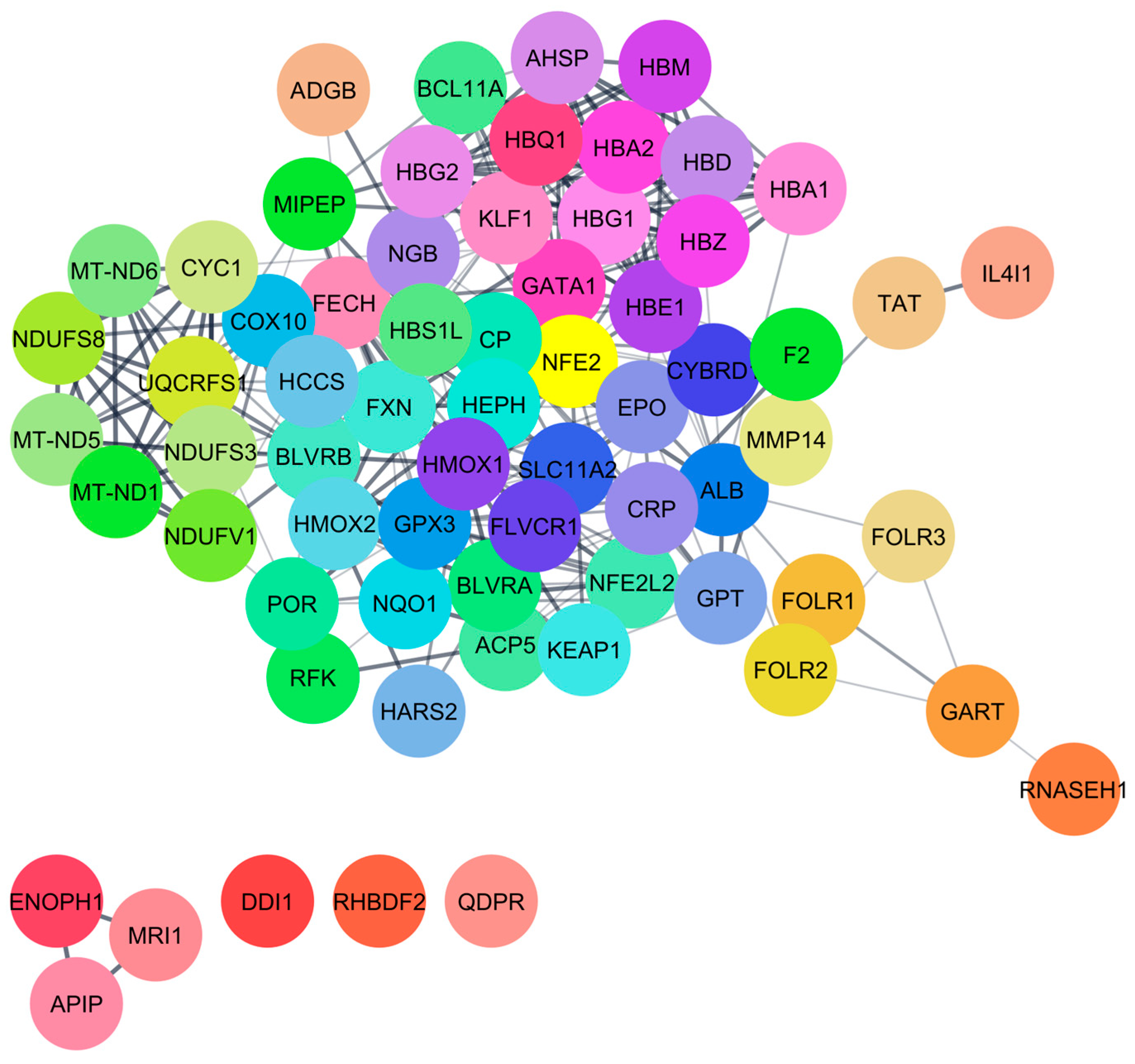
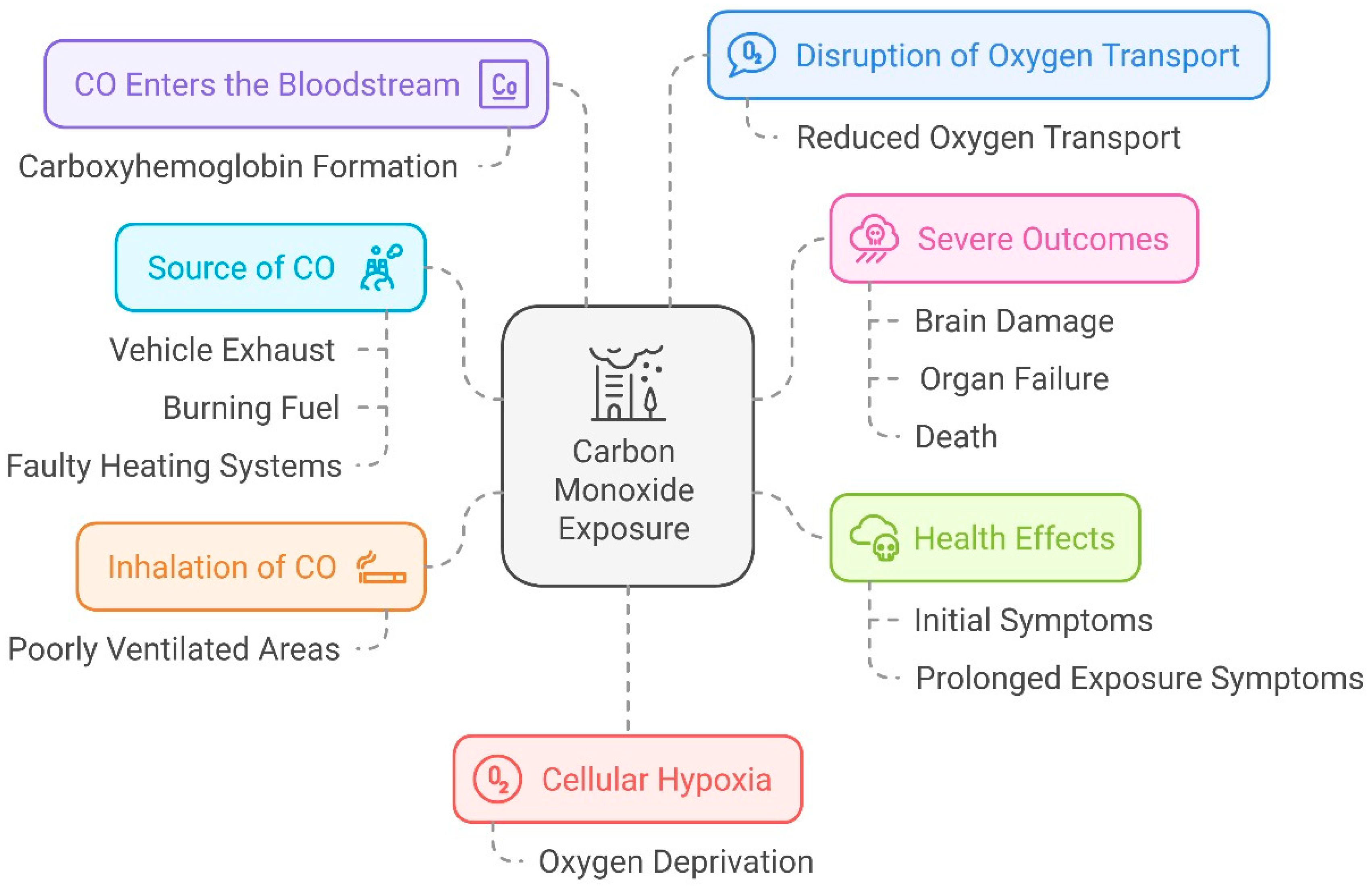
Protein–chemical interaction (PCI) analysis revealed ten proteins identified by STITCH: ADI1, BLVRB, CYGB, HBB, HBE1, HBG1, HBG2, HMOX1, HMOX2, and SLC46A1. Additionally, these proteins interacted with an additional 65 proteins identified by STRING, as shown in Figure 1. The identified proteins are involved in regulating crucial biological functions such as methionine synthesis (ADI1), heme metabolism (BLVRB), cellular protection (CYGB), etc. (Table 1). Notably, the interacted proteins involved in heme catabolism and oxidative stress defense (HMOX1 and HMOX2). At low concentrations (< 10 ppm), CO positively influences human health, including inducing erythropoiesis, hemoglobin mass, and methionine metabolism. However, at higher concentrations (> 10 ppm), CO negatively affects cytochrome c oxidase activity, alters cellular protection, impairs heme metabolism, etc. (Table 1). This damage can lead to serious health issues, including respiratory problems, brain damage, and impaired cognitive function, highlighting the importance of reducing CO emissions for public health (Figure 2).

Functional Annotation and Pathway Analysis of CO-Interacted Proteins

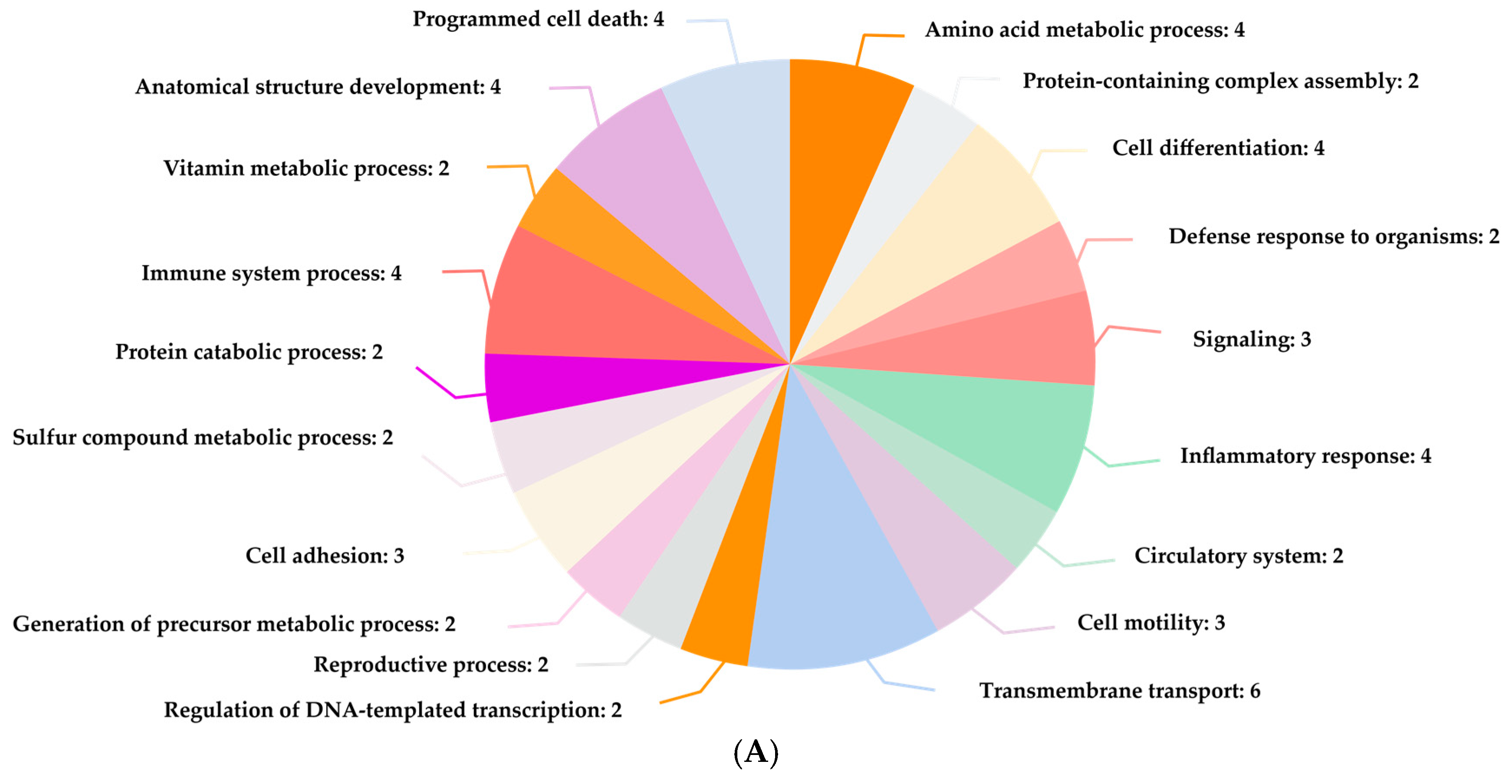
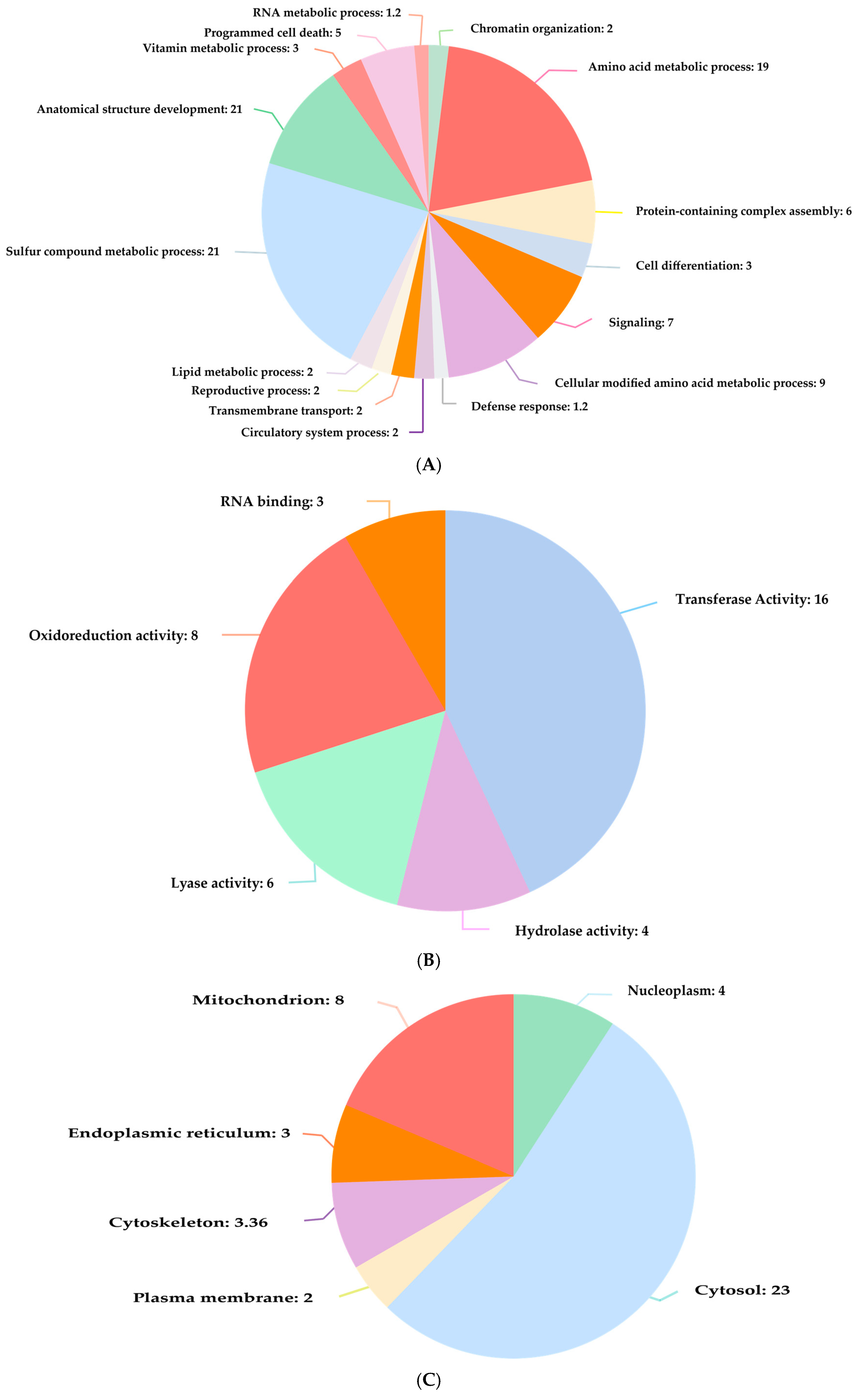
Functional annotation demonstrated that interacted proteins are involved in the various biological processes such as O2 transport, transmembrane transport, signaling, immune system process, and stress response (Figure 3A). This aligns with previous reports on the influence of CO on molecular functions like oxidoreductase activity, hydrolase activity, etc. (Figure 3B). Additionally, these proteins interact with various cellular components, including cytosol, extracellular space, and mitochondrion (Figure 3C). Furthermore, this compound is associated with numerous Reactome pathways, including iron uptake and transport, post-translational protein phosphorylation, synthesis of GPI-anchored proteins, platelet degranulation, and heme biosynthesis, listed in Table 3.

3.3. Identification of Human Proteins That Interacted with Hydrogen Sulfide

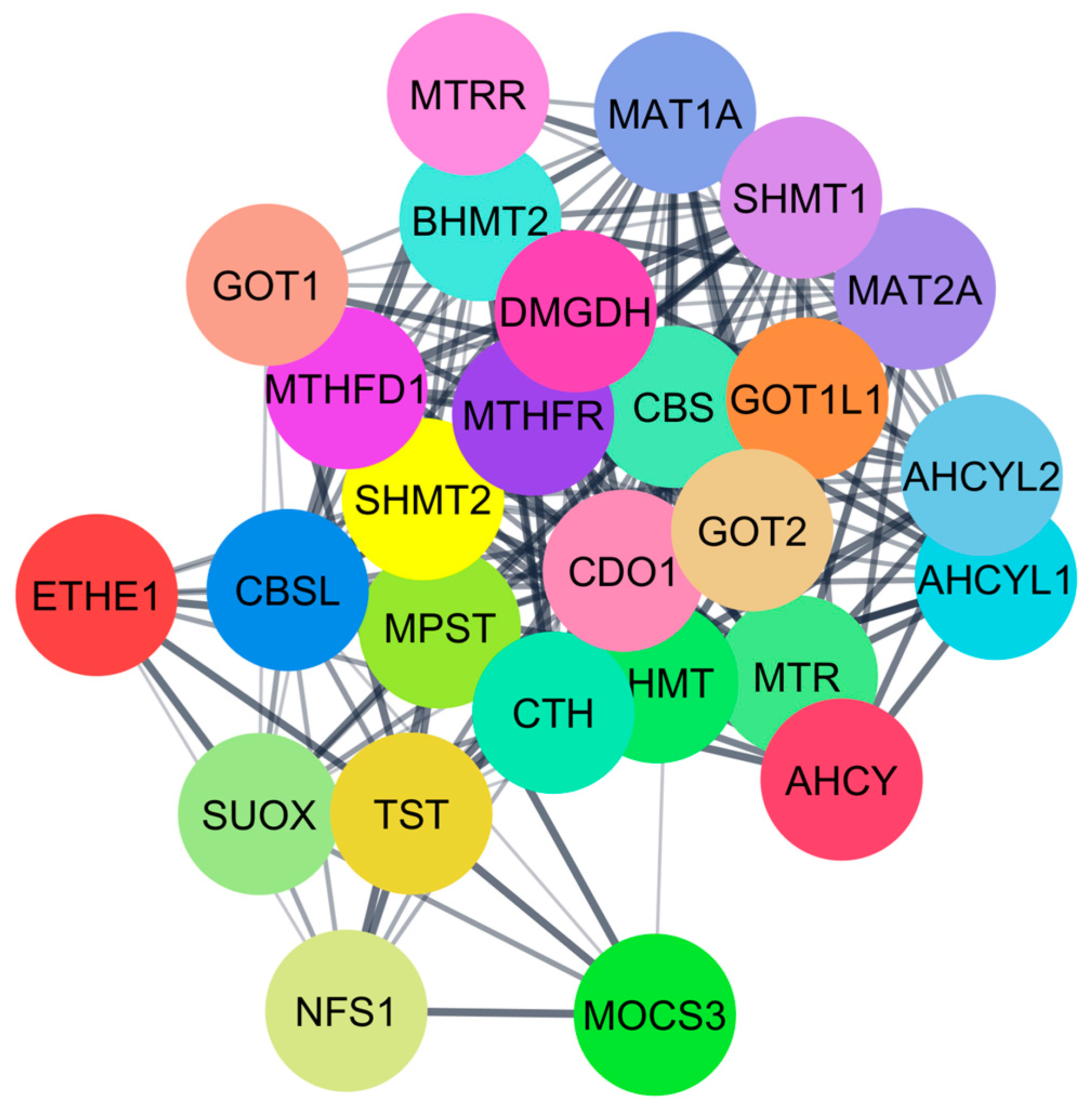
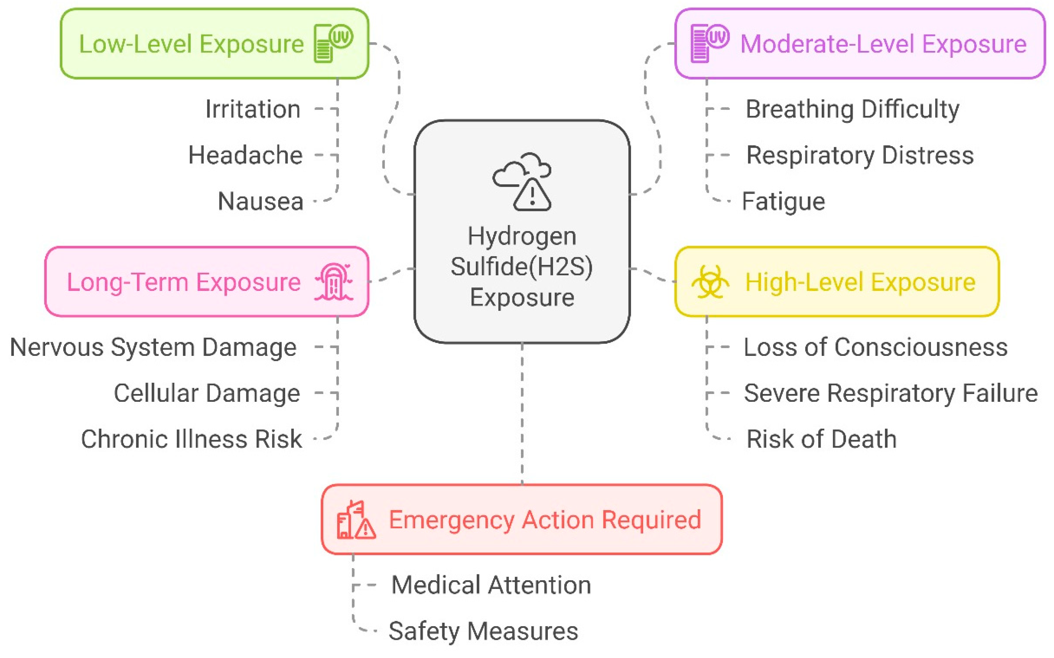
Five important proteins that interacted with H2S were found by PCI analysis: BHMT, CBS, CTH, MPST-3, and MTR-5. These proteins interacted with an additional 27 human proteins, identified from the STRING database through PPIs (Figure 4). These proteins play crucial roles in the regulation of human biological systems, including homocysteine metabolism (BHMT), methylation, DNA synthesis, folate metabolism (MTR-5), sulfur-containing compounds metabolism (MPST-3), etc. (Table 2). At lower concentrations (<100 µM), H2S positively impacts human health by influencing homocysteine metabolism and controlling the methionine cycle (Table 2). However, at elevated concentrations (>100 µM), it inhibits transcription and induces oxidation of these enzymes. Excessive hydrogen sulfide (H2S) can have detrimental effects on human health by causing damage to vital organs (Figure 5).

Functional Annotation and Pathway Analysis of H2S-Interacted Proteins

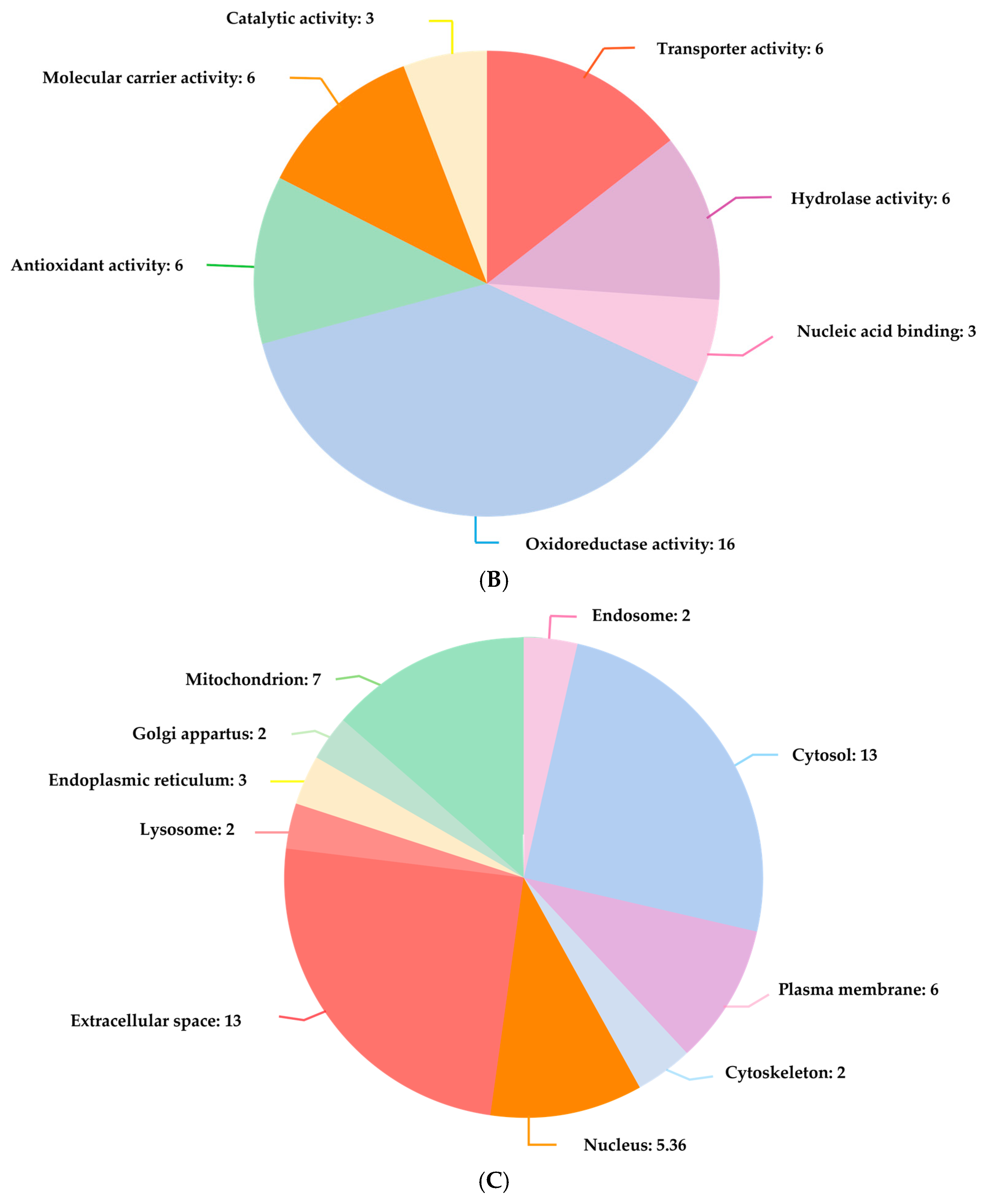
It has been shown that the functional annotation of H2S-interacted proteins is linked with numerous biological processes, cellular components, and molecular functions, including signaling, the metabolism of amino acids, programmed cell death, mitochondria, cytosol, sulfur transfer, and catalytic activity (Figure 6). Furthermore, this compound is associated with numerous Reactome pathways, including gluconeogenesis, the methionine salvage pathway, choline synthesis, and the methylation process, listed in Table 4.

3.4. Critical Human Proteins That Interacted with CO and H2S

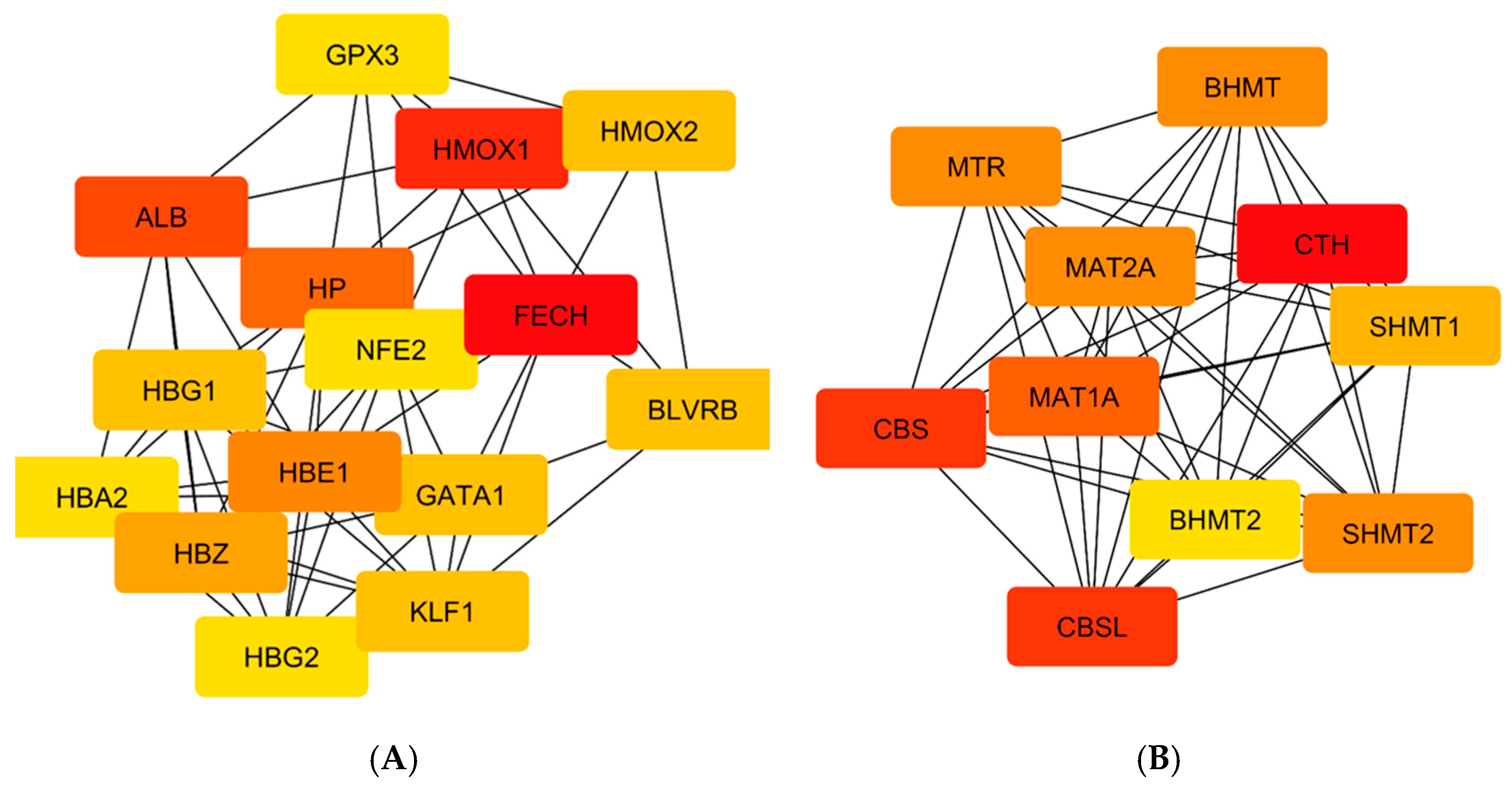
Table 5 shows interactions involving CO, a total of 65 proteins that were identified through STRING analysis, with 15 exhibiting the highest degree of centrality. It is important to highlight that FECH, HMOX1, and ALB emerged as significant proteins due to their remarkable degree of centrality, and they play essential roles in heme metabolism, transportation, and cellular immunity. Furthermore, in the H2S interactions, a total of 27 proteins were identified from the STRING database, with 9 showing the highest degree of centrality (Table 5). Notably, CTH, CBS, and CBSL were identified as key proteins based on their extreme degree of centrality, and they play essential roles in serine, cysteine, and sulfur-containing amino acid metabolism, transportation, and cellular immunity. The analysis of the critical proteins using the cytoHubba plugin of Cytoscape revealed that the most significant proteins (FECH, HMOX1, ALB, CTH, CBS, and CBSL) exhibited a deep red color, as illustrated in Figure 7.

3.5. Key Pathways That Are Regulated by Identified Critical Human Proteins

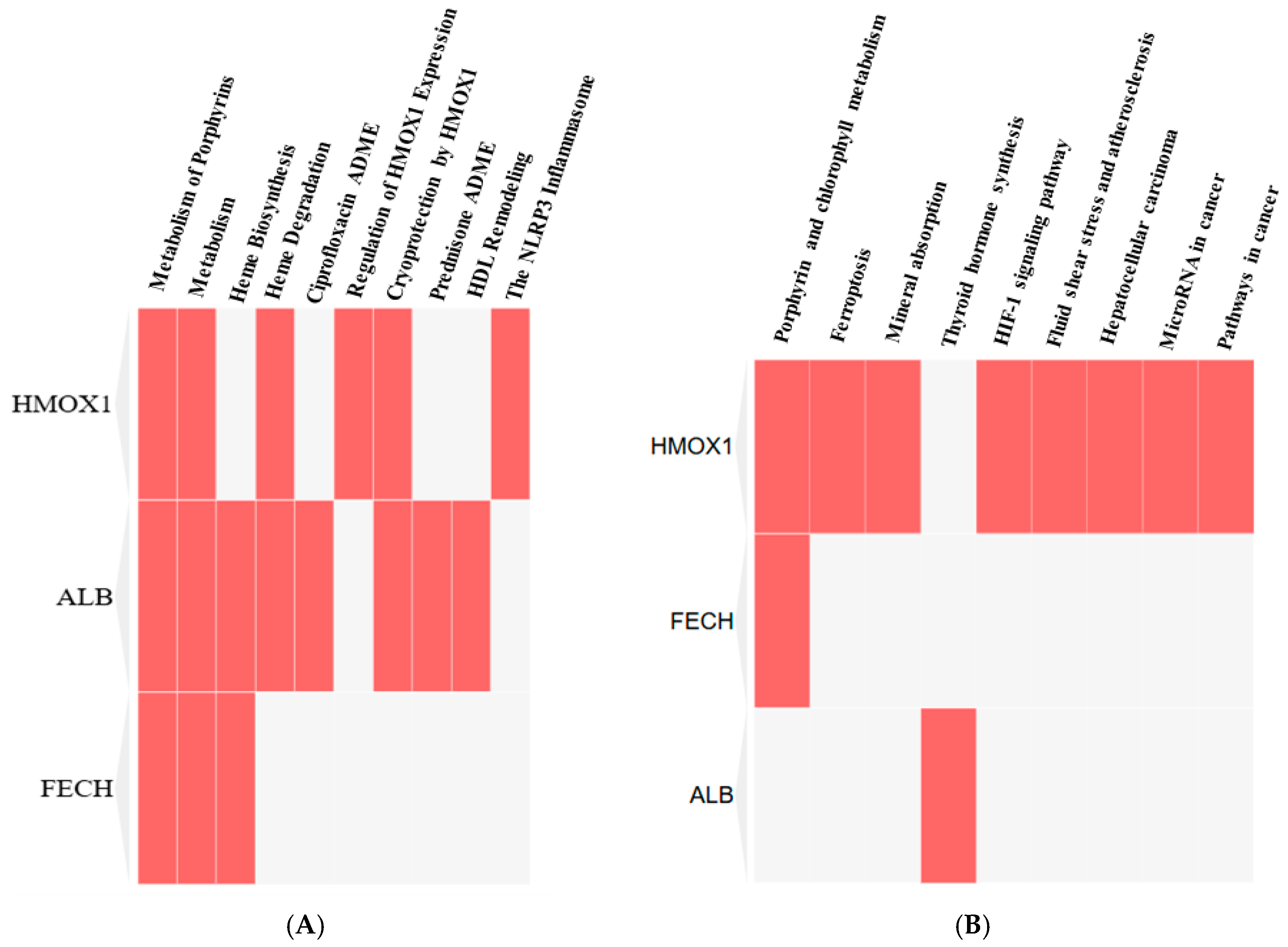
The Reactome clustergram of FECH, HMOX1, and ALB indicates that these proteins predominantly influence the 10 essential Reactome pathways (Figure 8A). However, the p-value analysis reveals a strong interaction of these proteins with porphyrin metabolism (Table 6). Alternately, the KEGG clustergram showed these proteins interacted with nine KEGG pathways (Figure 8B), but based on the p-value, it was shown that these proteins strongly regulate the metabolism of the porphyrin and chlorophyll pathways (Table 6).
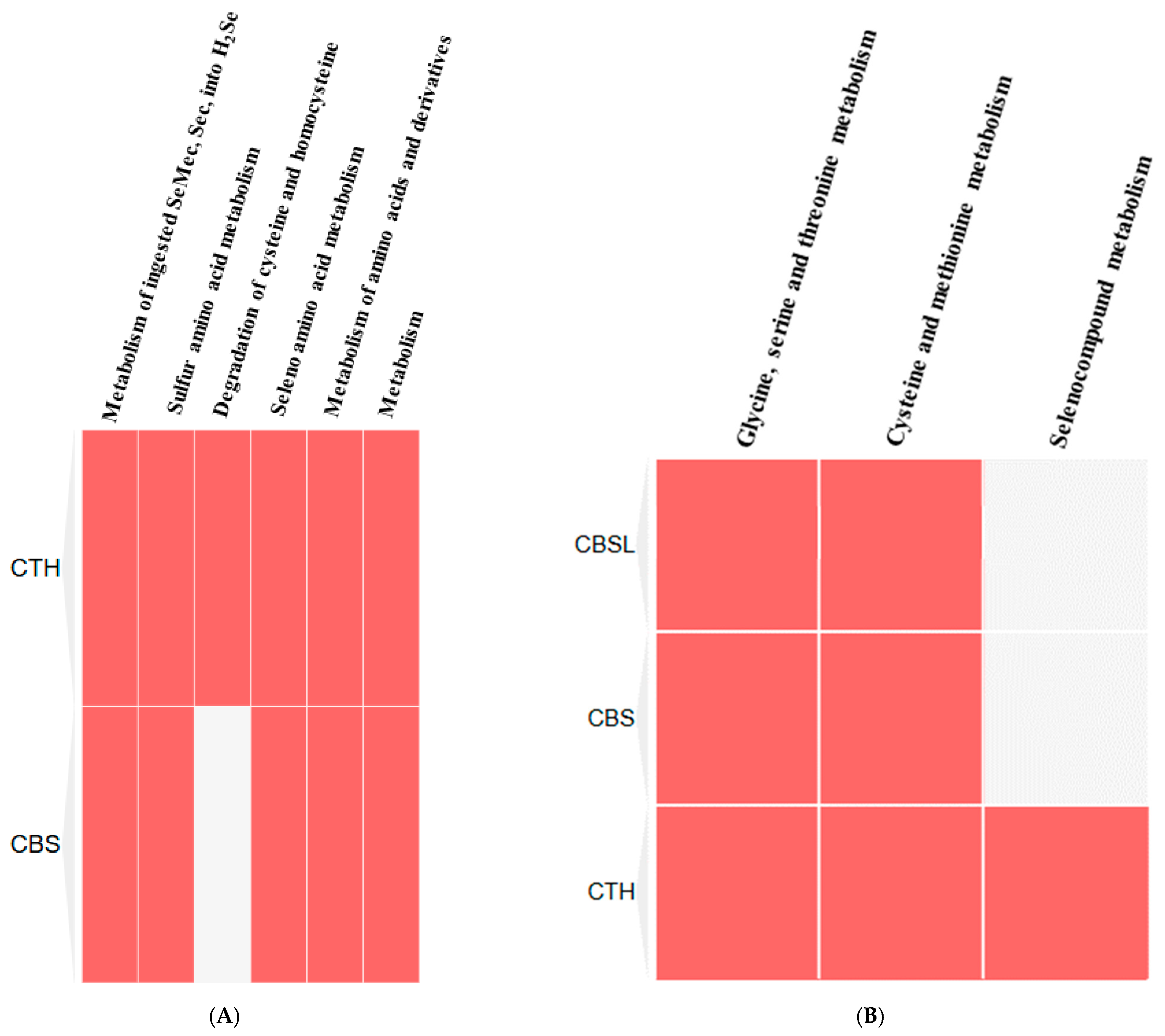
Alternately, the clustergram of the Reactome pathways of CTS and CBS showed that these proteins primarily regulate six crucial Reactome pathways (Figure 9A), but based on the p-value, it was shown that these proteins strongly interacted with the metabolism of ingested SeMet, Sec, and MeSec into H2Se (Table 7). Alternately, the KEGG clustergram showed these proteins interacted with three KEGG pathways (Figure 9B), but based on the p-value, it was shown that these proteins strongly regulated the glycine, serine, and threonine metabolism pathways (Table 7).

3.6. Hypothetical Direct Interactions Between CO and H2S and the Proteins Identified by STITCH

3.6.1. Collection and Preparations of Protein Structures and Ligands

The protein structures (PDBs) of ADI1, BLVRB, CYGB, HBB, HBE1, HBG1, HBG2, HMOX1, HMOX2, and SLC46A1 that interacted with CO and those of BHMT, CBS, CTH, MPST-3, and MTR-5 for H2S was collected from the AlphaFold Protein Structure Database, and then prepared and visualized by MGL tools (AutoDock visualizer) v.1.5.7.

3.6.2. SwissADME Results for CO and H2S Airborne Chemicals

The SwissADME identified the properties of two ligands (chemicals): 0.00Å2 TPSA (Topological Polar Surface Area) for CO and 25.30 Å2 for H2S; they have no rotatable bonds and have lead likenesses with poor GI absorption (Table 8). The results of Lipinski’s Rule of 5 for the CO and H2S ligands are shown in Table 9.

3.6.3. Results of Hypothetical Protein–Chemical Interactions

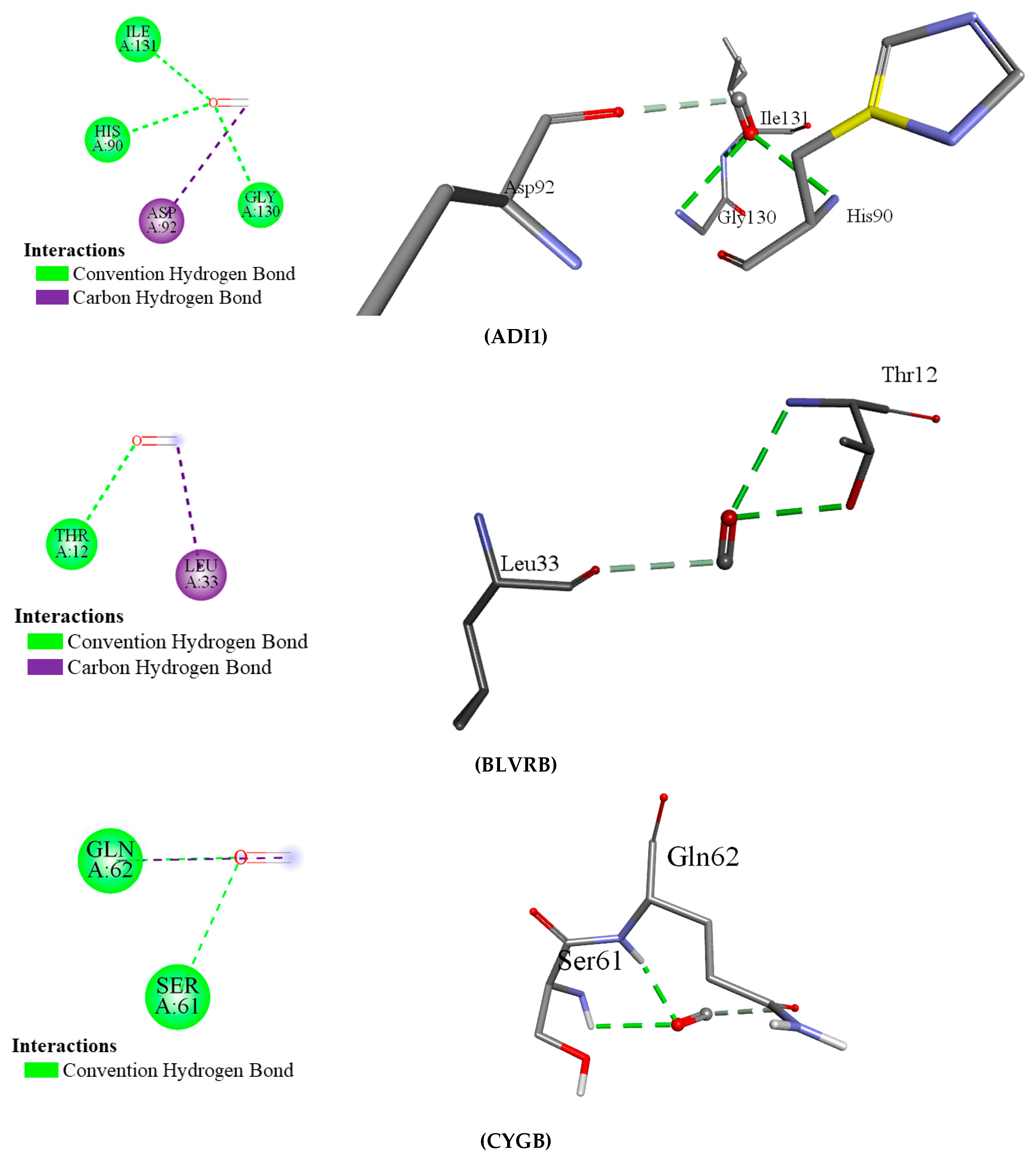
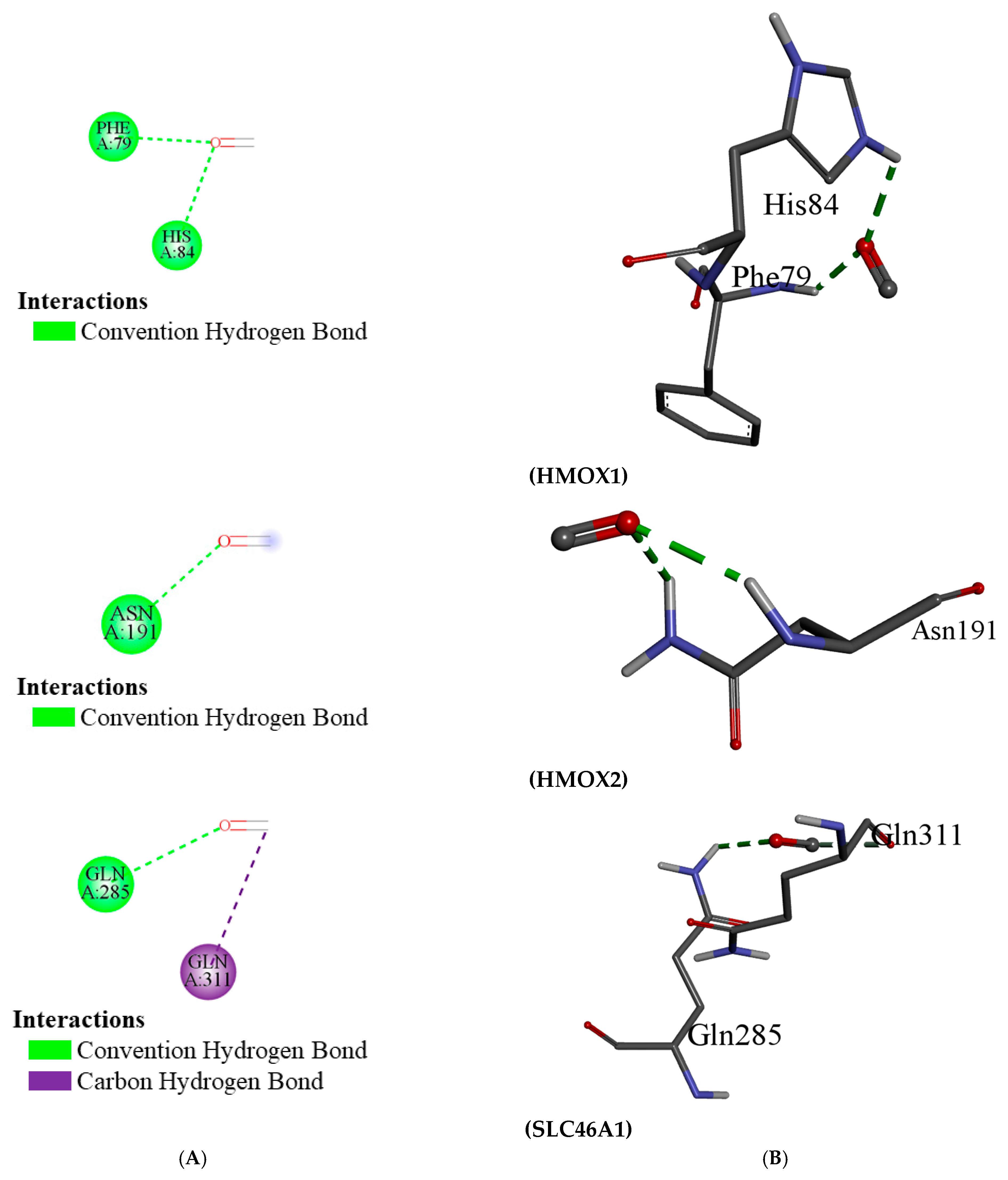
AutoDock Vina was used to dock the 2 ligands (CO and H2S) with the 14 targeted proteins. The docking results (binding energy/affinity) are demonstrated in Table 10. Among the proteins interacting with CO, ADI1 exhibited the highest binding energy (−1.9 kcal/mol). CO mainly interacted with glycine, isoleucine, histidine, and aspartate residues by the convention hydrogen bond and carbon–hydrogen bond. All the docking results are illustrated in Figure 10, but these interacting energies are inadequate for robust docking. Comparable findings were noted in the docking results of proteins interacting with H2S, and their binding energies are very low (−0.8 kcal/mol is the highest binding affinity); therefore, it was not visualized in Discover Studio.

4. Discussion

Carbon monoxide (CO) and hydrogen sulfide (H2S) are important gaseous molecules with significant positive and negative biological effects. Even though they are known to be poisonous in greater quantities (over 4 mg/m3 or 3.5 ppm), they are also essential for preserving cellular homeostasis and controlling several physiological functions [45]. With an emphasis on their interactions with important proteins, oxidative stress management, and consequences for vascular and metabolic health, this discussion will examine how CO and H₂S affect enzyme activity, cellular pathways, and metabolic activities.
The major effects of CO on biological systems are through the interaction with enzymes, proteins, and cellular pathways responsible for a variety of key physiological functions. Among the well-defined targets of CO are protein arginine methyltransferases, which catalyze protein methylation and control many cellular processes, from gene expression to cell signaling and cell cycle control.
Other important effects of CO involve interactions with proteins implicated in the regulation of oxidative stress and reactive oxygen species. CO modulates the activity of enzymes that are central in the oxidative balance, including HO-1 and HO-2, which are crucial for heme catabolism and the production of biliverdin and carbon monoxide itself. Of these, HO-1 possesses antioxidant properties and is induced by conditions of oxidative stress. CO, through the modulation of HO-1, may have protective effects against oxidative injury [46]. On the other hand, chronic CO exposure could interfere with cellular homeostasis and promote oxidative injury in conditions such as neurodegenerative disorders, mainly by breaking down the integrity of the blood–brain barrier [47]. Such breakdown in the integrity of the blood-brain barrier (BBB) may lead to enhanced neuroinflammatory responses and is considered an additional factor contributing to cognitive decline.
Further, CO is a player in the processes of vascular homeostasis. It modifies the functions of important oxygen-transport proteins, including cytoglobin and hemoglobin, implicated in oxygen transport and the maintenance of tissue oxygen levels. With the ability to modulate these proteins, CO modifies the delivery of oxygen to the tissues and the cellular responses to hypoxia; as a consequence, the blood flow is changed, as are tissue repair and immune responses [48]. Moreover, CO acts as an essential modulator of immune responses by tuning the production of inflammatory mediators with protective or pathogenic functions, depending on the context and length of exposure. While in an acute setting, CO can reduce inflammation, on the other hand, chronic exposure may increase inflammatory pathways that may lead to the development of chronic diseases like cardiovascular disorders and neurodegenerative conditions.
This study discovered that carbon monoxide interacts with multiple human proteins, genes, and enzymes that regulate vital functions of human health within tolerable concentrations, including methionine synthesis (ADI1), heme metabolism (BLVRB), and cellular protection (CYGB). Carbon monoxide is produced during heme metabolism by BLVRB enzymes, and extensive amounts of this airborne chemical alter cellular respiration [26]. Acireductone dioxygenase 1 (ADI1) is responsible for the methionine salvage pathway crucial for methionine synthesis and cellular stress responses [25]. Low CO levels (10–100 nM) influence methionine metabolism, but high CO levels (>100 µM) can alter enzyme metal centers, such as the iron (Fe2⁺) in ADI1, reducing catalytic activity [25]. Another primary interacted protein, cytoglobin (CYGB) protein, is responsible for cellular protection via antioxidant defense, lowering blood pressure, maintaining vascular health via nitric oxide regulation, and promoting cardiac cell survival [49]. Hemoglobin subunit epsilon 1 (HBE1) plays a crucial role in treating different hematologic disorders and blood cancer, but extensive CO negatively affects the functions of HBE1. Hemoglobin subunit gamma 1 (HBG1) plays a crucial role in fetal oxygen transport, but an extensive concentration of CO (>10 ppm) increases hemoglobin’s affinity for oxygen and causes impaired oxygen delivery to tissues. The enzyme betaine homocysteine S-methyltransferase (BHMT) interacts with H2S and is involved in the conversion of homocysteine into methionine via betaine methyl donor [36].
Like CO, hydrogen sulfide has also been recognized as an important gaseous molecule in biological systems with significant roles, primarily in regulating metabolic and redox processes. The role of H₂S is being reported to influence a lot of enzymes in sulfur metabolism, most especially in the trans-sulfuration pathway, which has an important role in the detoxification of homocysteine and in synthesizing major sulfur-containing biomolecules. Two important enzymes from the trans-sulfuration pathway are BHMT and CBS. Both of these enzymes are essential to converting homocysteine to cysteine and further synthesizing glutathione, an important antioxidant [50]. H2S, through its action on these enzymes, maintains the redox balance and thus supports the maintenance of cellular metabolism, reducing oxidative damage due to ROS. H2S has a beneficial impact on homocysteine metabolism through the induction of trans-sulfuration enzymes [37]. The enzyme cystathionine gamma-lyase (CTH) plays an essential role in homocysteine metabolism. A lower concentration (<10 µM) of H2S increases CTH function by inducing the feedback inhibition mechanism, but a higher concentration (>10 µM) of H2S decreases transcription and reduces the activity of the CTH enzyme (200 µM) [40]. MPST-3 plays a key role in the sulfur-containing compounds, and Nagahara [42] said the elevated levels (200 µM) of H2S induce oxidation of the MPST-3 enzyme and minimize its activity.
The possibility that H₂S can regulate oxidative stress is intriguing. Since it is a reducing molecule, H₂S can lessen oxidative damage by either directly increasing the cell’s antioxidant defense systems or by scavenging reactive oxygen species (ROS). For example, H₂S increases the expression and activity of glutathione peroxidase and other antioxidant enzymes, thus protecting the cell from oxidative damage [51]. This antioxidative role is highly valued in the context of diseases associated with inflammation and oxidative damage, such as cardiovascular diseases, diabetes, and neurodegenerative disorders.
Furthermore, H2S plays a role in energy production and metabolic efficiency. By influencing the activity of several enzymes like MAT and methionine synthase, which are responsible for synthesizing important metabolic intermediates, H2S provides a means to maintain energy homeostasis in the cell. Thus, a further suggestion was made about its ability to ensure an optimum metabolic outcome and to improve mitochondrial process efficiency [52]. These would have implications for CBS and cystathionine that further support the synthesis of sulfur and methionine, which are important in cellular growth, repair, and energy production. The beneficial roles played by H₂S in the regulation of oxidative stress and inflammation point toward its therapeutic potential in various conditions, particularly where such processes are dysregulated.
FECH, ALB, and HMOX1 are essential proteins for human health that play a significant role in regulating porphyrin and heme metabolism. These proteins have been identified as critical nodes based on their degree of centrality. Finding the most important nodes in a network based on the degree of centrality allows one to examine its topology, including its resilience and susceptibility to attacks [53]. Centrality measures employ graph theory and network analysis to evaluate an individual’s position within a protein network. Degree centrality, betweenness centrality, and closeness centrality are used to assess social influence inside networks [54]. Molecular docking interactions indicated the lowest binding energy, suggesting that CO and H2S have a weak interaction with these 14 proteins. Multiple investigations have explored the strong binding of CO with the heme pocket of myoglobin; however, we were unable to locate myoglobin within the STITCH database [55]. CO mostly binds with glycine and aspartate amino acids, and these are responsible for erythropoiesis and oxidative responses. Alternatively, a limited investigation was performed on the interactions of H2S with proteins; Nery et al. [56] reported comparable results regarding H2S interactions with hypoxia-inducible factors.
Both CO and H₂S are therapeutically very important. CO’s role in modulating vascular health, immune responses, and oxidative stress could represent targeted therapies for conditions like hypertension, vascular diseases, and neurodegenerative disorders. CO-releasing molecules, CORMs, have been pursued as active pharmaceuticals for the delivery of controlled amounts of CO for therapeutic applications, especially in diseases that feature excessive oxidative stress and inflammation.
However, although both gases possess therapeutic potential, dosing and delivery mechanisms are still big challenges. At high concentrations, both CO and H₂S are toxic; thus, it is very hard to translate their beneficial effects into clinical practice. For this reason, ongoing research is needed to develop ways to safely and effectively exploit these molecules in therapeutic settings. However, the therapeutic potential of these gases must be carefully harnessed to avoid their toxic effects, underscoring the importance of continued research in this area.

5. Conclusions

The increases in greenhouse gas emissions due to urbanization and industrial activities have been linked to a significant fall in air quality, now regarded as a foremost risk to public health globally. The emission of pollutants including particulate matter (PM), nitrogen dioxide (NO2), sulfur dioxide (SO2), carbon monoxide (CO), hydrogen sulfide, and volatile organic compounds (VOCs) has worsened air quality and is implicated in diverse health effects, predominantly on the respiratory and cardiovascular systems. These pollutants induce climate change and release a series of cellular and molecular effects in the human body, which lead to chronic diseases like asthma, chronic obstructive pulmonary disease (COPD), and heart disease. The priority in environmental health research has been to better understand these health effects at the molecular level. In particular, studies concentrating on the interactions of these pollutants with proteins at the molecular level will illuminate how these compounds throw biological functions out of gear and lead to the development of such diseases.
Air pollutants harm and benefit human health by interacting with cellular proteins. These cellular proteins are critical in maintaining normal physiological functions. Once inhaled, air pollutants bind to proteins, modifying their conformation, activity, or expression. In turn, this process disrupts essential biological functions such as immune responses, oxidative stress regulation, and cell signaling. Oxidative stress, a prominent mechanism through which air pollutants cause damage, is mediated largely by proteins involved in antioxidant defense mechanisms. Chemistry–protein interaction analysis is a very innovative tool to unveil the biological effects of air pollution. Scientists can get insight into the molecular pathways that are implicated after exposure by identifying the specific proteins that interact with different pollutants. In recent years, advances in bioinformatics and computational biology have allowed researchers to analyze the vast data associated with these chemical–protein interactions.
The research discusses the interaction between carbon monoxide (CO) and various proteins subsequently modifying several biological functions like methionine biosynthesis, heme metabolism, and cell protection; for instance, CO interacts with acireductone dioxygenase 1 (ADI1) and biliverdin reductase B (BLVRB), which has various negative effects on several cellular and metabolic functions. CO at low concentrations (about 10–15 ppm) seems to enhance erythropoiesis and affects methionine metabolism, while higher concentrations (>50 ppm) interfere with the proper functioning of hemoglobin, meaning cellular respiration is affected, and oxidative stresses occur. Such interactions may bring serious implications for health, such as respiratory and cognitive defects. In addition, the involved proteins may link with different biological pathways such as heme biosynthesis, platelet degranulation, and oxidative stress defense, thus emphasizing that CO exposure can have a very far-reaching effect.
Hydrogen sulfide (H2S) also inhibits key enzymes involved in the metabolism of homocysteine and sulfur-containing compounds and methylation processes. H2S inhibits essential enzymes such as betaine homocysteine S-methyltransferase (BHMT) and cystathionine beta-synthase (CBS), which disrupts proper metabolism. A low dose of H2S stimulates homocysteine metabolism and the methionine cycle, while a higher concentration gradually oxidizes enzymes, inhibits transcription, and damages further cellular function and metabolism. The critical pathways’ interaction with H2S mainly takes part in sulfur metabolism, methylation, and the regulation of homocysteine.
Also identified were critical proteins with high centralities in the network of protein interactions for both CO and H2S. For CO, involved in heme metabolism, cellular immunity, and stress response are proteins such as FECH, HMOX1, and ALB, while for H2S, CTH, CBS, and CBSL are involved in sulfur metabolism, amino acid regulation, and redox balance. The study further elaborates on these proteins’ regulatory roles in diverse biochemical processes, such as porphyrin metabolism and sulfur amino acid metabolism, as well as the regulation of gene expression, all of which emphasize how CO and H2S exert profound effects on human well-being.
In conclusion, molecular interactions between pollutants and proteins, in terms of their biological effects, are undoubtedly of great importance for understanding the health consequences of air pollution. Large strides forward in bioinformatics and computational biology, including the STITCH 4.0 and STRING 9.0 tools, have made it possible to elucidate the chemical–protein interaction networks and protein–protein interaction pathways and elaborate the molecular mechanisms of air-pollution-related diseases. Further digging into these networks and the disruption caused to biological processes may help to determine health outcomes, suggest new therapeutic approaches, and possibly prevent or ameliorate air pollution’s harmful effects. This type of research is certainly of the utmost importance in matters of public health and will form a stronger basis for understanding how the environment can influence human disease.
6. Future Direction/Recommendations

Moreover, the study shows the urgent need for further research into the health effects of air pollution at the molecular level. As air pollutants interact with human proteins, future research needs to be oriented toward a deeper understanding of the biochemical networks. Guided by the results arising from this study, several recommendations are possible:
  • Expand the study of molecular interaction: While this study used STITCH 4.0 and STRING 9.0 to investigate the interaction of proteins with H2S and CO pollutants, future studies need to be expanded in scope to other common pollutants such as particulate matter, nitrogen dioxide, and sulfur dioxide. These are common in many industrial and urban areas and affect protein function, leading to a variety of diseases. Advanced computational models can be utilized in simulating their interaction with proteins, hence enabling the researchers to find more specific pathways affected by these pollutants.
  • Investigation into long-term exposure effects: Most studies generally focus on acute exposure to such pollutants, but their long-term exposures have significant cumulative health effects. It is crucial to determine how chronic exposure to these pollutants alters protein functions over time and how such alterations may lead to the onset of various chronic diseases such as asthma, cardiovascular diseases, and even cancer. Longitudinal studies following the molecular changes in exposed individuals can offer a better understanding of such long-term effects.
  • Multidisciplinary engagement: The nature of the challenge in studying protein–protein interaction networks require merging expertise from bioinformatics, molecular biology, environmental science, and epidemiology in designing studies that connect changes at the molecular level to population-level measurable health outcomes; this could be further extended in collaborative work toward improving the predictive ability of models for forecasting health risk from environmental pollution data.
  • Public health and policy implications: The understanding of molecular interactions between pollutants and proteins can help in the development of targeted therapies. Governments and public health organizations should prioritize research that links environmental pollution to specific health conditions and implement policies that reduce exposure. Such policy measures may include strict regulation of emissions, increased monitoring of air quality, and public health campaigns to raise community awareness about the risks of pollution.
  • Development of therapeutic interventions: The identification of key proteins affected by pollutants will provide the window for developing therapeutic interventions. Drug development should be based on mitigating the harmful effects of pollutants on the proteins. Further research should be conducted to develop chemicals or interventions that can protect critical proteins from pollution-induced damage and assist in reducing the chance of developing pollution-related disorders.
Addressing the health effects of air pollution necessitates a multitherapy approach: detailed molecular research, public health campaigns, and policy alteration. Understanding the interaction of pollutants with proteins opens new avenues to prevent or treat diseases associated with air pollution.

Supplementary Materials

The following supporting information can be downloaded at: https://www.mdpi.com/article/10.3390/ijerph22030418/s1, Figure S1: Nitrogen oxides (NOX), Ozone (O3), Volatile CO2, and Sulfur dioxide (SO2) with human proteins were found using STITCH 4.0; Figure S2: Functional annotation study revealed NOX’s impacts on diverse biological processes (A), cellular component localization (B), and molecular functions (C) of distinct proteins in Homo sapiens; Figure S3: Functional annotation study revealed Ozone (O3)’s impacts on diverse biological processes (A), molecular functions localization (B), and Cellular Component (C) of distinct proteins in Homo sapiens; Figure S4: Functional annotation study revealed Volatile Carbon Dioxide’s impacts on diverse biological processes (A), molecular functions localization (B), and Cellular Component (C) of distinct proteins in Homo sapiens; Figure S5: Functional annotation study revealed SO2’s impacts on diverse biological processes (A), cellular component localization (B), and molecular functions (C) of distinct proteins in Homo sapiens; Figure S6: 3D structure of proteins that interacted with CO and H2S identified by STITCH; Table S1: Protein-protein interaction (PPI) of NOx identified through STRING 9.1; Table S2: Protein-protein interaction (PPI) of Ozone (O3) identified through STRING 9.1; Table S3: Protein-protein interaction (PPI) of Volatile Carbon Dioxide identified through STRING 9.1; Table S4: Protein-protein interaction (PPI) of Sulphur Dioxide (SO2) identified through STRING 9.1; Table S5: Degree centrality, closeness centrality, and betweenness centrality of CO interacted proteins identified by STRING; Table S6: Degree centrality, closeness centrality, and betweenness centrality of H2S interacted proteins identified by STRING.

Author Contributions

Design and conceptualization, M.R.S.; methodology, M.R.S., H.H.M. and M.S.H.; software, M.R.S. and D.S.; validation, M.E.U., M.F.H. and M.K.M.; formal analysis, M.R.S.; investigation, M.E.U. and M.F.H.; data curation, A.A.I.S.; writing—original draft preparation, M.R.S., H.H.M. and M.S.H.; writing—review and editing, M.R.S.; supervision, M.R.S.; funding acquisition, A.A.I.S. All authors have read and agreed to the published version of the manuscript.

Funding

This research has received funding from the NHMRC Investigator Grant, Center for Personalized Nanomedicine, Australian Institute for Bioengineering & Nanotechnology (AIBN), The University of Queensland, Brisbane, QLD 4072, Australia: APP1175047.

Institutional Review Board Statement

Not applicable.

Informed Consent Statement

Not applicable.

Data Availability Statement

All research data are provided in the current article.

Acknowledgments

The authors are grateful to the Australian Institute for Bioengineering and Nanotechnology (AIBN), University of Queensland, for providing the financial support.

Conflicts of Interest

The authors declare no conflicts of interest.

Abbreviations

STITCHSearch Tool for Interacting Chemicals
STRINGSearch Tool for the Retrieval of Interacting Genes/Proteins
PPIProtein–protein interaction
PCIProtein–chemical interaction
ACP5Acid phosphatase 5
CRPC-reactive protein
ALBAlbumin
BLVRBBiliverdin reductase B
RFKRiboflavin kinase gene
ADGBAndroglobin
NGBNeuroglobin
FECHFerrochelatase
AHSPAlpha hemoglobin stabilizing protein
HBQ1Hemoglobin subunit theta 1
HBA2Hemoglobin subunit alpha 2
HBZHuman T-cell leukemia virus type 1 (HTLV-1) basic leucine zipper
KLF1Kruppel-like factor 1
HBE1Hemoglobin subunit epsilon 1
BCL11AB-cell lymphoma/leukemia 11A
NFE2Nuclear factor erythroid 2
GATA1GATA binding protein 1
HBMHemojuvelin
HBG2Hemoglobin, gamma-2
HBG1Hemoglobin, gamma-1
HBA1Hemoglobin alpha subunit 1
HBDHemoglobin D
HMOX1Heme oxygenase 1
EPOErythropoietin gene
CPCeruloplasmin gene
BLVRABilirubin- very large acidic protein gene
MMP14Matrix metallopeptidase 14
FOLR2Folate receptor 2
FOLR3Folate receptor 3
HPRHaptoglobin related protein
NQO1NAD(P)H quinone dehydrogenase 1
KEAP1Kelch-like ECH-associated protein 1
FOLR1Folate receptor 1
NFE2L2Nuclear factor, erythroid 2-like 2
TATTyrosine aminotransferase gene
GPX3Glutathione peroxidase 3
GPTGlutamate pyruvate transaminase
F2Prothrombin gene
HPHaptoglobin
APIPAdenosine phosphoribosyltransferase-interacting protein
MRI1Mitochondrial ribosomal protein S1
ENOPH1Enolase phosphatase 1
HBS1LRibosomal rescue factor HBS1L
MIPEPMitochondrial intermediate peptidase
HARS2Histidyl-tRNA synthetase 2
PORP450 oxidoreductase
HMOX2Heme oxygenase-2
MT-ND6Mitochondrial NADH dehydrogenase subunit 6
MT-ND5Mitochondrial NADH dehydrogenase subunit 5
MT-ND1Mitochondrial NADH dehydrogenase subunit 1
NDUFS8NADH: Ubiquinone oxidoreductase subunit S8
NDUFV1NADH dehydrogenase (ubiquinone) flavoprotein 1
CYC1Cytochrome c, isoform 1
UQCRFS1Ubiquinol-cytochrome c reductase, rieske Fe-S protein 1
NDUFS3NADH: Ubiquinone oxidoreductase subunit S3
COX10Cytochrome c oxidase subunit 10
HCCSHepatocerebral cytopathy scanning gene
CYBRD1Cytochrome B reductase 1
SLC11A2Solute carrier family 11-member 2
HEPHHepatocyte expressed PH domain-containing gene
FXNFrataxin
FLVCR1Feline leukemia virus subgroup C receptor 1
GARTGlycinamide ribonucleotide transformylase
RNASEH1Ribonuclease H1
IL4I1Interleukin-4 induced 1
GOT1L1Glutamate–oxaloacetate transaminase 1-like 1
GOT2Glutamate–oxaloacetate transaminase 2
SUOXSulfite oxidase
NFS1Nuclear Fragile X Mental Retardation Syndrome-1
GOT1Glutamate-oxaloacetate transaminase 1
ETHE1Ethylmalonic Encephalopathy 1 gene
MOCS3Molybdenum cofactor sulfurase 3
MPSTMercaptopyruvate sulfotransferase
TSTThiosulfate sulfotransferase
MTRMethionine synthase reductase gene
BHMTBetaine-homocysteine methyltransferase
CBSCystathionine beta-synthase gene
CTHCystathionine gamma-lyase
BHMT2Betaine-homocysteine methyltransferase 2
AHCYL1Adenosylhomocysteinase Like 1
AHCYL2Adenosylhomocysteinase Like 2
CBSLCystathionine beta synthase like
MAT1AMethionine adenosyltransferase 1A
SHMT2Serine hydroxymethyltransferase 2
MAT2AMethionine adenosyltransferase 2A
MTHFRMethylenetetrahydrofolate reductase
SHMT1Serine hydroxymethyltransferase 1
MTHFD1NAD-dependent methylenetetrahydrofolate dehydrogenase 1
MTRRMethionine synthase reductase
DMGDHDimethylglycine dehydrogenase
CDO1Cysteine dioxygenase 1
AHCYAdenosylhomocysteinase

References

  1. Grieger, K.D. Understanding and Assessing Potential Environmental Risks of Nanomaterials: Emerging Tools for Emerging Risks; Technical University of Denmark: Kongens Lyngby, Denmark, 2011; Volume 17, pp. 43–46. [Google Scholar]
  2. Hussain, S.; Reza, M. Environmental damage and global health: Understanding the impacts and proposing mitigation strategies. J. Big-Data Anal. Cloud Comput. 2023, 8, 1–21. [Google Scholar]
  3. Maji, S.; Ahmed, S.; Kaur-Sidhu, M.; Mor, S.; Ravindra, K. Health risks of major air pollutants, their drivers and mitigation strategies: A review. Air Soil Water Res. 2023, 16, 11786221231154659. [Google Scholar] [CrossRef]
  4. Kaur, R.; Pandey, P. Air pollution, climate change, and human health in Indian cities: A brief review. Front. Sustain. Cities. 2021, 3, 705131. [Google Scholar] [CrossRef]
  5. Yang, W.; Omaye, S.T. Air pollutants, oxidative stress and human health. Mutat. Res./Genet. Toxicol. Environ. Mutagen. 2009, 674, 45–54. [Google Scholar] [CrossRef]
  6. Manisalidis, I.; Stavropoulou, E.; Stavropoulos, A.; Bezirtzoglou, E. Environmental and health impacts of air pollution: A review. Front. Public Health 2020, 8, 14. [Google Scholar] [CrossRef]
  7. Wu, Q.; Achebouche, R.; Audouze, K. Computational systems biology as an animal-free approach to characterize toxicological effects of persistent organic pollutants. ALTEX-Altern. Anim. Exp. 2020, 37, 287–299. [Google Scholar] [CrossRef] [PubMed]
  8. Kuhn, M.; von Mering, C.; Campillos, M.; Jensen, L.J.; Bork, P. STITCH: Interaction networks of chemicals and proteins. Nucleic Acids Res. 2007, 36, D684–D688. [Google Scholar] [CrossRef] [PubMed]
  9. Snider, J.; Kotlyar, M.; Saraon, P.; Yao, Z.; Jurisica, I.; Stagljar, I. Fundamentals of protein interaction network mapping. Mol. Syst. Biol. 2015, 11, 848. [Google Scholar] [CrossRef]
  10. Dixit, N.; Patel, M.; Motwani, H.; Patel, S.K.; Rawal, R.M.; Solanki, H.A. Deciphering the latent biomarkers in HBV and HDV- associated Hepatocellular carcinoma—An integrated bioinformatics analysis. Hum. Gene 2024, 40, 201287. [Google Scholar] [CrossRef]
  11. Sharma, J.; Parsai, K.; Raghuwanshi, P.; Ali, S.A.; Tiwari, V.; Bhargava, A.; Mishra, P.K. Emerging role of mitochondria in airborne particulate matter-induced immunotoxicity. Environ. Pollut. 2021, 270, 116242. [Google Scholar] [CrossRef]
  12. Bebek, G.; Yang, J. PathFinder: Mining signal transduction pathway segments from protein-protein interaction networks. BMC Bioinform. 2007, 8, 335. [Google Scholar] [CrossRef] [PubMed]
  13. Fotis, C.; Antoranz, A.; Hatziavramidis, D.; Sakellaropoulos, T.; Alexopoulos, L.G. Network-based technologies for early drug discovery. Drug Discov. Today 2018, 23, 626–635. [Google Scholar] [CrossRef] [PubMed]
  14. Sidak, D.; Schwarzerová, J.; Weckwerth, W.; Waldherr, S. Interpretable machine learning methods for predictions in systems biology from omics data. Front. Mol. Biosci. 2022, 9, 926623. [Google Scholar] [CrossRef]
  15. Nibbe, R.K.; Chowdhury, S.A.; Koyutürk, M.; Ewing, R.; Chance, M.R. Protein–protein interaction networks and subnetworks in the biology of disease. Wiley Interdiscip. Rev. Syst. Biol. Med. 2011, 3, 357–367. [Google Scholar] [CrossRef] [PubMed]
  16. Richards, A.L.; Eckhardt, M.; Krogan, N.J. Mass spectrometry-based protein–protein interaction networks for the study of human diseases. Mol. Syst. Biol. 2021, 17, e8792. [Google Scholar] [CrossRef]
  17. Audouze, K.; Juncker, A.S.; Roque, F.J.; Krysiak-Baltyn, K.; Weinhold, N.; Taboureau, O.; Jensen, T.S.; Brunak, S. Deciphering diseases and biological targets for environmental chemicals using toxicogenomics networks. PLoS Comput. Biol. 2010, 6, e1000788. [Google Scholar] [CrossRef] [PubMed]
  18. Osman, A.A.; Omar, A.A.A.; Khatun, M.A.; Sheikh, M.R.; Alam, M.S.; Zaman, M.M.U.; Abedin, M.T.; Sayed, M.A. Biochemical analysis of Leonurus sibiricus and prediction of its bioactive compound-protein interactions in Homo sapiens. Biores. Commun. 2024, 10, 1398–1412. [Google Scholar] [CrossRef]
  19. Miah, M.L.; Hossain, M.F.; Uddin, M.E.; Rahman, M.H.; Barua, H.; Osman, A.A.; Khan, M.A.A.; Sheikh, M.R. Proteomic insights into the health impacts of cigarette smoking: Analyzing nicotine, PAHs, aromatic amines, and aldehydes. Biores. Commun. 2025, 11, 1651–1678. [Google Scholar] [CrossRef]
  20. Santana Maldonado, C.; Weir, A.; Rumbeiha, W.K. A comprehensive review of treatments for hydrogen sulfide poisoning: Past, present, and future. Toxicol. Mech. Methods 2023, 33, 183–196. [Google Scholar] [CrossRef]
  21. Mistry, D.; Wise, R.P.; Dickerson, J.A. DiffSLC: A graph centrality method to detect essential proteins of a protein-protein interaction network. PLoS ONE 2017, 12, e0187091. [Google Scholar] [CrossRef]
  22. Ashtiani, M.; Salehzadeh-Yazdi, A.; Razaghi-Moghadam, Z.; Hennig, H.; Wolkenhauer, O.; Mirzaie, M.; Jafari, M. A systematic survey of centrality measures for protein-protein interaction networks. BMC Syst. Biol. 2018, 12, 80. [Google Scholar] [CrossRef]
  23. Hanwell, M.D.; Curtis, D.E.; Lonie, D.C.; Vandermeersch, T.; Zurek, E.; Hutchison, G.R. Avogadro: An advanced semantic chemical editor, visualization, and analysis platform. J. Cheminf. 2012, 4, 17. [Google Scholar] [CrossRef] [PubMed]
  24. Dhorajiwala, T.M.; Halder, S.T.; Samant, L. Comparative in silico molecular docking analysis of L-Threonine-3-Dehydrogenase, a protein target against African trypanosomiasis using selected phytochemicals. J. Appl. Biotechnol. Rep. 2019, 6, 101–108. [Google Scholar] [CrossRef]
  25. Ghio, A.J.; Stonehuerner, J.G.; Dailey, L.A.; Richards, J.H.; Madden, M.D.; Deng, Z.; Nguyen, N.-B.; Callaghan, K.D.; Yang, F.; Piantadosi, C.A. Carbon monoxide reversibly alters iron homeostasis and respiratory epithelial cell function. Am. J. Respir. Cell Mol. Biol. 2008, 38, 715–723. [Google Scholar] [CrossRef] [PubMed]
  26. O’Brien, L.; Hosick, P.A.; John, K.; Stec, D.E.; Hinds, T.D. Biliverdin reductase isozymes in metabolism. Trends Endocrinol. Metab. 2015, 26, 212–220. [Google Scholar] [CrossRef]
  27. Reeder, B.J. Insights into the function of cytoglobin. Biochem. Soc. Trans. 2023, 51, 1907–1919. [Google Scholar] [CrossRef]
  28. Schmidt, W.F.J.; Hoffmeister, T.; Haupt, S.; Schwenke, D.; Wachsmuth, N.B.; Byrnes, W.C. Chronic exposure to low-dose carbon monoxide alters hemoglobin mass and V˙O2max. Med. Sci. Sports Exerc. 2020, 52, 1879–1887. [Google Scholar] [CrossRef]
  29. Park, S.Y.; Lee, S.-J.; Cho, H.J.; Kim, J.-T.; Yoon, H.R.; Lee, K.H.; Kim, B.Y.; Lee, Y.; Lee, H.G. Epsilon-globin HBE1 enhances radiotherapy resistance by down-regulating BCL11A in colorectal cancer cells. Cancers 2019, 11, 498. [Google Scholar] [CrossRef]
  30. Hariharan, P.; Nadkarni, A. Insight of fetal to adult hemoglobin switch: Genetic modulators and therapeutic targets. Blood Rev. 2021, 49, 100823. [Google Scholar] [CrossRef]
  31. Lee, W.-H.; Kipp, Z.A.; Pauss, S.N.; Martinez, G.J.; Bates, E.A.; Badmus, O.O.; Stec, D.E.; Hinds, T.D. Heme oxygenase, biliverdin reductase, and bilirubin pathways regulate oxidative stress and insulin resistance: A focus on diabetes and therapeutics. Clin. Sci. 2025, 139, 171–198. [Google Scholar] [CrossRef]
  32. Pae, H.; Oh, G.; Choi, B.; Chae, S.; Kim, Y.-M.; Chung, K.; Chung, H. Carbon monoxide produced by heme oxygenase-1 suppresses T cell proliferation via inhibition of IL-2 production. J. Immun. 2004, 172, 4744–4751. [Google Scholar] [CrossRef] [PubMed]
  33. Ryter, S.W.; Choi, A.M.K. Targeting heme oxygenase-1/carbon monoxide for therapeutic modulation of inflammation. J. Lab. Clin. Med. 2016, 167, 7–34. [Google Scholar] [CrossRef]
  34. Zhang, X.; Li, S.; Liu, H.; Bai, H.; Liu, Q.; Yang, C.; Fan, P. Heme oxygenase 2 genetic variants alter hormonal and metabolic traits in polycystic ovary syndrome. Endocr. Connect. 2024, 13, e230463. [Google Scholar] [CrossRef] [PubMed]
  35. Li, H.; Wang, D.; Wu, H.; Shen, H.; Lv, D.; Zhang, Y.; Lu, H.; Yang, J.; Tang, Y.; Li, M. SLC46A1 contributes to hepatic iron metabolism by importing heme in hepatocytes. Metab. Clin. Exp. 2020, 110, 154306. [Google Scholar] [CrossRef] [PubMed]
  36. Evans, J.C.; Huddler, D.P.; Jiracek, J.; Castro, C.; Millian, N.S.; Garrow, T.A.; Ludwig, M.L. Betaine-homocysteine methyltransferase. Structure 2002, 10, 1159–1171. [Google Scholar] [CrossRef]
  37. Sbodio, J.I.; Snyder, S.H.; Paul, B.D. Regulators of the transsulfuration pathway. Br. J. Pharmacol. 2019, 176, 583–593. [Google Scholar] [CrossRef]
  38. Zhu, H.; Blake, S.; Chan, K.T.; Pearson, R.B.; Kang, J. Cystathionine β-synthase in physiology and cancer. Biomed. Res. Int. 2018, 2018, 3205125. [Google Scholar] [CrossRef]
  39. Fernandes, D.G.F.; Nunes, J.; Tomé, C.S.; Zuhra, K.; Costa, J.M.F.; Antunes, A.M.M.; Giuffrè, A.; Vicente, J.B. Human cystathionine γ-lyase is inhibited by s-nitrosation: A new crosstalk mechanism between NO and H2S. Antioxidants 2021, 10, 1391. [Google Scholar] [CrossRef]
  40. Jia, G.; Li, H.; Gan, H.; Wang, J.; Zhu, Z.; Wang, Y.; Ye, Y.; Shang, X.; Niu, W. Persulfidation of human cystathionine γ-lyase inhibits its activity: A negative feedback regulation mechanism for H2S production. Antioxidants 2024, 13, 1402. [Google Scholar] [CrossRef]
  41. Shibuya, N.; Tanaka, M.; Yoshida, M.; Ogasawara, Y.; Togawa, T.; Ishii, K.; Kimura, H. 3-Mercaptopyruvate sulfurtransferase produces hydrogen sulfide and bound sulfane sulfur in the brain. Antioxid. Redox Signal. 2009, 11, 703–714. [Google Scholar] [CrossRef]
  42. Nagahara, N. Multiple role of 3-mercaptopyruvate sulfurtransferase: Antioxidative function, H2S and polysulfide production and possible SOx production. Br. J. Pharmacol. 2018, 175, 577–589. [Google Scholar] [CrossRef] [PubMed]
  43. Elmore, C.L.; Wu, X.; Leclerc, D.; Watson, E.D.; Bottiglieri, T.; Krupenko, N.I.; Krupenko, S.A.; Cross, J.C.; Rozen, R.; Gravel, R.A.; et al. Metabolic derangement of methionine and folate metabolism in mice deficient in methionine synthase reductase. Mol. Genet. Metab. 2007, 91, 85–97. [Google Scholar] [CrossRef] [PubMed]
  44. Tyagi, N.; Moshal, K.S.; Sen, U.; Vacek, T.P.; Kumar, M.; Hughes, W.M.; Kundu, S.; Tyagi, S.C. H2S protects against methionine–induced oxidative stress in brain endothelial cells. Antioxid. Redox Signal. 2009, 11, 25–33. [Google Scholar] [CrossRef] [PubMed]
  45. Trotter, S. Law, Practice and Prevention of CO: CO-Gas Safety. Available online: https://www.co-gassafety.co.uk/resources/law-practice-prevention/ (accessed on 23 February 2025).
  46. Ryter, S.W. Heme oxygenase-1: An anti-inflammatory effector in cardiovascular, lung, and related metabolic disorders. Antioxidants. 2022, 11, 555. [Google Scholar] [CrossRef]
  47. Bi, M.; Zhang, M.; Guo, D.; Bi, W.; Liu, B.; Zou, Y.; Li, Q. N-Butylphthalide alleviates blood–brain barrier impairment in rats exposed to carbon monoxide. Front. Pharmacol. 2016, 7, 394. [Google Scholar] [CrossRef]
  48. Haldar, S.M.; Kar, S.; Kumari, S. Carbon monoxide in vascular homeostasis: Regulation of oxygen transporters and tissue responses. J. Physiol. 2014, 592, 4847–4862. [Google Scholar]
  49. Liu, X.; El-Mahdy, M.A.; Boslett, J.; Varadharaj, S.; Hemann, C.; Abdelghany, T.M.; Ismail, R.S.; Little, S.C.; Zhou, D.; Thuy, L.T.; et al. Cytoglobin regulates blood pressure and vascular tone through nitric oxide metabolism in the vascular wall. Nat. Commun. 2017, 8, 14807. [Google Scholar] [CrossRef]
  50. Kabil, O.; Motl, N.; Banerjee, R. H2S and its role in redox signaling. Biochim. Biophys. Acta Proteins Proteom. 2014, 1844, 1355–1366. [Google Scholar] [CrossRef]
  51. Szabó, C. Hydrogen sulphide and its therapeutic potential. Nat. Rev. Drug Discov. 2007, 6, 917–935. [Google Scholar] [CrossRef]
  52. Paul, B.D.; Snyder, S.H.; Kashfi, K. Effects of hydrogen sulfide on mitochondrial function and cellular bioenergetics. Redox Biol. 2021, 38, 101772. [Google Scholar] [CrossRef]
  53. Ugurlu, O. Comparative analysis of centrality measures for identifying critical nodes in complex networks. J. Comput. Sci. 2022, 62, 101738. [Google Scholar] [CrossRef]
  54. Saxena, A.; Iyengar, S. Centrality measures in complex networks: A survey. arXiv 2020, arXiv:2011.07190. [Google Scholar]
  55. Lim, M.; Jackson, T.A.; Anfinrud, P.A. Binding of CO to myoglobin from a heme pocket docking site to form nearly linear Fe-C-O. Science 1995, 269, 962–966. [Google Scholar] [CrossRef]
  56. Nery, W.D.S.; Campos, C.L.M.D.; Silva, C.T.D.; Lima, C.D.; Sousa, P.S.D.A.; Mota, M.D.C.; Veras, L.M.C.; Souza, L.K.M.D. Analysis of the interaction between hydrogen sulfide and receptors of hypoxia-inducible factors through molecular docking: An in-silico experiment. Int. J. Health Sci. 2024, 4, 2–8. [Google Scholar]
Figure 1. Interacted proteins of CO identified from the STRING database.
Ijerph 22 00418 g001
Figure 2. CO affects several organs and their functions.
Ijerph 22 00418 g002
Figure 3. Functional annotation study revealed CO-interacted proteins involved in various (A) biological processes, (B) molecular functions of distinct proteins, and (C) cellular component localization in Homo sapiens.
Ijerph 22 00418 g003aIjerph 22 00418 g003b
Figure 4. Proteins that interacted with H2S were identified by STRING.
Ijerph 22 00418 g004
Figure 5. Hydrogen sulfide (H2S) affects several human organs.
Ijerph 22 00418 g005
Figure 6. Functional annotation study revealed H2S-interacted proteins involved in various biological processes (A), molecular functions (B), and cellular component localization (C).
Ijerph 22 00418 g006
Figure 7. Critical nodes of human proteins that interacted with CO (A) and H2S (B); deep red color indicating the most crucial nodes (FECH, HMOX1, ALB, CBS, CBSL, and CTH).
Ijerph 22 00418 g007
Figure 8. Clustergram analysis of Reactome (A) and KEGG (B) pathways regulated by FECH, HMOX1, and ALB.
Ijerph 22 00418 g008
Figure 9. Clustergram analysis of Reactome (A) and KEGG (B) pathways regulated by CTH, CBS, and CBSL.
Ijerph 22 00418 g009
Figure 10. (A) 2D interaction of CO with target proteins. (B) 3D interaction of CO with target proteins using AutoDock Vina and visualized by Discovery Studio.
Ijerph 22 00418 g010aIjerph 22 00418 g010bIjerph 22 00418 g010c
Table 1. Comprehensive analysis of carbon monoxide (CO) impacts on various aspects of biological systems, including enzymes, pathways, proteins, and body functions.
ProteinFunctionPositive Effects of CO on Protein FunctionsNegative Effects of CO on Protein Functions
Acireductone dioxygenase 1 (ADI1) Responsible for the methionine salvage pathway, which is crucial for methionine synthesis and cellular stress responses. Low CO levels (1–20 ppm) influence methionine metabolism and associated pathways, affecting cellular homeostasis and ADI1. High CO levels (>20 ppm) can alter enzyme metal centers, such as the iron (Fe2⁺) in ADI1, reducing catalytic activity. It may impair cellular metabolism by altering the methionine salvage pathway [25].
Biliverdin reductase B (BLVRB) Responsible for heme metabolism, antioxidant defense, and cellular signaling.UnknownBy attaching to its heme-iron core, CO can reduce cytochrome c oxidase, therefore interfering with cellular respiration and perhaps influencing cellular metabolic processes by blocking BLVRB function [26].
Cytoglobin (CYGB) proteinResponsible for cellular protection via antioxidant defense, maintaining vascular health via nitric oxide regulation and promoting cardiac cell survival [27]. UnknownUnknown
Hemoglobin subunit beta (HBB) It is an essential component of hemoglobin and is present in red blood cells; it transports oxygen throughout the body. Lower concentration (approximately 5%) of CO positively affects HBB protein and enhances erythropoiesis and hemoglobin mass [28]. Higher concentration of CO increases hemoglobin’s affinity for oxygen and causes impaired oxygen delivery to tissues.
Hemoglobin subunit epsilon 1 (HBE1) Plays a crucial role in embryonic development and is used in treating different hematologic disorders and blood cancers [29]. Unknown CO negatively affects the functions of the HBE1 protein by binding with the hemoglobin subunit and causing hypoxia.
Hemoglobin subunit gamma 1 (HBG1) Plays a crucial role in fetal oxygen transport [30]. Unknown Higher concentration of CO (50–150 ppm) increases hemoglobin’s affinity for oxygen and causes impaired oxygen delivery to tissues.
Hemoglobin subunit gamma 2 (HBG2) Plays a crucial role in fetal oxygen transport [30]. UnknownHigher concentration (50–150 ppm) of CO increases hemoglobin’s affinity for oxygen and causes impaired oxygen delivery to tissues by affecting HBG2 functions.
The enzyme heme oxygenase 1 (HMOX1) Essential for biliverdin production from heme breakdown and oxidative stress protection [31]. Lower CO (10–15 ppm) concentration regulates immunological responses by inhibiting the proliferation of T cells and the generation of interleukin-2 [32]. Higher CO (>50 ppm) concentration can reduce HMOX1 activity and disturb its positive roles, causing possible toxicity and more oxidative stress from the heme-accumulating effect [33].
Heme oxygenase 2 (HMOX2)Essential for biliverdin, free ion, and CO production from heme breakdown [34]. Lower (10–15 ppm) concentration of CO regulates immunological responses by inhibiting the proliferation of T cells and the generation of interleukin-2 [32]. Higher CO (>50 ppm) concentration can reduce HMOX2 activity and disturb its positive roles [33].
Solute carrier family 46 member 1 (SLC46A1) Plays a crucial role in the transport and metabolism of folate and other molecules across cell membranes (intestines and central nervous system) [35]. UnknownHigher concentration of CO (>150 ppm) affects SLC46A1 activity by altering the proton-coupled folate transporter (PCFT).
Table 2. Comprehensive analysis of the impacts of hydrogen sulfide (H2S) on various aspects of biological systems, including enzymes, pathways, proteins, and body functions.
ProteinFunctionPositive Effects of H2S on Protein FunctionsNegative Effects of H2S on Protein Functions
The enzyme betaine homocysteine S-methyltransferase (BHMT) Involved in the conversion of homocysteine into methionine via betaine methyl donor [36]. H2S has a beneficial impact on homocysteine metabolism through the induction of trans-sulfuration enzymes [37]. Unknown.
Cystathionine beta-synthase (CBS) Plays a crucial role in homocysteine metabolism, redox balance, and H2S production [38]. Unknown.Unknown.
The enzyme cystathionine gamma-lyase (CTH) Plays an essential role in homocysteine metabolism with the production of cysteine, ammonia, and α-ketobutyrate [39]. A lower concentration (<10 µM) of H2S increases CTH function by inducing the feedback inhibition mechanism [40]. Higher concentrations (>10 µM) of H2S decrease the transcription and activity of the CTH enzyme (200 µM).
3-Mercaptopyruvate sulfurtransferase (MPST-3)Plays a key role in the metabolism of sulfur-containing compounds (Thiols, sulfides, etc.) and H2S production [41]. The minimum amount (<100 µM) of H2S plays a crucial role in influencing the activity of the MPST-3 enzyme [42]. Elevated levels (200 µM) of H2S induce oxidation of the MPST-3 enzyme and minimize its activity [42].
Methionine synthase reductase isoform 5 (MTR-5) It is a key enzyme of homocysteine to methionine conversion, methylation, DNA synthesis, and folate metabolism [43]. The minimum amount of H2S (10 nM to 100 nM) positively influenced homocysteine catabolism and maintained the overall functions of the methionine cycle [44]. Unknown.
Table 3. Pathways associated with the CO-interacted proteins.
Reactome IDPathway’s NameReactome IDPathway’s Name
R-HSA-917937Iron uptake and transportR-HSA-9818749Regulation of NFE2L2 gene expression
R-HSA-1592389Activation of Matrix MetalloproteinasesR-HSA-114608Platelet degranulation
R-HSA-1247673Erythrocytes take up oxygen and release carbon dioxideR-HSA-611105Respiratory electron transport
R-HSA-6807878COPI-mediated anterograde transportR-HSA-8964058HDL remodeling
R-HSA-425410Metal ion transportersR-HSA-6785807Interleukin-4 and interleukin-13 signaling
R-HSA-1442490Collagen degradationR-HSA-379726Mitochondrial tRNA aminoacylation
R-HSA-9700645ALK mutants bind TKIsR-HSA-1268020Mitochondrial protein import
R-HAS-163125Post-translational modification of GPI-anchored proteins R-HSA-9662834CD163 mediating an anti-inflammatory response
R-HSA-1234158Regulation of gene expression by hypoxia-inducible factorR-HSA-2168880Scavenging of heme from plasma
R-HSA-6799198Complex I biogenesisR-HSA-189451Heme biosynthesis
R-HSA-5655799Defective SLC40A1 causes hemochromatosis 4 (HFE4) R-HSA-9679191Potential therapeutics for SARS
R-HSA-9707587Regulation of HMOXI1 expression and activity R-HSA-8964208Phenylalanine metabolism
R-HSA-3299685Detoxification of reactive oxygen speciesR-HSA-9617828FOXO-mediated transcription of cell cycle genes
R-HSA-203615eNOS activationR-HSA-5689880Ub-specific processing proteases
R-HSA-1362409Mitochondrial iron–sulfur cluster biogenesisR-HSA-9818027NFE212 regulating detoxification enzymes
R-HSA-9707564Cytoprotection by HMOX1R-HSA-189483Heme metabolism
R-HSA-167827The proton buffering modelR-HSA-8957275Post-translational protein phosphorylation
R-HSA-9755511KEAP-1NFE2L2 pathwayR-HSA-379726Mitochondrial tRNA aminoacylation
R-HSA-8981607Intracellular oxygen transportR-HSA-173623Classical antibody-mediated complement activation
R-HSA-196757Metabolism of folate and pteridinesR-HSA-196843Vitamin B2 (riboflavin) metabolism
R-HSA-5694530Cargo concentration in the ERR-HSA-8951664Neddylation
R-HSA-73817Purine ribonucleoside monophosphate biosynthesisR-HSA-159418Recycling of bile acids and salts
R-HSA-1237044Erythrocytes take up carbon dioxide and release oxygenR-HSA-983231Factors involved in platelet production
R-HSA-8980692RHOA GTPase cycleR-HSA-167827The proton-buffering model
R-HSA-211897Cytochrome P450—arranged by substrate typeR-HSA-9749641Aspirin ADME
R-HSA-9757110Prednisone ADMER-HSA-429958miRNA decay by 3’ to 5′ exoribonuclease
R-HSA-9793528Ciprofloxacin ADMER-HSA-173623Classical antibody-mediated complement activation
R-HSA-6798695Neutrophil degradationR-HSA-9627069Regulation of the apoptosome activity
R-HSA-1237112Methionine salvage pathwayR-HSA-5619048Defective SLC11A2 causes hypochromic microcytic anemia
Table 4. Pathways associated with H2S-interacted proteins.
Reactome IDPathwayReactome IDPathway
R-HSA-5579024Defective MAT1A causes MATDR-HSA-8963693Aspartate and asparagine metabolism
R-HSA-1237112Methionine salvage pathwayR-HSA-1614635Sulfur amino acid metabolism
R-HSA-9013407RHOH GTPase cycleR-HSA-70263Gluconeogenesis
R-HSA-9013408RHOG GTPase cycleR-HSA-2408508Metabolism of ingested SeMet, Sec, MeSec into H2Se
R-HSA-196757Metabolism of folate and pteridinesR-HSA-3359469Defective MTR causes HMAG
R-HSA-156581MethylationR-HSA-71262Carnitine synthesis
R-HSA-94758Molybdenum cofactor biosynthesisR-HSA-9759218Cobalamin (Cbl) metabolism
R-HSA-8964539Glutamate and glutamine metabolismR-HSA-6798163Choline catabolism
R-HSA-1614517Sulfide oxidation to sulfateR-HSA-425381Bicarbonate transporters
R-HSA-1362409Mitochondrial iron–sulfur cluster biogenesisR-HSA-389661Glyoxylate metabolism and glycine degradation
Table 5. Critical human proteins with degree centrality that interacted with CO or H2S.
Carbon MonoxideHydrogen Sulfide
RankProteinDegreeRankProteinDegree
1FECH261CTH25
2HMOX1252CBS24
3ALB243CBSL24
4HP214MAT1A20
5HBE1195SHMT219
6HBZ186MAT2A19
7GATA1177MTR19
8HBG1178BHMT19
9BLVRB179SHMT118
10HMOX217
11KLF117
12HBA216
13NFE216
14HBG216
15GPX316
Table 6. Reactome and KEGG clustering pathways of FECH, HMOX1, and ALB that interacted with CO.
RankReactome Pathwayp-ValueRelated Protein
1Metabolism of porphyrins2.457 × 10−9FECH, ALB, and HMOX1
2Heme biosynthesis0.000001364FECH and ALB
3Heme degradation 0.000001799ALB and HMOX1
4Cytoprotection by HMOX1 0.00002562ALB and HMOX1
5Cellular response to chemical stress 0.0003025ALB and HMOX1
6Ciprofloxacin ADME 0.0007498ALB
7Regulation of HMOX1 expression and activity 0.0007498HMOX1
8Metabolism0.001295FECH, ALB, and HMOX1
9Prednisone ADME0.001499ALB
10HDL remodeling 0.001499ALB
KEGG pathway
1Porphyrin and chlorophyll metabolism0.00001353FECH and HMOX1
2Ferroptosis0.006138HMOX1
3Mineral absorption0.008973HMOX1
4Thyroid hormone synthesis0.01121ALB
5HIF-1 signaling pathway0.01626HMOX1
6Fluid shear stress and atherosclerosis0.02071HMOX1
7Hepatocellular carcinoma0.02499HMOX1
8MicroRNAs in cancer0.04579HMOX1
9Pathways in cancer0.07756HMOX1
Table 7. Reactome and KEGG clustering pathways of CBS, CTH, and CSBL that interacted with H2S.
RankPathwayp-ValueRelated Protein
1Metabolism of ingested SeMet, Sec, MeSec into H2Se 4.199 × 10⁻⁷CBS and CTH
2Sulfur-containing amino acid metabolism0.000005665 CBS and CTH
3Selenoamino acid metabolism0.0001103CBS and CTH
4Metabolism of amino acids and derivatives0.0009472CBS and CTH
5Metabolism of cysteine and homocysteine0.002248CTH
6Metabolism0.03307CBS and CTH
KEGG pathway
1Glycine, serine, and threonine metabolism 7.410 × 10⁻⁹CBS, CTH, and CSBL
2Cysteine and methionine metabolism1.470 × 10−8CBS, CTH, and CSBL
3Selenocompound metabolism0.002548CTH
Table 8. Properties of ligands identified with SwissADME.
Ligand NameFilters
Physicochemical PropertiesLipophilicityPharmacokineticsWater SolubilityLead Likeness
TPSA (Å2)No. of Rotatable BondsConsensus Log pGI Absorption
Carbon monoxide0.000−0.31 LowVery solubleNo
Hydrogen sulfide25.300−0.28LowSolubleNo
Table 9. Results of Lipinski’s Rule of 5 for the CO and H2S ligands.
Ligand NameProperty
Mass (mg/mol)H DonorH AcceptorLog pMolar Refractivity
Carbon monoxide312.00 56−0.05310177.14
Hydrogen sulfide34.00 000.11 10.38
Table 10. AutoDock Vina Docking Results- mode and binding affinity for airborne chemicals (CO and H2S) with identified (by STITCH) 14 proteins.
Carbon Monoxide
ProteinModeBinding Affinity (kcal/mol)
Acireductone dioxygenase 1 (ADI1)1−1.9
2−1.6
Biliverdin reductase B (BLVRB)1−1.8
2−1.7
Cytoglobin (CYGB)1−1.7
2−1.5
Hemoglobin subunit beta (HBB)1−1.6
2−1.5
Hemoglobin subunit epsilon 1 (HBE1)1−1.6
2−1.5
Hemoglobin subunit gamma 1 (HBG1)1−1.6
2−1.5
Hemoglobin subunit gamma-21−1.6
2−1.6
Heme oxygenase 1 (HMOX1)1−1.9
2−1.6
Heme oxygenase 21−1.7
2−1.6
Solute carrier family 46 member 1 (SLC46A1)1−1.8
2−1.7
Hydrogen sulfide
Betaine-homocysteine methyltransferase (BHMT)1−0.8
2−0.7
Cystathionine beta-synthase (CBS)1−0.8
2−0.7
Cystathionine γ-lyase 1−0.7
2−0.7
Mercaptopyruvate sulfurtransferase 3 (MPST-3)1−0.8
2−0.7
Disclaimer/Publisher’s Note: The statements, opinions and data contained in all publications are solely those of the individual author(s) and contributor(s) and not of MDPI and/or the editor(s). MDPI and/or the editor(s) disclaim responsibility for any injury to people or property resulting from any ideas, methods, instructions or products referred to in the content.