
The Impact of a Pulsed Light Stream on the Quality and Durability of the Cold-Stored Longissimus Dorsal Muscle of Pigs

 ,
,  , , and
Abstract
1. Introduction
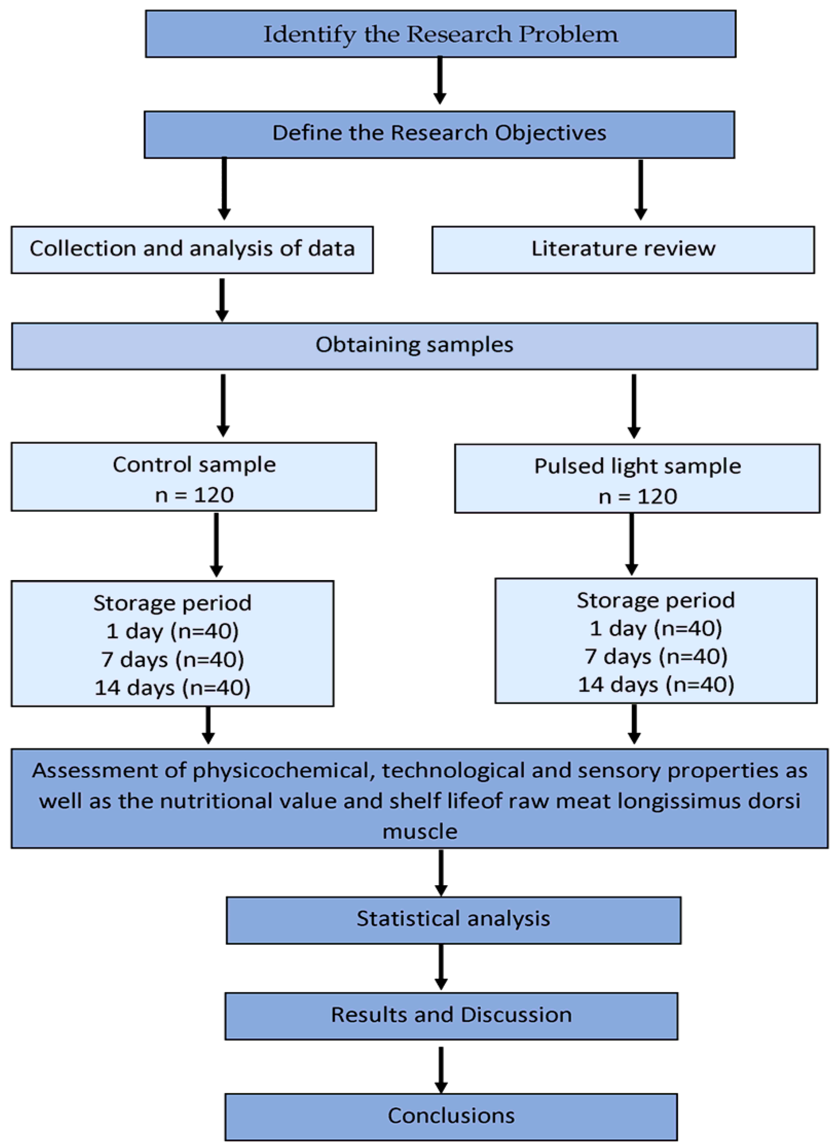
2. Materials and Methods
2.1. Samples Origin and Preparation
- analysis of chemical composition (content: protein, fat, water, minerals, and salts);
- measurements of pH, water activity, forced drip, thermal drip, oxidation-reduction potential, TBARS index, microbiological analysis-total microbial counts;
- determination of color, browning index (BI), and heme pigment content;
- determination of texture parameters and shear force;
- sensory analysis (aroma: intensity and desirability, juiciness, tenderness, taste: intensity and desirability).
2.2. Chemical Composition
2.3. Physicochemical and Microbiological Properties
2.4. Color Measurement and Heme Pigment Content
2.5. Texture Measurement
2.6. Sensory Properties
2.7. Statistical Analysis
3. Results and Discussion
4. Conclusions
Author Contributions
Funding
Institutional Review Board Statement
Informed Consent Statement
Data Availability Statement
Conflicts of Interest
References
- Misra, N.N.; Cheorum, J. Applications of cold plasma technology for microbiological safety in meat industry. Trends Food Sci. Technol. 2007, 64, 74–86. [Google Scholar] [CrossRef]
- Behsnian, D.; Butz, P.; Greiner, R.; Lautenschlager, R. Process-induced undesirable compounds: Chances of non-thermal approaches. Meat Sci. 2014, 98, 392–403. [Google Scholar] [CrossRef]
- Heinrich, V.; Zunabovic, M.; Varzakas, T.; Bergmair, J.; Kneifel, W. Pulsed light treatment of different food types with a special focus on meat: A critical review. Crit. Rev. Food Sci. Nutr. 2016, 56, 591–613. [Google Scholar] [CrossRef] [PubMed]
- Gomez-Lopez, V.M.; Ragaert, P.; Debevere, J.; Devlieghere, F. Pulsed light for food decontamination: A review. Trends Food Sci. Tech. 2007, 18, 464–473. [Google Scholar] [CrossRef]
- Food and Drug Administration. Pulsed Light for the Treatment of Food, 21CFR179.4; Food and Drug Administration: Silver Spring, MD, USA, 1996. [Google Scholar]
- Rowan, N.J.; Mac Gregor, S.J.; Andson, J.G.; Furcre, R.A.; McIllvney, L.; Frish, O. Pulsed-light inactivation of food-related microorganisms. Appl. Environ. Microb. 1999, 65, 1312–1315. [Google Scholar] [CrossRef]
- Marquenie, D.; Geeraerd, A.H.; Lammertyn, J.; Soontjens, C.; Van Impe, J.F.; Michielis, C.W.; Nicolaï, B.M. Combinations of pulsed white light and UV-C or mild heat treatment to inactivate conidia of Botrytis cinerea and Monilia fructigena. Int. J. Food Microbiol. 2003, 85, 185–196. [Google Scholar] [CrossRef]
- Sharma, R.R.; Demirci, A. Inactivation of Escherichia coli O157:H7 on inoculated alfalfa seeds with pulsed ultraviolet light and response surface modeling. J. Food Sci. 2003, 68, 1448–1453. [Google Scholar] [CrossRef]
- Choi, M.S.; Cheigh, C.I.; Jeong, E.; Shin, J.K.; Chung, M.S. Nonthermal sterilization of Listeria monocytogenes in infant foods by intense pulsed-light treatment. J. Food Eng. 2010, 97, 504–509. [Google Scholar] [CrossRef]
- Ngadi, M.O.; Latheef, M.B.; Kassama, L. Emerging technologies for microbial control in food processing. In Green Technologies in Food Production; Boye, J.I., Arcand, Y., Eds.; Springer: Berlin/Heidelberg, Germany, 2012; pp. 363–411. [Google Scholar]
- Roberts, P.; Hope, A. Virus inactivation by high intensity broad spectrum pulsed light. J. Virol Methods. 2003, 110, 61–65. [Google Scholar] [CrossRef]
- Barba, F.J.; Sant’Ana, A.S.; Orlien, V.; Koubaa, M. Innovative Food Preservation Technologies. Deactivation of Spoilage and Pathogenic Microorganisms; Academic Press; Elsevier: Cambridge, MA, USA, 2018. [Google Scholar]
- Elmnasser, N.; Guillou, S.; Leroi, F.; Orange, N.; Bakhrouf, A.; Federighi, M. Pulsed-light system as a novel food decontamination technology: A review. Cancer J. Microbiol. 2007, 53, 813–821. [Google Scholar] [CrossRef]
- Hierro, E.; Barroso, E.; De La Hoz, L.; Ordonez, J.A.; Manzano, S.; Fernandez, M. Efficacy of pulsed light for shelf-life extension and inactivation of Listeria monocytogenes on ready-to-eat cooked meat products. Innov. Food Sci. Emerg. Technol. 2011, 12, 275–281. [Google Scholar] [CrossRef]
- Cabedo, L.; Picart, I.; Barrot, L.; Teixidó, I.; Canelles, A. Prevalence of Listeria monocytogenes and Salmonella in ready-to-eat food in Catalonia, Spain. J. Food Prot. 2008, 71, 855–859. [Google Scholar] [CrossRef]
- Pysz, M.; Pisulewski, P.M.; Leszczyńska, T. The influence of pulse and continuous microwave field affecting on nutrition value and antioxidant properties of germinated soybean seeds. ŻYWNOŚĆ. Nauka. Technologia. Jakość. 2006, 1, 102–116. (In Polish) [Google Scholar]
- Rajkovic, A.; Tomasevic, I.; Smigic, N.; Uyttendaele, M.; Radovanovic, R.; Devlieghere, F. Pulsed UV light as an intervention strategy against Listeria monocytogenes and Escherichia coli O157:H7 on the surface of a meat slicing knife. J. Food Eng. 2010, 100, 446–451. [Google Scholar] [CrossRef]
- Hierro, E.; Ganan, M.; Barroso, B.; Fernandes, M. Pulsed light treatment for the inactivation of selected pathogens and the shelf-life extension of beef and tuna Carpaccio. Int. J. Food Microbiol. 2012, 158, 42–48. [Google Scholar] [CrossRef]
- Charles, F.; Vidal, V.; Olive, F.; Filgueiras, H.; Sallanon, H. Pulsed light treatment as new method to maintain physical and nutritional quality of fresh-cut mangoes. Innov. Food Sci. Emerg. Technol. 2013, 18, 190–195. [Google Scholar] [CrossRef]
- Ramos-Villarroel, A.Y.; Martín-Belloso, O.; Soliva-Fortuny, R. Combined effects of malic acid dip and pulsed light treatments on the inactivation of Listeria innocua and Escherichia coli on fresh-cut produce. Food Control 2015, 52, 112–118. [Google Scholar] [CrossRef]
- Duarte-Molina, F.; Gomez, P.L.; Agueda Castro, M.; Alzamora, S.M. Storage quality of strawberry fruit treated by pulsed light: Fungal decay, water loss and mechanical properties. Innov. Food Sci. Emerg. 2016, 34, 267–274. [Google Scholar] [CrossRef]
- Bhavya, M.L.; Hebbar, U. Pulsed light processing of foods for microbial safety. Food Qual. Saf. 2017, 1, 187–201. [Google Scholar] [CrossRef]
- Ortega-Rovas, E. Characterization and processing relevance of food particulate materials. Part. Part. Syst. Charact. 2012, 29, 192–203. [Google Scholar] [CrossRef]
- Palmieri, L.; Cacace, D. High intensity pulsed light technology. In Emerging Technologies for Food Processing; Academic Press: Cambridge, MA, USA, 2005; pp. 279–306. [Google Scholar] [CrossRef]
- PN-ISO 1442; Meat and Meat Products—Determination of Moisture Content (Reference Method). Polish Committee for Standardization: Warsaw, Poland, 2000.
- PN-A-04018:1975/Az3; Agricultural Food Products. Determination of Nitrogen by the Kjeldahl Method and Expressing as Protein. Polish Committee for Standardization: Warsaw, Poland, 2002.
- PN-ISO 1444; Meat and Meat Products—Determination of Free Fat Content. Polish Committee for Standardization: Warsaw, Poland, 2000.
- PN-A-82112:1973 Az 1; Meat and Meat Products—Determination of Chloride Content. Polish Committee for Standardization: Warsaw, Poland, 2002.
- PN-ISO 936; Meat and Meat Products—Determination of Total Ash. Polish Committee for Standardization: Warsaw, Poland, 2000.
- PN-EN ISO 4833; Food and Feed Microbiology—Horizontal Method for the Determination of Microbial Counts. Plate Method at 30 Degrees C. Polish Committee for Standardization: Warsaw, Poland, 2004.
- Pikul, J.; Leszczyński, D.E.; Kumerow, F.A. Evaluation of three modified TBA methods for measuring lipid oxidation in chicken meat. J. Agric. Food Chem. 1989, 37, 1309–1313. [Google Scholar] [CrossRef]
- Znaniecki, P. Outline of Circulation, Assessment and Processing of Raw Materials of Animalorigin; PWRiL: Warsaw, Poland, 1983; pp. 226–227. (In Polish) [Google Scholar]
- Van Oeckel, M.J.; Warnants, N.; Boucqueé, C.V. Comparison of different methods for measuring water holding capacity and juiciness of pork versus online screening methods. Meat Sci. 1999, 51, 313–320. [Google Scholar] [CrossRef]
- Pérez-López, A.J.; Noguera-Artiaga, L.; López-Miranda González, S.; Gómez-San Miguel, P.; Ferrández, B.; Carbonell-Barrachina, A.A. Acrylamide content in French fries prepared with vegetable oils enriched with β-cyclodextrin or β-cyclodextrin-carvacrol complexes. LWT Food Sci. Technol. 2021, 148, 111765. [Google Scholar] [CrossRef]
- Cserhalmi, Z.S.; Sass-Kiss, A.; Toth-Markus, M.; Lechner, N. Study of pulsed electric field treated citrus juices. Innov. Food Sci. Emerg. Technol. 2006, 7, 49–54. [Google Scholar] [CrossRef]
- Krzywicki, K. The determination of heam pigments in meat. Meat Sci. 1982, 7, 29–36. [Google Scholar] [CrossRef]
- Baryłko-Pikielna, N.; Matuszewska, I. Sensory Testing of Food. Basics—Methods—Application; Polish Society of Food Technologists: Wrocław, Poland, 2009. (In Polish) [Google Scholar]
- ISO 8586-2; Sensory Analysis. General Guidance for the Selection, Training and Monitoring of Assessors. International Organization for Standardization: Geneva, Switzerland, 2008.
- ISO 8587; Sensory Analysis. Methodology. International Organization for Standardization: Geneva, Switzerland, 2006.
- PN-EN ISO 8589; General Guidelines for the Design of a Sensory Analysis Laboratory. Polish Committee for Standardization: Warsaw, Poland, 2010.
- Hwang, H.J.; Park, B.W.; Chung, M.S. Comparison of microbial reduction effect of intense pulsed light according to growth stage and population density of Escherichia coli ATCC 25922 using a double Weibull model. Food Res. Int. 2023, 164, 112353. [Google Scholar] [CrossRef] [PubMed]
- Albert, T.; Braun, P.G.; Saffaf, J.; Wiacek, C. Physical methods for the decontamination of meat. Surf. Curr. Clin. Microbiol. Rep. 2021, 8, 9–20. [Google Scholar] [CrossRef]
- Wekhof, A.; Trompeter, F.J.; Franken, O. Pulsed UV disintegration (PUVD): A new sterilisation mechanism for packaging and broad medical-hospital applications. In Proceedings of the First International Conference on Ultraviolet Technologies, Washington, DC, USA, 14–16 June 2001. [Google Scholar]
- Kramer, B.; Wunderlich, J.; Muranyi, P. Inactivation of Listeria innocua on packaged meat products by pulsed light. Food Pack. Shelf Life 2019, 21, 100353. [Google Scholar] [CrossRef]
- Ganan, M.; Hierro, E.; Hospital, X.F.; Barroso, E.; Fernández, M. Use of pulsed light to increase the safety of ready-to-eat cured meat products. Food Control 2013, 32, 512–517. [Google Scholar] [CrossRef]
- Pałach, Z. Water activity of choosen food-stuff groups. Jakość Żywności 2008, 4, 22–26. (In Polish) [Google Scholar]
- Holownia, K.; Chinnan, M.S.; Reynolds, A.E. Pink color defect in poultry white meat as affected by endogenous conditions. J. Food Sci. 2003, 68, 742–747. [Google Scholar] [CrossRef]
- Rödel, W.; Scheuer, R. Das Redoxpotential bei Fleisch und Fleischerzeugnissen. Fleischwirtschaft 1998, 78, 974–981. [Google Scholar]
- German, J.B. Food processing and lipid oxidation. Adv. Exp. Med. Biol. 1999, 459, 23–50. [Google Scholar] [CrossRef]
- Henry-Costa, F. Irradiation effects on meat: A review. Rev. Ciências Agrar. 2009, 12, 255–262. [Google Scholar] [CrossRef]
- Tomasevic, I.; Djekic, I.; Novaković, S.; Barba, F.; Lorenzo, J.M. The feasibility of pulsed light processing in the meat industry. IOP Conf. Ser. Earth Environ. Sci. 2019, 333, 012034. [Google Scholar] [CrossRef]
- Tarlagdis, B.G.; Watts, B.M.; Younathan, M.T. A distillation method for the quantitative determination of malonaldehyde in rancid foods. J. Am. Oil Chem. Soc. 1960, 37, 44–48. [Google Scholar] [CrossRef]
- Koch, F.; Wiacek, C.; Braun, P.G. Pulsed light treatment for the reduction of Salmonella Typhimurim and Yersinia enterocolitica on pork skin and pork loin. Intl. J. Food Microbiol. 2019, 292, 64–71. [Google Scholar] [CrossRef] [PubMed]
- Rajkovic, A.; Tomasevic, I.; De Meulenaer, B.; Devlieghere, F. The effect of pulsed UV light on Escherichia coli O157:H7, Listeria monocytogenes, Salmonella typhimurium, Staphylococcus aureus and staphylococcal enterotoxin A on sliced fermented salami and its chemical quality. Food Control 2017, 73, 829–837. [Google Scholar] [CrossRef]
- Nicorescu, I.; Nyugen, B.; Chevalier, S.; Orange, N. Effect of pulsed light on the organoleptic propertis and shelf-life extension of pork and salmon. Food Control 2014, 44, 138–145. [Google Scholar] [CrossRef]
- Mancini, R.A.; Hunt, M. Current research in meat color. Meat Sci. 2005, 71, 100–121. [Google Scholar] [CrossRef]
- Troy, D.J.; Kerry, J.P. Consumer perception and the role of science in the meat industry. Meat Sci. 2010, 86, 214–226. [Google Scholar] [CrossRef]
- Valous, N.A.; Mendoza, F.; Sun, D.W.; Allen, P. Colour calibration of a laboratory computer vision system for quality evaluation of pre-sliced hams. Meat Sci. 2009, 81, 132–141. [Google Scholar] [CrossRef] [PubMed]
- Warris, P.D.; Brown, S.N.; Paściak, P. The colour of the adductor as a predictor of pork quality in the loin. Meat Sci. 2006, 73, 565–569. [Google Scholar] [CrossRef]
- Bąk, T.; Kondratowicz, J.; Denaburski, J. Zmiany Fizykochemiczne Mięsa Wieprzowego Normalnego Oraz z Wadami PSE i DFD mrożonego Metodą Owiewową i za Pomocą Ciekłego Dwutlenku Węgla. Mat. Konf. Nauk; Agrobiznes w regionie południowo-wschodniej Polski: Rzeszow, Poland, 1998; pp. 51–61. [Google Scholar]
- Kondartowicz, J.; Matusevičius, P. Właściwości technologiczne mięsa wieprzowego zamrożonego przy użyciu ciekłego azotu i metodą owiewową w różnym czasie od uboju. Żywność. Nauka. Technologia. Jakość 2003, 4, 173–183. [Google Scholar]
- Przybylski, W.; Koćwin-Podsiadła, M.; Kaczorek, S.; Krzęcio, E. Zależność pomiędzy wartością potencjału glikolitycznego mięśnia LD, mierzonego przyżyciowo u świń a pH końcowym mięsa i jego wydajnością technologiczną. Mat. XXVII Ses. Nauk. KTChiŻ PAN Szczec. 1996, 48–52. [Google Scholar]
- Cabeza, M.C.; de la Hoz, L.; Velasco, R.; Cambero, M.I.; Ordóňez, J.A. Safety and quality of ready-to-eat dry cured sausages subjected to E-beam radiation. Meat Sci. 2009, 83, 320–327. [Google Scholar] [CrossRef] [PubMed]
- Campus, M.; Flores, M.; Martinez, A.; Toldra, F. Effect of high pressure treatment on color, microbial and chemical characteristics of dry cured loin. Meat Sci. 2008, 80, 1174–1181. [Google Scholar] [CrossRef]
- Cava, R.; Ladero, L.; González, S.; Carrasco, A.; Ramirez, M.R. Effect of pressure and holding time on color, protein and lipid oxidation of sliced dry-cured Iberian ham and loin during refrigerated storage. Innov. Food Sci. Emerg. Technol. 2009, 10, 76–81. [Google Scholar] [CrossRef]
- Houser, T.A.; Sebranek, J.G.; Núňez Maisonet, W.; Cordray, J.C.; Ahn, D.U.; Dixon, P.M. Irradiation-induced cured ham color fading and regeneration. Food Sci. 2005, 70, 280–285. [Google Scholar] [CrossRef]
- Karamucki, T. Studies on the Formation of Colour of the Porcine Longissimus Lumborum Muscle and the Usefulness of Selected Methods of its Measurement for Pork Quality Assessment; Rozprawy: Szczecin, Poland, 2008. [Google Scholar]
- Lindahl, G.; Lundström, K.; Tornberg, E. Contribution of pigment content, mioglobin forms and internal reflectance to the colour of pork loin and ham from pure breed pigs. Meat Sci. 2001, 59, 141–151. [Google Scholar] [CrossRef]
- Karamucki, T.; Gardzielewska, J.; Jakubowska, M.; Rybak, K.; Garczewska, J. The relationship between colour and pH in cold-stored quail breast muscle. Ann. Anim. Sci. 2012, 13, 401–413. [Google Scholar] [CrossRef]
- Tomasevic, I. The effect of intense light pulses on the sensory quality and instrumental color of meat from different animal breeds. Biotechnol Anim Husb. 2015, 31, 273–281. [Google Scholar] [CrossRef]
- Tomasevic, I.; Rajkovic, A. The sensory quality of meat, game, poultry, seafood and meat products as affected by intense light pulses. A systematic review. Procedia Food Sci. 2015, 5, 285–288. [Google Scholar] [CrossRef]
- Wambura, P.; Verghese, M. Effect of pulsed ultraviolet light on quality of sliced ham. LWT Food Sci. Technol. 2011, 44, 2173–2179. [Google Scholar] [CrossRef]
- Genot, C. Congélation et Qualité de la Viande; Inra Editions: Paris, France, 2002. [Google Scholar]
- Fletcher, D.L. Poultry meat quality. World’s Poult. Sci. J. 2002, 58, 131–145. [Google Scholar] [CrossRef]
- Chang, H.J.; Wang, Q.; Zhou, G.H.; Xu, X.L.; Li, C.B. Influence of weak organic acids and sodium chloride marination on characteristics of connective tissue collagen and textural properties of beef semitendinosus muscle. J. Text. Stud. 2010, 41, 279–301. [Google Scholar] [CrossRef]
- Migdał, W.; Wojtysiak, D.; Palka, K.; Natonek-Wiśniewska, M.; Duda, I.; Nowocień, A. Chemical composition and texture parameters of some selected muscles of the polish landrace fatteners slaughtered at different age. Żywność. Nauka. Technologia. Jakość 2007, 6, 277–284. (In Polish) [Google Scholar]
- Cierach, M.; Borzyszkowski, M.; Niedźwiedź, J. Wołowina kulinarna—Czynniki przyżyciowe a jakość. Przem. Spoż. 2009, 8, 58–63. [Google Scholar]
- Bratcher, C.L.; Johnson, D.D.; Littell, R.C.; Gwartney, B.L. The effects of quality grade, aging, and location within muscle on Warner-Bratzler shear force in beef muscles of locomotion. Meat Sci. 2005, 70, 279–284. [Google Scholar] [CrossRef]
- White, A.; O’Sullivan, A.; Troy, D.J.; O’Neill, E.E. Effects of electrical stimulation, chilling temperature and hot-boning on the tenderness of bovine muscles. Meat Sci. 2006, 73, 196–203. [Google Scholar] [CrossRef]
- Baryłko-Pikielna, N. Sensory analysis in food quality assurance. Przem. Spoż. 1998, 12, 25–50. [Google Scholar]
- Ozer, N.P.; Demerci, A. Inactivation of Escherichia coli O157:H7 and Listeria monocytogenes inoculated on raw salmon fillets by pulsed UV-light treatment. Int. J. Food Sci. Technol. 2006, 41, 354–360. [Google Scholar] [CrossRef]


| Specification | Cold Storage Period (Days) | |||||
|---|---|---|---|---|---|---|
| 1 | 7 | 10 | ||||
| K | S | K | S | K | S | |
| Protein (%) | 20.85 ± 0.31 | 20.75 ± 0.31 | 20.25 ± 0.31 | 20.37 ± 0.31 | 20.43 ± 0.31 | 20.32 ± 0.31 |
| Fat (%) | 7.05 ± 1.46 | 7.16 ± 1.12 | 7.25 ± 1.21 | 7.19 ± 1.45 | 7.34 ± 1.32 | 7.26 ± 1.13 |
| Water (%) | 71.01 ± 1.28 | 71.23 ± 1.32 | 71.43 ± 1.45 | 71.35 ± 1.67 | 70.95 ± 1.63 | 70.98 ± 1.84 |
| Minerals (%) | 1.49 ± 0.08 | 1.50 ± 0.30 | 1.46 ± 0.16 | 1.54 ± 0.40 | 1.52 ± 0.16 | 1.58 ± 0.13 |
| Salts (%) | 0.57 ± 0.02 | 0.57 ± 0.02 | 0.61 ± 0.07 | 0.65 ± 0.15 | 0.49 ± 0.06 | 0.48 ± 0.03 |
| Specification | Cold Storage Period (Days) | |||||
|---|---|---|---|---|---|---|
| 1 | 7 | 10 | ||||
| K | S | K | S | K | S | |
| pH | 5.55 ± 0.05 | 5.53 ± 0.07 | 5.55 ± 0.02 | 5.54 ± 0.04 | 5.61 ± 0.12 | 5.58 ± 0.07 |
| Water activity | 0.976 ab ± 0.003 | 0.980 ab ± 0.004 | 0.975 ab ± 0.003 | 0.977 ab ± 0.001 | 0.976 a ± 0.004 | 0.957 b ± 0.010 |
| Thermal drip (%) | 21.10 ± 1.17 | 21.64 ± 2.28 | 22.88 ± 2.77 | 20.63 ± 2.11 | 19.47 ± 1.76 | 18.51 ± 1.18 |
| Forced drip (cm2) | 2.83 ± 0.70 | 2.66 ± 0.41 | 2.11 ± 0.78 | 3.73 ± 0.86 | 3.57 ± 0.14 | 2.79 ± 0.05 |
| TBARS index (mg MDA/kg) | 0.46 a ± 0.09 | 0.35 b ± 0.05 | 0.61 a ± 0.11 | 0.52 b ± 0.03 | 0.56 ab ± 0.05 | 0.57 ab ± 0.10 |
| Oxidation-reduction potential (mV) | 358.50 ab ± 5.02 | 365.57 ab ± 5.62 | 443.67 a ± 14.10 | 414.67 b ± 1.67 | 414.83 ab ± 12.03 | 404.30 ab ± 6.30 |
| Total numer of microorganisms (CFU/g) | 6.82 × 10² ± 5.57 × 10² | 5.30 × 10² ± 1.94 × 10² | 7.50 × 10³ ± 4.54 × 10² | 3.63 × 10³ ± 1.31 × 10² | 3.10 × 10⁶ ± 2.43 × 10⁵ | 1.59 × 10⁶ ± 1.33 × 10⁵ |
| Specification | Cold Storage Period (Days) | |||||
|---|---|---|---|---|---|---|
| 1 | 7 | 10 | ||||
| K | S | K | S | K | S | |
| L* | 53.87 ab ± 3.67 | 52.90 ab ± 2.60 | 54.86 ab ± 2.34 | 57.14 ab ± 4.12 | 53.18 a ± 5.91 | 58.51 b ± 3.70 |
| a* | 11.42 ± 0.76 | 10.84 ± 0.48 | 13.06 ± 1.70 | 12.60 ± 1.52 | 11.67 ± 1.49 | 11.83 ± 1.13 |
| b* | 6.84 ± 0.77 | 7.71 ± 1.86 | 8.48 ± 1.28 | 8.30 ± 1.02 | 8.12 ± 1.12 | 8.65 ± 1.08 |
| Browning index (BI) | 28.46 ± 2.51 | 30.17 ± 3.05 | 33.51 ± 2.34 | 31.19 ± 2.89 | 32.00 ± 2.71 | 30.23 ± 3.12 |
| ∆E | 1.43 | 2.33 | 5.36 | |||
| MB (%) | 60.05 ± 6.63 | 45.80 ± 10.93 | 51.29 ± 6.21 | 48.90 ± 8.43 | 43.45 ± 12.65 | 42.98 ± 21.37 |
| METMB (%) | 17.19 ± 7.25 | 30.00 ± 11.65 | 28.63 ± 9.38 | 27.44 ± 6.97 | 30.82 ± 8.17 | 21.03 ± 8.70 |
| MBO (%) | 22.76 ± 2.68 | 24.20 ± 9.13 | 20.08 ± 5.19 | 23.66 ± 7.07 | 25.73 ± 10.59 | 35.99 ± 18.33 |
| OZB (mg/kg) | 6.84 ± 0.87 | 7.21 ± 1.14 | 7.01 ± 1.46 | 7.32 ± 0.91 | 7.09 ± 1.32 | 7.24 ± 0.96 |
| Specification | Cold Storage Period (Days) | |||||
|---|---|---|---|---|---|---|
| 1 | 7 | 10 | ||||
| K | S | K | S | K | S | |
| Hardness 1 (N) | 91.91 ab ± 6.03 | 140.02 ab ± 5.21 | 156.34 ab ± 4.80 | 131.34 ab ± 3.76 | 132.35 a ± 8.82 | 47.23 b ± 4.89 |
| Hardness 2 (N) | 44.53 a ± 3.02 | 80.63 b ± 4.79 | 105.40 ab ± 8.58 | 83.65 ab ± 3.45 | 83.44 a ± 6.00 | 35.19 b ± 6.55 |
| Rigidity 5 (N) | 28.15 ± 2.02 | 43.10 ± 9.36 | 28.96 ± 1.46 | 26.51 ± 1.24 | 9.94 ± 1.98 | 5.30 ± 1.08 |
| Rigidity 8 (N) | 68.06 ab ± 5.47 | 110.13 ab ± 6.72 | 104.26 ab ± 4.28 | 78.40 ab ± 3.43 | 67.53 a ± 4.65 | 20.95 b ± 1.76 |
| Adhesiveness (mJ) | 1.33 ± 0.08 | 2.01 ± 0.01 | 2.39 ± 0.06 | 1.53 ± 0.02 | 1.86 ± 0.09 | 2.14 ± 0.03 |
| Cohesiveness | 0.19 ± 0.05 | 0.27 ± 0.06 | 0.19 ± 0.08 | 0.26 ± 0.03 | 0.26 ± 0.08 | 0.23 ± 0.06 |
| Springiness(mm) | 2.41 ± 0.58 | 2.92 ± 0.43 | 3.08 ± 0.37 | 3.94 ± 0.32 | 4.03 ± 0.71 | 3.60 ± 0.59 |
| Resilience | 0.14 ± 0.01 | 0.11 ± 0.02 | 0.15 ± 0.05 | 0.13 ± 0.06 | 0.21 ± 0.08 | 0.24 ± 0.03 |
| Gumminess (N) | 11.85 ± 1.10 | 15.92 ± 1.61 | 29.62 ± 2.46 | 35.94 ± 3.43 | 34.44 ± 2.78 | 15.08 ± 1.17 |
| Chewiness (mJ) | 30.80 ab ± 2.60 | 48.01 ab ± 2.81 | 91.98 ab ± 3.92 | 142.36 ab ± 2.06 | 144.72 a ± 5.71 | 39.47 b ± 5.74 |
| Shear force (N/cm2) | 102.10 a ± 20.71 | 69.41 b ± 19.77 | 81.40 ab ± 20.42 | 70.06 ab ± 9.52 | 64.07 ab ± 7.13 | 68.43 ab ± 2.30 |
| Specification | Cold Storage Period (Days) | |||||
|---|---|---|---|---|---|---|
| 1 | 7 | 10 | ||||
| K | S | K | S | K | S | |
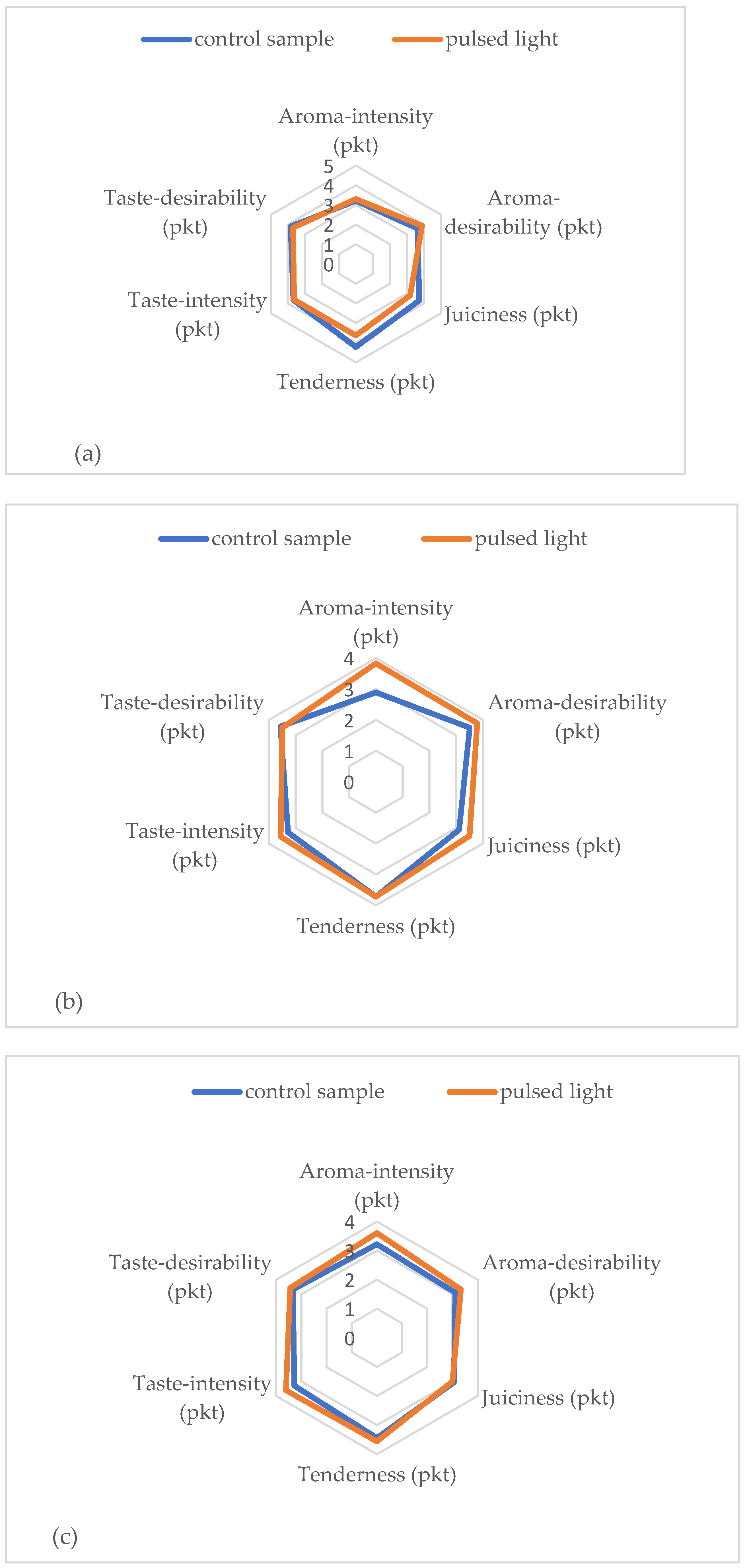
| Aroma-intensity (pkt) | 3.22 ± 0.71 | 3.31 ± 0.88 | 2.89 ± 0.70 | 3.83 ± 0.43 | 3.22 ± 0.62 | 3.61 ± 0.60 |
| Aroma-desirability (pkt) | 3.61 ± 0.49 | 3.88 ± 0.69 | 3.50 ± 0.71 | 3.78 ± 0.51 | 3.11 ± 0.55 | 3.33 ± 0.66 |
| Juiciness (pkt) | 3.72 ± 0.06 | 3.19 ± 0.70 | 3.11 ± 0.36 | 3.50 ± 0.66 | 3.06 ± 0.53 | 3.00 ± 0.61 |
| Tenderness (pkt) | 4.22 ± 0.71 | 3.63 ± 0.83 | 3.72 ± 0.51 | 3.72 ± 0.79 | 3.44 ± 0.85 | 3.56 ± 0.63 |
| Taste-intensity (pkt) | 3.67 ± 0.43 | 3.63 ± 0.44 | 3.28 ± 0.87 | 3.56 ± 0.73 | 3.28 ± 0.36 | 3.61 ± 0.42 |
| Taste-desirability (pkt) | 3.83 ± 0.35 | 3.69 ± 0.46 | 3.57 ± 0.95 | 3.50 ± 0.56 | 3.33 ± 0.50 | 3.44 ± 0.58 |
Disclaimer/Publisher’s Note: The statements, opinions and data contained in all publications are solely those of the individual author(s) and contributor(s) and not of MDPI and/or the editor(s). MDPI and/or the editor(s) disclaim responsibility for any injury to people or property resulting from any ideas, methods, instructions or products referred to in the content. |
© 2023 by the authors. Licensee MDPI, Basel, Switzerland. This article is an open access article distributed under the terms and conditions of the Creative Commons Attribution (CC BY) license (https://creativecommons.org/licenses/by/4.0/).
Share and Cite
Duma-Kocan, P.; Rudy, M.; Gil, M.; Stanisławczyk, R.; Żurek, J.; Zaguła, G. The Impact of a Pulsed Light Stream on the Quality and Durability of the Cold-Stored Longissimus Dorsal Muscle of Pigs. Int. J. Environ. Res. Public Health 2023, 20, 4063. https://doi.org/10.3390/ijerph20054063
Duma-Kocan P, Rudy M, Gil M, Stanisławczyk R, Żurek J, Zaguła G. The Impact of a Pulsed Light Stream on the Quality and Durability of the Cold-Stored Longissimus Dorsal Muscle of Pigs. International Journal of Environmental Research and Public Health. 2023; 20(5):4063. https://doi.org/10.3390/ijerph20054063
Chicago/Turabian StyleDuma-Kocan, Paulina, Mariusz Rudy, Marian Gil, Renata Stanisławczyk, Jagoda Żurek, and Grzegorz Zaguła. 2023. "The Impact of a Pulsed Light Stream on the Quality and Durability of the Cold-Stored Longissimus Dorsal Muscle of Pigs" International Journal of Environmental Research and Public Health 20, no. 5: 4063. https://doi.org/10.3390/ijerph20054063
APA StyleDuma-Kocan, P., Rudy, M., Gil, M., Stanisławczyk, R., Żurek, J., & Zaguła, G. (2023). The Impact of a Pulsed Light Stream on the Quality and Durability of the Cold-Stored Longissimus Dorsal Muscle of Pigs. International Journal of Environmental Research and Public Health, 20(5), 4063. https://doi.org/10.3390/ijerph20054063