
Biological Mechanisms behind Wischnewsky Spots Finding on Gastric Mucosa: Autopsy Cases and Literature Review


 ,
,  ,
, 

Abstract
:1. Introduction
2. Materials and Methods
2.1. Forensic Investigations
2.2. Literature Review
3. Results
3.1. Cases Presentation
3.1.1. Case 1
3.1.2. Case 2
3.1.3. Case 3
3.2. Literature Review
3.3. Scene Investigation and Autopsy Findings
3.3.1. Case 1
3.3.2. Case 2
3.3.3. Case 3
3.3.4. Analysis of Cases
4. Discussion
5. Conclusions
- -
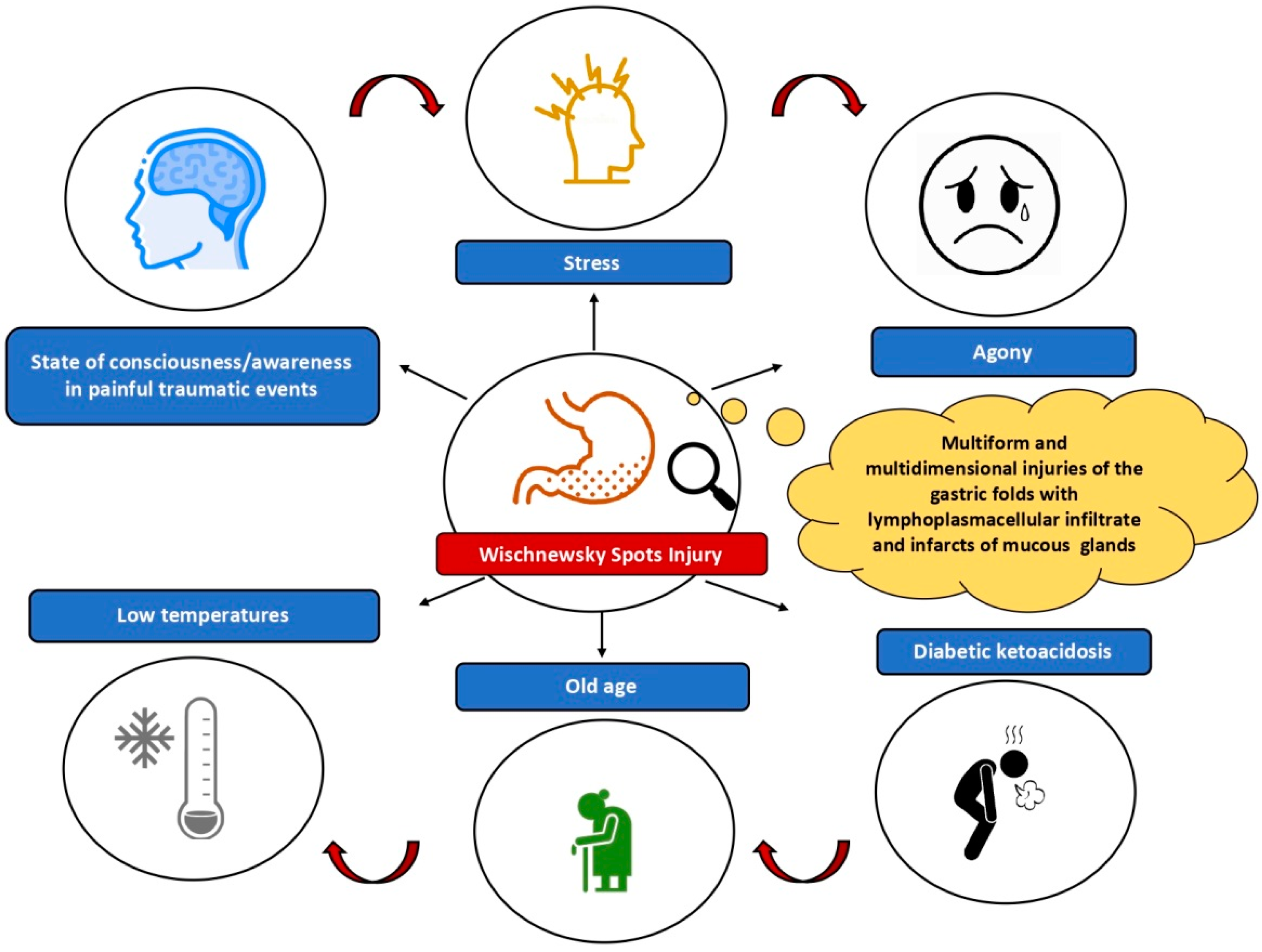
- Hypothermia;
- -
- Physical stress with prolonged agony caused by concomitant and particularly painful traumatic events (impalement, traumatic fractures with reduced mobility);
- -
- Activation of gastrin and pepsinogen correlated to low temperatures;
- -
- Old age;
- -
- Diabetic ketoacidosis.
- -
- In this regard, we encourage the publication of further forensic cases of hypothermia or WS finding by suggesting, for the purpose of homogeneity between the studies, to:
- -
- Describe an accurate analysis of the scene where the body was found (rivers, sea, mountains, cold environments, etc.) reporting the ambient temperature;
- -
- Report patient’s age, circumstantial data, hide and die phenomena, any stress factors at the time of death, previous comorbidities;
- -
- Describe macroscopic and microscopic autopsy data, with any other signs of hypothermia;
- -
- Perform post-mortem laboratory investigations, including vitreous glucose and β-hydroxybutyrate, to investigate the correlation with diabetic ketoacidosis.
Author Contributions
Funding
Institutional Review Board Statement
Informed Consent Statement
Data Availability Statement
Conflicts of Interest
References
- Takauji, S.; Hayakawa, M. Intensive care with extracorporeal membrane oxygenation rewarming in accident severe hypothermia (ICE-CRASH) study: A protocol for a multicentre prospective, observational study in Japan. BMJ Open 2021, 11, e052200. [Google Scholar] [CrossRef]
- Le May, M.; Osborne, C.; Russo, J.; So, D.; Chong, A.Y.; Dick, A.; Froeschl, M.; Glover, C.; Hibbert, B.; Marquis, J.F.; et al. Effect of Moderate vs Mild Therapeutic Hypothermia on Mortality and Neurologic Outcomes in Comatose Survivors of Out-of-Hospital Cardiac Arrest: The CAPITAL CHILL Randomized Clinical Trial. JAMA 2021, 326, 1494–1503. [Google Scholar] [CrossRef]
- Bright, F.; Winskog, C.; Byard, R.W. Wischnewski spots and hypothermia: Sensitive, specific, or serendipitous? Forensic. Sci. Med. Pathol. 2013, 9, 88–90. [Google Scholar] [CrossRef]
- Flabouris, K.; Russell, P.; Wills, S. Wischnewski spots in a case of accidental hypothermia. Clin. Case Rep. 2021, 9. [Google Scholar] [CrossRef] [PubMed]
- Almeida, B.A.; Santos, I.R.; Henker, L.C.; Lorenzett, M.P.; Ferrari, F.E.; Surita, L.E.; Panziera, W.; Pavarini, S.P.; Driemeier, D. Wischnewski Spots and Black Oesophagus in Suspected Fatal Hypothermia in a Brown Howler Monkey (Alouatta guariba clamitans) and a Rabbit (Oryctolagus cuniculus). J. Comp. Pathol. 2021, 186, 18–22. [Google Scholar] [CrossRef]
- Dickinson, G.M.; Maya, G.X.; Lo, Y.; Jarvis, H.C. Hypothermia-related Deaths: A 10-year Retrospective Study of Two Major Metropolitan Cities in the United States. J. Forensic. Sci. 2020, 65, 2013–2018. [Google Scholar] [CrossRef] [PubMed]
- Duval, I.; Doberentz, E.; Madea, B. Lethal hypothermia due to impalement. Forensic. Sci. Int. 2020, 314, 110397. [Google Scholar] [CrossRef] [PubMed]
- Yang, C.; Sugimoto, K.; Murata, Y.; Hirata, Y.; Kamakura, Y.; Koyama, Y.; Miyashita, Y.; Nakama, K.; Higashisaka, K.; Harada, K.; et al. Molecular mechanisms of Wischnewski spot development on gastric mucosa in fatal hypothermia: An experimental study in rats. Sci. Rep. 2020, 10, 1877. [Google Scholar] [CrossRef] [PubMed]
- McCarthy, S.; Garland, J.; Hensby-Bennett, S.; Philcox, W.; Kesha, K.; Stables, S.; Tse, R. Black Esophagus (Acute Necrotizing Esophagitis) and Wischnewsky Lesions in a Death from Diabetic Ketoacidosis: A Possible Underlying Mechanism. Am. J. Forensic. Med. Pathol. 2019, 40, 192–195. [Google Scholar] [CrossRef] [PubMed]
- Garland, J.; Philcox, W.; Kesha, K.; McCarthy, S.; Lam, L.; Palmiere, C.; O’Regan, T.; Stables, S.; Tse, R. Biochemical Differences Between Vitreous Humor and Cerebral Spinal Fluid in a Death From Diabetic Ketoacidosis. Am. J. Forensic. Med. Pathol. 2019, 40, 188–191. [Google Scholar] [CrossRef] [PubMed]
- Tse, R.; Garland, J.; Kesha, K.; Triggs, Y.; Yap, Z.; Stables, S. Basal Subnuclear Vacuolization, Armanni-Ebstein Lesions, Wischnewsky Lesions, and Elevated Vitreous Glucose and β-Hydroxybuyrate: Is It Hypothermia, Diabetic Ketoacidosis, or Both? Am. J. Forensic. Med. Pathol. 2018, 39, 279–281. [Google Scholar] [CrossRef]
- Stern, A.W.; Vieson, M.D. Wischnewsky-like spots in fatal cases of canine hypothermia. Int. J. Leg. Med. 2017, 131, 1639–1641. [Google Scholar] [CrossRef] [PubMed]
- Doberentz, E.; Markwerth, P.; Madea, B. Fatty degeneration in renal tissue in cases of fatal accidental hypothermia. Rom. J. Leg. Med. 2017, 25, 152–157. [Google Scholar] [CrossRef]
- Ortmann, J.; Doberentz, E.; Kernbach-Wighton, G.; Madea, B. Lethal hypothermia after firing a suicidal shot to the head in a car. Arch. Kriminol. 2016, 238, 188–197. [Google Scholar] [PubMed]
- Kanchan, T.; Babu Savithry, K.S.; Geriani, D.; Atreya, A. Wischnewski spots in fatal burns. Med. Leg. J. 2016, 84, 52–55. [Google Scholar] [CrossRef] [PubMed]
- Clark, K.H.; Stoppacher, R. Gastric Mucosal Petechial Hemorrhages (Wischnewsky Lesions), Hypothermia, and Diabetic Ketoacidosis. Am. J. Forensic. Med. Pathol. 2016, 37, 165–169. [Google Scholar] [CrossRef] [PubMed]
- Palmiere, C.; Teresiński, G.; Hejna, P. Postmortem diagnosis of hypothermia. Int. J. Leg. Med. 2014, 128, 607–614. [Google Scholar] [CrossRef] [PubMed] [Green Version]
- Bright, F.M.; Winskog, C.; Tsokos, M.; Walker, M.; Byard, R.W. Issues in the diagnosis of hypothermia: A comparison of two geographically separate populations. J. Forensic. Leg. Med. 2014, 22, 30–32. [Google Scholar] [CrossRef] [PubMed]
- Zivković, V.; Nikolić, S. Philemon and Baucis, Diogenes and syllogomania, Wischnewski and hypothermia—Gastric mucosal lesions in partially mummified bodies. Med. Sci. Law. 2014, 54, 177–180. [Google Scholar] [CrossRef] [PubMed]
- Bright, F.; Winskog, C.; Walker, M.; Byard, R.W. Why are Wischnewski spots not always present in lethal hypothermia? The results of testing a stress-reduced animal model. J. Forensic. Leg. Med. 2013, 20, 785–787. [Google Scholar] [CrossRef] [PubMed]
- Bright, F.; Gilbert, J.D.; Winskog, C.; Byard, R.W. Additional risk factors for lethal hypothermia. J. Forensic. Leg. Med. 2013, 20, 595–597. [Google Scholar] [CrossRef] [PubMed]
- Preuss, J.; Dettmeyer, R.; Poster, S.; Lignitz, E.; Madea, B. The expression of heat shock protein 70 in kidneys in cases of death due to hypothermia. Forensic. Sci. Int. 2008, 176, 248–252. [Google Scholar] [CrossRef] [PubMed]
- Tsokos, M.; Rothschild, M.A.; Madea, B.; Rie, M.; Sperhake, J.P. Histological and immunohistochemical study of Wischnewsky spots in fatal hypothermia. Am. J. Forensic. Med. Pathol. 2006, 27, 70–74. [Google Scholar] [CrossRef]
- Preuss, J.; Thierauf, A.; Dettmeyer, R.; Madea, B. Wischnewsky’s spots in an ectopic stomach. Forensic. Sci. Int. 2007, 169, 220–222. [Google Scholar] [CrossRef] [PubMed]
- Landeira-Fernandez, J. Analysis of the cold-water restraint procedure in gastric ulceration and body temperature. Physiol. Behav. 2004, 82, 827–833. [Google Scholar] [CrossRef] [PubMed]
- Mant, A.K. Some postmortem observations in accidental hypothermia. Med. Sci. Law 1964, 4, 44–46. [Google Scholar] [CrossRef] [PubMed]
- Mizukami, H.; Shimizu, K.; Shiono, H.; Uezono, T.; Sasaki, M. Forensic diagnosis of death from cold. Leg. Med. 1999, 1, 204–209. [Google Scholar] [CrossRef]
- Haba, K.; Yamamoto, H.; Takada, M. Wishnewski’s flecks and local cold injuries of accidental hypothermia. Res. Pract. Forensic. Med. 1989, 32, 283–289. [Google Scholar]
- Vincent, G.P.; Paré, W.P.; Prenatt, J.E.D. Aggression, body temperature and stress ulcer. Physiol. Behav. 1984, 32, 265–268. [Google Scholar] [CrossRef]
- Kiang-Ulrich, M.; Horvath, S.M. Age-related differences in response to acute cold challenge (−10 °C) in male F344 rats. Exp. Gerontol. 1985, 20, 201–209. [Google Scholar] [CrossRef]

| Case | Sex | Age | Comorbidities | Scene Finding | Main Autopsy Findings |
|---|---|---|---|---|---|
| 1 | M | 81 | Dementia | Steep hill | Head trauma |
| 2 | F | 83 | Dementia | Impervious area to the banks of a river | Vertebral fracture |
| 3 | M | 85 | Schizophrenia | Recovery of the body in a river | Vertebral fracture |
Publisher’s Note: MDPI stays neutral with regard to jurisdictional claims in published maps and institutional affiliations. |
© 2022 by the authors. Licensee MDPI, Basel, Switzerland. This article is an open access article distributed under the terms and conditions of the Creative Commons Attribution (CC BY) license (https://creativecommons.org/licenses/by/4.0/).
Share and Cite
Sacco, M.A.; Abenavoli, L.; Juan, C.; Ricci, P.; Aquila, I. Biological Mechanisms behind Wischnewsky Spots Finding on Gastric Mucosa: Autopsy Cases and Literature Review. Int. J. Environ. Res. Public Health 2022, 19, 3601. https://doi.org/10.3390/ijerph19063601
Sacco MA, Abenavoli L, Juan C, Ricci P, Aquila I. Biological Mechanisms behind Wischnewsky Spots Finding on Gastric Mucosa: Autopsy Cases and Literature Review. International Journal of Environmental Research and Public Health. 2022; 19(6):3601. https://doi.org/10.3390/ijerph19063601
Chicago/Turabian StyleSacco, Matteo Antonio, Ludovico Abenavoli, Cristina Juan, Pietrantonio Ricci, and Isabella Aquila. 2022. "Biological Mechanisms behind Wischnewsky Spots Finding on Gastric Mucosa: Autopsy Cases and Literature Review" International Journal of Environmental Research and Public Health 19, no. 6: 3601. https://doi.org/10.3390/ijerph19063601
APA StyleSacco, M. A., Abenavoli, L., Juan, C., Ricci, P., & Aquila, I. (2022). Biological Mechanisms behind Wischnewsky Spots Finding on Gastric Mucosa: Autopsy Cases and Literature Review. International Journal of Environmental Research and Public Health, 19(6), 3601. https://doi.org/10.3390/ijerph19063601