Article

Associations between Muscle-Tendon Morphology and Functional Movements Capacity, Flexibility, and Balance in Older Women

by
Pornpimol Muanjai
1,2,*,
Juntip Namsawang
1,2,
Danguole Satkunskienė
3 and
Sigitas Kamandulis
3
1
Department of Physical Therapy, Allied Health Sciences Faculty, Burapha University, Chonburi 20131, Thailand
2
Exercise and Nutrition Innovation and Sciences Research Unit, Burapha University, Chonburi 20131, Thailand
3
Institute of Sport Science and Innovations, Lithuanian Sports University, 44221 Kaunas, Lithuania
*
Author to whom correspondence should be addressed.
Int. J. Environ. Res. Public Health 2022, 19(23), 16099; https://doi.org/10.3390/ijerph192316099
Submission received: 17 October 2022 / Revised: 24 November 2022 / Accepted: 29 November 2022 / Published: 1 December 2022
(This article belongs to the Special Issue Physical Activity for Special Populations)

Abstract

:
Loss of functional movement capacity in older adults is related to adverse changes in musculotendinous morphology, but this relationship is poorly understood. This study examined the associations between musculotendinous morphology and functional movements, flexibility, and balance ability. Ninety-nine older women (66.6 ± 4.6 years, body mass index 23.5 ± 3.3 kg∙m–2) were recruited from Chonburi Province, Thailand. During one 90-min visit, muscle ultrasound imaging of vastus lateralis, biceps femoris, and medial gastrocnemius muscles, and tendon ultrasonography of the Achilles tendon and patellar tendon were performed. Measures were also obtained for the straight leg raise, passive dorsiflexion, balance, and functional tests (Five Times Sit to Stand (5TSTS), Timed Up and Go (TUG), 10-Meter Fast Walk Test (10-MFWT), and 6-Minute Walk Test (6-MWT)). The results specify that functional movement performance correlates most strongly with medial gastrocnemius muscle thickness (5TSTS (r = −0.26), TUG (r = −0.44), 10-MFWT (r = 0.41), and 6-MWT (r = 0.48) all p < 0.05) and that vastus lateralis muscle thickness and medial gastrocnemius muscle thickness correlate positively with balance ability (r = 0.24, 0.34; p < 0.05) and negatively with fear of falling. It appears that muscle mass, rather than other morphological parameters, such as muscle quality or fascicle length, is the main factor affecting the susceptibility of older women to frailty.

1. Introduction

Loss of functional capacity in older adults leads to declines in social connections, quality of life, and health disorders, including increased frailty and mortality. Older adults’ movement functions deteriorate because of various causes, including muscle atrophy and diminished strength, balance, and cardiovascular fitness [1]. In addition, soft tissue stiffness increases because of increases in collagen cross-linking [2], extracellular water content within a muscle, and intramuscular adipose tissue [3], as well as decreases in collagen fibril diameter [4]. These alterations contribute to a decline in joint flexibility, as evidenced by decreases in the range of motion (ROM), which can affect balance and cause functional limitations and a decline in quality of life as people age [5].
Muscle morphology can be detected by ultrasound (US) imaging, which is now widely used to assess musculotendinous quality and quantity for identifying sarcopenia in older adults [6,7]. Madden et al. [8] reported that muscle thickness (MT), as measured by US imaging, is closely related to muscle mass as measured by bioimpedance and grip strength. Independent of muscle mass, echo intensity (EI) can be used to determine muscle quality in assessing muscle function in older adults [9]. Age-related biological processes affect muscle composition by increasing adipose tissue accumulation and fibrosis and altering the distribution of water content and the extracellular matrix [10]. These muscle changes are reflected by increased EI, which provides a marker of lower-quality muscle [10,11]. Changes in fascicle length (FL) can also contribute to alterations in body function, e.g., shorter and more pennate fascicles cause slower muscle shortening [12].
In assessing functional capacity in older adults, the contribution of muscle morphology other than muscle mass remains poorly understood compared to other functional fitness measures. The aim of this study was to investigate whether muscle–tendon morphology and functional movements, flexibility, and balance ability are related in older women. We predicted that two major elements, namely loss of FL and muscular quality, would be evident in older women, which may increase the susceptibility to frailty. Identifying the characteristics most predictive of older people’s functional capacity will provide useful information for more precise recommendations for senior fitness programs.

2. Materials and Methods

2.1. Participants

Ninety-nine older women (66.6 ± 4.6 years, body mass index (BMI) 23.5 ± 3.3 kg∙m−2) participated in this study. They were recruited from Chonburi Province, Thailand. The main inclusion criteria were age 60–80 years, completion of the Physical Activity Readiness Questionnaire, and the ability to walk independently. The exclusion criteria were neuromuscular or skeletal injury of the lower extremities or lower back in the preceding 6 months, inability to follow instructions during the assessments, having moderate pain (visual analog scale > 3/10), having currently any analgesic drugs, and obesity (BMI > 29.9 kg∙m−2). The characteristics of the participants are presented in Table 1. Exercise volume was calculated by exercise time per day multiplied by the number of days per week in the past month. Participants were made aware of the testing requirements and provided written informed consent. The study was approved by the Research and Innovation Administration Division of Burapha University Ethics Committee (IRB1-007/2565, approved 4 January 2022) and followed the principles of the Declaration of Helsinki.

2.2. Study Design

This was a cross-sectional study. Data were collected from February through July 2022 in the Physical Therapy Department, Allied Health Science Faculty, Burapha University, Chonburi, Thailand. All data were collected with a single visit lasting 60–90 min. US imaging of the vastus lateralis (VL), long head of bicep femoris (BF), and medial gastrocnemius (MG) muscles, and tendon ultrasonography of the Achilles tendon (AT) and patellar tendon (PT) were performed. All analyses of muscle morphology were performed on the dominant leg, which was defined as the kicking leg [13]. ROM, balance, and functional movement were assessed using the Five Times Sit to Stand (5TSTS), Timed Up and Go (TUG), 10-Meter Fast Walk Test (10-MFWT), and 6-Minute Walk Test (6-MWT) in the order listed. The same investigators performed all assessments with 2 min rest intervals between tests. After the investigators’ demonstration, each participant was encouraged to perform a practice trial of one repetition, followed by the complete protocol for each performance test.

2.3. Measurements

2.3.1. Musculotendinous Ultrasound Imaging

B-mode US (M5 series, Shenzhen, Mindray Bio-Medical, China) with a linear 4 cm and 7.5 MHz probe (MSK preset) was used to capture the muscle and tendon images of the dominant leg. For VL and BF, the US probe was located at 50% of the distance between the anterior superior iliac spine and the superior pole of the patella in the supine and prone positions, respectively. In the prone position, images of MG were taken at a location corresponding to the largest circumference of the calf. Each muscle was captured by two images in the transverse and longitudinal views using US gel and minimal pressure for the probe. The US settings of 10 MHz, gain of 54 dB, and dynamic range of 60 with a depth of 5 cm and 4 cm (thigh and leg, respectively) were kept consistent across all participants.
MT and EI were analyzed using ImageJ software (National Institutes of Health, Bethesda, MD, USA). MT was defined as the distance between the superficial and deep aponeuroses on transverse images, except for BF MT, which was analyzed using a longitudinal image. Subcutaneous adipose thickness was identified as the line between the skin and muscle interface. The raw EI representing muscle quality was analyzed using gray-scale analysis (measured in arbitrary units with black = 0 and white = 255). The corrected EI was calculated as the raw EI plus the subcutaneous adipose thickness multiplied by 40.5278 [14]. Resting FL (FL0) and passive stretching FL (FLP) was predicted by drawing an extrapolated line of visible fascicles between the superficial and deep aponeuroses using Tracker 5.0.7 [15], and the magnitude of FL strain (FLs) was calculated as FLP − FL0. The average of two images for MT, EI, FL0, and FLs, and the FLs/FL0 ratio were analyzed. The US settings and analysis in this study were performed as described previously [4,16].
The cross-sectional area (CSA, in cm2) of leg tendons was measured in the transverse view for two images of each tendon at the medial malleolus level for AT with the participant in the prone position and the foot placed against the wall (neutral position) [17]. PT images were taken at the midpoint from tibial tuberosity to the patella apex while the participant sat in a chair relaxed with the knee flexed at 90° [18]. The anatomical CSA from the tendon images was digitized manually using the polygon function to outline the visible border of each tendon, including the fascia, using ImageJ software. The average of two images for CSA was analyzed.

2.3.2. Flexibility

The passive straight leg raise (SLR) test was used to assess hamstring flexibility, with the participant lying in a relaxed supine position and the leg raised passively. Before the leg raise, longitudinal images of the BF were captured for FL0 determination with the participant in the prone position. During the test, one examiner used a belted strain system to fix the opposite leg to the bed and to stabilize the pelvis to prevent posterior pelvic tilt. Another examiner positioned the inclinometer (Baseline Bubble Inclinometer, Fabrication Enterprises, Elmsford, NY, USA) using a foam fixation holder at the distal end of the tibia while one hand grasped the back of the calcaneus, and the other hand was placed over the knee to keep it straight. Passive motion of hip flexion was ended by the perception of firm resistance and/or onset of pelvic rotation [19]. The maximal angle was maintained for 5 s to allow data to be read from the inclinometer and to obtain a longitudinal image of the BF to measure FLP in the stretched position. This test was performed three times with a 15 s rest between each repetition. The average of the two closest angles and SLR images were analyzed.
Passive dorsiflexion (PDF) was performed to assess calf ROM with the participant lying prone in a relaxed position and the foot placed over the bed’s edge. The examiner first positioned the participant’s foot in the neutral position (90°) using a standard goniometer (Mahidol University, Bangkok, Thailand) with the stationary arm placed parallel to the fibular head, and the movable arm placed parallel to the lateral side of the foot below the fifth metatarsal. Longitudinal images of MG were captured for FL0 at this initial position. Next, the examiner applied PDF without any rotation or deviations of the foot, with the knee kept straight [20,21]. The maximal angle was maintained for 5 s to read data from the inclinometer and to obtain a longitudinal image of the MG to measure FLP. This test was performed three times with a 15 s rest between each repetition. The average of the two closest angles and SLR images were analyzed.

2.3.3. Balance Performance

The single leg stance (SLS) test was performed by the participant standing on the dominant leg with the hands on the opposite shoulders. The examiner stood close to the participant during the test to ensure safety. The participants were asked to hold the stance as long as possible but no longer than 45 s. The test was performed twice, and the longest time was analyzed [22].
A questionnaire was used for fall prediction assessment, namely the Thai version of the Falls Efficacy Scale International (FES-I), which has a Cronbach’s alpha of 0.95 [23]. The Thai FES-I comprises 16 ability-related questions, rated using a four-point Likert-type scale, with 1 indicating no fear and 4 indicating the maximum fear of falling [24]. A total score of 16–21 points indicates no fear of falling, 22–27 points indicate a mild to moderate fear of falling, and 28–64 points indicate the greatest fear of falling.

2.3.4. Functional Performance

Functional leg strength and transitional movement abilities were measured using the 5TSTS. This test assesses how quickly a participant can shift five times from a seated to a fully standing posture and back again using a standard chair (43 cm in height and 47.5 cm in depth). For this and another functional test, a stopwatch (Casio HS-3V, Tokyo, Japan) was used to time the movement. The chair was located against the wall to prevent it from moving during the test. The test was repeated three times with 1 min rest in between, and the best time was analyzed [25].
The TUG was used to assess functional mobility and balance. Performance was measured as the fastest time taken from standing up from a standard chair with a backrest, walking straight for 3 m, turning, and returning to the chair. The test was repeated three times with 1 min rest in between, and the best time was analyzed [26].
The 10-MFWT was used to indicate functional mobility and gait. The participant walked at the quickest pace over a 10 m distance, and only the middle 3 m of the walk was timed. The test was performed twice with 1 min rest in between. The recorded time was used to calculate the walking speed in m∙s–1. In addition, the average of the two tests was analyzed [27].
The 6-MWT was used to measure aerobic capacity and endurance in submaximal exercise. The participant walked as far as possible for 6 min [28], and the maximal distance was recorded.

2.4. Statistical Analysis

The sample size was estimated from a previous study of 90 older adults [16], which reported small to large correlations between performance variables. The mean and standard deviation (SD) were computed for all descriptive variables. Before the correlational analyses, the raw data for MT, subcutaneous thickness, and tendon CSA were normalized by body weight. Pearson’s correlation (r) was calculated to assess the associations between muscle-tendon morphology (MT, EI, FL0, and FLS) and functional movement (5TSTS, TUG, 10-MFWT, 6-MWT performance), flexibility (SLR and PDF), and balance (SLS and FES-I) variables. Correlation coefficients of r = 0.10, 0.30, and 0.50 were accepted as indicating weak, moderate, and strong associations, respectively [29].
For each functional movement test (5TSTS, TUG, 10-MFWT, and 6-MWT), stepwise multiple linear regression analysis was used to analyze the data for musculotendinous US imaging, flexibility, and balance as independent variables. To account for multicollinearity, the variance inflation factor was calculated. All statistical procedures were performed using SPSS (version 23.0, IBM Corp., Armonk, NY, USA). An alpha level of p ≤ 0.05 was considered to be significant for all analyses.

3. Results

3.1. Performance Variables

The means for musculotendinous morphology, flexibility, balance, and physical function variables are shown in Table 2. The participants’ performance on these tests was better than that of 320 Thai women aged 60–67 years [27]: for the 5TSTS, less than the average of 13.2 s; for the TUG, less than the average of 9.9 s; for the 10-MFWT, faster than the average of 1.32 m/s; and for the 6-MWT, more than the average of 366 m.

3.2. Associations between Musculotendinous Morphology and Functional Performance

In the tests of physical function, small to moderate associations were found between MG MT and 5TSTS (r = −0.26, p = 0.01) and TUG time (r = −0.44, p < 0.001) (Table 3). Performance on the 10-MFWT correlated moderately with MG MT (r = 0.41, p < 0.001), and a faster 10-MFWT correlated weakly with a longer MG FL0 (r = 0.23, p = 0.022) and a longer BF FL0 (r = 0.22, p = 0.032). 6-MWT distance correlated positively with MG MT (r = 0.48, p < 0.001) and weakly with VL MT (r = 0.21, p = 0.038). By contrast, 6-MWT distance correlated negatively with the EI for VL (r = −0.20; p = 0.045) and MG (r = −0.27, p = 0.007).

3.3. Associations between Musculotendinous Morphology and Balance

In balance assessment, small to moderate correlations were found between VL and MG MT and SLS performance (r = 0.24 and 0.34, respectively; p < 0.05). SLS correlated negatively with the EI of VL (r = −0.34, p = 0.001) and positively with the CSA of AT and PT CSA (r = 0.34 and 0.22, respectively; p < 0.05). A higher FES-I score correlated negatively with VL (r = −0.23, p = 0.019) and MG MT (r = −0.21, p = 0.04).

3.4. Associations between Musculotendinous Morphology and Leg Flexibility

A larger SLR angle correlated slightly with BF FLP (r = 0.21, p = 0.039). PDF correlated negatively with MG FL0 (r = −0.42, p < 0.001) and positively with MG FLs (r = 0.50, p < 0.001) (Table 3). We found a moderate association between the SLR angle and BF MT (r = 0.33, p = 0.001). PDF angle correlated negatively with a subcutaneous thickness of the MG (r = −0.23, p = 0.021).

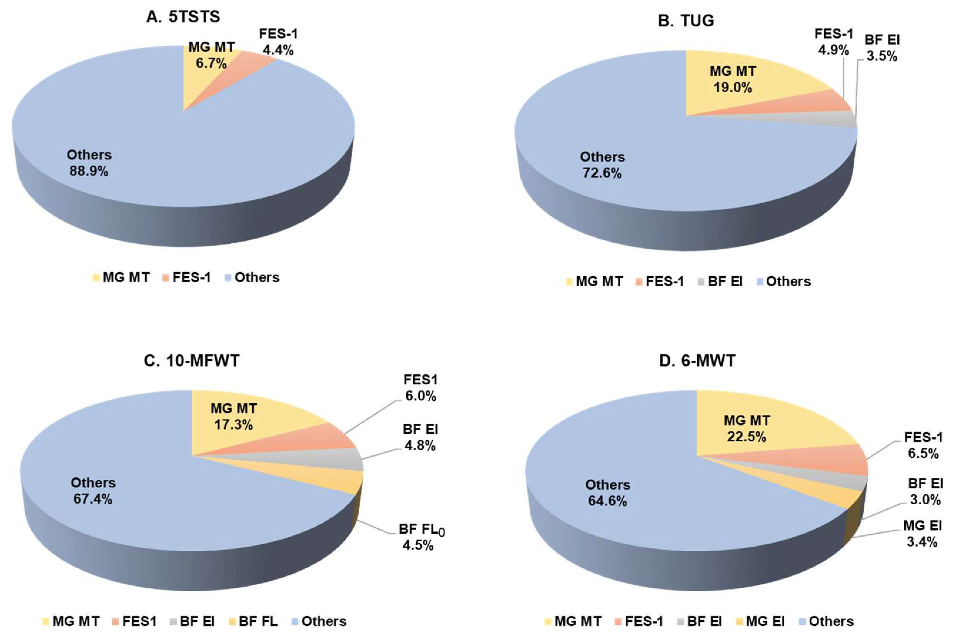
3.5. Functional Movement Performance Prediction

The contributions of the tested variables identified in the regression analyses for each functional performance are shown in Figure 1. For 5TSTS, the initial model regression indicated that MG MT explained only 6.7% of the variance (R2 = 0.067, F = 6.7, p = 0.011). After the addition of other variables, FES-I increased the explained variance (R2 = 0.110, F = 5.7, p = 0.005) (Figure 1A). MG MT also explained a small part of the variance for TUG performance (R2 = 0.190, F = 21.9, p < 0.001) and this increased to 27.5% when considering other variables, such as FES-I and BF muscle quality (R2 = 0.275, F = 11.5, p < 0.001) (Figure 1B). The regression analysis also showed that MT of MG could explain the variance of 10-MFWT speed (R2 = 0.173, F = 19.5, p < 0.001) and that the explained variance increased for the combination of MG MT, FES-I, BF EI, and BF FL0 (R2 = 0.326, F = 10.9, p < 0.001) (Figure 1C). The contribution of MG MT, FES-I, and muscle quality of BF and MG explained 35.4% of the variance for submaximal walking capacity, as measured by the 6-MWT distance (R2 = 0.354, F = 12.3, p < 0.001) (Figure 1D). Only 22.5% of the variance for the 6-MWT distance could be explained by US measurement of MG MT (R2 = 0.225, F = 27.1, p < 0.001).

4. Discussion

Loss of functional physical fitness in old age contributes to many health disorders. Here, we investigated the relationships between musculotendinous morphology, flexibility, balance, and physical function in nonobese older women. The main findings of the present study are that the MT of MG was most closely related to physical functional performance and that the MT of VL and MG were associated positively with balance ability and negatively with the fear of falling. However, the results of this study support only partially the assumption that muscle EI or FL is related to functional fitness, as reflected in the generalization that muscle mass, rather than other morphological parameters, is the main factor affecting the susceptibility to frailty in older women.
To measure the MT of VL and MG, we used US imaging, an indirect method for assessing age-related sarcopenia. Previous studies have shown good correlations between changes in MT, as measured with US, and the anatomical CSA, as measured with magnetic resonance imaging [30,31]. In addition, a strong association has been reported between MT and force production in a maximum voluntary contraction [32,33]. In the current study, MG MT was a crucial explanation for some variances in functional performance and correlated positively with functional performance. These findings highlight the need to maintain muscle mass to sustain functional physical fitness in older age.
The maximum distance of the 6-MWT and the time spent doing the TUG correlated most strongly with MT. Performance on these two tests worsens with age [34,35]. The MT of MG negatively correlated with increased fear of falling, as indicated by the FES-I score, which is also tangentially related to diminishing balance with advancing age [35,36]. We also found that the MT of MG correlated with fast walking speed. Studies of older people have shown that walking speed declines with age, most likely because of the decline in force production ability [37], changes in gait mechanics required for stability [38], or a combination of cognitive impairment and a history of falling [39].
The regression analysis results in our study suggest that a single measure may not be a good predictor of physical functional performance in older women. We found that MG MT could explain 17.3% of the variance for 10-MFWT speed and that this increased to 32.6% after the inclusion of MG MT, FES-I, BF EI, and BF FL0. Komforti et al. [16] noted that MG subcutaneous thickness and CSA explained 22.8% of the variance in gait speed, which increased to 45.5% after the inclusion of the 30 s chair stand and grip strength and heel rise test results.
In addition to MT, the EI of VL and MG correlated negatively with 6-MWT performance in the current study. Low EI values signify superior muscle quality [40], whereas high EI values are linked to muscle disease and dysfunction [41]. In our study, MG MT, BF EI, and MG EI explained 28.9% of the variance in 6-MWT performance when only US measurements were included. This finding is consistent with that of Cruz-Montecinos et al. [42], who found that 70% of the variance in the distance walked during the 6-MWT was accounted for by the rectus femoris EI, vastus intermedius EI, and vastus intermedius MT. In addition to its role in endurance events, ankle plantar flexor muscle quality is important for sit to stand and TUG performance in 21 aged people [43]. However, we did not observe significant correlations between EI and functional leg strength, FES-I score, or fast walking speed, although the correlations between EI and SLS and PDF were significant. Given the contradictory results, the value of muscle EI for predicting functional physical fitness in aged populations requires further study.
The current analysis found stronger relationships between FL and leg flexibility compared to FL and functional movement performance. This is not surprising because the functional testing was performed within the range of typical amplitudes and was straightforward. Our data are consistent with a recent report that only grip strength and balance were associated with the risk of falls but not muscle flexibility in 2130 community-dwelling older adults [44]. By contrast, other evidence suggests that low strength and flexibility may be linked to the risk of falls through increased gait variability in older adults [45]. In another study, myofascial release of hamstring tissue improved fast walking speed by increasing the ROM of the hip and knee joints [46]. Drew et al. [47] found a significant negative association between muscle flexibility, as measured by the Sit-and-Reach test, and running economy, which requires a stiffer musculotendinous structure. Leg flexibility increase may be significant in clinical situations, such as post-operative rehabilitation. A 5° increase in knee ROM was shown to increase walking distance by 50 m in the 6-MWT after total knee arthroplasty [48].
Performance on the SLS test correlated positively with the MT of VL and MG, EI of VL, and CSA of AT and PT. A higher FES-I score, associated with a decreased fear of falling, correlated negatively with VL and MG MT. The previous study also found a moderate to high correlation (r = 0.50–0.70) between FES-I and Berg Balance Scale and functional reach test in people with Parkinson’s disease [49]. These findings suggest that the tendon and muscle masses, as well as the knee extensors’ quality, may be crucial for balance in older people. This is consistent with Hill et al. [43], who showed that the MT and EI of the ankle plantar flexors and knee extensors are related to a decrease in postural sway during quiet standing in older men and women. Onat et al. [50] reported that the MT of the lower leg is related to balance ability in stroke patients. Our findings suggest that maintenance of muscle and tendon masses may preserve balance ability and should be primary targets when creating health promotion or rehabilitation programs for older women.
The present study has investigated the association between musculotendinous imaging and functional performance in a large older women cohort but did not include men and young participants which makes not possible direct comparison to other sex and age persons. Indeed, it is expected that MT are important to the older men to similar level as it is to the women despite well-established differences in intramuscular fat and function. Earlier study with seniors have documented a moderate correlation between sit to stand test and quadriceps thickness, but not for EI in both 37 men and 43 women [51]. The correlation between rectus femoris thickness and fast gait speed was also found in 42 normal and overweight older men [52]. In terms of age, the previous study showed no associations between leg muscle mass, strength and gait performance found in 20 young adults, whereas ankle plantar flexor strength had a high positive relation to maximal gait speed in 20 older adults [53]. This could be explained by the age-related loss of the fast fibers seen in older adults [54]. Although age-related reductions in muscle size, strength, and fiber denervation are well-established, comparing functional performance between young and old populations is still sometimes challenging since they use different assessment methods.

Limitation

Given that research participants were physically active, normal weight, and could complete the tests with above-average performance, one limitation of the present study is that the results are restricted to healthy and physically fit older women. Another limitation is that US analysis refers to two-dimensional measurements while the muscle and tendon morphology is three-dimensional. Because of the fascicle curvature and three-dimensional orientation, FL estimation is prone to error. However, we ensured accurate data collection by adhering to the FL measuring approach as outlined previously [55].

5. Conclusions

In summary, MT was found to be a more significant indicator of physical capacity than muscle quality or fascicle length in older, healthy, non-obese, and physically fit women. Hence, increasing MT should be a primary target for women’s fitness programs to prevent clinical issues such as frailty.

Author Contributions

Conceptualization, P.M. and S.K.; methodology, P.M.; formal analysis, P.M.; investigation, P.M. and J.N.; data curation, P.M. and D.S.; writing—original draft preparation, P.M.; writing—review and editing, P.M., J.N., D.S. and S.K.; supervision, S.K.; project administration, P.M.; funding acquisition, P.M. All authors have read and agreed to the published version of the manuscript.

Funding

The present study was fully financially supported by the Allied Health Sciences Research Fund and partly by the Burapha University Research and Development Fund.

Institutional Review Board Statement

The study was conducted in accordance with the Declaration of Helsinki, and approved by the Institutional Review Board of the Research and Innovation Administration Division of Burapha University Ethics Committee (IRB1-007/2565) on 4 January 2022.

Informed Consent Statement

Informed consent was obtained from all subjects involved in the study.

Data Availability Statement

Not applicable.

Acknowledgments

We would like to thank all the participants and Theerapat Nonnoi, Petprawprow Prommool, and Prakairung Jaikewthi who supported the project and David A Jones who admired the project topic work.

Conflicts of Interest

The authors declare no conflict of interest.

References

  1. Rogeri, P.S.; Zanella, R.; Martins, G.L.; Garcia, M.D.A.; Leite, G.; Lugaresi, R.; Gasparini, S.O.; Sperandio, G.A.; Ferreira, L.H.B.; Souza-Junior, T.P.; et al. Strategies to Prevent Sarcopenia in the Aging Process: Role of Protein Intake and Exercise. Nutrients 2021, 14, 52. [Google Scholar] [CrossRef] [PubMed]
  2. Avery, N.C.; Bailey, A.J. Enzymic and Non-Enzymic Cross-Linking Mechanisms in Relation to Turnover of Collagen: Relevance to Aging and Exercise. Scand. J. Med. Sci. Sports 2005, 15, 231–240. [Google Scholar] [CrossRef]
  3. Marcus, R.L.; Addison, O.; Kidde, J.P.; Dibble, L.E.; Lastayo, P.C. Skeletal Muscle Fat Infiltration: Impact of Age, Inactivity, and Exercise. J. Nutr. Health Aging 2010, 14, 362–366. [Google Scholar] [CrossRef] [PubMed] [Green Version]
  4. Miyamoto, N.; Kimura, N.; Hirata, K. Non-Uniform Distribution of Passive Muscle Stiffness within Hamstring. Scand. J. Med. Sci. Sports 2020, 30, 1729–1738. [Google Scholar] [CrossRef]
  5. Mecagni, C.; Smith, J.P.; Roberts, K.E.; O’Sullivan, S.B. Balance and Ankle Range of Motion in Community-Dwelling Women Aged 64 to 87 Years: A Correlational Study. Phys. Ther. 2000, 80, 1004–1011. [Google Scholar] [CrossRef] [PubMed] [Green Version]
  6. Rustani, K.; Kundisova, L.; Capecchi, P.L.; Nante, N.; Bicchi, M. Ultrasound Measurement of Rectus Femoris Muscle Thickness as a Quick Screening Test for Sarcopenia Assessment. Arch. Gerontol. Geriatr. 2019, 83, 151–154. [Google Scholar] [CrossRef] [PubMed]
  7. Albano, D.; Messina, C.; Vitale, J.; Sconfienza, L.M. Imaging of Sarcopenia: Old Evidence and New Insights. Eur. Radiol. 2020, 30, 2199–2208. [Google Scholar] [CrossRef] [PubMed]
  8. Madden, K.M.; Feldman, B.; Arishenkoff, S.; Meneilly, G.S. A Rapid Point-of-Care Ultrasound Marker for Muscle Mass and Muscle Strength in Older Adults. Age Ageing 2021, 50, 505–510. [Google Scholar] [CrossRef] [PubMed]
  9. Lopez, P.; Wilhelm, E.N.; Rech, A.; Minozzo, F.; Radaelli, R.; Pinto, R.S. Echo Intensity Independently Predicts Functionality in Sedentary Older Men. Muscle Nerve 2017, 55, 9–15. [Google Scholar] [CrossRef]
  10. Taniguchi, M.; Yamada, Y.; Fukumoto, Y.; Sawano, S.; Minami, S.; Ikezoe, T.; Watanabe, Y.; Kimura, M.; Ichihashi, N. Increase in Echo Intensity and Extracellular-to-Intracellular Water Ratio Is Independently Associated with Muscle Weakness in Elderly Women. Eur. J. Appl. Physiol. 2017, 117, 2001–2007. [Google Scholar] [CrossRef]
  11. Pillen, S.; Tak, R.O.; Zwarts, M.J.; Lammens, M.M.Y.; Verrijp, K.N.; Arts, I.M.P.; van der Laak, J.A.; Hoogerbrugge, P.M.; van Engelen, B.G.M.; Verrips, A. Skeletal Muscle Ultrasound: Correlation between Fibrous Tissue and Echo Intensity. Ultrasound Med. Biol. 2009, 35, 443–446. [Google Scholar] [CrossRef] [PubMed]
  12. Baxter, J.R.; Hullfish, T.J.; Chao, W. Functional Deficits May Be Explained by Plantarflexor Remodeling Following Achilles Tendon Rupture Repair: Preliminary Findings. J. Biomech. 2018, 79, 238–242. [Google Scholar] [CrossRef]
  13. Van Melick, N.; Meddeler, B.M.; Hoogeboom, T.J.; Nijhuis-van der Sanden, M.W.G.; Van Cingel, R.E.H. How to Determine Leg Dominance: The Agreement between Self-Reported and Observed Performance in Healthy Adults. PLoS ONE 2017, 12, e0189876. [Google Scholar] [CrossRef] [PubMed] [Green Version]
  14. Young, H.-J.; Jenkins, N.T.; Zhao, Q.; Mccully, K.K. Measurement of Intramuscular Fat by Muscle Echo Intensity. Muscle Nerve 2015, 52, 963–971. [Google Scholar] [CrossRef] [PubMed] [Green Version]
  15. Muanjai, P.; Mickevicius, M.; Sniečkus, A.; Sipavičienė, S.; Satkunskiene, D.; Kamandulis, S.; Jones, D.A. Low Frequency Fatigue and Changes in Muscle Fascicle Length Following Eccentric Exercise of the Knee Extensors. Exp. Physiol. 2020, 105, 502–510. [Google Scholar] [CrossRef]
  16. Komforti, D.; Joffe, C.; Magras, A.; Peller, A.; Garbe, E.; Garib, R.; Trapuzzano, A.; Dawson, N.; Stock, M.S. Does Skeletal Muscle Morphology or Functional Performance Better Explain Variance in Fast Gait Speed in Older Adults? Aging Clin. Exp. Res. 2021, 33, 921–931. [Google Scholar] [CrossRef]
  17. Schneebeli, A.; Visconti, L.; Cescon, C.; Clijsen, R.; Giardini, G.; Arizzio, M.E.; Barbero, M. Tendon Morphological Changes after a Prolonged Ski Race Can Be Detected by Ultrasound Echo Intensity. J. Foot Ankle Res. 2020, 13, 34. [Google Scholar] [CrossRef] [PubMed]
  18. Satkunskiene, D.; Mickevicius, M.; Snieckus, A.; Kamandulis, S. Leg Stiffness, Valgus Knee Motion, and Q-Angle Are Associated with Hypertrophic Soft Patella Tendon and Idiopathic Knee Pain in Adolescent Basketball Players. J. Sports Med. Phys. Fit. 2017, 57, 1003–1013. [Google Scholar] [CrossRef] [PubMed]
  19. Ayala, F.; Sainz de Baranda, P.; De Ste Croix, M.; Santonja, F. Criterion-Related Validity of Four Clinical Tests Used to Measure Hamstring Flexibility in Professional Futsal Players. Phys. Ther. Sport 2011, 12, 175–181. [Google Scholar] [CrossRef] [PubMed]
  20. Abdel-aziem, A.A.; Mohammad, W.S. Plantar-Flexor Static Stretch Training Effect on Eccentric and Concentric Peak Torque—A Comparative Study of Trained versus Untrained Subjects. J. Hum. Kinet. 2012, 34, 49–58. [Google Scholar] [CrossRef] [Green Version]
  21. Konrad, A.; Glashüttner, C.; Reiner, M.M.; Bernsteiner, D.; Tilp, M. The Acute Effects of a Percussive Massage Treatment with a Hypervolt Device on Plantar Flexor Muscles’ Range of Motion and Performance. J. Sports Sci. Med. 2020, 19, 690–694. [Google Scholar] [PubMed]
  22. Sipe, C.L.; Ramey, K.D.; Plisky, P.P.; Taylor, J.D. Y-Balance Test: A Valid and Reliable Assessment in Older Adults. J. Aging Phys. Act. 2019, 27, 663–669. [Google Scholar] [CrossRef]
  23. Thiamwong, L.; Suwanno, J. Fear of Falling and Related Factors in a Community-Based Study of People 60 Years and Older in Thailand. Int. J. Gerontol. 2017, 11, 80–84. [Google Scholar] [CrossRef]
  24. Namsawang, J.; Muanjai, P.; Luangpon, N.; Kiatkulanusorn, S. The Effects of Electrical Stimulation Program on Navicular Height, Balance, and Fear of Falling in Community-Dwelling Elderly. Int. J. Env. Res. Public Health 2021, 18, 9351. [Google Scholar] [CrossRef] [PubMed]
  25. Janyacharoen, T.; Yonglitthipagon, P.; Nakmareong, S.; Katiyajan, N.; Auvichayapat, P.; Sawanyawisuth, K. Effects of the Applied Ancient Boxing Exercise on Leg Strength and Quality of Life in Patients with Osteoarthritis. J. Exerc. Rehabil. 2018, 14, 1059–1066. [Google Scholar] [CrossRef] [PubMed] [Green Version]
  26. Jiménez-García, J.D.; Martínez-Amat, A.; De la Torre-Cruz, M.J.; Fábrega-Cuadros, R.; Cruz-Díaz, D.; Aibar-Almazán, A.; Achalandabaso-Ochoa, A.; Hita-Contreras, F. Suspension Training HIIT Improves Gait Speed, Strength and Quality of Life in Older Adults. Int. J. Sports Med. 2019, 40, 116–124. [Google Scholar] [CrossRef] [PubMed]
  27. Thaweewannakij, T.; Wilaichit, S.; Chuchot, R.; Yuenyong, Y.; Saengsuwan, J.; Siritaratiwat, W.; Amatachaya, S. Reference Values of Physical Performance in Thai Elderly People Who Are Functioning Well and Dwelling in the Community. Phys. Ther. 2013, 93, 1312–1320. [Google Scholar] [CrossRef] [PubMed] [Green Version]
  28. Boukabous, I.; Marcotte-Chénard, A.; Amamou, T.; Boulay, P.; Brochu, M.; Tessier, D.; Dionne, I.; Riesco, E. Low-Volume High-Intensity Interval Training Versus Moderate-Intensity Continuous Training on Body Composition, Cardiometabolic Profile, and Physical Capacity in Older Women. J. Aging Phys. Act. 2019, 27, 879–889. [Google Scholar] [CrossRef] [PubMed]
  29. Cohen, J. Statistical Power Analysis for the Behavioral Sciences; Academic Press: Cambridge, MA, USA, 2013; ISBN 978-1-4832-7648-9. [Google Scholar]
  30. Reeves, N.D.; Maganaris, C.N.; Narici, M.V. Ultrasonographic Assessment of Human Skeletal Muscle Size. Eur. J. Appl. Physiol. 2004, 91, 116–118. [Google Scholar] [CrossRef]
  31. Franchi, M.V.; Reeves, N.D.; Narici, M.V. Skeletal Muscle Remodeling in Response to Eccentric vs. Concentric Loading: Morphological, Molecular, and Metabolic Adaptations. Front Physiol. 2017, 8, 447. [Google Scholar] [CrossRef]
  32. Kelp, N.Y.; Gore, A.; Clemente, C.J.; Tucker, K.; Hug, F.; Dick, T.J.M. Muscle Architecture and Shape Changes in the Gastrocnemii of Active Younger and Older Adults. J. Biomech. 2021, 129, 110823. [Google Scholar] [CrossRef] [PubMed]
  33. Strasser, E.M.; Draskovits, T.; Praschak, M.; Quittan, M.; Graf, A. Association between Ultrasound Measurements of Muscle Thickness, Pennation Angle, Echogenicity and Skeletal Muscle Strength in the Elderly. Age 2013, 35, 2377–2388. [Google Scholar] [CrossRef] [Green Version]
  34. Al-Ghamdi, N.S.; Shaheen, A.A.M. Reference Values and Regression Equations for Predicting the 6-Minute Walk Distance in Saudi Adults Aged 50-80 Years: A Cross- Sectional Study. J. Back Musculoskelet. Rehabil. 2021, 34, 783–793. [Google Scholar] [CrossRef]
  35. Wu, H.; Wei, Y.; Miao, X.; Li, X.; Feng, Y.; Yuan, Z.; Zhou, P.; Ye, X.; Zhu, J.; Jiang, Y.; et al. Characteristics of Balance Performance in the Chinese Elderly by Age and Gender. BMC Geriatr. 2021, 21, 596. [Google Scholar] [CrossRef] [PubMed]
  36. Aartolahti, E.; Lönnroos, E.; Hartikainen, S.; Häkkinen, A. Long-Term Strength and Balance Training in Prevention of Decline in Muscle Strength and Mobility in Older Adults. Aging Clin. Exp. Res. 2020, 32, 59–66. [Google Scholar] [CrossRef] [PubMed] [Green Version]
  37. Thompson, B.J.; Conchola, E.C.; Palmer, T.B.; Stock, M.S. Effects of Aging on Maximal and Rapid Velocity Capacities of the Leg Extensors. Exp. Gerontol. 2014, 58, 128–131. [Google Scholar] [CrossRef] [PubMed]
  38. Winter, D.A.; Patla, A.E.; Frank, J.S.; Walt, S.E. Biomechanical Walking Pattern Changes in the Fit and Healthy Elderly. Phys. Ther. 1990, 70, 340–347. [Google Scholar] [CrossRef]
  39. Studenski, S.; Perera, S.; Patel, K.; Rosano, C.; Faulkner, K.; Inzitari, M.; Brach, J.; Chandler, J.; Cawthon, P.; Connor, E.B.; et al. Gait Speed and Survival in Older Adults. JAMA 2011, 305, 50–58. [Google Scholar] [CrossRef] [Green Version]
  40. Young, H.-J.; Southern, W.M.; Mccully, K.K. Comparisons of Ultrasound-Estimated Intramuscular Fat with Fitness and Health Indicators. Muscle Nerve 2016, 54, 743–749. [Google Scholar] [CrossRef]
  41. Manini, T.M.; Clark, B.C.; Nalls, M.A.; Goodpaster, B.H.; Ploutz-Snyder, L.L.; Harris, T.B. Reduced Physical Activity Increases Intermuscular Adipose Tissue in Healthy Young Adults. Am. J. Clin. Nutr. 2007, 85, 377–384. [Google Scholar] [CrossRef] [Green Version]
  42. Cruz-Montecinos, C.; Guajardo-Rojas, C.; Montt, E.; Contreras-Briceño, F.; Torres-Castro, R.; Díaz, O.; Cuesta-Vargas, A. Sonographic Measurement of the Quadriceps Muscle in Patients with Chronic Obstructive Pulmonary Disease: Functional and Clinical Implications. J. Ultrasound Med. 2016, 35, 2405–2412. [Google Scholar] [CrossRef] [PubMed] [Green Version]
  43. Hill, M.W.; Roberts, M.; Price, M.J.; Kay, A.D. Association between Knee Extensor and Ankle Plantarflexor Muscle Thickness and Echo Intensity with Postural Sway, Mobility and Physical Function in Older Adults. Exp. Gerontol. 2021, 150, 111385. [Google Scholar] [CrossRef] [PubMed]
  44. Lin, W.-S.; Hsu, N.-W.; Lee, M.-J.; Lin, Y.-Y.; Tsai, C.-C.; Pan, P.-J. Correlation Analysis of Physical Fitness and Its Impact on Falls in 2130 Community- Dwelling Older Adults: A Retrospective Cross-Sectional Study. BMC Geriatr. 2022, 22, 447. [Google Scholar] [CrossRef]
  45. Kang, H.G.; Dingwell, J.B. Separating the Effects of Age and Walking Speed on Gait Variability. Gait Posture 2008, 27, 572–577. [Google Scholar] [CrossRef] [PubMed]
  46. Itotani, K.; Kawahata, K.; Takashima, W.; Mita, W.; Minematsu, H.; Fujita, H. Myofascial Release of the Hamstrings Improves Physical Performance-A Study of Young Adults. Healthcare 2021, 9, 674. [Google Scholar] [CrossRef]
  47. Drew, R.C.; Sinoway, L.I.; White, M.J. The Two-Hour Marathon: Running Economy and Lower Body Flexibility. J. Appl. Physiol. 2011, 110, 284–285. [Google Scholar] [CrossRef] [PubMed]
  48. Van Onsem, S.; Verstraete, M.; Dhont, S.; Zwaenepoel, B.; Van Der Straeten, C.; Victor, J. Improved Walking Distance and Range of Motion Predict Patient Satisfaction after TKA. Knee Surg Sports Traumatol. Arthrosc. 2018, 26, 3272–3279. [Google Scholar] [CrossRef]
  49. Mehdizadeh, M.; Martinez-Martin, P.; Habibi, S.-A.; Fereshtehnejad, S.-M.; Abasi, A.; Niazi Khatoon, J.; Saneii, S.H.; Taghizadeh, G. Reliability and Validity of Fall Efficacy Scale-International in People with Parkinson’s Disease during On- and Off-Drug Phases. Parkinsons Dis. 2019, 2019, 6505232. [Google Scholar] [CrossRef]
  50. Onat, Ş.Ş.; Polat, C.S.; Gürçay, E.; Özcan, D.S.; Orhan, A. Muscle Architecture and Clinical Parameters in Stroke Patients: An Ultrasonographic Study. J. Clin. Ultrasound 2022, 50, 713–718. [Google Scholar] [CrossRef]
  51. Yoshiko, A.; Ogawa, M.; Shimizu, K.; Radaelli, R.; Neske, R.; Maeda, H.; Maeda, K.; Teodoro, J.; Tanaka, N.; Pinto, R.S.; et al. Chair Sit-to-Stand Performance Is Associated with Diagnostic Features of Sarcopenia in Older Men and Women. Arch. Gerontol. Geriatr. 2021, 96, 104463. [Google Scholar] [CrossRef]
  52. Giuliani, H.K.; Shea, N.W.; Gerstner, G.R.; Mota, J.A.; Blackburn, J.T.; Ryan, E.D. The Influence of Age and Obesity-Altered Muscle Tissue Composition on Muscular Dimensional Changes: Impact on Strength and Function. J. Gerontol. A Biol. Sci. Med. Sci. 2020, 75, 2286–2294. [Google Scholar] [CrossRef] [PubMed]
  53. Muehlbauer, T.; Granacher, U.; Borde, R.; Hortobágyi, T. Non-Discriminant Relationships between Leg Muscle Strength, Mass and Gait Performance in Healthy Young and Old Adults. Gerontology 2018, 64, 11–18. [Google Scholar] [CrossRef] [PubMed]
  54. Coletti, C.; Acosta, G.F.; Keslacy, S.; Coletti, D. Exercise-Mediated Reinnervation of Skeletal Muscle in Elderly People: An Update. Eur. J. Transl. Myol. 2022, 32, 10416. [Google Scholar] [CrossRef] [PubMed]
  55. Ando, R.; Taniguchi, K.; Saito, A.; Fujimiya, M.; Katayose, M.; Akima, H. Validity of Fascicle Length Estimation in the Vastus Lateralis and Vastus Intermedius Using Ultrasonography. J. Electromyogr. Kinesiol. 2014, 24, 214–220. [Google Scholar] [CrossRef] [PubMed]
Figure 1. The percentages of explained variance for functional performance variables. (A) 5TSTS performance, (B) TUG performance, (C) 10-MFWT performance, (D) 6-MWT performance. MT, muscle thickness; BF, biceps femoris; MG, medial gastrocnemius; FL0, resting fascicle length; FES-1, Falls Efficacy Scale-international; 5TSTS, five times sit to stand; TUG, timed up and go; 10-MFWT, 10 min fast walk test; 6-MWT, six minutes walk test.
Ijerph 19 16099 g001
Table 1. Participant characteristics (n = 99).
Participant Characteristics
Age (years)66.6 ± 4.6
60–65 years (n)47
66–70 years (n)34
71–75 years (n)14
76–80 years (n)4
Body mass (kg)57.3 ± 8.7
Height (cm)156.1 ± 4.7
BMI (kg∙m−2)23.5 ± 3.3
Exercise volume (min∙week−1)200 ± 213
Data are mean ± SD. BMI, body mass index.
Table 2. Means (±SD) for musculotendinous ultrasound imaging, flexibility, balance, and functional movement tests of the study participants (n = 99).
Ultrasound Imaging
VL MT (cm)1.81 ± 0.30
BF MT (cm)1.48 ± 0.32
MG MT (cm)1.53 ± 0.24
VL FL (cm)9.22 ± 1.85
BF FL0 (cm)10.95 ± 2.60
BF FLP (cm)18.98 ± 3.95
MG FL0 (cm)5.07 ± 0.79
MG FLP (cm)5.68 ± 0.75
Passive BF FLs/FL00.78 ± 0.38
Passive MG FLs/FL00.13 ± 0.09
Corrected VL EI (A.U.)101.4 ± 21.8
Corrected BF EI (A.U.)104.5 ± 17.2
Corrected MG EI (A.U.)83.7 ± 14.9
AT CSA (cm2)0.512 ± 0.114
PT CSA (cm2)0.662 ± 0.127
FlexibilityBalance
SLR (°)87.9 ± 11.0Single leg stance (s)30.0 ± 16.1
Passive DF (°)12.9 ± 4.9FES-1 (points)23.6 ± 6.6
Physical functions
5TSTS (s)6.32 ± 1.4610-MFWT (m∙s−1)1.92 ± 0.32
TUG (s)6.44 ± 1.106-MWT (m)500.8 ± 67.7
The data are presented as mean and SD. VL, vastus lateralis; MT, muscle thickness; BF, biceps femoris; MG, medial gastrocnemius; FL, fascicle length; FL0, resting fascicle length; FLP, passive fascicle length; FLs, strained fascicle length; EI, echo intensity; AT, Achilles tendon; PT, patella tendon; CSA, cross-sectional area; SLR, straight leg raise; DF, dorsiflexion; FES-1, Falls Efficacy Scale-international; 5TSTS, five times sit to stand; TUG, timed up and go; 10-MFWT, 10 min fast walk test; 6-MWT, six-minute walk test.
Table 3. Pearson correlation representing the association between musculotendinous morphology and other performance variables (n = 99).
SLRPDFSLSFES-15TSTSTUG10-MFWT6-MWT
VL MTnor0.170, 0.0930.227 *, 0.0240.235 *, 0.019−0.228 *, 0.023−0.144, 0.154−0.185, 0.0660.140, 0.1680.208 *, 0.038
BF MTnor0.331 **, 0.0010.068, 0.5080.093, 0.364−0.183, 0.074−0.061, 0.553−0.113, 0.269−0.019, 0.0850.080, 0.436
MG MTnor0.048, 0.6390.128, 0.2060.339 **, 0.001−0.207 *, 0.040−0.257 *, 0.010−0.444 **, 0.0000.411 **, 0.0000.480 **, 0.000
VL SubcuTnor0.099, 0.327−0.352 **, 0.000−0.130, 0.200−0.014, 0.894−0.013, 0.9010.031, 0.758−0.057, 0.557−0.088, 0.387
BF SubcuTnor0.166, 0.030−0.084, 0.4090.018, 0.8580.002, 0.9820.011, 0.915−0.117, 0.2480.047, 0.6420.080, 0.428
MG SubcuTnor0.029, 0.775−0.231 *, 0.021−0.030, 0.765−0.037, 0.718−0.083, 0.414−0.095, 0.3480.115, 0.257−0.111, 0.272
VL FL0.139, 0.1710.023, 0.825−0.164, 0.1070.016, 0.874−0.004, 0.968−0.122, 0.2300.084, 0.4100.010, 0.919
BF FL00.189, 0.0640.044, 0.667−0.045, 0.664−0.109, 0.2280.159, 0.1190.101, 0.324−0.217 *, 0.032−0.041, 0.689
MG FL0−0.056, 0.582−0.423 **, 0.0000.086, 0.397−0.163, 0.1070.028, 0.781−0.186, 0.0650.230 *, 0.0220.139, 0.171
BF FLs0.210 **, 0.039−0.093, 0.3670.031, 0.762−0.129, 0.207−0.087, 0.398−0.158, 0.1220.118, 0.2490.033, 0.749
MG FLs−0.053, 0.6010.499 **, 0.0000.069, 0.4990.079, 0.435−0.057, 0.5750.040, 0.692−0.123, 0.2240.000, 0.998
VL EIcor−0.055, 0.586−0.445 **, 0.000−0.342 **, 0.0010.167, 0.0990.045, 0.6560.126, 0.215−0.060, 0.553−0.202 *, 0.045
BF EIcor−0.009, 0.926−0.091, 0.372−0.101, 0.3190.015, 0.880−0.060, 0.555−0.116, 0.2550.135, 0.1840.108, 0.289
MG EIcor0.022, 0.830−0.189, 0.065−0.107, 0.2910.065, 0.520−0.009, 0.9290.142, 0.161−0.076, 0.457−0.268 **, 0.007
AT CSAnor0.024, 0.8130.054, 0.5960.338 **, 0.001−0.021, 0.837−0.010, 0.919−0.017, 0.8650.043, 0.6730.109, 0.281
PT CSAnor−0.036, 0.7230.231 *, 0.0220.221 *, 0.0290.014, 0.895−0.026, 0.802−0.132, 0.1960.046, 0.6500.159, 0.117
Data are presented as r, p value. * p < 0.05, ** p < 0.01. VL, vastus lateralis; MTnor, normalized muscle thickness; BF, biceps femoris; MG, medial gastrocnemius; SubcuTnor, normalized subcutaneous thickness; FL0, resting fascicle length; FLs, strained fascicle length; EIcor, corrected echo intensity; AT, Achilles tendon; PT, patella tendon; CSAnor, normalized cross-sectional area; SLR, straight leg raise; PDF, passive dorsiflexion; SLS, single leg stance; FES-1, Falls Efficacy Scale-international; 5TSTS, five times sit to stand; TUG, timed up and go; 10-MFWT, 10 min fast walk test; 6-MWT, six-minute walk test.
Publisher’s Note: MDPI stays neutral with regard to jurisdictional claims in published maps and institutional affiliations.

