Article

Chronic Community Exposure to Environmental Metal Mixtures Is Associated with Selected Cytokines in the Navajo Birth Cohort Study (NBCS)

1
Integrative Anthropological Sciences, University of California Santa Barbara, Santa Barbara, CA 93106, USA
2
Department of Anthropology, University of New Mexico, Albuquerque, NM 87131, USA
3
Academic Science Education and Research Training Program, Health Sciences Center, University of New Mexico, Albuquerque, NM 87131, USA
4
Health Sciences Center, College of Pharmacy, University of New Mexico, Albuquerque, NM 87131, USA
5
Department of Mathematics and Statistics, University of New Mexico, Albuquerque, NM 87131, USA
*
Author to whom correspondence should be addressed.
Int. J. Environ. Res. Public Health 2022, 19(22), 14939; https://doi.org/10.3390/ijerph192214939
Submission received: 20 October 2022 / Revised: 9 November 2022 / Accepted: 10 November 2022 / Published: 13 November 2022

Abstract

:
Many tribal populations are characterized by health disparities, including higher rates of infection, metabolic syndrome, and cancer—all of which are mediated by the immune system. Members of the Navajo Nation have suffered chronic low-level exposure to metal mixtures from uranium mine wastes for decades. We suspect that such metal and metalloid exposures lead to adverse health effects via their modulation of immune system function. We examined the relationships between nine key metal and metalloid exposures (in blood and urine) with 11 circulating biomarkers (cytokines and CRP in serum) in 231 pregnant Navajo women participating in the Navajo Birth Cohort Study. Biomonitored levels of uranium and arsenic species were considerably higher in participants than NHANES averages. Each biomarker was associated with a unique set of exposures, and arsenic species were generally immunosuppressive (decreased cellular and humoral stimulating cytokines). Overall, our results suggest that environmental metal and metalloid exposures modulate immune status in pregnant Navajo women, which may impact long-term health outcomes in mothers and their children.

1. Introduction

The legacy of mining from the Cold War Era has left several U.S. communities exposed to metal and metalloid contaminants (hereafter “metals”) with potential health consequences. The abandonment of more than 160,000 hard rock mines and >500,000 mine waste sites (Figure 1) [1] has resulted in 40% of watersheds in the western United States being contaminated with mine waste and related metals [2]. These sites are often located on or contiguous with the watersheds of tribal lands, such as the Navajo Nation. Although Navajo communities have long been concerned that environmental exposure to mine waste contributes to poor health outcomes among tribe members, no comprehensive characterization of the metal body burden of this population has been conducted prior to the Navajo Birth Cohort Study (NBCS). Here, to address potential immune modulation resulting from environmental exposures, we characterize the relationship between measured metal and cytokines concentrations among pregnant Navajo women participating in the Navajo Birth Cohort Study (NBCS).
Members of the Navajo Nation, located in the Four Corners Region of the Southwestern US (Figure 1), are exposed to the metal contaminants resulting from historic mining activities via several pathways. While active mining and milling on Navajo Nation for uranium ended in 1986, more than 500 abandoned mines and waste sites remain, containing multiple metals and metalloids, including arsenic (As), lead (Pb), cadmium (Cd), manganese (Mn), and sometimes mercury (Hg) [3,4,5]. Navajo community members are likely exposed to such wastes via multiple pathways, including the consumption of local water and crops [6], contact with contaminated soil and dust from mine features, and the inhalation of metals released from combustion for home heating and cooking and local power sources [7,8]. Exposure via drinking water is of particular concern, as >30% of Navajo members lack access to public water systems, and unregulated water sources exceed the maximum contaminant levels of the Safe Drinking Water Act for uranium by 13% and arsenic by 15% [9]. Even among major public water systems on Navajo Nation, many have been repeatedly out of compliance according to standards for one or more metals [10].
Metal exposures are well linked to several adverse health effects. Chronic low-dose exposures to uranium and arsenic in drinking water, for example, are associated with cancers [11] and various aspects of metabolic syndrome and autoimmunity [12,13], including chronic inflammation [14], cardiovascular diseases [15,16], and kidney damage [17]. Co-occurring metals such as lead, cadmium, manganese, and mercury are related to a similarly wide array of negative health outcomes, further including hypertension and neurodegenerative disease [7,18,19,20,21]. Prenatal exposures to metals are also related to elevated risks of infant mortality [22,23], stunted growth [24], and lung cancer in adulthood [25]. The health effects of metal exposure are, therefore, not localized to a single generation.
A primary mechanism by which metals influence the development of disease risk is likely their immunomodulatory effects [26,27]. Several metals are immunostimulatory [18,19,28,29] and others immunosuppressive [25,30], with effects often varying by dose, the chronicity of exposure, and individual factors, such as age and sex [18,31,32]. Uranium and arsenic exposures are linked to alterations in cellular and humoral immune responses in animals [33,34,35] and in humans with occupational metal exposure [36,37]. Less is understood, however, about the role of these metals in immune system alterations that occur in populations chronically exposed to environmental metals [38,39,40]. Studies in such systems are important to reveal the community-wide effects of metal exposures, the epidemiology of associated diseases, and steps for policy action. In addition, such studies in pregnant women lend insight into the maternal–fetal immune environment, a key factor in the developmental origins of health and disease [41,42].
In this study, we examine the potential immunomodulatory effects of specific environmental metals and metalloids among pregnant women participating in the Navajo Birth Cohort Study. Decades of research firmly establish that members of the Navajo Nation experience higher rates of severe respiratory infection [43], end-stage renal disease [44], metabolic syndrome [45,46,47], and infant mortality [48] relative to U.S. averages. Given Navajo members’ chronic low-level exposure to environmental metal mixtures [9,49], we expect that metal exposures play a role in observed health disparities. Specifically, we hypothesize that metal exposures exert immunomodulatory effects in exposed individuals. To test this, we pair participant biomonitoring (U, As and its species AsIII, MMA, and DMA; Hg, Pb, Cd, and Mn), and circulating cytokine and acute phase protein levels (IL-4, IL-6, IL-7, IL-10, IL-12, IL-17, IL-29, IFNα, IFNγ, TNFα, and CRP), to test the relationships of metal and metalloid exposures with the key mediators of innate, cellular, and humoral immune function.

2. Methods

2.1. Inclusion Criteria

The NBCS was initiated to address Navajo community members’ concerns about the potential health effects of chronic environmental exposure to uranium mine wastes. In 2013, the NBCS began recruiting pregnant women between 14 and 45 years of age who had lived on Navajo Nation for at least 5 years. To be included in the study, women had to be willing to deliver at a participating hospital, and have their child followed up for one year postnatally.

2.2. Survey Information

At enrollment, a survey of socioeconomic, demographic, lifestyle, and reproductive history information was administered by trained community health environmental research staff. The responses were entered into the research database RedCap in accordance with all privacy and security requirements of both the Navajo Nation (NNHRRB 11-323) and University of New Mexico institutional review boards (UNM HRPO 11-310). Women broadly represented several demographic categories (Table 1) and were recruited during all trimesters of pregnancy.

2.3. Biological Sample Collection

Trained hospital staff collected and prepared blood and urine samples. Samples intended for the metal biomonitoring analysis were collected using pre-screened metal-free cups, transfer pipettes, and Nalgene cryo-vials provided by the Center for Disease Control (CDC), National Center for Environmental Health (NCEH), and the Division of Laboratory Sciences (DLS). At enrollment or 36 weeks gestation prenatal visits, the participants provided 40–50 mL of urine in a sterile collection cup. Laboratory staff aliquoted 1.8 mL urine samples into separate Nalgene cryo-vials for multi-element metal, total arsenic, speciated As, creatinine, and mercury/iodine analysis. Hospital laboratory staff also collected peripheral blood via venipuncture and then allowed the blood to clot at room temperature for 30 to 40 min. Once clotted, laboratory staff centrifuged the blood tube at 2400 revolutions per minute for 15 min to separate the serum. Aliquots of serum (1.8 mL) were transferred to a 2.0 mL Nalgene cryo-vial. After processing the urine and serum samples, hospital staff placed all cryo-vials in a −80 °C freezer for storage. The samples were transferred from participating facilities on Navajo Nation on dry ice to freezer storage facilities at UNM. Chain of Custody forms were completed, reviewed, and validated at each stage of collection, storage, and analysis. The samples were stored, analyzed, and disposed of in accordance with participant wishes, as indicated on the consent forms. Samples not consumed during analysis will be returned to participants after the close of the study analysis period if they desire, in accordance with cultural practices.

2.4. Metal Biomonitoring

UNM laboratory staff examined the samples for quality control. Then, the urine vials and one serum vial were shipped on dry ice to the CDC Division of Laboratory Systems for analysis. Urine concentrations of metals and metalloids (U, As and its species Arsenite AsIII, Monomethylarsonic acid MMA, Dimethylarsinic acid DMA, Pb, Cd, and Mn) were measured using inductively coupled plasma dynamic-reaction cell mass spectrometry [ICP-DRC-MS Perkin Elmer NexION 300, CDC Method No. 3018.6-02, 3018A4-02, & 3031.1-01; [50,51,52,53]. HPLC was used for arsenic speciation prior to the ICP-MS analysis (CDC Method No. ITU003B). HPLC used an Agilent 1200 Series thermostatted column compartment (Item #G1316B) along with an Agilent Instant Pilot control module (Item #G4208-67001). Serum concentrations of the metals (Pb, Cd, Mn, Hg) were also measured using ICP-DRC-MS [CDC Method No. 3016.8-05] [54]. Metal exposures for Pb, Cd, and Mn were examined in both serum and urine, as their presence in the different media is known to vary by the dose and chronicity of exposure [55,56,57]. To determine if the ICP-MS runs were in control, the multi-rule quality control system (MRQCS) was implemented [58] based on the results of two to three custom-made and characterized urine bench quality control materials that were inserted at the start and end of each analytical run. All reference materials were made in-house at the CDC, according to NIST standards. The limit of detection for these elements in urine and serum ranged from 0.002 to 1.91 ng/mL, depending on the analyte and biological media (complete LoD by element in Table 2). The CDC’s acceptable percent of recovery ranges from 80 to 120%. Urinary metal concentrations were adjusted for sample creatinine concentrations, determined using a Roche/Hitachi Modular P Chemistry Analyzer.

2.5. Serum Cytokines

Eleven biomarkers, including 10 cytokines and 1 acute phase protein, were analyzed in the serum samples: interleukin-4 (IL-4); interleukin-6 (IL-6); interleukin-7 (IL-7); interleukin-10 (IL-10); interleukin-12p70 (IL-12); interleukin-17 (IL-17); and interleukin-29 (IL-29); interferon alpha (IFNα); interferon gamma (IFNγ); tumor necrosis factor alpha (TNFα); and C-reactive protein (CRP). These 11 markers were chosen for early analysis because they are indicative of immune responses from innate, cellular, and humoral branches, and their irregularities are associated with conditions of chronic inflammation and related cardiovascular and metabolic disease [59,60,61,62].
Cytokine measurements were performed using the Meso Scale Discovery multiplex four- or ten-spot 96-well electrochemiluminescence detection platform. Serum samples were diluted 1:2 in appropriate assay buffer and incubated on the plate for two hours. The plates were washed with PBS-Tween, and corresponding Sulfo-Tag secondary reagents were added. They were then incubated for an additional two hours. Plates were read using the MESO QuickPlex SQ 120 microplate reader and results calculated using the Discovery Workbench 4.0 software. Urine and serum metal measurements below the limit of detection (LoD) were replaced by the (LoD) divided by the square root of two.

2.6. Statistical Analysis

To broadly describe their associations, we evaluated correlations among the metals and between metal concentrations and cytokine levels (alpha = 0.05). To control for confounding variables in metal–cytokine relationships, we then used multivariate linear regression models. Metal biomonitoring and cytokine concentrations were log-transformed to reduce skewness. Certain cytokines varied by participant age and trimester in univariate regression; thus, we included these variables as analytical controls in all multivariate regressions (unpublished data). We further controlled for pre-pregnancy BMI class in multivariate models of CRP, as the two were strongly positively related. Cytokine values did not differ by education level, marital status, annual income, or fetal sex in univariate models, apart from weak relationships with IL-10, which were best accounted for by age; thus, these variables were not controlled for in multivariable modeling. We performed backwards selection from full models (including participant attributes and all log-transformed metals) based on AIC criteria with the R function “step”.

3. Results

3.1. Participant Metal Concentrations and Their Correlations

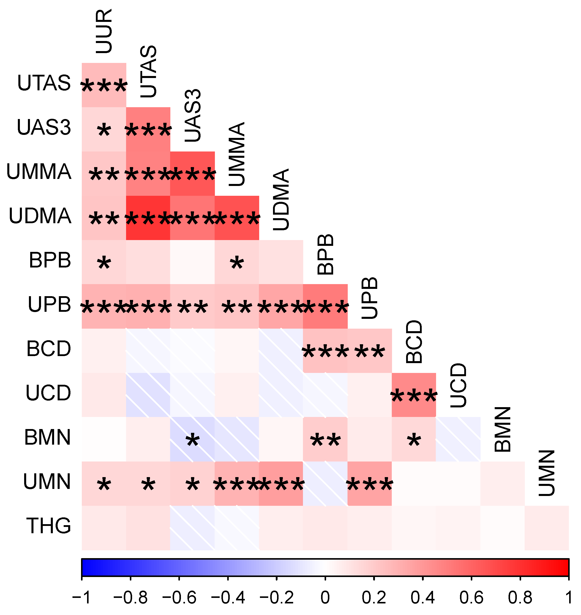
Four metal concentrations were on average higher among participants in this sample than in the NHANES participants: uranium, arsenic species (i.e., AsIII, MMA, DMA), cadmium, and manganese (Table 2). Concentrations that fell below the NHANES medians were those of lead and mercury. Metals demonstrated several non-transitive correlations among one another (Figure 2, Table S1). Urinary or blood lead concentrations were positively correlated with all metals except urinary cadmium, urinary magnesium, and mercury. Uranium was strongly and positively correlated with arsenic and its species, lead, and weakly with urinary magnesium. Arsenic and its species were also positively correlated with urinary manganese. Apart from its relationship with uranium, arsenic, and lead, manganese was positively correlated with cadmium. Mercury was unrelated to any metal, perhaps because of its overall low presence among the participants.

3.2. Participant Cytokine Levels and Correlations with Metal Exposures

The average cytokine levels among the study participants are summarized in Table S2. Concentrations of arsenic and its species, lead, cadmium, and manganese were correlated with several cytokines among the participants (Figure 3, Table 3). Most notably, arsenic species were negatively correlated with Th2 cytokine IL-4, inflammatory cytokines IL-6, IL-12, IL-29, and TNFα, and complemented CRP. The arsenic species DMA, however, was positively correlated with IFNα.

3.3. Multivariable Linear Regression Models

Due to the exploratory nature of this analysis, we did not correct for multiple testing and consider all the metal exposures in the best fit models to have notable relationships with cytokine levels, although we emphasize those with particularly strong relationships (p of ß < 0.05, Table 4). This helps target potentially important exposures for future analysis. All cytokines demonstrated relationships with metal exposures (Table 4). Although we have structured Table 4 according to model structure, where cytokine levels are responses, we have organized the presentation of the results and their discussion according to each metal and its profile of relationships with various cytokines.
Arsenic species, to which the participants had high exposures, demonstrated broadly immunosuppressive effects on cytokines (Table 4). Higher levels of total arsenic, AsIII, and MMA together corresponded with lower IL-4 and IL-29. Arsenic’s relationship with CRP depended on the species considered, as MMA and total arsenic had a strong negative association with CRP, whereas DMA had a strongly positive association. DMA further had a strong, positive relationship with IFNα. Counter to expectations, uranium demonstrated only a weakly negative relationship with IFNγ, despite the participants’ high exposure.
For certain metals, blood and urinary measures demonstrated opposite immunomodulatory effects on the same cytokine (Table 4). Blood lead, for example, was broadly inflammatory with strongly positive relationships with IL-10, IL-17, IFNγ, and TNFα; however, urinary lead decreased TNFα. Blood cadmium, further, appeared immunosuppressive in its negative relationship with IL-4, IL-29, and TNFα; however, urinary cadmium increased both IL-29 and TNFα. Other effects of the metals were more straightforward. Manganese demonstrated a general immunostimulatory influence, including strong positive relationships with IL-4, IL-12, and TNFα. Lastly, mercury exposures did not have any strong relationships with cytokines, apart from a weakly positive relationship with IFNγ.

4. Discussion

The aim of our study was to probe the immunomodulatory effects of long-term metal exposure among pregnant women participating in the Navajo Birth Cohort Study. This is particularly important as the abundance of environmental uranium and arsenic in the Navajo Nation has been a health concern among its members for decades, and because the prenatal immunological environment is a critical pathway for the developmental origins of health and disease. Here, we find that exposures to metal mine wastes demonstrate several strong relationships with inflammatory and autoimmune-related cytokines among pregnant women. In particular, arsenic appeared to be generally immunosuppressive, lowering IL-4 and IL-29, but with variable effects on inflammatory cytokines IFNα and CRP depending on its species. Overall, our results build on findings from Erdei et al. [12] and Ong et al. [63] to recommend future avenues for targeted examination of the effects of combination, dose, and chronicity of environmental metal exposures on immune function. Here, we discuss the consistency of our findings with the existing literature, their implications for health outcomes among current and future members of the Navajo Nation, and future directions of this work.

4.1. Immunomodulatory Effects of Metals and Comparisons with Previous Literature

The cytokine profile associated with arsenic exposure was nuanced, such that arsenic and early-state species had a general immunosuppressive effect (lower IL-4, IL-29, and CRP), but its end-state species appeared to increase certain inflammatory cytokines (DMA, higher IFNα and CRP). The immunosuppressive effects of arsenic and its species are well documented [25,34]: even at low doses, AsIII in particular corresponds with a greater disruption of T cell proliferation and inhibition of DNA repair [64], and its metabolite MMA plays a key role in the disruption of IL-7 signaling in mice [65]. Our findings of arsenic-related immunosuppression also align with evidence in Navajo women that arsenic exposure reduced the prevalence of autoantibodies [12]. Participant sex may further shape such findings. Women have greater methylation capacity of arsenic than men, which further accelerates during pregnancy, leading women to suffer the immunotoxicity of arsenic less severely [31,66]. The immunosuppressive effects of arsenic in our study are, thus, potentially reduced given our focus on women during pregnancy.
Although arsenic is known to suppress adaptive immunity, in some cases it can increase innate inflammatory responses. In mice, for example, chronic arsenic exposure reduced mature macrophage numbers and increased immature and pro-inflammatory macrophages [35]. Further, in humans, arsenic increased viral responses [26], inhibited DNA repair [67], and chronic low levels increased the expression of pro-inflammatory mediators [14]. This type of pro-inflammatory shift may lie at the root of our findings of positive relationships of DMA with IFNα.
Other relationships between biomonitored metals and cytokines in this study are consistent with evidence in previous literature. Lead’s strong positive relationship with inflammatory markers among the participants in this study (IL-10, IL-17, IFNγ, TNFα) is consistent with its particularly strong pro-inflammatory effects in women [18,68] and is suggestive of participants’ chronic exposure [69]. Concentrations of cadmium among the participants corresponded with lower IL-4, suggesting an immunological shift from a Th2 towards a Th1 response. Opposite directions of influence of blood and urinary cadmium on IL-29 and TNFα perhaps results from differences in chronicity that measures capture, where blood represents acute and urine long-term exposure [56]. These effects could also represent differences in dose in chronic vs acute exposures, as low cadmium can lead to proinflammatory effects and high cadmium exposure the opposite [70]. Finally, manganese appeared to be a general immunostimulant in our study (higher IL-4, IL-12, TNFα), consistent with the effects of chronic manganese exposure on increased inflammatory, humoral, and anti-tumor activity in murine models [27] and increased IL-1ß in pregnant women [71].

4.2. Implications for Health Outcomes

Our results demonstrate clear associations between environmental metal exposures and immune activity, suggesting that metals’ immunomodulatory effects combined with chronic and potentially life-long exposures contribute to health outcomes in current and future Navajo community members. Uranium and arsenic are primary concerns, given community members’ high exposures. Although uranium did not appear to modulate immunity, the broad immunosuppression of arsenic could pave the way for infectious disease and overreliance on pro-inflammatory immune responses. In combination with the immunostimulatory effects of lead and manganese (Figure 2), immunomodulation by metals likely contributes to the prevalence of disease in the Navajo Nation via distinct and complementary pathways [35]. Our study also highlights concerns of the intergenerational effects of maternal metal exposures on offspring. Arsenic and its species readily cross the placenta, increasing infant mortality [22,23] and later risks of lung cancer [25]. Arsenic, lead, and cadmium exposures during pregnancy all result in fetal immunosuppression and lower thymic volume [26]. In turn, prenatal exposures to uranium reduce offspring growth [24]. The potential reach of maternal metal exposures, therefore, is far and the potential consequences dire.

5. Limitations and Future Directions

We acknowledge that the sample size of this study was limited, including that biological samples were collected at a single time point. In addition, studies of environmental metal exposures are scarce, relative to studies of experimental and occupational exposures; therefore, definitive comparisons between our findings and previous work are few. Nevertheless, significant changes in circulating cytokines with metal biomonitoring concentrations strengthen the case that environmental metal exposure contributes to immune system disruption. Our findings encourage several additional approaches. A first approach is to determine whether findings are broadly generalizable to women by longitudinally analyzing a larger sample of women. This would include examining potential alterations in arsenic metabolism (e.g., AsIII to MMA to DMA) with chronicity of exposure. As individuals are exposed to distinct sets of metals in their environment, a second approach is to examine metals’ combined influences on immune function. This would include examining how potentially beneficial metal exposures, such as zinc [72], ameliorate the damaging effects of other metals. Finally, the findings urge implementing plans to reduce the risks that environmental metal exposures pose to personal and larger scale environmental health.

6. Conclusions

Using data from the Navajo Birth Cohort Study (NBCS), the aim of this study was to evaluate the immunomodulatory effects of environmental metal exposures among Navajo Nation community members. Highly correlated arsenic species in biomonitoring appear to be strong drivers of immunosuppression. Taken as a whole, our results suggest that chronic community-level exposure to mixed metals and metalloids on the Navajo Nation plays a role in altering immune responses. The findings of our study are relevant to both acute immunomodulation and its potential long-term and widespread consequences, informing both basic science and health policy.

Supplementary Materials

The following supporting information can be downloaded at: https://www.mdpi.com/article/10.3390/ijerph192214939/s1, Table S1: All significant correlations between biomonitored metals, arranged in descending order by Spearman’s rho; Table S2: Summary statistics for cytokine measurements (ng/pL).

Author Contributions

Conceptualization, D.M.; Data curation, L.L.; Formal analysis, N.T.G., J.O. and L.L.; Funding acquisition, D.M.; Methodology, N.T.G., J.O., L.L. and D.M.; Writing—original draft, N.T.G. and J.O.; Writing—review & editing, D.M. All authors have read and agreed to the published version of the manuscript.

Funding

This research was funded by CDC TS000135-05; NIH Office of the Director UG3/UH3D023344; NIH NIMHD P50MD015706, NIH NIEHS P50ES026102, and NIH K12 GM088021. The APC was funded by NIH Office of the Director UG3/UH3D023344.

Institutional Review Board Statement

The study was conducted according to the guidelines of the Declaration of Helsinki and approved by the Navajo Nation Human Research Review Board (NNHRRB 11-323 17 September 2019–17 September 2023) and the Institutional Review Board of the University of New Mexico (protocol code UNM HRPO 11-310 NBCS/ECHO+ 10 September 2019–30 May 2023, NBCS initial 2 June 2012).

Informed Consent Statement

Informed consent was obtained from all subjects involved in the study.

Data Availability Statement

In accordance with Tribal Sovereignty, any data connected with biological samples from tribal members can only be shared with appropriate tribal policies and permissions. Should other researchers wish to use these data at a future time for reanalysis or to explore additional hypotheses, they would need to request access to the data through application to the Navajo Nation Human Research Review Board, and their proposal would need to meet the guidelines for research in accordance with tribal policy.

Conflicts of Interest

The authors declare no conflict of interest.

References

  1. Lewis, J.; Hoover, J.; MacKenzie, D. Mining and Environmental Health Disparities in Native American Communities. Curr. Environ. Health Rep. 2017, 4, 130–141. [Google Scholar] [CrossRef] [PubMed] [Green Version]
  2. NNDWR. Safe Drinking Water Hauling Feasibility Study and Pilot Project. In Proceedings of the Uranium Contamination Stakeholders Workshop, Window Rock, AZ, USA, 1 January 2020. [Google Scholar]
  3. Hoover, J.H.; Coker, E.; Barney, Y.; Shuey, C.; Lewis, J. Spatial Clustering of Metal and Metalloid Mixtures in Unregulated Water Sources on the Navajo Nation—Arizona, New Mexico, and Utah, USA. Sci. Total Environ. 2018, 633, 1667–1678. [Google Scholar] [CrossRef] [PubMed]
  4. Pereira, R.; Marques, C.R.; Ferreira, M.J.S.; Neves, M.F.J.V.; Caetano, A.L.; Antunes, S.C.; Mendo, S.; Gonçalves, F. Phytotoxicity and Genotoxicity of Soils from an Abandoned Uranium Mine Area. Appl. Soil Ecol. 2009, 42, 209–220. [Google Scholar] [CrossRef]
  5. National Research Council. Potential Environmental Effects of Uranium Mining, Processing, and Reclamation. In Uranium Mining in Virginia: Scientific, Technical, Environmental, Human Health and Safety, and Regulatory Aspects of Uranium Mining and Processing in Virginia; National Academies Press: Washington, DC, USA, 19 December 2011. [Google Scholar]
  6. Samuel-Nakamura, C. Uranium and Other Heavy Metals in the Plant-Animal-Human Food Chain near Abandoned Mining Sites and Structures in an American Indian Community in Northwestern New Mexico. Ph.D. Thesis, UCLA, Los Angeles, CA, USA, 2013. [Google Scholar]
  7. Ebrahimi, M.; Khalili, N.; Razi, S.; Keshavarz-Fathi, M.; Khalili, N.; Rezaei, N. Effects of Lead and Cadmium on the Immune System and Cancer Progression. J. Environ. Health Sci. Eng. 2020, 18, 335–343. [Google Scholar] [CrossRef] [PubMed]
  8. Rowe, C.L.; Hopkins, W.A.; Congdon, J.D. Ecotoxicological Implications of Aquatic Disposal of Coal Combustion Residues In the United States: A Review. Environ. Monit. Assess. 2002, 80, 207–276. [Google Scholar] [CrossRef] [PubMed]
  9. Hoover, J.; Gonzales, M.; Shuey, C.; Barney, Y.; Lewis, J. Elevated Arsenic and Uranium Concentrations in Unregulated Water Sources on the Navajo Nation, USA. Expo. Health 2017, 9, 113–124. [Google Scholar] [CrossRef] [Green Version]
  10. Cowan, E. Inspections Show Navajo Utility Had Years of Violations. Arizona Daily Sun, 9 December 2016. [Google Scholar]
  11. Kurttio, P.; Komulainen, H.; Leino, A.; Salonen, L.; Auvinen, A.; Saha, H. Bone as a Possible Target of Chemical Toxicity of Natural Uranium in Drinking Water. Environ. Health Perspect. 2005, 113, 68–72. [Google Scholar] [CrossRef]
  12. Erdei, E.; Shuey, C.; Pacheco, B.; Cajero, M.; Lewis, J.; Rubin, R.L. Elevated Autoimmunity in Residents Living near Abandoned Uranium Mine Sites on the Navajo Nation. J. Autoimmun. 2019, 99, 15–23. [Google Scholar] [CrossRef]
  13. Hund, L.; Bedrick, E.J.; Miller, C.; Huerta, G.; Nez, T.; Ramone, S.; Shuey, C.; Cajero, M.; Lewis, J. A Bayesian Framework for Estimating Disease Risk Due to Exposure to Uranium Mine and Mill Waste on the Navajo Nation. J. R. Stat. Soc. Ser. A Stat. Soc. 2015, 178, 1069–1091. [Google Scholar] [CrossRef]
  14. Prasad, P.; Sinha, D. Low-Level Arsenic Causes Chronic Inflammation and Suppresses Expression of Phagocytic Receptors. Environ. Sci. Pollut. Res. Int. 2017, 24, 11708–11721. [Google Scholar] [CrossRef]
  15. Corlin, L.; Rock, T.; Cordova, J.; Woodin, M.; Durant, J.L.; Gute, D.M.; Ingram, J.; Brugge, D. Health Effects and Environmental Justice Concerns of Exposure to Uranium in Drinking Water. Curr. Environ. Health Rep. 2016, 3, 434–442. [Google Scholar] [CrossRef] [PubMed]
  16. James, K.A.; Byers, T.; Hokanson, J.E.; Meliker, J.R.; Zerbe, G.O.; Marshall, J.A. Association between Lifetime Exposure to Inorganic Arsenic in Drinking Water and Coronary Heart Disease in Colorado Residents. Environ. Health Perspect. 2015, 123, 128–134. [Google Scholar] [CrossRef] [PubMed] [Green Version]
  17. Zamora, M.L.; Tracy, B.L.; Zielinski, J.M.; Meyerhof, D.P.; Moss, M.A. Chronic Ingestion of Uranium in Drinking Water: A Study of Kidney Bioeffects in Humans. Toxicol. Sci. 1998, 43, 68–77. [Google Scholar] [CrossRef]
  18. Fenga, C.; Gangemi, S.; Di Salvatore, V.; Falzone, L.; Libra, M. Immunological Effects of Occupational Exposure to Lead (Review). Mol. Med. Rep. 2017, 15, 3355–3360. [Google Scholar] [CrossRef] [PubMed] [Green Version]
  19. Jaishankar, M.; Tseten, T.; Anbalagan, N.; Mathew, B.B.; Beeregowda, K.N. Toxicity, Mechanism and Health Effects of Some Heavy Metals. Interdiscip. Toxicol. 2014, 7, 60–72. [Google Scholar] [CrossRef] [PubMed] [Green Version]
  20. O’Neal, S.L.; Zheng, W. Manganese Toxicity Upon Overexposure: A Decade in Review. Curr. Environ. Health Rep. 2015, 2, 315–328. [Google Scholar] [CrossRef] [Green Version]
  21. Padula, A.M.; Ma, C.; Huang, H.; Morello-Frosch, R.; Woodruff, T.J.; Carmichael, S.L. Drinking Water Contaminants in California and Hypertensive Disorders in Pregnancy. Environ. Epidemiol. 2021, 5, e149. [Google Scholar] [CrossRef]
  22. Ahmed, S.M.; Noble, B.N.; Joya, S.A.; Ibn Hasan, M.O.S.; Lin, P.-I.; Rahman, M.L.; Mostofa, G.; Quamruzzaman, Q.; Rahman, M.; Christiani, D.C.; et al. A Prospective Cohort Study Examining the Associations of Maternal Arsenic Exposure With Fetal Loss and Neonatal Mortality. Am. J. Epidemiol. 2019, 188, 347–354. [Google Scholar] [CrossRef] [Green Version]
  23. Quansah, R.; Armah, F.A.; Essumang, D.K.; Luginaah, I.; Clarke, E.; Marfoh, K.; Cobbina, S.J.; Nketiah-Amponsah, E.; Namujju, P.B.; Obiri, S.; et al. Association of Arsenic with Adverse Pregnancy Outcomes/Infant Mortality: A Systematic Review and Meta-Analysis. Environ. Health Perspect. 2015, 123, 412–421. [Google Scholar] [CrossRef]
  24. Brugge, D.; Buchner, V. Health Effects of Uranium: New Research Findings. Rev. Environ. Health 2011, 26, 231–249. [Google Scholar] [CrossRef]
  25. Dangleben, N.L.; Skibola, C.F.; Smith, M.T. Arsenic Immunotoxicity: A Review. Environ. Health 2013, 12, 73. [Google Scholar] [CrossRef] [PubMed] [Green Version]
  26. Rychlik, K.A.; Sillé, F.C.M. Environmental Exposures during Pregnancy: Mechanistic Effects on Immunity. Birth. Defects Res. 2019, 111, 178–196. [Google Scholar] [CrossRef] [PubMed]
  27. Wang, C.; Zhang, R.; Wei, X.; Lv, M.; Jiang, Z. Chapter Seven—Metalloimmunology: The Metal Ion-Controlled Immunity. In Advances in Immunology; Dong, C., Jiang, Z., Eds.; Advances in Immunology in China—Part B; Academic Press: Cambridge, MA, USA, 2020; Volume 145, pp. 187–241. [Google Scholar]
  28. Bigazzi, P.E. Metals and Kidney Autoimmunity. Environ. Health Perspect. 1999, 107, 753–765. [Google Scholar] [CrossRef] [Green Version]
  29. Shrivastava, R.; Upreti, R.K.; Seth, P.K.; Chaturvedi, U.C. Effects of Chromium on the Immune System. FEMS Immunol. Med. Microbiol. 2002, 34, 1–7. [Google Scholar] [CrossRef] [PubMed]
  30. Jadhav, S.H.; Sarkar, S.N.; Ram, G.C.; Tripathi, H.C. Immunosuppressive Effect of Subchronic Exposure to a Mixture of Eight Heavy Metals, Found as Groundwater Contaminants in Different Areas of India, through Drinking Water in Male Rats. Arch. Environ. Contam. Toxicol. 2007, 53, 450–458. [Google Scholar] [CrossRef]
  31. Ferrario, D.; Gribaldo, L.; Hartung, T. Arsenic Exposure and Immunotoxicity: A Review Including the Possible Influence of Age and Sex. Curr. Environ. Health Rep. 2016, 3, 1–12. [Google Scholar] [CrossRef]
  32. Marth, E.; Jelovcan, S.; Kleinhappl, B.; Gutschi, A.; Barth, S. The Effect of Heavy Metals on the Immune System at Low Concentrations. Int. J. Occup. Med. Environ. Health 2001, 14, 375–386. [Google Scholar]
  33. Dublineau, I.; Souidi, M.; Gueguen, Y.; Lestaevel, P.; Bertho, J.M.; Manens, L.; Delissen, O.; Grison, S.; Paulard, A.; Monin, A.; et al. Unexpected Lack of Deleterious Effects of Uranium on Physiological Systems Following a Chronic Oral Intake in Adult Rat. BioMed Res. Int. 2014, 2014, 181989. [Google Scholar] [CrossRef] [Green Version]
  34. Gera, R.; Singh, V.; Mitra, S.; Sharma, A.K.; Singh, A.; Dasgupta, A.; Singh, D.; Kumar, M.; Jagdale, P.; Patnaik, S.; et al. Arsenic Exposure Impels CD4 Commitment in Thymus and Suppress T Cell Cytokine Secretion by Increasing Regulatory T Cells. Sci. Rep. 2017, 7, 7140. [Google Scholar] [CrossRef] [Green Version]
  35. Medina, S.; Lauer, F.T.; Castillo, E.F.; Bolt, A.M.; Ali, A.-M.S.; Liu, K.J.; Burchiel, S.W. Exposures to Uranium and Arsenic Alter Intraepithelial and Innate Immune Cells in the Small Intestine of Male and Female Mice. Toxicol. Appl. Pharmacol. 2020, 403, 115155. [Google Scholar] [CrossRef]
  36. Assad, N.; Sood, A.; Campen, M.J.; Zychowski, K.E. Metal-Induced Pulmonary Fibrosis. Curr. Environ. Health Rep. 2018, 5, 486–498. [Google Scholar] [CrossRef] [PubMed]
  37. Lawrence, D.A.; McCabe, M.J.J. Immunomodulation by Metals. Int. Immunopharmacol. 2002, 2, 293–302. [Google Scholar] [CrossRef]
  38. Gonzales, M.; Erdei, E.; Hoover, J.; Nash, J. A Review of Environmental Epidemiology Studies in Southwestern and Mountain West Rural Minority Populations. Curr. Epidemiol. Rep. 2018, 5, 101–113. [Google Scholar] [CrossRef] [PubMed]
  39. Hoover, J.; Erdei, E.; Nash, J.; Gonzales, M. A Review of Metal Exposure Studies Conducted in the Rural Southwestern and Mountain West Region of the United States. Curr. Epidemiol. Rep. 2019, 6, 34–49. [Google Scholar] [CrossRef] [Green Version]
  40. Guéguen, Y.; Roy, L.; Hornhardt, S.; Badie, C.; Hall, J.; Baatout, S.; Pernot, E.; Tomasek, L.; Laurent, O.; Ebrahimian, T.; et al. Biomarkers for Uranium Risk Assessment for the Development of the CURE (Concerted Uranium Research in Europe) Molecular Epidemiological Protocol. Radiat. Res. 2017, 187, 107–127. [Google Scholar] [CrossRef]
  41. Bauman, M.D.; Van de Water, J. Translational Opportunities in the Prenatal Immune Environment: Promises and Limitations of the Maternal Immune Activation Model. Neurobiol. Dis. 2020, 141, 104864. [Google Scholar] [CrossRef]
  42. Gluckman, P.D.; Hanson, M.A.; Buklijas, T. A Conceptual Framework for the Developmental Origins of Health and Disease. J. Dev. Orig. Health Dis. 2010, 1, 6–18. [Google Scholar] [CrossRef] [Green Version]
  43. Watt, J.P.; O’Brien, K.L.; Benin, A.L.; Whitney, C.G.; Robinson, K.; Parkinson, A.J.; Reid, R.; Santosham, M. Invasive Pneumococcal Disease among Navajo Adults, 1989–1998. Clin. Infect. Dis. 2004, 38, 496–501. [Google Scholar] [CrossRef]
  44. Hochman, M.E.; Watt, J.P.; Reid, R.; O’Brien, K.L. The Prevalence and Incidence of End-Stage Renal Disease in Native American Adults on the Navajo Reservation. Kidney Int. 2007, 71, 931–937. [Google Scholar] [CrossRef] [Green Version]
  45. Harmon, M.E.; Campen, M.J.; Miller, C.; Shuey, C.; Cajero, M.; Lucas, S.; Pacheco, B.; Erdei, E.; Ramone, S.; Nez, T.; et al. Associations of Circulating Oxidized LDL and Conventional Biomarkers of Cardiovascular Disease in a Cross-Sectional Study of the Navajo Population. PLoS ONE 2016, 11, e0143102. [Google Scholar] [CrossRef] [Green Version]
  46. Hutchinson, R.N.; Shin, S. Systematic Review of Health Disparities for Cardiovascular Diseases and Associated Factors among American Indian and Alaska Native Populations. PLoS ONE 2014, 9, e80973. [Google Scholar] [CrossRef] [PubMed]
  47. Schumacher, C.; Ferucci, E.D.; Lanier, A.P.; Slattery, M.L.; Schraer, C.D.; Raymer, T.W.; Dillard, D.; Murtaugh, M.A.; Tom-Orme, L. Metabolic Syndrome: Prevalence among American Indian and Alaska Native People Living in the Southwestern United States and in Alaska. Metab. Syndr. Relat. Disord. 2008, 6, 267–273. [Google Scholar] [CrossRef] [PubMed] [Green Version]
  48. Hunter, L.C.M.; Lewis, J.; Peter, D.; Begay, M.-G.; Ragin-Wilson, A. The Navajo Birth Cohort Study. J. Environ. Health 2015, 78, 4. [Google Scholar]
  49. Hoover, J.H.; Erdei, E.; Begay, D.; Gonzales, M.; Jarrett, J.M.; Cheng, P.-Y.; Lewis, J. Exposure to Uranium and Co-Occurring Metals among Pregnant Navajo Women. Environ. Res. 2020, 190, 109943. [Google Scholar] [CrossRef] [PubMed]
  50. Jarrett, J.; Jones, R.; Caldwell, K.; Verdon, C. Total Urine Arsenic Measurements Using Inductively Coupled Plasma Mass Spectrometry with a Dynamic Reaction Cell. At. Spectrosc. 2007, 28, 113–122. [Google Scholar]
  51. Caldwell, K.; Hartel, J.; Jarrett, J.; Jones, R. Inductively Coupled Plasma Mass Spectrometry to Measure Multiple Toxic Elements in Urine in NHANES 1999–2000. At. Spectrosc. 2005, 26, 1–7. [Google Scholar]
  52. CDC. Urine Multi-Element ICP-DRC-MS Laboratory Procedure Manual DLS 3018.6-02 & 3018A.4-02 2014. 11 November. Available online: https://wwwn.cdc.gov/nchs/data/nhanes/2015-2016/labmethods/UTAS_UTASS_UM_UMS_I_MET.PDF (accessed on 9 November 2022).
  53. CDC. Arsenic, Chromium, and Nickel in Urine by ICP-MS Manual DLS 3031.1-01 2018. 11 November. Available online: https://wwwn.cdc.gov/nchs/data/nhanes/2017-2018/labmethods/UTAS-J-UCM-J-UNI-J-MET-508.pdf (accessed on 9 November 2022).
  54. CDC. Blood Multi-Element Analysis for Cadmium, Lead, Manganese, Mercury, and Selenium by ICP-DRC-MS Laboratory Procedure Manual DLS 3016.8-05 2019. 11 November. Available online: https://wwwn.cdc.gov/nchs/data/nhanes/2019-2020/labmethods/PBCD-K-PBY-K-R-MET-508.pdf (accessed on 9 November 2022).
  55. Adams, S.V.; Newcomb, P.A. Cadmium Blood and Urine Concentrations as Measures of Exposure: NHANES 1999–2010. J. Expo. Sci. Environ. Epidemiol. 2014, 24, 163–170. [Google Scholar] [CrossRef] [Green Version]
  56. Flora, S.J.S. Chapter 29—Metals. In Biomarkers in Toxicology; Gupta, R.C., Ed.; Academic Press: Boston, MA, USA, 2014; pp. 485–519. ISBN 978-0-12-404630-6. [Google Scholar]
  57. Gulson, B.L.; Cameron, M.A.; Smith, A.J.; Mizon, K.J.; Korsch, M.J.; Vimpani, G.; McMichael, A.J.; Pisaniello, D.; Jameson, C.W.; Mahaffey, K.R. Blood Lead–Urine Lead Relationships in Adults and Children. Environ. Res. 1998, 78, 152–160. [Google Scholar] [CrossRef]
  58. Caudill, S.P.; Schleicher, R.L.; Pirkle, J.L. Multi-Rule Quality Control for the Age-Related Eye Disease Study. Stat. Med. 2008, 27, 4094–4106. [Google Scholar] [CrossRef]
  59. Lee, Y.W.; Kim, P.H.; Lee, W.H.; Hirani, A.A. Interleukin-4, Oxidative Stress, Vascular Inflammation and Atherosclerosis. Biomol. Ther. 2010, 18, 135–144. [Google Scholar] [CrossRef] [Green Version]
  60. McGeachy, M.J.; Cua, D.J.; Gaffen, S.L. The IL-17 Family of Cytokines in Health and Disease. Immunity 2019, 50, 892–906. [Google Scholar] [CrossRef] [PubMed]
  61. Rodrigues, K.F.; Pietrani, N.T.; Bosco, A.A.; Campos, F.M.F.; Sandrim, V.C.; Gomes, K.B.; Rodrigues, K.F.; Pietrani, N.T.; Bosco, A.A.; Campos, F.M.F.; et al. IL-6, TNF-α, and IL-10 Levels/Polymorphisms and Their Association with Type 2 Diabetes Mellitus and Obesity in Brazilian Individuals. Arch. Endocrinol. Metab. 2017, 61, 438–446. [Google Scholar] [CrossRef] [PubMed] [Green Version]
  62. Wang, J.-M.; Huang, A.-F.; Xu, W.-D.; Su, L.-C. Insights into IL-29: Emerging Role in Inflammatory Autoimmune Diseases. J. Cell. Mol. Med. 2019, 23, 7926–7932. [Google Scholar] [CrossRef] [PubMed] [Green Version]
  63. Ong, J.; Erdei, E.; Rubin, R.L.; Miller, C.; Ducheneaux, C.; O’Leary, M.; Pacheco, B.; Mahler, M.; Henderson, P.N.; Pollard, K.M.; et al. Mercury, Autoimmunity, and Environmental Factors on Cheyenne River Sioux Tribal Lands. Autoimmune Dis. 2014, 2014, 325461. [Google Scholar] [CrossRef]
  64. Xu, H.; Wang, X.; Burchiel, S.W. Toxicity of Environmentally-Relevant Concentrations of Arsenic on Developing T Lymphocyte. Environ. Toxicol. Pharmacol. 2018, 62, 107–113. [Google Scholar] [CrossRef]
  65. Ezeh, P.C.; Xu, H.; Lauer, F.T.; Liu, K.J.; Hudson, L.G.; Burchiel, S.W. Monomethylarsonous Acid (MMA+3) Inhibits IL-7 Signaling in Mouse Pre-B Cells. Toxicol. Sci. 2016, 149, 289–299. [Google Scholar] [CrossRef] [PubMed] [Green Version]
  66. Lindberg, A.-L.; Rahman, M.; Persson, L.-Å.; Vahter, M. The Risk of Arsenic Induced Skin Lesions in Bangladeshi Men and Women Is Affected by Arsenic Metabolism and the Age at First Exposure. Toxicol. Appl. Pharmacol. 2008, 230, 9–16. [Google Scholar] [CrossRef]
  67. Dashner-Titus, E.J.; Schilz, J.R.; Simmons, K.A.; Duncan, T.R.; Alvarez, S.C.; Hudson, L.G. Differential Response of Human T-Lymphocytes to Arsenic and Uranium. Toxicol. Lett. 2020, 333, 269–278. [Google Scholar] [CrossRef]
  68. Metryka, E.; Chibowska, K.; Gutowska, I.; Falkowska, A.; Kupnicka, P.; Barczak, K.; Chlubek, D.; Baranowska-Bosiacka, I. Lead (Pb) Exposure Enhances Expression of Factors Associated with Inflammation. Int. J. Mol. Sci. 2018, 19, 1813. [Google Scholar] [CrossRef] [Green Version]
  69. Dobrakowski, M.; Boroń, M.; Czuba, Z.P.; Kasperczyk, A.; Machoń-Grecka, A.; Kasperczyk, S. Cytokines Related to Three Major Types of Cell-Mediated Immunity in Short- and Long-Term Exposures to Lead Compounds. J. Immunotoxicol. 2016, 13, 770–774. [Google Scholar] [CrossRef]
  70. Marth, E.; Barth, S.; Jelovcan, S. Influence of Cadmium on the Immune System. Description of Stimulating Reactions. Cent. Eur. J. Public Health 2000, 8, 40–44. [Google Scholar] [PubMed]
  71. Aung, M.T.; Meeker, J.D.; Boss, J.; Bakulski, K.M.; Mukherjee, B.; Cantonwine, D.E.; McElrath, T.F.; Ferguson, K.K. Manganese Is Associated with Increased Plasma Interleukin-1β during Pregnancy, within a Mixtures Analysis Framework of Urinary Trace Metals. Reprod. Toxicol. 2020, 93, 43–53. [Google Scholar] [CrossRef] [PubMed]
  72. Cooper, K.L.; King, B.S.; Sandoval, M.M.; Liu, K.J.; Hudson, L.G. Reduction of Arsenite-Enhanced Ultraviolet Radiation-Induced DNA Damage by Supplemental Zinc. Toxicol. Appl. Pharmacol. 2013, 269, 81–88. [Google Scholar] [CrossRef] [PubMed]
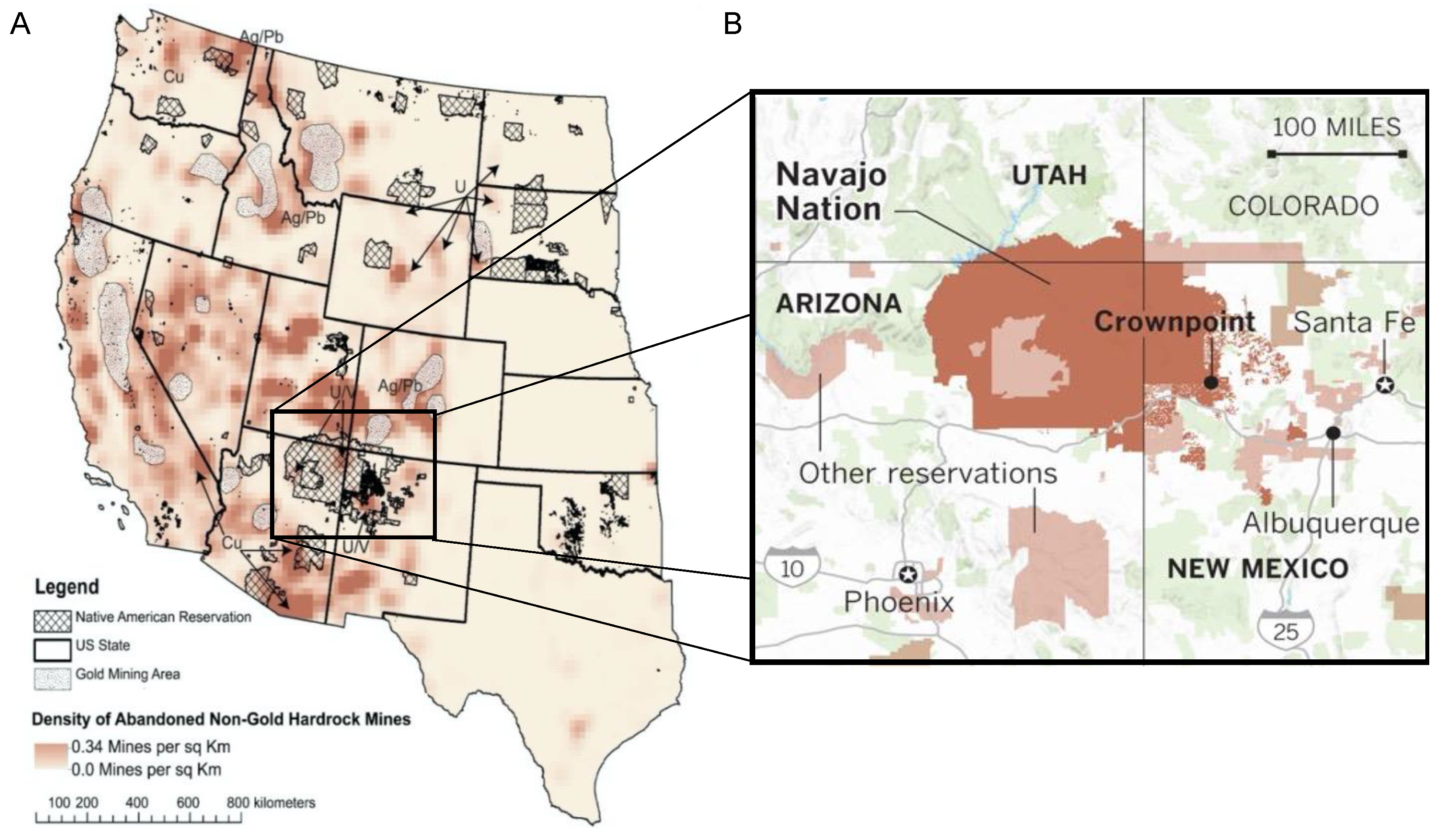
Figure 1. (A) Map of the western United States showing the locations of Native American reservations and the density of non-gold hard rock mines (adapted from ref [1]). (B) US Census Bureau map of Four Corners Region with Navajo Nation marked in dark brown: its area is approximately 27,000 square miles, roughly equal to that of West Virginia.
Ijerph 19 14939 g001
Figure 2. Spearman’s correlations between metal biomonitoring results. Correlation coefficient is designated by color with blues designating negative correlations and reds designating positive correlations. Asterisks denote significant correlations at the p < 0.05 (*), 0.01 (**), and 0.001 (***).
Ijerph 19 14939 g002
Figure 3. Spearman’s correlations between metal biomonitoring and biomarkers. Correlation coefficient is designated by color with blues designating negative correlations and reds designating positive correlations. Asterisks denote significant correlations at the p < 0.05 (*), 0.01 (**), and 0.001 (***).
Ijerph 19 14939 g003
Table 1. Summary of sociodemographic characteristics of study cohort (n = 231).
Variable
Maternal age (years)Mean (SD):27 (6.2)
CategoryResult: n (%) *
Education levelNo high school diploma97 (57.1)
High school diploma73 (42.9)
Household income<$20,000/year54 (39.4)
>$20,000/year83 (60.6)
Marital statusMarried or living with a partner112 (78.9)
Not married or living with a partner30 (21.1)
Pre-pregnancy BMIUnderweight or normal52 (26.4)
Overweight70 (35.5)
Obese75 (38.1)
Trimester at sample collection1st34 (14.7)
2nd97 (42)
3rd100 (43.3)
Sex of childFemale107 (49.3)
Male110 (50.7)
* Some data not available for all participants, % of participants with available data. NOTE: No difference in variables was observed between the subset of individuals selected for this study analysis and the NBCS as a whole.
Table 2. Summary statistics for concentrations of exposure biomonitoring, including limit of detection (LoD).
Abbr.Metal/MetaboliteMatrixUnitsnLoDMean (SD)Median (IQR)NHANES
Median (95th%Tile)
Factor Navajo vs.
NHANES *
UURUraniumUrineµg/dL2160.0020.03 (0.13)0.02 (0.01–0.03)0.005 (0.031)
UTASTotal arsenicUrineµg/dL2140.267.15 (6.12)5.56 (4.15–8.08)5.74 (49.9)0.97
UAS3Arsenite (AsIII)Urineµg/L2130.120.48 (0.39)0.38 (0.19–0.62)0.12 (1.11)3.17×
UMMAMonomethylarsonic acidUrineµg/L2130.20.52 (0.45)0.38 (0.21–0.68)0.28 (1.45)1.36×
UDMADimethylarsinic acidUrineµg/L2131.915.29 (3.61)4.25 (3.07–6.52)2.95 (12)1.44×
BPBLeadBloodµg/dL2120.070.41 (0.4)0.32 (0.25–0.42)0.88 (2.89)0.36
UPBLeadUrineµg/L2160.030.37 (0.56)0.28 (0.19–0.39)0.32 (1.38)0.88
BCDCadmiumBloodµg/L2120.10.34 (0.17)0.31 (0.24–0.4)0.27 (1.35)1.15×
UCDCadmiumUrineµg/L2160.0360.25 (0.18)0.22 (0.14–0.32)0.179 (1.08)1.23×
BMNManganeseBloodµg/L2120.9920.07 (6.82)19 (15–24.64)9.2 (16.1)2.07×
UMNManganeseUrineµg/L2150.130.37 (0.45)0.23 (0.15–0.41)0.13 (0.28)1.77×
THGTotal mercuryBloodµg/L2120.280.42 (0.27)0.34 (0.2–0.51)0.74 (4.66)0.46
* Factor indicates the magnitude of Navajo medians relative to NHANES medians. Metals or metalloids in bold have a sample median value higher than the NHANES median.
Table 3. Summary of significant (p < 0.05) Spearman’s correlations between cytokines and biomonitored metals and metalloids.
CytokineMetalRhopn
IL-4UAS3−0.340211
UMMA−0.230.001211
BMN0.150.033209
IL-6UAS3−0.150.032211
BMN0.180.009209
IL-10BPB0.150.032209
UCD−0.150.028214
IL-12UAS3−0.140.047211
BMN0.160.023209
IL-29UAS3−0.190.006203
IFNαUDMA0.180.011203
IFNγUCD0.140.035214
TNFαUAS3−0.150.035211
UPB−0.160.017214
BMN0.160.023209
CRPUTAS−0.180.009204
UAS3−0.160.019203
UMMA−0.170.018203
UDMA−0.160.021203
Table 4. Summary of final multivariable models of the relationship between biomonitored metals and metalloids (UUR, UTAS, AsIII, DMA, and MMA, BPB, UPB, BMN, UMN, THG) and cytokines after variable selection.
CytokinenPredictors Retained in Multivariate ModelEstimateStd Errorp
IL-4203age0.0130.0080.114
log(UAS3) *−0.2280.0580
log(BCD)−0.2610.1130.022
log(BMN)0.310.1560.049
IL-6203log(UAS3)−0.1250.0650.054
log(BMN)0.3290.1720.057
IL-7203log(BCD)0.10.0610.103
IL-10203age−0.020.0080.017
log(UDMA)−0.1830.0980.064
log(UPB)−0.1760.120.143
log(BPB)0.4220.1340.002
log(UMN)0.1390.0750.066
IL-12203log(UTAS)0.170.1070.112
log(UAS3)−0.1130.0690.103
log(BCD)−0.2480.1320.062
log(UCD)0.1420.0880.107
log(BMN)0.3860.170.024
IL-17203log(BPB)0.30.1280.02
log(BCD)−0.2660.1440.066
IL-29196log(UAS3)−0.5520.1810.003
log(UPB)0.5450.2580.036
log(BCD)−0.9960.3920.012
log(UCD)0.5050.2530.047
log(BMN)0.7780.4780.105
IFNα196log(UTAS)−0.510.3050.096
log(UDMA)0.9790.3160.002
log(UMN)−0.2210.1570.16
log(THG)0.2840.1990.155
IFNγ203trim.L−0.2480.1120.028
trim.Q−0.050.0940.595
log(UUR)−0.0960.0620.123
log(BPB)0.2550.1050.017
log(THG)0.1730.0940.066
TNFα203age−0.0070.0050.164
log(UPB)−0.1830.060.003
log(BPB)0.1760.0740.017
log(BCD)−0.1470.0710.04
log(UCD)0.1520.0530.004
log(BMN)0.1860.0890.037
CRP171log(UTAS)−0.8090.240.001
log(UMMA)−0.3240.1620.047
log(UDMA)0.6670.2890.022
log(BPB)0.2730.1710.111
* Strong immunomodulatory effects (p of estimate < 0.05) are in bold.
Publisher’s Note: MDPI stays neutral with regard to jurisdictional claims in published maps and institutional affiliations.