Protocol

Description of ROM-SPORT I Battery: Keys to Assess Lower Limb Flexibility

1
Department of Physical Activity and Sport, Faculty of Sport Sciences, CEIR Campus Mare Nostrum (CMN), University of Murcia, 30720 Murcia, Spain
2
Locomotor System and Sport Research Group (E0B5-07), University of Murcia, 30720 Murcia, Spain
Int. J. Environ. Res. Public Health 2022, 19(17), 10747; https://doi.org/10.3390/ijerph191710747
Submission received: 13 July 2022 / Revised: 10 August 2022 / Accepted: 26 August 2022 / Published: 29 August 2022
(This article belongs to the Special Issue Lower Extremity Diseases, Injuries and Public Health)

Abstract

:
Limited range of motion (ROM) is considered one of the most important intrinsic and modifiable risk factors for the most common sports-related injuries. In addition, controlling and monitoring an athlete’s ROM is a strategy to achieve optimal ROM and improve athletic performance in sports, especially those that require high ROM in the major joints. Therefore, assessing ROM (pre-participation, during a rehabilitation process, on return to play, etc.) is important not only as a method to prevent sports injuries, but also as a quantitative determinant of the potential of athletic performance. However, despite the variety of different ROM assessment methods described in the literature, there is no consensus on which methods are best suited for this goal. Recently, the ROM-SPORT I battery has been shown to have advantages over other ROM assessment methods. This tool has not yet been fully described in detail for researchers, sports professionals, and clinicians to learn. The main objective of this study is to describe the ROM-SPORT I battery tests in detail using the following criteria: test description, simplicity of the test procedure, low need for human and material resources, predictive validity, and reliability.

1. Introduction

Sporting activities play an important role in our society today [1]. Millions of people around the world play some kind of sport (either at amateur or professional level) for many reasons, including enjoyment and fun, relaxation, socialisation, and maintaining or improving fitness and health. However, playing a sport also carries a certain risk of injury [2]. Previous epidemiological studies have shown that sports-related injuries account for 10–19% of all cases of acute injury in the emergency department [1,3].
Several research studies and centres of sports medicine and science emphasise the importance of regular pre-participation and in-season assessments to identify the primary and modifiable factors that may predispose athletes to injury. This is the most effective way to prevent and reduce the number and severity of sports injuries [2,4,5,6,7,8]. Identifying athletes at high risk of injury therefore allows for the implementation of specific interventions that directly target the critical factors related to the mechanisms of injury [6,8].
Limited range of motion (ROM) due to a lack of muscle flexibility has been shown to be one of the most important predictors of common sports injuries, such as groin pain (restricted hip abduction [9,10,11], lateral rotation [9], medial rotation [12,13], and total rotation [9] ROMs); tendinopathies of the patella (restricted hip flexion with the knee extended [10,12,14] and ankle dorsiflexion ROMs [15,16]) and Achilles (restricted ankle dorsiflexion ROM [17]); strains of the hamstrings (restricted hip flexion with the knee extended [18,19], knee flexion [18], and ankle dorsiflexion ROMs [20]) and quadriceps (restricted knee flexion ROM [18,19]); rupture of the anterior cruciate ligament (restricted lateral [21,22] and medial hip rotation ROMs [21,23,24]); low back pain (restricted hip flexion with the knee extended [25,26], hip extension [27], hip lateral rotation [28,29], hip internal rotation [26,29], and knee flexion [30] ROMs) and knee pain (restricted hip extension [31] and flexion ROMs [32]). In addition, certain sports such as taekwondo, diving, figure skating, and gymnastics require high ROM values in order for athletes to successfully perform the technical actions most highly rated by the judges [33] and improve their physical performance [34,35]. It therefore seems clear that ROM should be assessed in athletes as an intervention strategy to optimise their athletic performance and reduce their risk of injury. Flexibility has been shown to be specific to each joint, muscle action, or specific movement [25,26]. For this reason, the assessment of flexibility in athletes has shown very different results depending on the sport [36,37,38]. For example, artistic gymnastics, rhythmic gymnastics, and swimming jumps had the highest values for hip flexion with the knee extended ROM and the lowest values for water polo and marathons [38]. Shoulder and ankle ROM have higher values in speed swimmers than in other athletes [39]. Ultramarathon runners who cover a longer distance in competition maintain baseline values for hip flexion with the knee extended ROM and stride length during competition [40]. Goalkeepers have higher ROM values than outfield players only for knee flexion, hip flexion with the knee extended, and hip abduction [41,42]. The dominant limb of soccer players has higher values for knee flexion ROM than the non-dominant limb [43,44]. Rowers with more years of experience show higher values for hip flexion with the knee extended ROM than rowers with less experience and a lower competition level [45]. International dancers and gymnasts have higher ROM values in the shoulder (flexion and extension), hip (flexion, extension, and abduction), ankle (from dorsiflexion to planar flexion), and trunk (from hyperextension to full flexion) than national athletes and beginners [46]. International kickboxers have a higher ROM of hip adduction and ankle dorsiflexion than national fighters [47]. Medal athletes in Taekwondo show higher flexibility than non-medal athletes in passive and active hip flexion with the knee extended test and in the hip abduction test [48]. Furthermore, it has recently been shown that flexibility depends on age and maturation [49,50].
There is a large number of published tests to assess the ROM of the major joints of the lower extremities (hip, knee, and ankle). There are different methods of assessing ROM, e.g., passive (e.g., Straight Leg Raise Test (hip flexion ROM)) or active (e.g., Walking Step Test (ankle dorsiflexion ROM)), and/or using single (Thomas Test (hip extension ROM)) or multiple (Deep Back Squat (hip flexion ROM)) tests. In addition, numerous measuring instruments have been proposed to measure ROM directly (goniometer, inclinometer, etc.) or indirectly (tape measure, video camera, etc.) in degrees.
However, despite the large number of published ROM tests, there is currently no consensus on which examination tests are most appropriate for assessing the major joints of the lower limbs. Identifying criterion-referenced assessment tests and promoting their use in sports medicine and competitive sports would allow clinicians, physiotherapists, and sports professionals to standardise the assessment and monitoring of ROM. This would also facilitate the identification of athletes who are at risk of injury and/or whose ROM values are insufficient to achieve a higher level of technical athletic performance.
Knowledge of ROM monitoring can also lead to the application of training interventions to improve the athlete’s ROM values (e.g., stretching or foam rolling) [51]. In addition, it would be possible in research to more reliably investigate the role of ROM in the development of acute and overuse-related musculoskeletal pathologies associated with restricted ROM values (establishing normal and restricted cut-off values) and to support the efficacy of different treatments (e.g., stretch training, massage, or self-myofascial release exercises) to maintain and/or improve ROM [52,53]. For all these purposes, Hopkins et al. [54,55,56] suggests that the selection of diagnostic reference tests should first be based on the criteria of high validity and reliability, and then emphasise the simplicity and universality of the procedure.
Recently, the measurement method of the ROM-SPORT I battery tests has been shown to have advantages over other methods published in the literature [57]. However, no detailed description of the ROM-SPORT I battery tests has been published. Therefore, the main objective of this study is to describe the ROM-SPORT I battery tests in detail using the following criteria: test description, simplicity of the test procedure, low need for human and material resources, predictive validity, and reliability.

2. Description of the Tests of ROM-SPORT Battery I

The current study proposes to perform 11 assessment tests in the extended version of ROM-SPORT I battery to obtain the ROM measurements of the major joints of the lower extremities. The following ROM measurements would be taken at the hip: flexion with the knee extended (Figure 1), flexion with the knee flexed (Figure 2), extension with the knee relaxed (Figure 3), adduction with 90° hip flexion (Figure 4), abduction with the knee extended (Figure 5), abduction with 90° hip flexion (Figure 6), internal rotation with the knee flexed (Figure 7), and external rotation (Figure 8) with the knee flexed. In the knee, flexion is assessed (Figure 9), while in the ankle, dorsiflexion is measured with the knee extended (Figure 10) and flexed (Figure 11).
In the shortened version of the ROM-SPORT I battery, 7 tests are performed to obtain the following ROM measurements: in the hip, flexion with the knee extended and extension with the knee relaxed; in the knee, flexion; and in the ankle, dorsiflexion with the knee extended and flexed. These tests were chosen for the shortened version of ROM-SPORT I battery because they measure the ROMs most commonly reported as restricted in athletes [57] and the general population [36,58].
The procedure for each test in the ROM-SPORT I battery is described in detail in the following Figure 1, Figure 2, Figure 3, Figure 4, Figure 5, Figure 6, Figure 7, Figure 8, Figure 9, Figure 10 and Figure 11.
Figure 1. Passive hip flexion with the knee extended test (hamstrings).
Ijerph 19 10747 g001
Figure 2. Passive hip flexion with the knee flexed test (gluteus maximus).
Ijerph 19 10747 g002
Figure 3. Passive hip extension with the knee relaxed test (iliopsoas).
Ijerph 19 10747 g003
Figure 4. Passive hip adduction with the 90° hip flexion test (piriformis); * [59,60].
Ijerph 19 10747 g004
Figure 5. Passive hip abduction with the knee extended test (adductors).
Ijerph 19 10747 g005
Figure 6. Passive hip abduction with the 90° hip flexion test (adductors monoarticular).
Ijerph 19 10747 g006
Figure 7. Passive internal hip rotation test (external rotators).
Ijerph 19 10747 g007
Figure 8. Passive external hip rotation test (internal rotators).
Ijerph 19 10747 g008
Figure 9. Passive knee flexion test (quadriceps).
Ijerph 19 10747 g009
Figure 10. Ankle dorsiflexion with the knee extended test (gastrocnemius).
Ijerph 19 10747 g010
Figure 11. Ankle dorsiflexion with the knee flexed test (soleus).
Ijerph 19 10747 g011

3. Highlights of the ROM-SPORT I Procedure

3.1. Familiarisation and Warm-Up Phase

Before assessing ROM, the athlete should perform 8–10 min of warm-up training with cardio (jogging or cycling; 50–70 W and 60–80 rpm; light intensity, 10–12 Borg scale) according to the recommendations [61,62] and 60–90 s of dynamic flexibility of the major lower limb muscles (gluteus maximus, hamstring, adductors, iliopsoas, quadriceps, external and internal rotators of the hip, gastrocnemius and soleus) [63]. Previous studies have reported that 60–180 s of static stretching produces a lasting change in the viscoelastic properties of muscles during a 20–30 min rest period [63]. The application of the ROM-SPORT I battery to an athlete takes less than 20 min [57]. The positions used for the stretching exercises should be similar to those used for the different tests to reduce possible learning bias in the results.
Warm-up exercises were performed because: (1) all tests required a large muscle tension stimulus; (2) warm-ups reduce the effects of muscle stretch by repeated trials during data collection; and (3) they reduce the variability and standard error of measurements by minimising the effects of different muscle temperatures on muscle flexibility [64,65].
After the warm-up, the ROM assessment is performed.

3.2. Human Resources

One of the disadvantages of the ROM-SPORT I battery is that the ROM assessment of the hip and knee joints requires two trained evaluators: one to ensure that the athlete remains in the correct position during the test manoeuvres (assistant evaluator) and another to perform the test (main evaluator). However, only one evaluator is required for the assessment of the ankle ROM. The use of two evaluators to perform the tests seems to limit the practical application of these measurement methods in a sporting and clinical context. As these measurement methods are simple to use, the role of the assistant evaluator (ensuring adequate stabilisation of the pelvis and other body segments during all tests) could be taken on by any postgraduate student or sports coach conducting one or two 10-min training sessions (statement based on the authors’ extensive experience).

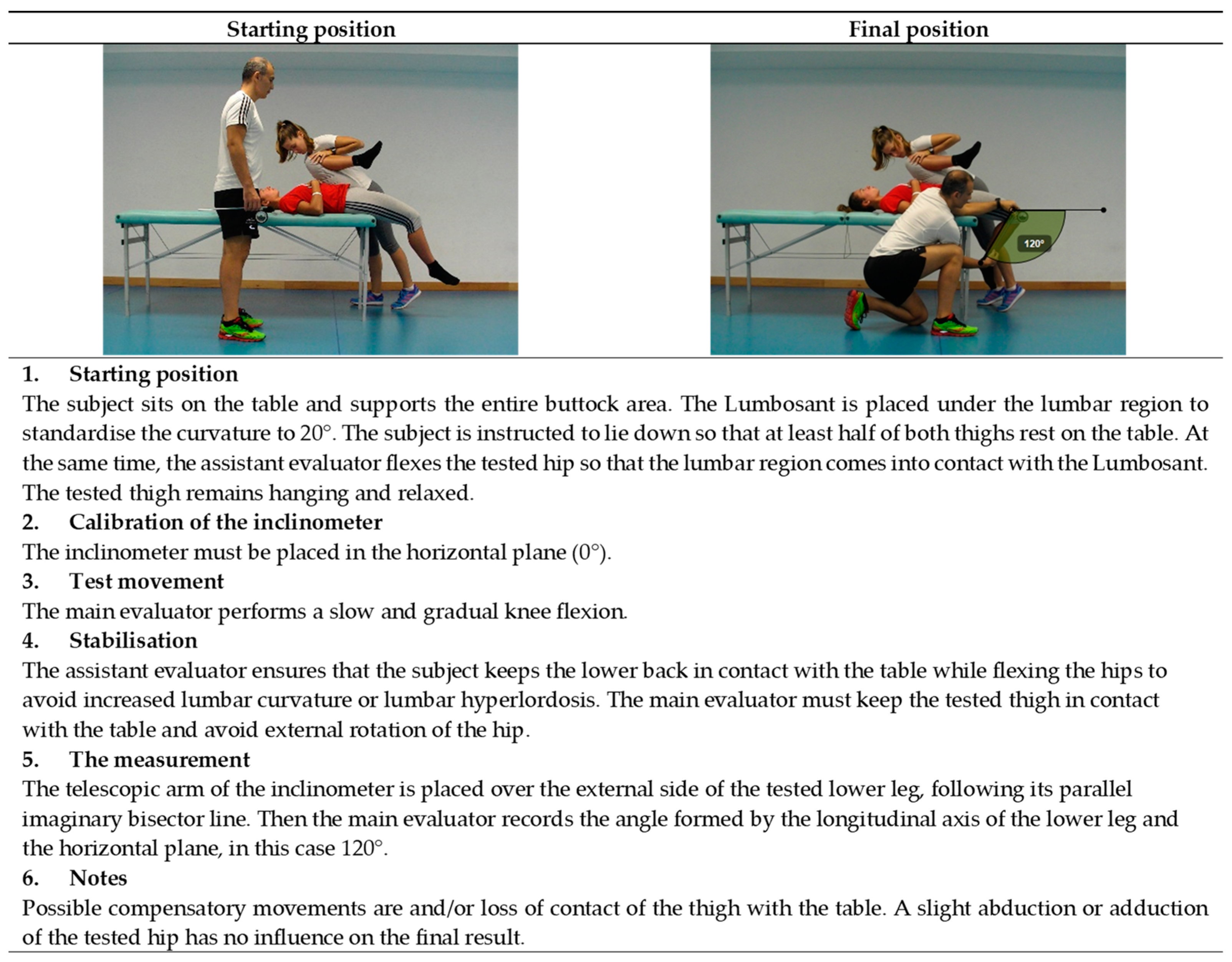
3.3. Athlete’s Starting Position

The athlete’s starting position is the neutral or zero-degree position. Seven of the eleven tests of the ROM-SPORT I battery are performed in the supine position (hip and knee ROM), with the exception of the hip rotation and the ankle ROM tests. Therefore, depending on the position of the athlete, the tests can be performed in the following order: standing (ankle dorsiflexion ROM tests), prone (hip rotation tests), and supine (hip ROM tests), or the other way round. This order of ROM test performance helps to shorten the duration of the evaluation session. The competencies of the main and the assistant evaluator are brief and clearly defined. The main evaluator checks the correct starting position of the athlete and performs the target movement of the assessment, while the assistant examiner checks the compensatory movements with his hands and the Lumbosant.
Finally, a table and a standard box (about 30.5 cm high) are used to position the athletes for the tests. The use of a box to position the athletes for the ankle dorsiflexion with the knee flexed ROM test allows the evaluator and the athletes to adopt a comfortable position during the measurement procedure of the ankle dorsiflexion ROM [66].

3.4. Measuring Instrument and Its Calibration

The starting position of the athlete; the competences, capabilities, and skills of the evaluators; and the use of the measuring instrument lead to a simple and fast ROM measurement. An inclinometer (ISOMED, Inc, Portland, OR, USA) with a telescopic arm is used as the main measuring instrument for all ROM assessment tests, except for the hip abduction with the knee extended ROM test, which requires a long-arm goniometer. A lower-back protection support, or Lumbosant (Imucot Traumatología SL, Murcia, Spain), is used in the hip and knee ROM tests to standardise the lordotic curve (20°) during the assessment manoeuvres [67,68,69].
The use of an inclinometer with a telescopic arm has the advantage that no body landmarks need to be marked, since the maximum ROM values can be determined as the angle that the longitudinal axis of the moving body segment (imaginary line of the lateral bisector (sagittal movements) or anterior bisector (frontal movements) of the mobilised body segment) makes with the vertical or horizontal plane. Thus, the initial and final positions can be systematically and repeatedly determined with precision [58]. The inclinometer with a telescopic arm becomes a single-arm goniometer, which has the advantage of having a plane of gravity; this allows for better measuring accuracy and a higher measuring speed [57,69].
Depending on the movement, the inclinometer is calibrated with gravity at 0° vertically (adduction and abduction with 90° hip flexion; internal and external rotation with flexed knee; ankle dorsiflexion with extended and flexed knee) or horizontally (flexion with extended and flexed knee; extension with relaxed knee and knee flexion).

3.5. Movement of the Assessment

In the ROM-SPORT I battery procedure, a maximum of passive movement is used in all tests. The rationale for using passive manoeuvres is for the following two reasons: Firstly, in several active tests, the peak ROM depends on the athlete’s muscle strength (mainly psoas, hamstrings, quadriceps) and the ability to simultaneously contract the agonist muscles and relax the antagonist muscles being measured [70]. This severely limits the use of active testing in individuals with low physical conditions, such as children and adolescents [71]. Furthermore, the different strength levels of athletes of different sexes and different sports do not allow for a comparison of the flexibility profiles [72]. Secondly, the athlete’s motivation has been shown to influence the result of an active ROM test, leading to intraindividual variability or a source of measurement error [58].
For the ankle dorsiflexion tests ROM, the body weight itself should be loaded in order to obtain a maximum passive measurement [51]. In addition, all selected passive tests specifically measure the main movement of the joint, i.e., the extensibility of the target muscle. Linear and functional tests (e.g., shoulder mobility test, overhead squat test, floor-to-toe distance test) have more disadvantages than angular tests for assessing muscle extensibility. These tests are significantly influenced by the anthropometric measures and intermuscular coordination of the subjects, which negatively affects the validity of the results.

3.6. Stabilisation

Stabilisation of the athlete, which refers to the athlete’s starting position and the control of compensations during maximal passive movements, is a fundamental phase of the ROM-SPORT I battery procedure in order to obtain an accurate measurement. In this sense, and in relation to the pelvis, it has been shown that significant compensatory movements of the pelvis occur during hip and knee movements (sagittal (posterior and anterior tilting), frontal (hike/upward tilt or drop/downward tilting) and transversal planes (backwards or forwards)) [25,67,73,74,75].
Consequently, these compensatory movements that increase ROM could alter the validity (false negative) of the measurement obtained [57,67]. Moreover, the pelvic tilt starts right at the beginning of the movement and gradually increases. The task of the assistant evaluator is to ensure the correct stability of the initial position, of a specific body segment, or of the pelvis throughout the assessment manoeuvre in order to avoid or minimise any compensatory movements that could increase and bias the final value.
Therefore, knowledge of the athlete’s starting position and control of the compensations, especially by the assistant evaluator, is essential.

3.7. Criteria for the End of the Range of Motion

Following the scientific literature, the endpoint for each ROM test is determined by one or more of the following criteria: (1) The main evaluator is unable to continue the stretching manoeuvre due to increased resistance of the muscle being tested; (2) The athlete feels a strong but tolerable stretch, just before the onset of pain; (3) One or both evaluators (main and assistant) have detected a palpable and perceptible compensatory movement that could increase the ROM [57,68].

3.8. Measurement

At the end of the maximum passive movement, the main evaluator places the telescopic arm of the inclinometer on the longitudinal axis of the moving body segment (imaginary line of the lateral bisector (sagittal movements) or the anterior bisector (frontal movements) of the mobilised body segment). The main evaluator then measures the angle formed by the longitudinal axis of the mobilised body segment and the line of gravity at 0°. At least two measurements per ROM test (dominant and non-dominant) and body limb in randomised order are recommended to obtain a reliable measurement. The mean value for each test is considered the final (true) ROM value [57,68,76]. In cases where a deviation of more than 5% was observed in the ROM values between the two attempts of a ROM test, an additional attempt is made. The two most closely related trials are used to calculate the true ROM value, provided the deviation in the new trial is <5%. If this is not the case, the evaluator would have to check the procedure for possible errors or circumstances that could explain the deviation. There was a 10 s break between the two trials and 30 s break between the tests. The athletes were tested in sportswear and without shoes.

3.9. Notes

Based on the scientific literature and the authors’ extensive previous experience, certain assessment skills have been reinforced in detail at the different phases of the procedure of each test, which influence the effectiveness of the procedure and the measurement result.
In summary, compared to other ROM assessment procedures, the use of the highlights of the ROM-SPORT I battery procedure contributes to a very fast implementation of the ROM test. The test duration of the ROM-SPORT I battery varies between about 15 (shortened version) and 20 (extended version) minutes. If the warm-up phase is omitted (e.g., in some clinical contexts), the duration is reduced to 7–11 min, although variability between sessions may increase by ±2°–4°. The estimated time for testing ROM with a telescopic arm inclinometer is 1 min per ROM test.

4. The Validity of the ROM-SPORT I Battery

With this in mind, all tests selected for the ROM-SPORT I battery have been validated by American medical organizations [58,77] and accepted by sports medicine and science handbooks [58,78] based on anatomical knowledge and extensive clinical experience (content validity or expert judge). The validity of an assessment test can be defined as the accuracy with which its measurement matches the true value, i.e., the extent to which it fulfils its objective [55]. Previous studies have demonstrated criterion validity of the gold standard [67,79,80,81]. In particular, control of compensatory movements or end-of-motion criteria are key elements of good validity.
After the evaluation of ROM, however, there is another important aspect to consider that is closely related to the concept of validity: The interpretation of the results is obtained on the basis of the values of a norm (normative validity) and a criterion (criterion validity). Normative validity values are used to compare the measurement of a ROM test with the normal values of a specific population or the flexibility profile of a sport. It is important to establish values that allow sports professionals to determine an athlete’s performance potential. In this sense, the research group “E0B5-07 Musculoskeletal System and Sport” of the University of Murcia has determined the flexibility profile using the ROM-SPORT I battery in soccer [49,82,83], futsal [37,44,84,85], basketball [86], handball [37,87], inline hockey [88,89], duathlon [90], taekwondo [91,92], and equestrian athletes [93].
Criterion-related values (criterion validity) refer to a cut-off value that indicates a high risk of a specific type of injury. It is important to establish values that allow clinicians, physiotherapists, and sports physicians to identify athletes at risk of injury. This knowledge would enable the early application of individualised training programmes to improve (athletes with borderline values or at high risk of injury) or maintain (athletes with normal values or at low risk of injury) baseline ROM levels and thus minimise the impact of one of the most important risk factors on the likelihood of lower limb injury. Table 1 shows the evidence-based cut-off values of several of the 11 tests selected for ROM-SPORT I battery that can be used to classify the ROM measures as restricted/high risk of injury or normal/low risk of injury. It should be noted, however, that there is no consensus on what constitutes normal and “high injury risk” values for some of the ROM measures. For this reason, several values for each category and measurement are given in Table 1, depending on the study, and should therefore be interpreted with some caution.

5. Reliability of the ROM-SPORT I Battery

Reliability is about the reproducibility of a measurement, i.e., whether the application of the assessment method can consistently produce the same results under the same conditions. In clinical or sports assessments, the reliability of a measurement is determined by human factors (the evaluator’s experience or training in administering the test, variations in assessment methodology, and individual-related variability) and/or the instrument used. Based on the fact that the most commonly used instruments for estimating ROM (goniometer and inclinometer) have proven to be reliable [57,69,76], the reliability of lower limb ROM measurements depends mainly on human factors [76]. Two different aspects of human reliability-related factors should be discussed before considering an appropriate measure for sports and clinical purposes: inter-evaluator reliability and intra- evaluator reliability [102]. Intra-evaluator reliability provides information on the extent to which multiple measurements taken at different times for the same test by the same evaluator are similar [103]. Intra-evaluator reliability can be determined if there are short (generally less than 3 h: intra-session) or long (generally more than 24 h: inter-session) intervals between test sessions. Longer periods between assessments (e.g., two weeks) are very important in clinical and sports contexts as they allow clinicians and sports professionals to monitor the performance or health status of their athletes and make informed decisions about whether a real change has occurred between testing sessions following the application of a training intervention [55]. The studies that analysed the intra-evaluator reliability of the values from the ROM-SPORT I battery using long time intervals between sessions (>24 h) showed moderate to high inter-session reliability scores (Table 2). Therefore, researchers, clinicians, and sports professionals can be 95% confident that a change between two measurements of more than 4°–7° for the ROM values from the ROM-SPORT I battery is likely to indicate a real change (determined by the statistical minimal detectable change with a 95% confidence interval [MDC95%]).
The key element for the reliability of the measurement obtained with the ROM-SPORT I battery is the use of the inclinometer as a measuring tool (Table 2). This fact, confirmed in several studies using other methods, has shown that the inclinometer is extremely reliable for lower limb ROM measurements (intraclass correlation index (ICC) > 0.90) and does not have the disadvantage of the goniometer, which requires precise positioning of its arms while the goniometer is moved simultaneously with the limb [58,106]. Finally, in contrast to other, more sophisticated devices (video cameras or isokinetic dynamometers), the cost of an inclinometer is relatively low (about 150 €).
For an accurate assessment of ROM, athletes should not have performed any vigorous physical activity in the previous 48 h. In addition, practical familiarization with the tests and a warm-up before athletes perform the tests will improve the accuracy of the measurements. This also applies to training examiners with the tests and the ROM-SPORT I battery procedure.

6. Practical Applications of the ROM-SPORT I Battery

The ROM-SPORT I battery can also be used in sports, clinical, and research settings with the following objectives:
To accurately quantify the ROM measurements of the hip, knee and ankle, and increased tolerance to stretch [107,108].
To determine the possibility of physical–technical sport performance in athletes who participate in sports that require technical skills with a high ROM (e.g., taekwondo, diving, figure skating, and gymnastics) [36,38,109].
To identify athletes with muscle tightness that results in limited ROM (or high risk of injury), especially in soft tissue injuries [44,88].
To quantify the effectiveness of intervention programs or significant chronic ROM changes (e.g., stretching and foam rolling intervention) aimed at maintaining or improving muscle flexibility in both healthy and injured individuals [110].
In physical therapy processes, to determine whether the ROM of the injured joint has been fully restored, which may contribute to a safe return to play (athletes) or activities of daily living (general population) [111].
For future research, it would be necessary for more studies to use the ROM-SPORT I battery to define the ROM cut-off values for different groups in terms of sex, age, physical condition, type of sport, etc., to identify what values would be considered normal and which ones are considered of high risk for injury.

7. Conclusions

The eleven tests of the ROM-SPORT I battery described here seem to be the most appropriate for assessing the ROM of the major joints of the lower limbs. The use of the telescopic arm of the inclinometer and the Lumbosant, as well as the competence of the evaluators, are key elements for accurate, reproducible, and rapid measurement. The ROM-SPORT I battery has criterion-related values to identify individuals at high risk for a specific type of injury.

Funding

This research received no external funding.

Institutional Review Board Statement

The study was conducted according to the guidelines of the Declaration of Helsinki and approved by Ethics and Research Committee of the University of Murcia (Spain) (ID: 1702/2017).

Informed Consent Statement

Informed consent was obtained from all subjects involved in the study.

Acknowledgments

We are grateful to Cristina Cuello for her critical reading of the manuscript and our colleagues Pilar Sainz de Baranda, Fernando Santonja and Francisco Ayala from the research group “E0B5-07 Musculoskeletal System and Sport” at the University of Murcia for their contributions to the design and development of the ROM-SPORT I battery.

Conflicts of Interest

The authors declare no conflict of interest.

References

  1. Baarveld, F.; Visser, C.; Kollen, B.; Backx, F. Sports-related injuries in primary health care. Fam. Pract. 2011, 28, 29–33. [Google Scholar] [CrossRef]
  2. Bahr, R.; Holme, I. Risk Factors for Sports Injuries—A Methodological Approach; British Association of Sport and Excercise Medicine: Auckley, UK, 2003; Volume 37. [Google Scholar]
  3. Lindqvist, K.; Timpka, T.; Bjurulf, P. Injuries during leisure physical activity in a Swedish municipality. Scand. J. Soc. Med. 1996, 24, 282–292. [Google Scholar] [CrossRef]
  4. Baechle, T.R.; Earle, R.W. Essentials of Strength Training and Conditioning; Human kine.: Champaign, IL, USA, 2008. [Google Scholar]
  5. American College of Sports Medicine. ACSM’s Health-Related Physical Fitness Assessment Manual; Lippincott Williams & Wilkins: Baltimore, MD, USA, 2013. [Google Scholar]
  6. Thacker, S.; Gilchrist, J.; Stroup, D.; Kimsey, C.; Thacker, S.; Gilchrist, J.; Stroup, D.; Kimsey, C. The Impact of Stretching on Sports Injury Risk: A Systematic Review of the Literature. Med. Sci. Sports Exerc. 2004, 36, 371–378. [Google Scholar] [CrossRef]
  7. Ekstrand, J.; Spreco, A.; Bengtsson, H.; Bahr, R. Injury rates decreased in men’s professional football: An 18-year prospective cohort study of almost 12 000 injuries sustained during 1.8 million hours of play. Br. J. Sports Med. 2021, 55, 1084–1092. [Google Scholar] [CrossRef]
  8. Hägglund, M.; Waldén, M.; Ekstrand, J. Risk factors for lower extremity muscle injury in professional soccer: The UEFA injury study. Am. J. Sports Med. 2013, 41, 327–335. [Google Scholar] [CrossRef]
  9. Verrall, G.; Slavotinek, J.; Barnes, P.; Esterman, A.; Oakeshott, R.; Spriggins, A. Hip joint range of motion restriction precedes athletic chronic groin injury. J. Sci. Med. Sport 2007, 10, 463–466. [Google Scholar] [CrossRef]
  10. Arnason, A.; Sigurdsson, S.; Gudmundsson, A.; Holme, I.; Engebretsen, L.; Bahr, R. Risk Factors for Injuries in Football. Am. J. Sports Med. 2004, 32, 5S–16S. [Google Scholar] [CrossRef]
  11. Emery, C.; Meeuwisse, W. Risk factors for groin injuries in hockey. Med. Sci. Sports Exerc. 2001, 33, 1423–1433. [Google Scholar] [CrossRef]
  12. Ibrahim, A.; Murrell, G.; Knapman, P. Adductor Strain and Hip Range of Movement in Male Professional Soccer Players. J. Orthop. Surg. 2007, 15, 46–49. [Google Scholar] [CrossRef]
  13. Nevin, F.; Delahunt, E. Adductor squeeze test values and hip joint range of motion in Gaelic football athletes with longstanding groin pain. J. Sci. Med. Sport 2014, 17, 155–159. [Google Scholar] [CrossRef]
  14. Okamura, S.; Wada, N.; Tazawa, M.; Sohmiya, M.; Ibe, Y.; Shimizu, T.; Usuda, S.; Shirakura, K. Injuries and disorders among young ice skaters: Relationship with generalized joint laxity and tightness. Open Access J. Sports Med. 2014, 5, 191–195. [Google Scholar] [CrossRef] [PubMed]
  15. Backman, L.; Danielson, P. Low range of ankle dorsiflexion predisposes for patellar tendinopathy in junior elite basketball players: A 1-year prospective study. Am. J. Sports Med. 2011, 39, 2626–2633. [Google Scholar] [CrossRef]
  16. Scattone Silva, R.; Nakagawa, T.H.; Ferreira, A.L.G.; Garcia, L.C.; Santos, J.E.M.; Serrão, F.V. Lower limb strength and flexibility in athletes with and without patellar tendinopathy. Phys. Ther. Sport 2016, 20, 19–25. [Google Scholar] [CrossRef] [PubMed]
  17. Wilder, R.; Sethi, S. Overuse injuries: Tendinopathies, stress fractures, compartment syndrome, and shin splints. Clin. Sports Med. 2004, 23, 55–81. [Google Scholar] [CrossRef]
  18. Witvrouw, E.; Danneels, L.; Asselman, P.; D’Have, T.; Cambier, D. Muscle flexibility as a risk factor for developing muscle injuries in male professional soccer players: A prospective study. Am. J. Sports Med. 2003, 31, 41–46. [Google Scholar] [CrossRef]
  19. Ekstrand, J.; Gillquist, J. The frequency of muscle tightness and injuries in soccer players. Am. J. Sports Med. 1982, 10, 75–78. [Google Scholar] [CrossRef]
  20. Gabbe, B.; Finch, C.; Wajswelner, H.; Bennell, K. Predictors of lower extremity injuries at the community level of Australian football. Clin. J. Sport Med. 2004, 14, 56–63. [Google Scholar] [CrossRef]
  21. Tainaka, K.; Takizawa, T.; Kobayashi, H.; Umimura, M. Limited hip rotation and non-contact anterior cruciate ligament injury: A case–control study. Knee 2014, 21, 86–90. [Google Scholar] [CrossRef]
  22. Lopes, O.; Gomes, J.; Freitas Spinelli, L. Range of motion and radiographic analysis of the hip in patients with contact and non-contact anterior cruciate ligament injury. Knee Surg. Sports Traumatol. Arthrosc. 2016, 24, 2868–2873. [Google Scholar] [CrossRef]
  23. VandenBerg, C.; Crawford, E.; Enselman, E.; Robbins, B.; Wojtys, E.; Bedi, A. Restricted Hip Rotation Is Correlated With an Increased Risk for Anterior Cruciate Ligament Injury. Arthrosc. J. Arthrosc. Relat. Surg. 2017, 33, 317–325. [Google Scholar] [CrossRef]
  24. Bedi, A.; Warren, R.; Wojtys, E.; Oh, Y.; Ashton-Miller, J.; Oltean, H.; Kelly, B. Restriction in hip internal rotation is associated with an increased risk of ACL injury. Knee Surg. Sports Traumatol. Arthrosc. 2016, 24, 2024–2031. [Google Scholar] [CrossRef] [PubMed]
  25. Cejudo, A.; Centenera-Centenera, J.; Santonja-Medina, F. The Potential Role of Hamstring Extensibility on Sagittal Pelvic Tilt, Sagittal Spinal Curves and Recurrent Low Back Pain in Team Sports Players: A Gender Perspective Analysis. Int. J. Environ. Res. Public Health 2021, 18, 8654. [Google Scholar] [CrossRef] [PubMed]
  26. Vad, V.; Bhat, A.; Basrai, D.; Gebeh, A.; Aspergren, D.; Andrews, J. Low Back Pain in Professional Golfers: The Role of Associated Hip and Low Back Range-of-Motion Deficits. Am. J. Sports Med. 2004, 32, 494–497. [Google Scholar] [CrossRef]
  27. Moreno-Pérez, V.; López-Valenciano, A.; Ayala, F.; Fernandez-Fernandez, J.; Vera-Garcia, F. Comparison of hip extension and rotation ranges of motion in young elite tennis players with and without history of low back pain. J. Back Musculoskelet. Rehabil. 2019, 32, 629–638. [Google Scholar] [CrossRef]
  28. Cejudo, A.; Moreno-Alcaraz, V.J.; Izzo, R.; Santonja-Medina, F.; Sainz de Baranda, P. External and Total Hip Rotation Ranges of Motion Predispose to Low Back Pain in Elite Spanish Inline Hockey Players. Int. J. Environ. Res. Public Health 2020, 17, 4858. [Google Scholar] [CrossRef]
  29. Van Dillen, L.; Bloom, N.; Gombatto, S.; Susco, T. Hip rotation range of motion in people with and without low back pain who participate in rotation-related sports. Phys. Ther. Sport 2008, 9, 72–81. [Google Scholar] [CrossRef]
  30. Kanchanomai, S.; Janwantanakul, P.; Pensri, P.; Jiamjarasrangsi, W. A prospective study of incidence and risk factors for the onset and persistence of low back pain in Thai university students. Asia-Pac. J. Public Health 2015, 27, NP106–NP115. [Google Scholar] [CrossRef]
  31. Roach, S.; San Juan, J.; Suprak, D.; Lyda, M.; Boydston, C. Patellofemoral pain subjects exhibit decreased passive hip range of motion compared to controls. Int. J. Sports Phys. Ther. 2014, 9, 468–475. [Google Scholar]
  32. Witvrouw, E.; Van Tiggelen, D.; Willems, T. Risk factors and prevention of anterior knee pain. In Anterior knee pain and patellar instability. Anterior Knee Pain and Patellar Instability; Springer: London, UK, 2006; pp. 135–145. [Google Scholar]
  33. Sainz de Baranda, P.; Cejudo, A.; Ayala, F.; Santonja, F. Perfil óptimo de flexibilidad del miembro inferior en jugadoras de fútbol sala. Rev. Int. Med. Cienc. Act. Fis. Deporte 2015, 15, 647–662. [Google Scholar] [CrossRef]
  34. Gonzalo-Skok, O.; Serna, J.; Rhea, M.; Marín, P. Relationships between functional movement tests and performance tests in young elite male basketball players. Int. J. Sports Phys. Ther. 2015, 10, 628–638. [Google Scholar]
  35. Panoutsakopoulos, V.; Kotzamanidou, M.C.; Papaiakovou, G.; Kollias, I.A. The Ankle Joint Range of Motion and Its Effect on Squat Jump Performance with and without Arm Swing in Adolescent Female Volleyball Players. J. Funct. Morphol. Kinesiol. 2021, 6, 14. [Google Scholar] [CrossRef]
  36. Hogg, J.; Schmitz, R.; Nguyen, A.-D.; Shultz, S.J. Lumbo-Pelvic-Hip Complex Passive Hip Range-of-Motion Values Across Sex and Sport. J. Athl. Train. 2018, 53, 560–567. [Google Scholar] [CrossRef] [Green Version]
  37. Cejudo, A.; Robles-Palazón, F.; Sainz De Baranda, P. Fútbol sala de élite: Diferencias de flexibilidad según sexo. E-Balonmano.Com. Rev. De Cienc. Del Deporte 2019, 15, 37–48. [Google Scholar]
  38. Canda Moreno, A.; Gómez Martín, A.; Heras Gómez, E. Valoración de la flexibilidad de tronco mediante el test del cajón en diferentes modalidades deportivas. Selección 2004, 13, 148–154. [Google Scholar]
  39. Sprague, H. Relationship of certain physical measurements to swimming speed. Am. Alliance Health Phys. Educ. Recreat. 1976, 47, 810–814. [Google Scholar] [CrossRef]
  40. Pilianidis, T.; Douda, H.; Smilios, I.; Nikolaidis, K.; Tsarouchas, E.; Tokmakidis, S. Changes in muscular flexibility and performance during an ultra-marathon. J. Hum. Mov. Stud. 2002, 43, 417–427. [Google Scholar]
  41. Oberg, B.; Ekstrand, J.; Moller, M.; Gillquist, J. Muscle strength and flexibility in different positions of soccer players. Int. J. Sports Med. 1984, 5, 213–216. [Google Scholar] [CrossRef]
  42. López-Valenciano, A.; Ayala, F.; Vera-García, F.; De Ste Croix, M.; Hernández-Sánchez, S.; Ruiz-Pérez, I.; Cejudo, A.; Santonja, F. Comprehensive profile of hip, knee and ankle ranges of motion in professional football players. J. Sports Med. Phys. Fit. 2019, 59, 102–109. [Google Scholar] [CrossRef]
  43. Fousekis, K.; Tsepis, E.; Poulmedis, P.; Athanasopoulos, S.; Vagenas, G. Intrinsic risk factors of non-contact quadriceps and hamstring strains in soccer: A prospective study of 100 professional players. Br. J. Sports Med. 2011, 45, 709–714. [Google Scholar] [CrossRef]
  44. Cejudo, A.; Ruiz-Pérez, I.; Hernández-Sánchez, S.; De Ste Croix, M.; Sainz de Baranda, P.; Ayala, F. Comprehensive Lower Extremities Joints Range of Motion Profile in Futsal Players. Front. Psychol. 2021, 12, 658996. [Google Scholar] [CrossRef]
  45. Battista, R.; Pivarnik, J.; Dummer, G.; Sauer, N.; Malina, R. Comparisons of physical characteristics and performances among female collegiate rowers. J. Sports Sci. 2007, 25, 651–657. [Google Scholar] [CrossRef]
  46. Gannon, L.; Bird, H.; Gan Non, L. The quantification of joint laxity in dancers and gymnasts The quantification of joint laxity in dancers and gymnasts. J. Sports Sci. 1999, 17, 743–750. [Google Scholar] [CrossRef]
  47. Sánchez-Sánchez, J.; Pérez, A.; Boada, P.; García, M.; Moreno, C.; Carretero, M. Estudio de la flexibilidad de luchadores de kickboxing de nivel internacional. Arch. Med. Deporte 2014, 31, 85–91. [Google Scholar]
  48. De la Fuente, A.; Gómez-Landero, L. Motor differences in cadet taekwondo athletes according to competition level. Rev. Int. Med. Cienc. Act. Fis. Deporte 2019, 19, 63–75. [Google Scholar] [CrossRef]
  49. Robles-Palazón, F.; Ayala, F.; Cejudo, A.; De Ste Croix, M.; Sainz de Baranda, P.; Santonja, F. Effects of age and maturation on lower extremity range of motion in male youth soccer players. J. Strength Cond. Res. 2020, 36, 1417–1425. [Google Scholar] [CrossRef]
  50. Lloyd, R.; Oliver, J.; Faigenbaum, A.; Myer, G.; Croix, M. Chronological age vs. biological maturation: Implications for exercise programming in youth. J. Strength Cond. Res. 2014, 28, 1454–1464. [Google Scholar] [CrossRef]
  51. Gómez-Jiménez, F.; Ayala, F.; Cejudo, A.; Sainz de Baranda, P.; Santonja, F. Efecto del nivel de experiencia clínica del examinador sobre la validez de criterio y fiabilidad inter-sesión de cinco medidas del rango de movimiento de la flexión. Cuad. De Psicol. Del Deporte 2015, 15, 123–134. [Google Scholar] [CrossRef]
  52. Iwata, M.; Yamamoto, A.; Matsuo, S.; Hatano, G.; Miyazaki, M.; Fukaya, T.; Fujiwara, M.; Asai, Y.; Suzuki, S. Dynamic Stretching Has Sustained Effects on Range of Motion and Passive Stiffness of the Hamstring Muscles. J. Sci. Med. Sport 2019, 18, 13–20. [Google Scholar]
  53. De Souza, A.; Sanchotene, C.; da Silva Lopes, C.; Beck, J.; da Silva, A.; Pereira, S.; Ruschel, C. Acute effect of 2 self-myofascial release protocols on hip and ankle range of motion. J. Sport Rehabil. 2019, 28, 159–164. [Google Scholar] [CrossRef]
  54. Hopkins, W. How to interpret changes in an athletic performance test. Sport Sci. 2004, 8, 1–7. [Google Scholar]
  55. Hopkins, W. Measures of Reliability in Sports Medicine and Science. Sports Med. 2000, 30, 1–15. [Google Scholar] [CrossRef]
  56. Hopkins, W.; Marshall, S.; Batterham, A.; Hanin, J. Progressive Statistics for Studies in Sports Medicine and Exercise Science. Med. Sci. Sports Exerc. 2009, 41, 3–12. [Google Scholar] [CrossRef]
  57. Cejudo, A.; Sainz de Baranda, P.; Ayala, F.; De Ste Croix, M.; Santonja-Medina, F. Assessment of the Range of Movement of the Lower Limb in Sport: Advantages of the ROM-SPORT I Battery. Int. J. Environ. Res. Public Health 2020, 17, 7606. [Google Scholar] [CrossRef]
  58. Gerhardt, J.; Cocchiarella, L.; Lea, R. The Practical Guide to Range of Motion Assessment; American Medical Association: Chicago, IL, USA, 2002. [Google Scholar]
  59. Palmer, M.; Epler, M. Fundamentos de Las Técnicas de Evaluación Musculoesquelética; Paidotribo: Barcelona, Spain, 2002. [Google Scholar]
  60. Alter, M. Los Estiramientos; Paidotribo: Barcelona, Spain, 2004. [Google Scholar]
  61. Bishop, D. Warm Up I Potential Mechanisms and the Effects of Passive Warm Up on Exercise Performance. Sports Med. 2003, 33, 439–454. [Google Scholar] [CrossRef]
  62. Bishop, D. Warm-up II: Performance changes following active warm up and how to structure the warm-up. Sport Med. 2003, 33, 483–498. [Google Scholar] [CrossRef]
  63. Murphy, J.; Di Santo, M.; Alkanani, T.; Behm, D. Aerobic activity before and following short-duration static stretching improves range of motion and performance vs. a traditional warm-up Impact of 10-Minute Interval Roller Massage on Performance and Active Range of Motion View project Metastability Stre. Appl. Physiol. Nutr. Metab. 2010, 35, 679–690. [Google Scholar] [CrossRef]
  64. Castro-Pinero, J.; Chillon, P.; Ortega, F.; Montesinos, J.; Sjostrom, M.; Ruiz, J. Criterion-related validity of sit-and-reach and modified sit-and-reach test for estimating hamstring flexibility in children and adolescents aged 6–17 years. Int. J. Sports Med. 2009, 30, 658–662. [Google Scholar] [CrossRef]
  65. Dixon, J.; Keating, J. Variability in straight leg raise measurements. Physiotherapy 2000, 86, 361–370. [Google Scholar] [CrossRef]
  66. Cejudo, A.; Sainz De Baranda, P.; Ayala, F.; Santonja, F. A simplified version of the weight-bearing ankle lunge test: Description and test-retest reliability. Man. Ther. 2014, 19, 355–359. [Google Scholar] [CrossRef]
  67. Santonja-Medina, F.; Santonja-Renedo, S.; Cejudo, A.; Ayala, F.; Ferrer, V.; Pastor, A.; Collazo-Diéguez, M.; Rodríguez-Ferrán, O.; Andújar, P.; Sainz de Baranda, P. Straight Leg Raise Test: Influence of Lumbosant© and Assistant Examiner in Hip, Pelvis Tilt and Lumbar Lordosis. Symmetry 2020, 12, 927. [Google Scholar] [CrossRef]
  68. Cejudo, A.; Sainz de Baranda, P.; Ayala, F.; Santonja, F. Test-retest reliability of seven common clinical tests for assessing lower extremity muscle flexibility in futsal and handball players. Phys. Ther. Sport 2015, 16, 107–113. [Google Scholar] [CrossRef]
  69. Clapis, P.; Davis, S.; Davis, R. Reliability of inclinometer and goniometric measurements of hip extension flexibility using the modified Thomas test. Physiother. Theory Pract. 2008, 24, 135–141. [Google Scholar] [CrossRef]
  70. Fredriksen, H.; Dagfinrud, H.; Jacobsen, V.; Maehlum, S. Passive knee extension test to measure hamstring muscle tightness. Scand. J. Med. Sci. Sports 1997, 7, 279–282. [Google Scholar] [CrossRef]
  71. Ayala, F.; Sainz de Baranda, P.; De Ste Croix, M.; Santonja, F. Reproducibility and criterion-related validity of the sit and reach test and toe touch test for estimating hamstring flexibility in recreationally active young adults. Phys. Ther. Sport 2012, 13, 219–226. [Google Scholar] [CrossRef]
  72. Hahn, T.; Foldspang, A.; Vestergaard, E.; Ingemann-Hansen, T. Active knee joint flexibility and sports activity. Scand. J. Med. Sci. Sports 1999, 9, 74–80. [Google Scholar] [CrossRef]
  73. Bogduk, N.; Pearcy, M.; Hadfield, G. Anatomy and biomechanics of psoas major. Clin. Biomech. 1992, 7, 109–119. [Google Scholar] [CrossRef]
  74. Kendall, F.; McCreary, E.; Provance, P.; Rodgers, M.; Romani, W. Muscles: Testing and Function with Posture and Pain; Lippincott Williams & Wilkins: Baltimore, MD, USA, 2005; ISBN 0781747805. [Google Scholar]
  75. Norris, C.; Matthews, M. Correlation between hamstring muscle length and pelvic tilt range during forward bending in healthy individuals: An initial evaluation. J. Bodyw. Mov. Ther. 2006, 10, 122–126. [Google Scholar] [CrossRef]
  76. Hayen, A.; Dennis, R.; Finch, C. Determining the intra-and inter-observer reliability of screening tools used in sports injury research. J. Sci. Med. Sport 2007, 10, 201–210. [Google Scholar] [CrossRef] [Green Version]
  77. Greene, W.; Heckman, J. Clinical Assessment of Joint Movement; Edika Med.: Barcelona, Spain, 1997. [Google Scholar]
  78. Magee, D. Orthopedic Physical Assessment; Elsevier Health Sciences: Philadelphia, PA, USA, 2013. [Google Scholar]
  79. Enwemeka, C. Radiographic verification of knee goniometry. Scand. J. Rehabil. Med. 1986, 18, 47–49. [Google Scholar]
  80. Gogia, P.; Braatz, J.; Rose, S.; Norton, B. Reliability and Validity of Goniometric Measurements at the Knee. Phys. Ther. 1987, 67, 192–195. [Google Scholar] [CrossRef]
  81. Vigotsky, A.; Lehman, G.; Beardsley, C.; Contreras, B.; Chung, B.; Feser, E. The modified Thomas test is not a valid measure of hip extension unless pelvic tilt is controlled. PeerJ 2016, 4, e2325. [Google Scholar] [CrossRef] [PubMed]
  82. Cejudo, A. El perfil óptimo de flexibilidad en jóvenes jugadores de fútbol durante su periodo sensible del desarrollo físico. Batería ROM-SPORT. JUMP 2020, 2, 16–25. [Google Scholar] [CrossRef]
  83. Cejudo, A.; Robles-Palazón, F.; Ayala, F.; De Ste Croix, M.; Ortega-Toro, E.; Santonja, F.; Sainz de Baranda, P. Age-related differences in flexibility in soccer players 8-19 years old. PeerJ 2019, 7, e6236. [Google Scholar] [CrossRef]
  84. Sainz De Baranda, P.; Cejudo, A.; Ayala, F.; Santonja, F. Perfil de flexibilidad de la extremidad inferior en jugadoras senior de fútbol sala. Rev. Española De Educ. Física Y Deportes 2015, 409, 35–48. [Google Scholar]
  85. Cejudo, A.; Sainz De Baranda, P.; Ayala, F.; Santonja, F. Normative data of lower-limb muscle flexibility in futsal players. Rev. Int. Med. Cienc. Act. Fis. Deporte 2014, 14, 509–525. [Google Scholar]
  86. Cejudo, A. Lower Extremity Flexibility Profile in Basketball Players: Gender Differences and Injury Risk Identification. Int. J. Environ. Res. Public Health 2021, 18, 11956. [Google Scholar] [CrossRef]
  87. Cejudo, A.; Sainz De Baranda, P.; Ayala, F.; Santonja, F. Perfil de flexibilidad de la extremidad inferior en jugadores senior de balonmano. Cuad. De Psicol. Del Deporte 2014, 14, 111–120. [Google Scholar] [CrossRef]
  88. Cejudo, A.; Moreno-Alcaraz, V.; Izzo, R.; Robles-Palazón, F.J.; Sainz de Baranda, P.; Santonja-Medina, F. Flexibility in Spanish Elite Inline Hockey Players: Profile, Sex, Tightness and Asymmetry. Int. J. Environ. Res. Public Health 2020, 17, 3295. [Google Scholar] [CrossRef]
  89. Cejudo, A.; Moreno-Alcaraz, V.; De Ste Croix, M.; Santonja-Medina, F.; Sainz de Baranda, P. Lower-Limb Flexibility Profile Analysis in Youth Competitive Inline Hockey Players. Int. J. Environ. Res. Public Health 2020, 17, 4338. [Google Scholar] [CrossRef]
  90. Cejudo, A.; Ruiz, I.; Sainz de Baranda, P.; Ayala, F. Rango de movimiento de la extremidad inferior en atletas de duatlón. SPORT TK-Rev. EuroAmericana De Cienc. Del Deporte 2013, 2, 31–40. [Google Scholar] [CrossRef]
  91. Cejudo, A.; San Cirilo-Soriano, B.; Robles Palazón, F.J.; Sainz de Baranda, P. Análisis del perfil de flexibilidad en jóvenes taekwondistas. Rev. De Artes Marciales Asiáticas 2018, 11, 30–33. [Google Scholar] [CrossRef] [Green Version]
  92. Lichota, M.; Plandowska, M.; Mil, P. The Shape of Anterior-Posterior Curvatures of the Spine in Athletes Practising Selected Sports. Pol. J. Sport Tour. 2011, 2, 112–121. [Google Scholar] [CrossRef]
  93. Cejudo, A.; Ginés-Díaz, A.; Sainz de Baranda, P. Asymmetry and Tightness of Lower Limb Muscles in Equestrian Athletes: Are They Predictors for Back Pain? Symmetry 2020, 12, 1679. [Google Scholar] [CrossRef]
  94. Kibler, W.; McQueen, C. Fitness evaluations and fitness findings in competitive junior tennis players. Clin. Sports Med. 1988, 7, 403–416. [Google Scholar] [CrossRef]
  95. L’hermette, M.; Polle, G.; Tourny-Chollet, C.; L’hermette, M. Hip passive range of motion and frequency of radiographic hip osteoarthritis in former elite handball players. Br. J. Sports Med. 2006, 40, 45–49. [Google Scholar] [CrossRef] [PubMed]
  96. Holla, J.; Van Der Leeden, M.; Roorda, L.; Bierma-Zeinstra, S.; Damen, J.; Dekker, J.; Steultjens, M. Diagnostic accuracy of range of motion measurements in early symptomatic hip and/or knee osteoarthritis. Arthritis Care Res. 2012, 64, 59–65. [Google Scholar] [CrossRef]
  97. Young, S.; Harris, A.; Safran, M. Hip Range of Motion and Association With Injury in Female Professional Tennis Players Hip Systematic Review View project New Zealand Rotator Cuff Registry View project. Artic. Am. J. Sports Med. 2014, 42, 2654–2658. [Google Scholar] [CrossRef]
  98. Peterson, F.; Kendall, E.; Geise, P. Kendall’s Músculos. Pruebas, Funciones y Dolor Postural; Marbán, Ed.: Madrid, Spain, 2005. [Google Scholar]
  99. Shah, S.; Testa, E.; Gammal, I.; Sullivan, J.; Gerland, R.; Goldstein, J.; Cohn, R. Hip Range of Motion: Which Plane of Motion Is More Predictive of Lower Extremity Injury in Elite Soccer Players? A Prospective Study. J. Surg. Orthop. Adv. 2019, 28, 201–208. [Google Scholar] [PubMed]
  100. Peat, G.; Thomas, E.; Duncan, R.; Wood, L.; Wilkie, R.; Hill, J.; Hay, E.; Croft, P. Estimating the probability of radiographic osteoarthritis in the older patient with knee pain. Arthritis Rheum. 2007, 57, 794–802. [Google Scholar] [CrossRef]
  101. Malliaras, P.; Cook, J.; Kent, P. Reduced ankle dorsiflexion range may increase the risk of patellar tendon injury among volleyball players. J. Sci. Med. Sport 2006, 9, 304–309. [Google Scholar] [CrossRef]
  102. Kottner, J.; Gajewski, B.; Streiner, D. Guidelines for Reporting Reliability and Agreement Studies (GRRAS). Int. J. Nurs. Stud. 2011, 48, 659–660. [Google Scholar] [CrossRef] [PubMed]
  103. Prather, H.; Harris-Hayes, M.; Hunt, H.; Steger-May, K.; Clohisy, J. Hip range of motion and provocative physical examination tests reliability and agreement in asymptomatic volunteers. PMR J. Inj. Funct. Rehabil. 2010, 2, 888–895. [Google Scholar] [CrossRef]
  104. Ayala, F.; Sainz de Baranda, P.; De Ste Croix, M.; Santonja, F. Absolute reliability of five clinical tests for assessing hamstring flexibility in professional futsal players. J. Sci. Med. Sport 2012, 15, 142–147. [Google Scholar] [CrossRef] [PubMed]
  105. Cejudo, A.; Ayala, F.; Sainz de Baranda, P.; Santonja, F. Reliability of two methods of clinical examination of the flexibility of the hip adductor muscles. Int. J. Sports Phys. Ther. 2015, 10, 976–983. [Google Scholar] [PubMed]
  106. Reese, N.; Bandy, W. Use of an inclinometer to measure flexibility of the iliotibial band using the Ober test and the modified Ober test: Differences in magnitude and reliability of measurements. J. Orthop. Sports Phys. Ther. 2003, 33, 326–330. [Google Scholar] [CrossRef] [PubMed]
  107. LaRoche, D.; Connolly, D. Effects of stretching on passive muscle tension and response to eccentric exercise. Am. J. Sports Med. 2006, 34, 1000–1007. [Google Scholar] [CrossRef] [PubMed]
  108. Law, R.; Harvey, L.; Nicholas, M.; Tonkin, L.; Se Sousa, M.; Finniss, D. Stretch exercises increase tolerance to stretch in patients with chronic musculoskeletal pain: A randomized controlled trial. Am. Phys. Ther. Assoc. 2009, 89, 1016–1026. [Google Scholar] [CrossRef]
  109. Rubini, E.; Costa, A.; Gomes, P. The Effects of Stretching on Strength Performance. Sports Med. 2007, 37, 213–224. [Google Scholar] [CrossRef]
  110. Ayala, F.; Sainz De Baranda, P. Calidad metodológica de los programas de estiramiento: Revisión sistemática. Int. J. Med. Sci. Phys. Act. Sport 2013, 13, 163–181. [Google Scholar]
  111. Kim, G.; Kim, H.; Kim, W.; Kim, J. Effect of stretching-based rehabilitation on pain, flexibility and muscle strength in dancers with hamstring injury: A single-blind, prospective, randomized clinical trial. J. Sports Med. Phys. Fit. 2017, 59, 1287–1295. [Google Scholar] [CrossRef]
Table 1. High risk of injury cut-off values of the tests selected for the ROM-SPORT I battery.
ROM TestRestricted ROM/High Risk of Injury
(Predictive Validity)
Hip flexion with the knee extended≤68° [19]
≤70–71° [14,25,94]
≤88° [18]
Hip flexion with the knee flexed≤111° [95]
≤114° [96]
≤120° [59]
Hip extension with the knee relaxed<0° [97,98]
≤13° [19]
Hip adduction with the 90° hip flexion≤19° [95]
≤20° [98]
≤26° [93]
Hip abduction with the knee extended≤28° [19]
≤45° [10,58,77]
Abduction with the 90° hip flexion≤50° [58]
≤80° [77]
Hip internal rotation≤23° [23]
≤28–30° [22,24,29,99]
Hip external rotation≤24–26° [9,13,95]
Knee flexion≤120–121° [18,100]
≤128° [93]
≤132° [19,96]
Ankle dorsi-flexion with the knee extended≤17° [19]
Ankle dorsi-flexion with the knee flexed≤28° [19]
≤35–37° [15,16]
≤45° [101]
°: degrees; numbers in brackets represent the specific reference that has reported the cut-off value.
Table 2. Investigative studies that examined the intra-evaluator reliability of the assessment tests selected for ROM-SPORT I battery over a longer period of time (>one day).
Reference/SampleTestHuman and Material ResourcesProcedureN° of Measurements and Time IntervalResults
Ayala et al. (2012) [104]
M (n = 70)
Recreational athletes
Hip flexion with the knee extended2 evaluators
Inclinometer
Lumbosant
5 min cicloergometer and stretching
2 trials, mean
3 sessions
4 weeks
SEM = 4.1°
ICC = 0.88
Cejudo et al. (2014) [66]
M (n = 24)
F (n = 26)
Recreational athletes
Ankle dorsiflexion with the knee flexedOne evaluator
Inclinometer
No warm-up
2 trials, mean
3 sessions
2 weeks
SEM = 1.3°
MDC95% = 3.8°
ICC = 0.95
Cejudo et al. (2015) [68]
M (n = 60)
F (n = 30)
Futsal players
Handball players
(a) Hip flexion with the knee flexed
(b) Hip flexion with the knee extended
(c) Hip extension with the knee relaxed
(d) Hip abduction with the knee extended
(e) Knee flexion
2 evaluators
Inclinometer
Lumbosant
5 min jogging and stretching
2 trials, mean
3 sessions
2 weeks
(a) SEM = 2.5°; MDC95% = 6.9°
(b) SEM = 1.9°; MDC95% = 5.3°
(c) SEM = 1.3°; MDC95% = 3.6°
(d) SEM = 1.8°; MDC95% = 5.0°
(e) SEM = 2.8°; MDC95% = 7.8°
Cejudo et al. (2015) [105]
M (n = 25)
F (n = 25)
Recreational athletes
(a) Hip abduction with the knee extended
(b) Hip abduction with the 90° hip flexion
Two evaluators
Inclinometer
No warm-up
2 trials, mean
4 sessions
2 weeks
(a) SEM = 2.0°; MDC95% = 5.5°
(b) SEM = 2.1°; MDC95 = 5.8°
Cejudo et al.
(unpublished data)
M (n = 40)
F (n = 18)
Recreational athletes
(a) Hip adduction with the 90° hip flexion
(b) Hip internal rotation with the knee flexed
(c) Hip external rotation with the knee flexed
Two evaluators
Inclinometer
No warm-up
2 trials, mean
4 sessions
6–8 day apart
(a) SEM = 1.8°; MDC95% = 4.5°
(b) SEM = 1.9°; MDC95% = 5.5°
(c) SEM = 2.1°; MDC95% = 5.9°
SEM: standard error of the measure; MDC95%: minimal detectable change at 95% confidence interval; ICC: intraclass correlation index; SE: standard error; M: male; F: female.
Publisher’s Note: MDPI stays neutral with regard to jurisdictional claims in published maps and institutional affiliations.

