
Motion Analysis Focusing on Rotational Movements of Professional Female Baseball Pitchers: Comparison with Male University Baseball Pitchers

 , , ,
, , ,
Abstract
1. Introduction
2. Material and Methods
2.1. Participants
2.2. Methods
2.3. Parameters
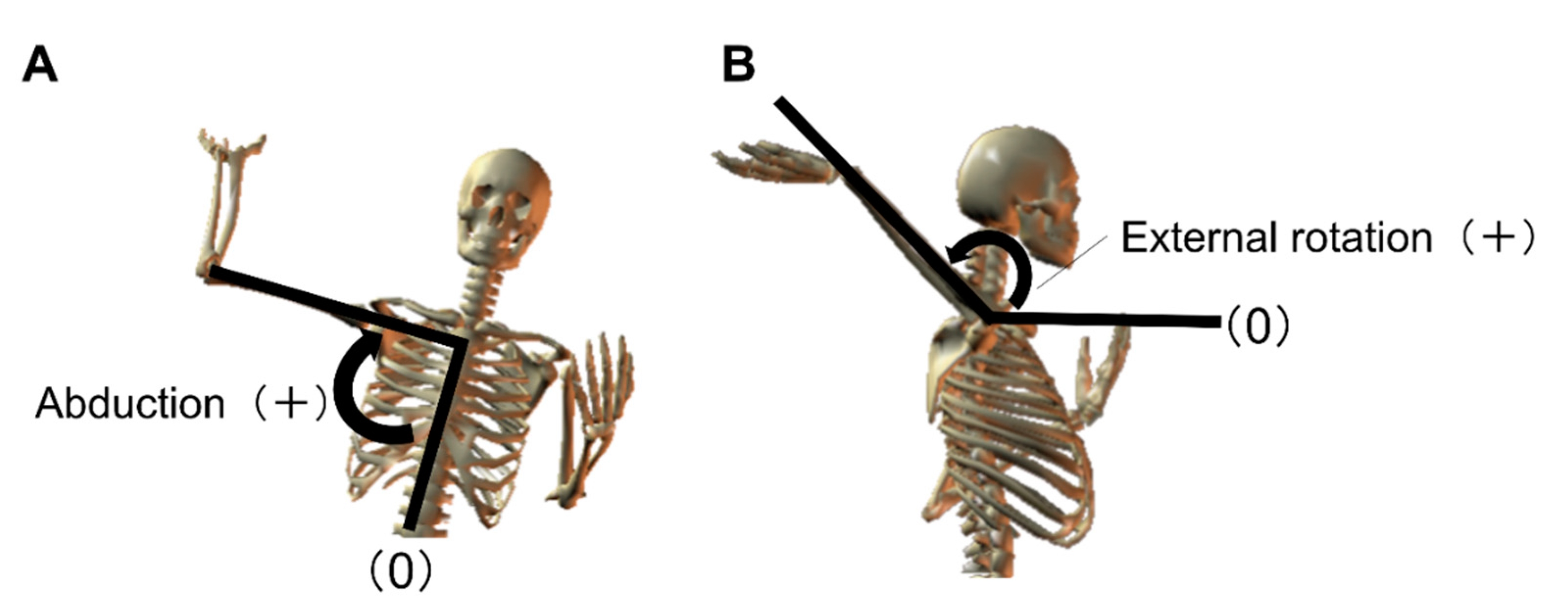
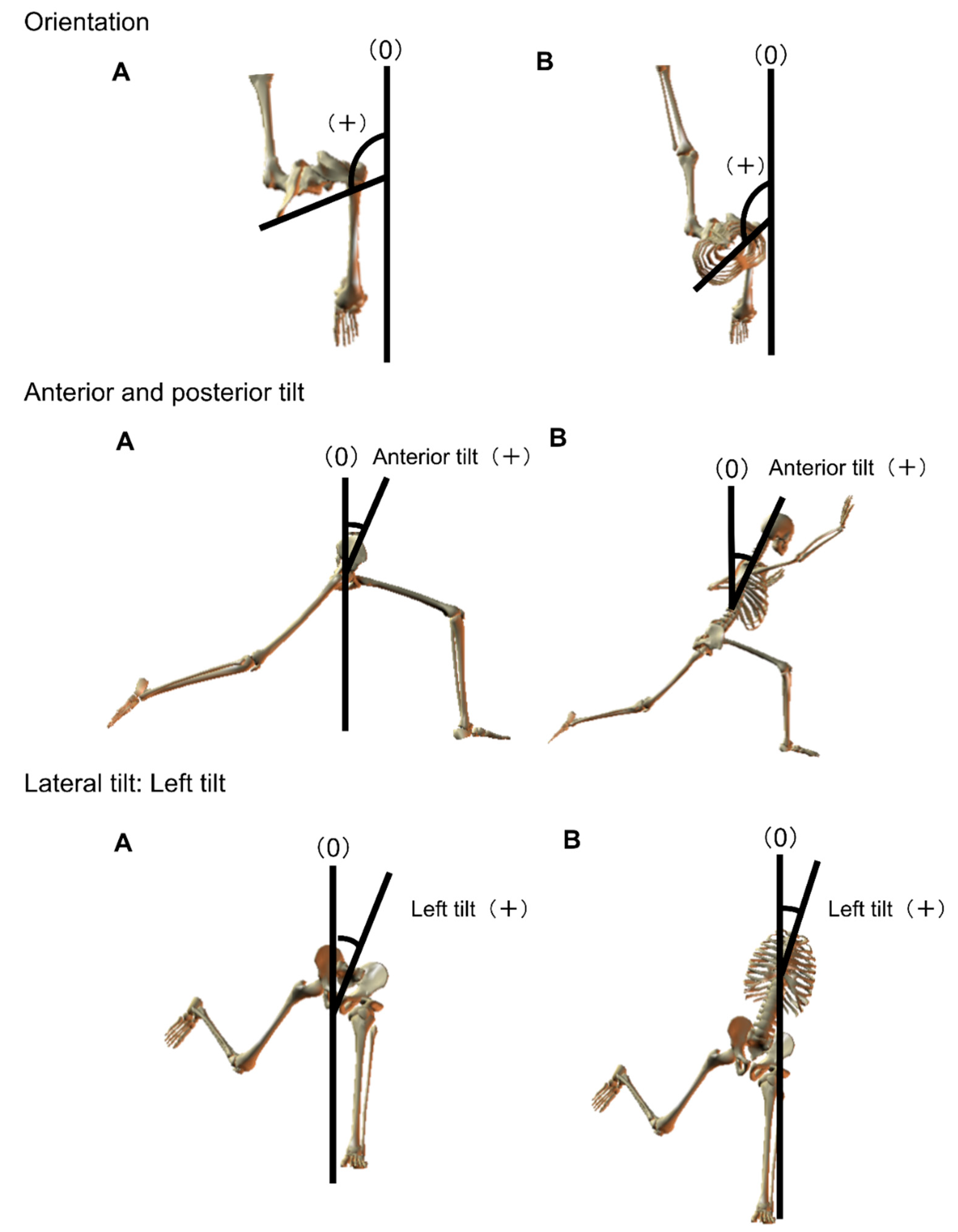
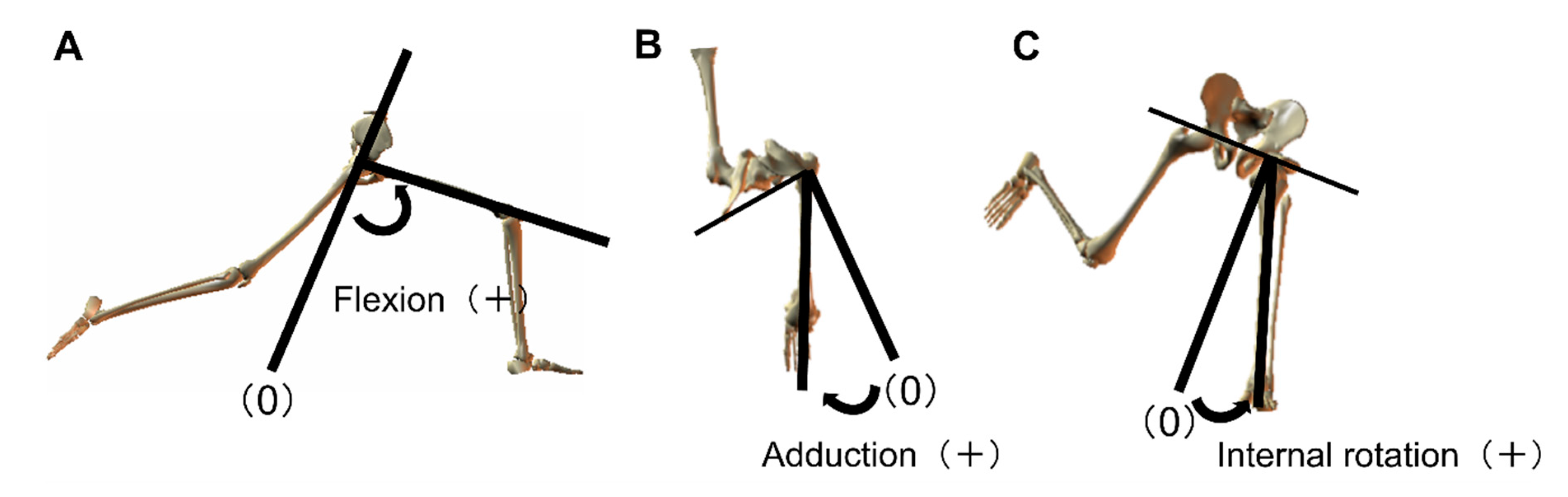
2.4. Definition
2.5. Statistical Analysis
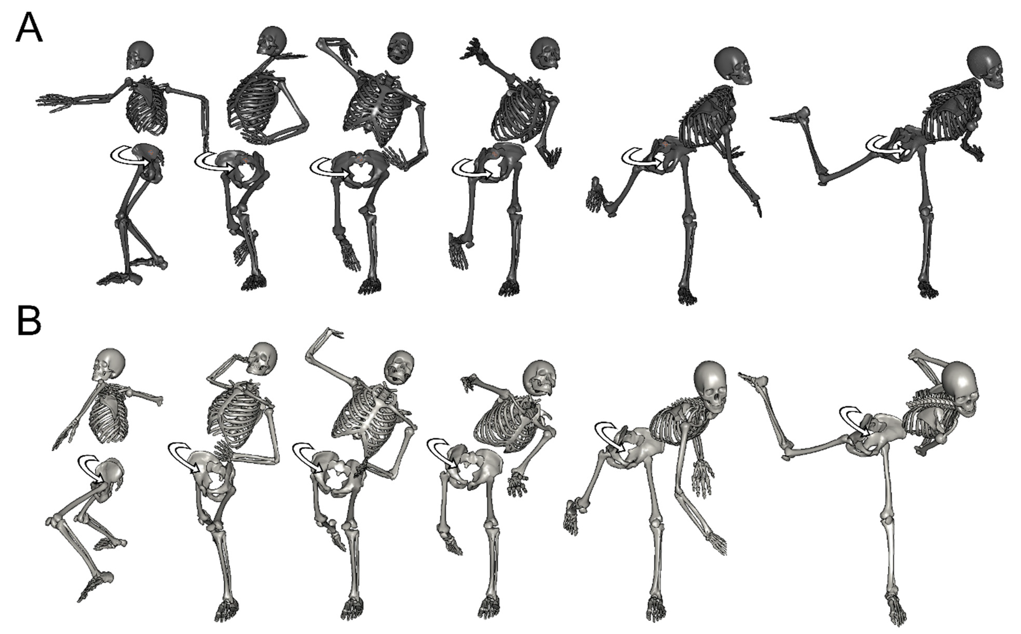
3. Results
4. Discussion
4.1. External Rotation Angle of the Shoulder
4.2. Pelvic and Thorax Orientation Angle
4.3. Anterior or Lateral Tilt Angle of the Pelvis and Thorax
4.4. Transmitting the Energy
5. Conclusions
Author Contributions
Funding
Institutional Review Board Statement
Informed Consent Statement
Data Availability Statement
Conflicts of Interest
References
- Japan High School Girls Baseball Federation. 2021. Available online: http://www.jhgbf.org/jhgbf_kameikou/ (accessed on 10 December 2017).
- Sauers, E.L.; Huxel Bliven, K.C.; Johnson, M.P.; Falsone, S.; Walters, S. Hip and glenohumeral rotational range of motion in healthy professional baseball pitchers and position players. Am. J. Sport. Med. 2014, 42, 430–436. [Google Scholar] [CrossRef] [PubMed]
- Walch, G.; Boileau, P.; Noel, E.; Donell, S.T. Impingement of the deep surface of the supraspinatus tendon on the posterosuperior glenoid rim: An arthroscopic study. J. Shoulder Elb. Surg. 1992, 1, 238–245. [Google Scholar] [CrossRef]
- Stodden, D.F.; Fleisig, G.S.; McLean, S.P.; Lyman, S.L.; Andrews, J.R. Relationship of Pelvis and Upper Torso Kinematics to Pitched Baseball Velocity. J. Appl. Biomech. 2001, 17, 164–172. [Google Scholar] [CrossRef]
- Aragon, V.J.; Oyama, S.; Oliaro, S.M.; Padua, D.A.; Myers, J.B. Trunk-rotation flexibility in collegiate softball players with or without a history of shoulder or elbow injury. J. Athl. Train. 2012, 47, 507–513. [Google Scholar] [CrossRef] [PubMed]
- Matsui, T.; Morihara, T.; Azuma, Y.; Seo, K.; Hiramoto, M.; Kida, Y.; Takashima, M.; Horii, M.; Kubo, T. The prediction of pitching injuries of the shoulder and elbow by comparing the ROM between dominant and non-dominant side on neck/trunk rotations and hip internal rotation. Jpn. J. Phys. Fit. Sports Med. 2013, 62, 223–226. [Google Scholar] [CrossRef][Green Version]
- Nissen, C.W.; Westwell, M.; Ounpuu, S.; Patel, M.; Solomito, M.; Tate, J. A biomechanical comparison of the fastball and curveball in adolescent baseball pitchers. Am. J. Sports Med. 2009, 37, 1492–1498. [Google Scholar] [CrossRef] [PubMed]
- Miyazaki, S.; Ishida, Y.; Totoribe, K.; Kawahara, K.; Chosa, E. Physical movement and characteristics of muscle activity around the shoulder girdle during the throwing motion—Comparison of normal shoulder and throwing injured shoulder. Jpn. J. Clin. Biomech. 2011, 32, 167–172. [Google Scholar]
- Jobe, F.W.; Bradley, J.P. Rotator cuff injuries in baseball. Prevention and rehabilitation. Sports Med. 1988, 6, 378–387. [Google Scholar] [CrossRef] [PubMed]
- Fleisig, G.S.; Barrentine, S.W.; Zheng, N.; Escamilla, R.F.; Andrews, J.R. Kinematic and kinetic comparison of baseball pitching among various levels of development. J. Biomech. 1999, 32, 1371–1375. [Google Scholar] [CrossRef]
- Wu, G.; Siegler, S.; Allard, P.; Kirtley, C.; Leardini, A.; Rosenbaum, D.; Whittle, M.; D’Lima, D.D.; Cristofolini, L.; Witte, H.; et al. ISB recommendation on definitions of joint coordinate system of various joints for the reporting of human joint motion–Part I: Ankle, hip, and spine. Int. Soc. Biomech. J. Biomech. 2002, 35, 543–548. [Google Scholar] [CrossRef]
- Wu, G.; van der Helm, F.C.; Veeger, H.E.; Makhsous, M.; Van Roy, P.; Anglin, C.; Nagels, J.; Karduna, A.R.; McQuade, K.; Wang, X.; et al. ISB recommendation on definitions of joint coordinate systems of various joints for the reporting of human joint motion—Part II: Shoulder, elbow, wrist and hand. J. Biomech. 2005, 38, 981–992. [Google Scholar] [CrossRef] [PubMed]
- Hellmann, F.; Verdi, M.; Schlemper, B.R., Jr.; Caponi, S. 50th anniversary of the Declaration of Helsinki: The double standard was introduced. Arch. Med. Res. 2014, 45, 600–601. [Google Scholar] [CrossRef] [PubMed]
- Dun, S.; Fleisig, G.S.; Loftice, J.; Kingsley, D.; Andrews, J.R. The relationship between age and baseball pitching kinematics in professional baseball pitchers. J. Biomech. 2007, 40, 265–270. [Google Scholar] [CrossRef] [PubMed]
- Aguinaldo, A.L.; Chambers, H. Correlation of throwing mechanics with elbow valgus load in adult baseball pitchers. Am. J. Sports Med. 2009, 37, 2043–2048. [Google Scholar] [CrossRef] [PubMed]
- Ito, H.; Nakazato, K.; Watarai, K.; Nakajima, H. The role of trunk movement during throwing motion—Trunk movement and throwing injury of the upper extremity. J. Jpn. Soc. Clin. Sports Med. 2001, 9, 332–339. [Google Scholar]
- Seo, K.; Morihara, T.; Matsui, T.; Azuma, Y.; Hiramoto, M.; Kida, Y.; Yamada, Y.; Nakamura, Y.; Houjou, T.; Morii, M.; et al. Three-dimensional motion analysis in high school pitchers focused on the dominant lower extremity. J. Jpn. Soc. Clin. Sports Med. 2013, 21, 618–622. [Google Scholar]


| Female Professional Baseball Pitchers (Mean ± SD, N = 15) | Male University Baseball Pitchers (Mean ± SD, N = 14) | p-Value | |
|---|---|---|---|
| Age (years) | 21.7 ± 3.2 | 19.9 ± 0.8 | 0.06 |
| Height (cm) | 162.5 ± 5.1 | 176.4 ± 3.0 | 0.01 * |
| Body mass (Kg) | 59.0 ± 6.6 | 73.1 ± 3.0 | 0.00 * |
| Years of baseball experience | 14.5 ± 3.2 | 11.6 ± 2.1 | 0.01 * |
| Years of experience as a pitcher | 12.4 ± 3.5 | 8.6 ± 3.1 | 0.01 * |
| Female Professional Baseball Pitchers | Male University Baseball Pitchers | |||
|---|---|---|---|---|
| MER | BR | MER | BR | |
| Abduction (°) | 94.8 ± 8.3 | 92.5 ± 8.5 | 100.1 ± 11.0 | 96.4 ± 8.9 |
| External rotation (°) | 161.8 ± 12.4 | 129.8 ± 13.3 | 166.0 ± 13.5 | 132.9 ± 12.2 |
| Female Professional Baseball Pitchers | Male University Baseball Pitchers | |||
|---|---|---|---|---|
| MER | BR | MER | BR | |
| Orientation | ||||
| Pelvis (°) | 93.7 ± 4.7 | 99.5 ± 5.4 | 85.4 ± 5.4 | 92.4 ± 5.8 |
| Thorax (°) | 102.9 ± 6.7 | 114.4 ± 6.3 | 93.7 ± 6.3 | 104.9 ± 7.3 |
| Thorax-pelvis (°) | 9.3 ± 5.9 | 14.8 ± 5.8 | 8.3 ± 7.1 | 12.5 ± 6.8 |
| Female Professional Baseball Pitchers | Male University Baseball Pitchers | |||
|---|---|---|---|---|
| MER | BR | MER | BR | |
| Anterior and posterior tilt | ||||
| Pelvis (°) | 35.6 ± 5.5 | 36.7 ± 5.9 | 37.4 ± 14.2 | 37.4 ± 14.7 |
| Thorax (°) | 17.0 ± 5.1 | 26.5 ± 5.1 | 24.0 ± 9.2 | 33.1 ± 8.6 |
| Thorax-pelvis (°) | −18.7 ± 8.2 | −10.2 ± 7.9 | −13.3 ± 16.6 | −4.3 ± 17.2 |
| Lateral tilt | ||||
| Pelvis (°) | 1.2 ± 5.2 | 1.6 ± 4.6 | 8.8 ± 6.0 | 9.6 ± 6.7 |
| Thorax (°) | 24.4 ± 6.5 | 28.5 ± 7.9 | 30.4 ± 6.5 | 33.3 ± 6.6 |
| Thorax-pelvis (°) | 23.2 ± 8.9 | 26.9 ± 10.3 | 21.5 ± 7.7 | 23.8 ± 8.1 |
| Female Professional Baseball Pitchers | Male University Baseball Pitchers | |||
|---|---|---|---|---|
| MER | BR | MER | BR | |
| Flexion (°) | 99.4 ± 10.3 | 98.2 ± 10.5 | 105.3 ± 18.1 | 101.4 ± 19.0 |
| Adduction (°) | 7.2 ± 6.9 | 10.5 ± 7.1 | −4.7 ± 5.1 | −0.6 ± 4.6 |
| Internal rotation (°) | 19.1 ± 11.4 | 17.5 ± 12.0 | 32.7 ± 11.5 | 31.6 ± 13.2 |
Publisher’s Note: MDPI stays neutral with regard to jurisdictional claims in published maps and institutional affiliations. |
© 2021 by the authors. Licensee MDPI, Basel, Switzerland. This article is an open access article distributed under the terms and conditions of the Creative Commons Attribution (CC BY) license (https://creativecommons.org/licenses/by/4.0/).
Share and Cite
Azuma, Y.; Matsui, T.; Hiramoto, M.; Hashimoto, R.; Matsuzawa, K.; Miyazaki, T.; Seo, K.; Watanabe, Y.; Kida, N.; Kai, Y.; et al. Motion Analysis Focusing on Rotational Movements of Professional Female Baseball Pitchers: Comparison with Male University Baseball Pitchers. Int. J. Environ. Res. Public Health 2021, 18, 13342. https://doi.org/10.3390/ijerph182413342
Azuma Y, Matsui T, Hiramoto M, Hashimoto R, Matsuzawa K, Miyazaki T, Seo K, Watanabe Y, Kida N, Kai Y, et al. Motion Analysis Focusing on Rotational Movements of Professional Female Baseball Pitchers: Comparison with Male University Baseball Pitchers. International Journal of Environmental Research and Public Health. 2021; 18(24):13342. https://doi.org/10.3390/ijerph182413342
Chicago/Turabian StyleAzuma, Yoshikazu, Tomoyuki Matsui, Machiko Hiramoto, Ruo Hashimoto, Kanta Matsuzawa, Tetsuya Miyazaki, Kazuya Seo, Yuya Watanabe, Noriyuki Kida, Yoshihiro Kai, and et al. 2021. "Motion Analysis Focusing on Rotational Movements of Professional Female Baseball Pitchers: Comparison with Male University Baseball Pitchers" International Journal of Environmental Research and Public Health 18, no. 24: 13342. https://doi.org/10.3390/ijerph182413342
APA StyleAzuma, Y., Matsui, T., Hiramoto, M., Hashimoto, R., Matsuzawa, K., Miyazaki, T., Seo, K., Watanabe, Y., Kida, N., Kai, Y., & Morihara, T. (2021). Motion Analysis Focusing on Rotational Movements of Professional Female Baseball Pitchers: Comparison with Male University Baseball Pitchers. International Journal of Environmental Research and Public Health, 18(24), 13342. https://doi.org/10.3390/ijerph182413342