Article

Effect of Different Exercise Types on the Cross-Sectional Area and Lumbar Lordosis Angle in Patients with Flat Back Syndrome

1
Department of Sports Science, Dongguk University, 123, Dongdae-ro, Gyeongju-si 38066, Korea
2
Division of Sports Medicine, Department of Orthopedic Surgery, Samsung Medical Center, 81, Irwon-ro, Gangnam-gu, Seoul 06351, Korea
3
Department of Health Rehabilitation, Osan University, 45, Cheonghak-ro, Osan-si 18119, Korea
4
Sports Medicine Center, Sunsoochon Hospital, 76, Olympic-ro, Songpa-gu, Seoul 05556, Korea
5
Department of Sports Science, Hanyang University, 55, Hanyang Daehak-ro, Sangnok-gu, Ansan-si 15588, Korea
*
Author to whom correspondence should be addressed.
The author (Yong-Gon Seo) contributed equally to write this work with first author (Won-Moon Kim).
Int. J. Environ. Res. Public Health 2021, 18(20), 10923; https://doi.org/10.3390/ijerph182010923
Submission received: 20 September 2021 / Revised: 4 October 2021 / Accepted: 14 October 2021 / Published: 17 October 2021
(This article belongs to the Special Issue Exercise Physiology and Performance)

Abstract

:
Flat back syndrome (FBS) is a sagittal imbalance wherein the normal spinal curvature is reduced. This study aimed to compare the effects of different exercise programs on the cross-sectional area (CSA) of the lumbar muscles, lumbar lordosis angle (LLA), lumbar disability, and flexibility in patients with FBS. Thirty-six females with flexible FBS were randomly allocated to the corrective exercise group (CEG, n = 12), resistance exercise group (REG, n = 12), and physical therapy group (PTG, n = 12). CEG and REG patients participated in a 12-week exercise intervention for 60 min three times per week. The CSA, LLA, Oswestry disability index (ODI), and sit-and-reach test were measured before and after intervention. CSA showed a significant difference between groups (p < 0.01), with CEG and REG demonstrating a significant increase (p < 0.05 and p < 0.05, respectively). LLA showed a significant difference between groups (p < 0.001); CEG showed a higher increase than did REG (p < 0.01) and PTG (p < 0.001). ODI also showed a significant difference between groups (p < 0.001), being lower in CEG than in REG (p < 0.001) and PTG (p < 0.001). Lumbar flexibility significantly improved in all groups, albeit with a significant difference (p < 0.001). Although corrective and resistance exercise programs effectively improve these parameters, corrective exercise is superior to other interventions for patients with FBS.

1. Introduction

Flat back syndrome (FBS) is a type of sagittal imbalance in the spine, and it is characterized by loss of the lordotic curve [1]. Although the pathogenic mechanism remains unclear, poor posture and lack of exercise are major causes of spinal deformity [2,3]. The normal curve of the spine is known to have a buffering effect against gravity and provides optimal, coordinated body movements [4].
An abnormal spinal curvature is associated with musculoskeletal and nervous systems problems [5]. Long-standing spinal imbalance is known to cause scoliosis, pelvic malalignment syndrome, tension-type headache, and nerve entrapment syndrome [6]. Therefore, it is very important to keep the optimal curve of the spine, and intensive intervention is required for abnormal alignment in patients with FBS.
Various interventions, including brace wearing, surgical correction, and exercise therapy, have been used to correct an abnormal spinal curvature. In the past, a surgical approach was mainly used to correct the spinal curve in patients with FBS [2]. The intervention for correcting the spinal curvature is influenced by various factors such as the patient’s age, sex, stage of spinal disease, and etiology of the disease [7].
Several studies have reported that exercise therapy as a nonsurgical approach can improve an abnormal spinal curvature in FBS [3,5,8]. A previous study [9] reported a significant improvement in the curvature of the cervical, thoracic, and lumbar vertebrae after exercise intervention. Another study conducted by Harrison and Oakley [1] reported a significant change in the spinal alignment and pain scale score after the lumbar renal traction method and manual therapy. However, most of the studies have focused on parameters such as the spinal curve, function, pain, and muscle strength, and few have compared different exercise programs for FBS.
Therefore, this study aimed to investigate the effects of different exercise programs on the cross-sectional area (CSA) of the lumbar muscle, the lumbar lordosis angle (LLA), lumbar function, and flexibility in patients with FBS.

2. Materials and Methods

2.1. Study Design

The G-power program (Heinrich Heine University Düsseldorf, Düsseldorf, Ger-many, version 3.1.9.4) [10] was used to determine the sample size using the effect size = 0.25, α = 0.05, power = 0.80%, the number of groups = 3, and the number of measurements = 2. The minimal sample size was calculated as 42. Therefore, 42 patients with diagnosed with FBS were enrolled, but six patients refused to participate due to personal reasons before starting this study. Finally, thirty-six female patients with flexible FBS participated in this study. The inclusion criteria were as follows: no improvement in symptoms including back pain for the past 3 months despite drug therapy, a thoracic kyphosis angle ≤30° of Cobb’s angle [9], and moderate disability with back pain (over 3 in numeric pain rating scale) according to the Oswestry disability index (ODI) (21–40%). Patients were excluded from the study if they had previously other spinal disease or had other musculoskeletal disease or had undergone spine surgery.
The participants were randomly assigned into a corrective exercise group (CEG, n = 12), a resistance exercise group (REG, n = 12), and a physical therapy group (PTG, n = 12) according to the order of enrollment in the study. All patients received sufficient explanations regarding the study and subsequently signed an informed consent form before participation. This study was conducted according to the guidelines of the Declaration of Helsinki, and all procedures involving patients were approved by the Ethics Committee of the relevant institution (IRB No. DGU-20200027).

2.2. Outcome Assessments

All assessments in this study were conducted before and after exercise intervention with the same method.

2.3. Cobb’s Angle

The LLA was evaluated using lateral radiographs (REX-52R; Listem, Donghwari, Korea) obtained with the patient standing. Cobb’s angle [11] was measured as the angle between the extended line along the upper border of L1 and the extended line along the lower border of L5 (Figure 1).

2.4. Cross-Sectional Area

The CSA of the lumbar muscle was measured using computed tomography (CT) (Sytec-Sri; GE, Boston, MA, USA). One radiologist evaluated all the images and measurements to ensure reliability. The patients laid in the supine position with the knee joint flexed at 25°–30°, and images were acquired for a total of 2 to 4 min with maintenance of a natural breathing pattern. CT parameters to acquire images of the CSA of the lower border of the fourth lumbar vertebral body were as follows: voltage, 120 kV (peak); current, 240 mA/s; thickness, 5 mm; and field of view, 35–40 cm. On the monitor of the picture archiving and communication system, the CSA (cm2) of the paraspinal lumbar muscles including longissimus, iliocostalis, and multifidus was analyzed and calculated (Figure 2).

2.5. Oswestry Disability Index

The ODI is a scale designed to measure disability in daily life caused by lower back pain. The scale consists of 10 items, including pain intensity, personal care, lifting objects, sitting, walking, sleeping, standing, sexual activity, travel, and social life. Each item is scored from 0 to 5, with 0 representing no discomfort and 5 representing the most severe discomfort. A higher score represents more severe disability [12].

2.6. Sit-and-Reach Test

The sit-and-reach test was conducted using the DWR-OT1038 device (Dawoori, Anseong-si, Korea). The patients sat with their legs together, knees extended, and upper body erect. With their hands placed side by side or overlapping, and while exhaling and bending their upper body forward, the patients reached forward with their arms as far as possible and pushed an indicator. The participant was instructed to maintain the farthest position for 3 s, and the examiner recorded the position of the indicator at this point [13].

2.7. Exercise Programs

In this study, corrective and resistance exercises were performed by two groups, whereas the third group received only physical therapy. The corrective exercise program was based on the principles of the Schroth exercises and focused on correction of thoracolumbar spine. The program was revised by referring to the findings of previous studies [14,15]. The exercise program was conducted for 60 min three times per week for 12 weeks. The exercise intensity was prescribed according to a rating of perceived exertion (RPE) of 13 with “somewhat hard” to 15 with “hard”. The resting time between each exercise set was 60 s. The detailed corrective exercise program is summarized in Table 1.
The resistance exercise program was consisted of trunk exercise and whole body muscle strength exercise, and the whole body exercise was performed with elastic resistance bands (Performance Health, Akron, OH, USA). This program was modified from previous studies [16,17]. The initial exercise intensity in elastic resistance bands was set at 30–40% of one-repetition maximum and was progressively increased up to 60–70% during the intervention. The resting time between each set was 60 s. Details of the resistance exercise program are summarized in Table 2.
Physical therapy was conducted by a physical therapist for 60 min. It included 20 min of ultrasound therapy at a frequency of 1 MHz and an intensity of 1.6 W/cm2, 20 min of interferential current therapy, and 20 min of hot pack application in the thoracolumbar spine region.

2.8. Statistical Analysis

The Kolmogorov–Smirnov test was used to confirm normal distribution of the data. Means and standard deviations were used for descriptive statistics of all parameters. One-way analysis of variance was used to determine the differences in clinical outcomes among the three groups. Post-hoc analysis was performed when there was a significant difference between groups. All statistical analyses were performed using SPSS Statistics version 22.0 (IBM, Armonk, NY, USA), and the level of significance was set at p < 0.05.

3. Results

Thirty-six patients with FBS participated in this study, and there were no significant differences in the baseline characteristics of the three groups (Table 3).
With regard to the CSA of the lumbar muscle, there was a significant difference among the three groups (p < 0.01). Post hoc analysis showed a significant increase in the CEG and REG, relative to that in the PTG (p < 0.05, p < 0.05, respectively). The LLA also showed a significant difference among groups (p < 0.001), with a significantly higher value in the CEG than in the REG (p < 0.01) and PTG (p < 0.001). In addition, the REG showed a more significant improvement than did the PTG (p < 0.001).
The ODI was also significantly different among groups (p < 0.001). Post hoc analysis showed that the ODI was significantly lower in the CEG than in the REG (p < 0.001) and PTG (p < 0.001), while it was significantly lower in the REG than in the PTG (p < 0.001).
Changes in flexibility were significantly different among groups (p < 0.001). Post hoc analysis revealed that the CEG and REG showed a more significant increase than did the PTG (p < 0.001, p < 0.001, respectively). There were significant improvements in the CSA, the LLA, the ODI, and flexibility after the interventions in the CEG (p < 0.001) and REG (p < 0.001), whereas there was no significant improvement in any parameter except flexibility (p < 0.05) in the PTG (CSA, p = 0.725; LLA, p = 0.491; ODI, p = 0.136, respectively; Table 4).

4. Discussion

Flat back syndrome is associated with malalignment of the spinal curve, resulting in a forward head posture and lower back pain [18]. This study compared the effects of different exercise types on the CSA, the LLA, the ODI, and flexibility of the spine and found that compared with physical therapy, both corrective and resistance exercises are effective in improving these parameters in patients with FBS.
The deep lumbar muscles play an important role in maintaining control and stability of the spinal column [19]. The presence of a flat back is associated with malalignment in the spine, which could cause dysfunction of the deep lumbar muscles and result in chronic low back pain and deep muscle atrophy [20,21]. In the present study, the CSA of the lumbar muscles showed a more significant increase in the CEG and REG than in the PTG. This result is similar to the findings of Cho et al. [22], who demonstrated that lumbar extension exercises can increase the CSA of the deep paraspinal muscles, including the longissimus, iliocostalis, spinalis, and multifidus. This suggests that improvement of the lordotic curve in the lumbar region after exercise intervention contributes to a decrease in the overload on the lumbar vertebrae and increases the lumbar muscle activity, resulting in an increase in the CSA of the lumbar muscles. This mechanism is supported by previous studies reporting the positive effects of the Schroth and mobilization exercises on muscle activity [19,23]. Further study is needed to confirm whether corrective exercise or resistance exercise would be more beneficial in terms of an increase in the CSA of the lumbar muscle in patients with FBS.
The ideal curvature of the spine in the sagittal plane serves to reduce loads on the vertebral discs and any shock to the spine, and it allows effective action of the spinal muscles [24]. An abnormal LLA caused by thoracolumbar kyphosis can have negative effects on the overall biomechanics of the spine [25]. Decreased thoracolumbar motion is associated with the range of motion of the lumbar spine, and it can reduce lumbar lordosis [26]. In the present study, corrective and resistance exercises resulted in greater improvements in the LLA than did physical therapy alone. However, the corrective exercise program was the most effective in improving lumbar lordosis. Several studies have reported that corrective exercise programs improved the LLA [22,27]. Particularly, several exercise modes, including mobilization, lumbar stabilization exercise, and the Schroth exercises are beneficial interventions to improve the LLA [23,28]. We speculate that the corrective exercises applied in this study, which comprised mobilization and the Schroth exercises, may have improved the LLA in patients with FBS.
Several studies [28,29,30] have reported that lumbar stabilization exercises reduce lumbar functional disability measured using the ODI in patients with chronic low back pain. In this study, the CEG and REG showed a lower ODI score than did the PTG, and the corrective exercise program, which included lumbar stabilization exercises, was superior to the other interventions for patients with FBS. One previous study [31] reported that flat back syndrome can be associated with functional limitations after surgical intervention. Most previous studies on lumbar functional disability have focused on patients with chronic low back pain or flat back who undergo surgery. To the best of our knowledge, this is the first study evaluating functional disability in patients with functional flat back. Further studies with larger samples are required to confirm our findings.
In a previous study, hamstring flexibility was significantly lesser in patients with low back pain than in healthy patients [32]. Tightness of the hamstring muscle is associated with the lumbar spinal curve, and it results in loss of the lordotic curve in the lumbar region [33,34]. Our results indicated that exercise interventions and physical therapy improved the flexibility of the hamstring and erector spinae muscles in patients with FBS, with the CEG and REG showing a greater increase in flexibility. Thus, corrective and resistance exercises could be beneficial in terms of improved flexibility in patients with FBS. Further research can confirm the mechanism underlying the improved flexibility.
This study has some limitations. First, all the participants were female, and our results may not be applicable to all populations, particularly because of differences in the prevalence of FBS between women and men. Therefore, further studies should confirm differences in these results between male and female patients. Second, the sample size was small, even though the patients were divided into three groups. A larger sample is necessary to validate our findings.

5. Conclusions

Corrective and resistance exercises are both effective in improving the CSA of the lumbar muscles, the LLA, the ODI, and flexibility, with better effects than those of simple physical therapy. However, corrective exercise programs seem to be the most appropriate intervention for patients with FBS.

Author Contributions

Conceptualization, W.-M.K. and Y.-G.S.; methodology, Y.-G.S., Y.-J.P. and H.-S.C.; investigation, W.-M.K. and C.-H.L.; writing—original draft, W.-M.K. and Y.-G.S.; writing—review and editing, Y.-G.S. All authors have read and agreed to the published version of the manuscript.

Funding

This research received no external funding.

Institutional Review Board Statement

The study was conducted according to the guidelines of the Declaration of Helsinki, and approved by the Institutional Review Board of Dongguk University institutional review board (DGU IRB 20200027).

Informed Consent Statement

Informed consent was obtained from all subjects involved in the study.

Data Availability Statement

No new data were created or analyzed in this study. Data sharing is not applicable to this article.

Conflicts of Interest

The authors declare no conflict of interest.

References

  1. Harrison, D.E.; Oakley, P.A. Non-operative corrective of flat back syndrome using lumbar extension traction: A CBP® case series of two. J. Phys. Ther. Sci. 2018, 30, 1131–1137. [Google Scholar] [CrossRef] [PubMed] [Green Version]
  2. Farcy, J.P.; Schwab, F.J. Management of flatback and related kyphotic decompensation syndromes. Spine 1997, 22, 2452–2457. [Google Scholar] [CrossRef] [PubMed]
  3. Vaughn, D.W.; Brown, E.W. The influence of an in-home based therapeutic exercise program on thoracic kyphosis angles. J. Back Musculoskelet Rehabil. 2007, 20, 155–165. [Google Scholar] [CrossRef] [Green Version]
  4. Lau, K.T.; Cheung, K.Y.; Chan, K.B.; Chan, M.H.; Lo, K.Y.; Chiu, T.T. Relationships between sagittal postures of thoracic and cervical spine, presence of neck pain, neck pain severity and disability. Man Ther. 2010, 15, 457–462. [Google Scholar] [CrossRef] [PubMed]
  5. Kamaz, M.; Kıreşi, D.; Oğuz, H.; Emlik, D.; Levendoğlu, F. CT measurement of trunk muscle areas in patients with chronic low back pain. Diagn. Interv. Radiol. 2007, 13, 144–148. [Google Scholar]
  6. Klein, R.G.; Eek, B.C.; DeLong, W.B.; Mooney, V. A randomized double-blind trial of dextrose-glycerine-phenol injections for chronic, low back pain. J. Spinal Disord. 1993, 6, 23–33. [Google Scholar] [CrossRef]
  7. Bautmans, L.; Van Arken, J.; Van Mackelenberg, M.; Mets, T. Rehabilitation using manual mobilization for thoracic kyphosis in elderly postmenopausal patients with osteoporosis. J. Rehabil. Med. 2010, 42, 129–135. [Google Scholar] [CrossRef] [PubMed] [Green Version]
  8. Giglio, C.A.; Volpon, J.B. Development and evaluation of thoracic kyphosis and lumbar lordosis during growth. J. Child. Orthop. 2007, 1, 187–193. [Google Scholar] [CrossRef] [Green Version]
  9. Cho, H.S.; Kim, C.H. The effects of customized spinal corrective exercise program on the spinal curvature change and posture balance ability for flat back syndrome. J. Korean Soc. Wellness 2020, 15, 409–417. [Google Scholar] [CrossRef]
  10. Faul, F.; Erdfelder, E.; Buchner, A.; Lang, A.G. Statistical power analyses using G*Power 3.1: Tests for correlation and regression analyses. Behav. Res. Methods 2009, 41, 1149–1160. [Google Scholar] [CrossRef] [Green Version]
  11. Cobb, J. Outline for the study of scoliosis. Instr. Course Lect. 1947, 5, 261–275. [Google Scholar]
  12. Fairbank, J.C.; Pynsent, P.B. The Oswestry Disability Index. Spine 2000, 25, 2940–2953. [Google Scholar] [CrossRef]
  13. French, G.; Grayson, C.; Sanders, L.; William, T.; Willard, M. A comparative analysis of the traditional sit-and-reach test and the R.S. Smith sit-and-reach design. Corinth. J. Stud. Res. Ga. Coll. 2016, 17, 74–80. [Google Scholar]
  14. Negrini, S.; Fusco, C.; Minozzi, S.; Atanasio, S.; Zaina, F.; Romano, M. Exercises reduce the progression rate of adolescent idiopathic scoliosis: Results of a comprehensive systematic review of the literature. Disabil. Rehabil. 2008, 30, 772–785. [Google Scholar] [CrossRef]
  15. Yoo, W.G. Effect of individual strengthening exercises for anterior pelvic tilt muscles on back pain, pelvic angle, and lumbar ROMs of a LBP patient with flat back. J. Phys. Ther. Sci. 2013, 25, 1357–1358. [Google Scholar] [CrossRef] [PubMed] [Green Version]
  16. Iversen, V.M.; Vasseljen, O.; Mork, P.J.; Berthelsen, I.R.; Børke, J.B.; Berheussen, G.F.; Tveter, A.T.; Salvesen, Ø.; Fimland, M.S. Resistance training in addition to multidisciplinary rehabilitation for patients with chronic pain in the low back: Study protocol. Contemp. Clin. Trials Commun. 2017, 6, 115–121. [Google Scholar] [CrossRef] [PubMed]
  17. Iversen, V.M.; Vasseljen, O.; Mork, P.J.; Gismervik, S.; Bertheussen, G.F.; Salvesen, Ø.; Fimland, M.S. Resistance band training or general exercise in multidisciplinary rehabilitation of low back pain? A randomized trial. Scand. J. Med. Sci. Sports 2018, 28, 2074–2083. [Google Scholar] [CrossRef] [PubMed] [Green Version]
  18. Quek, J.; Pua, Y.H.; Clark, R.A.; Bryant, A.L. Effects of thoracic kyphosis and forward head posture on cervical range of motion in older adults. Man Ther. 2013, 18, 65–71. [Google Scholar] [CrossRef] [PubMed]
  19. Park, S.Y.; Shim, J.H. Effect of 8 weeks of Schroth exercise (three-dimensional convergence exercise) on pulmonary function, Cobb’s angle, and erector spinae muscle activity in idiopathic scoliosis. J. Korea Converg. Soc. 2014, 5, 61–68. [Google Scholar] [CrossRef] [Green Version]
  20. Kim, K.T.; Lee, J.H. Sagittal imbalance. J. Korean Soc. Spine Surg 2009, 16, 142–151. [Google Scholar] [CrossRef]
  21. Fortin, M.; Macedo, L.G. Multifidus and paraspinal muscle group cross-sectional areas of patients with low back pain and control patients: A systematic review with a focus on blinding. Phys. Ther. 2013, 93, 873–888. [Google Scholar] [CrossRef]
  22. Cho, J.H.; Lee, K.H.; Lim, S.T.; Chun, B.O. Comparison of muscle cross-sectional area and lumbar muscle strength according to degenerative spinal diseases. Asian J. Kinesiol. 2020, 22, 1–10. [Google Scholar] [CrossRef]
  23. Lehnert-Schroth, C. Three-Dimensional Treatment for Scoliosis: Physiotherapeutic Method for Deformities of the Spine; Martindale Press: Palo Alto, CA, USA, 2007. [Google Scholar]
  24. Kim, W.J.; Song, D.G.; Lee, J.W.; Kang, J.W.; Park, K.Y.; Koo, J.Y.; Kwon, W.C.; Choy, W.S. Proximal junctional problems in surgical treatment of lumbar degenerative sagittal imbalance patients and relevant risk factors. J. Korean Soc. Spine Surg. 2013, 20, 156–162. [Google Scholar] [CrossRef] [Green Version]
  25. Lee, C.S.; Kang, S.S. Spino-pelvic parameters in adult spinal deformities. J. Korean Orthop. Assoc. 2016, 51, 9–29. [Google Scholar] [CrossRef]
  26. Haussler, K.K. Anatomy of the thoracolumbar vertebral region. Vet. Clin. N. Am. Equine Pract. 1999, 15, 13–26. [Google Scholar] [CrossRef]
  27. Cho, I.; Jeon, C.; Lee, S.; Lee, D.; Hwangbo, G. Effects of lumbar stabilization exercise on functional disability and lumbar lordosis angle in patients with chronic low back pain. J. Phys. Ther. Sci. 2015, 27, 1983–1985. [Google Scholar] [CrossRef] [Green Version]
  28. Parveen, A.; Nuhmani, S.; Hussain, M.E.; Khan, M.H. Effect of lumbar stabilization exercises and thoracic mobilization with strengthening exercises on pain level, thoracic kyphosis, and functional disability in chronic low back pain. J. Complement. Integr. Med. 2020, 18, 419–424. [Google Scholar] [CrossRef]
  29. Suh, J.H.; Kim, H.; Jung, G.P.; Ko, J.Y.; Ryu, J.S.; Kang, H. The effect of lumbar stabilization and walking exercises on chronic low back pain. A randomized controlled trial. Medicine 2019, 98, e16173. [Google Scholar] [CrossRef] [PubMed]
  30. França, F.R.; Burke, T.N.; Hanada, E.S.; Marques, A.P. Segmental stabilization and muscular strengthening in chronic low back pain—a comparative study. Clinics 2010, 65, 1013–1017. [Google Scholar] [CrossRef] [Green Version]
  31. Choi, J.H.; Jang, J.S.; Yoo, K.S.; Shin, J.M.; Jang, I.T. Functional limitations due to stiffness after long-level spinal instrumented fusion surgery to correct lumbar degenerative flat back. Spine 2018, 43, 1044–1051. [Google Scholar] [CrossRef]
  32. Hasarangi, L.; Jayawardana, D.G. Comparison of hamstring flexibility between patients with chronic lower back pain and the healthy individuals at the National Hospital of Sri Lanka. Biomed. J. Sci. Tech. Res. 2018, 5, 4410–4413. [Google Scholar] [CrossRef]
  33. Stokes, I.A.; Abery, J.M. Influence of the hamstring muscles on lumbar spine curvature in sitting. Spine 1980, 5, 525–528. [Google Scholar] [CrossRef] [PubMed]
  34. McCarthy, J.J.; Betz, R.R. The relationship between tight hamstring and lumbar hypolordosis in children with cerebral palsy. Spine 2000, 25, 211–213. [Google Scholar] [CrossRef] [PubMed]
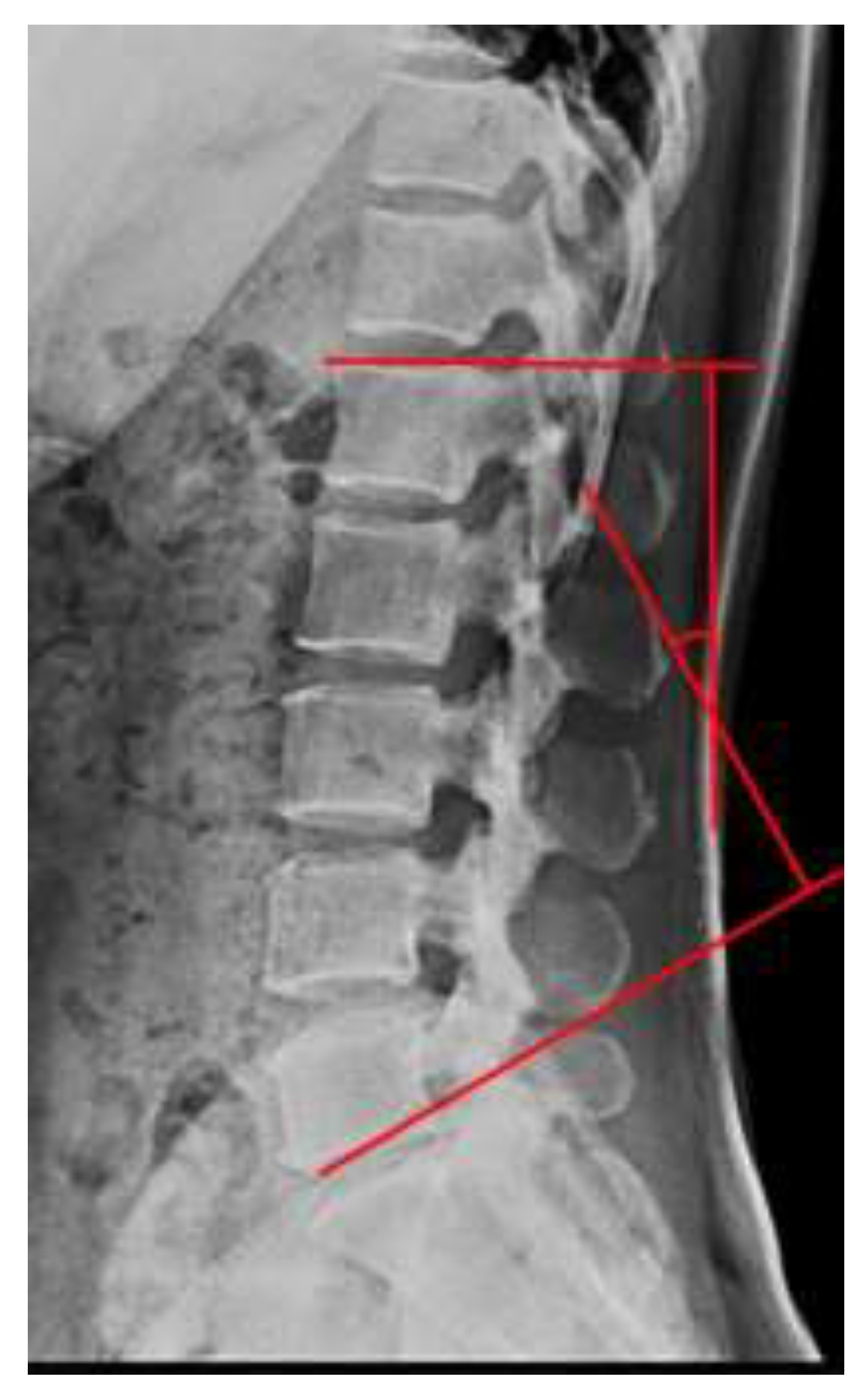
Figure 1. X-ray measurement of the lumbar lordosis angle (Cobb’s method).
Ijerph 18 10923 g001
Figure 2. Computed tomography measurement of the cross-sectional area of the paraspinal lumbar muscle.
Ijerph 18 10923 g002
Table 1. The corrective exercise program.
Corrective Exercise Program
Exercise TypeExercise ModeTimeIntensity
Warm-upStretching for upper and lower body5 minRPE
(10–13)
Mobilization
exercise for
correction
Anterior/posterior pelvis exercises using the Schroth breathing pattern30 minRPE
(13–15)
15–20 Reps
3 Sets
Thoracolumbar spine mobilization exercises using the Schroth breathing pattern
Lumbosacral spine mobilization exercises using the Schroth breathing pattern
Thoracic kyphosis mobilization exercises using the Schroth breathing pattern
Thoracolumbar lordosis mobilization exercises using the Schroth breathing pattern
Corrective
exercise for the thoracolumbar spine
Thoracolumbar corrective exercises using an exercise ball30 minRPE
(13–15)
15–20 Reps
3 Sets
Thoracolumbar corrective exercises using Pilates rings
Thoracolumbar corrective exercises using dumbbells
Thoracolumbar corrective exercises using tubing
Thoracolumbar corrective exercises using slings
Cool-downStretching for upper and lower body5 minRPE
(10–13)
RPE, rating of perceived exertion; Reps, repetitions.
Table 2. The resistance exercise program.
Resistance Exercise Program
Exercise TypeExercise ModeTimeIntensity
Warm-upStretching for upper and lower body5 minRPE
(10–13)
Trunk exercisePlanks to strengthen the trunk muscles30 minRPE
(13–15)
15–20 Reps
3 Sets
Side planks to strengthen the trunk muscles
Functional planks to strengthen the trunk muscles
Upper/lower body muscle-strengthen exercise with elastic resistance bandsScapular retraction exercise40 min1RM of 30%–40% to 60–70% 15–20 Reps
3 Sets
Push-up plus exercise
Lat pull-down
Squats
Lunges
Step-ups
Cool-downStretching of upper and lower body5 minRPE
(10–13)
RM, repetition maximum; RPE, rating of perceived exertion; Reps, repetitions.
Table 3. Baseline characteristics of participants.
CharacteristicsCEGREGPTGp-Value
Numbers121212-
Age (years)38.83 ± 3.4939.67 ± 2.8439.83 ± 3.070.078
Height (cm)159.03 ± 3.42161.99 ± 3.29161.77 ± 3.790.085
Weight (kg)63.40 ± 6.1161.89 ± 4.1961.05 ± 4.840.528
BMI (kg/m2)23.60 ± 2.1423.58 ± 1.7223.59 ± 1.750.999
Data are presented as mean ± standard deviation. CEG, corrective exercise group; REG, resistance exercise group; PTG, physical therapy group; BMI, body mass index.
Table 4. Changes in clinical outcomes after the interventions.
VariableTimeCEGREGPTGFPost Hoc
CSA (cm2)Pre20.30 ± 4.7415.55 ± 2.5319.83 ± 3.455.519 **a > c *
b > c *
Post24.53 ± 4.34 †††23.78 ± 2.49 †††19.96 ± 3.75
LLA (°)Pre33.17 ± 1.8533.50 ± 1.6232.75 ± 2.0932.960 ***a > c ***
a > b **
b > c ***
Post40.25 ± 2.73 †††37.17 ± 1.95 †††32.5 ± 2.32
ODIPre25.00 ± 1.8123.58 ± 1.9324.17 ± 1.5331.788 ***a > c ***
a > b ***
b > c *
Post14.58 ± 3.68 †††20.67 ± 2.45 †††23.50 ± 1.98
Flexibility (cm)Pre0.67 ± 6.73−0.17 ± 6.811.50 ± 4.9628.997 ***a > c ***
b > c ***
Post14.25 ± 3.60 †††14.92 ± 4.60 †††2.50 ± 5.14
Values are presented as mean ± standard deviation. CEG, corrective exercise group; REG, resistance exercise group; PTG, physical therapy group; CSA, cross-sectional area; LLA, lumbar lordosis angle; ODI, Oswestry disability index; a, CEG; b, REG; c, PTG; p < 0.05, ††† p < 0.001: paired t test; * p < 0.05, ** p < 0.01, *** p < 0.001: one-way analysis of variance.
Publisher’s Note: MDPI stays neutral with regard to jurisdictional claims in published maps and institutional affiliations.