Article

A Rapid Culture Method for the Detection of Campylobacter from Water Environments

Veterinary Research Institute, 621 00 Brno-Medlánky, Czech Republic
*
Author to whom correspondence should be addressed.
Int. J. Environ. Res. Public Health 2021, 18(11), 6098; https://doi.org/10.3390/ijerph18116098
Submission received: 17 May 2021 / Revised: 27 May 2021 / Accepted: 2 June 2021 / Published: 5 June 2021
(This article belongs to the Special Issue New Advances in Environmental Microbiology)

Abstract

:
The natural environment and water are among the sources of Campylobacter jejuni and Campylobacter coli. A limited number of protocols exist for the isolation of campylobacters in poorly filterable water. Therefore, the goal of our work was to find a more efficient method of Campylobacter isolation and detection from wastewater and surface water than the ISO standard. In the novel rapid culture method presented here, samples are centrifuged at high speed, and the resuspended pellet is inoculated on a filter, which is placed on Campylobacter selective mCCDA agar. The motile bacteria pass through the filter pores, and mCCDA agar suppresses the growth of background microbiota on behalf of campylobacters. This culture-based method is more efficient for the detection and isolation of Campylobacter jejuni and Campylobacter coli from poorly filterable water than the ISO 17995 standard. It also is less time-consuming, taking only 72 h and comprising three steps, while the ISO standard method requires five or six steps and 144–192 h. This novel culture method, based on high-speed centrifugation, bacterial motility, and selective cultivation conditions, can be used for the detection and isolation of various bacteria from water samples.

Graphical Abstract

1. Introduction

The genus Campylobacter comprises 33 species. These are Gram-negative, microaerophilic, motile, curved or spiral-shaped bacteria [1]. Thermophilic species, such as Campylobacter jejuni and Campylobacter coli, are the most frequently reported ones associated with human gastrointestinal infections [2,3,4].
Campylobacteriosis is a leading foodborne gastroenteritis of bacterial origin in Europe [2]. The course of the disease is characterised by nausea, fever, abdominal cramps, watery to bloody diarrhoea, and vomiting [3]. In severe cases, campylobacteriosis is associated with serious postinfectious complications, such as peripheral nervous system damage, Guillain–Barré syndrome, and reactive arthritis [5,6].
C. jejuni and C. coli are frequently detected as commensal bacteria in chickens [2,7,8]. Despite the fact that poultry is considered to be a major source of campylobacter infections in humans, multiple other sources exist, including other domestic and wild animals and wild birds [9,10,11]. Recently, it has been reported that agricultural waste used in waste-to-energy processes is an important source of campylobacters [12]. Faeces of wildlife, such as deer and other ruminants, may also act as a source of campylobacters in the environment [9]. Waterborne Campylobacter spp. are assumed to originate from animal faeces, agricultural leaks, and wastewater contamination. C. jejuni and C. coli are often present in aquatic environments, which may provide a further source of infections [13,14]. Several studies have shown that campylobacters can also be detected in drinking water [15,16,17] and in untreated wastewater [13,14,18]. Bacteria such as C. jejuni and C. coli can be released by wastewater treatment plants into the environment [19]. Exposure to surface water for leisure activities and consumption of unwashed raw fruits and vegetables that have been irrigated or in contact with contaminated water also pose potential risks [20,21]. Therefore, detecting thermotolerant campylobacters in water samples is an important task for public health professionals.
There are only a limited number of protocols for Campylobacter detection from turbid water samples. Stagnant turbid water can often contain organic pollution (leaves, mud, water algae, protozoa, wood pieces, etc.), and the filtration process becomes rather complicated in actual practice, as sedimenting or floating impurities are unsuitable for filtration [22]. ISO standard 17995, a protocol from 2005 [23], was only applicable to filterable water. The standard was modified in 2019 and is suitable for all water types now. The ISO standard declares that users who intend to employ this method are expected to verify its performance for the particular matrix under their own laboratory conditions. Therefore, we used filters with a smaller pore size (0.22 μm) for more effective “catching” of campylobacters from water environments. We supposed that filtration using higher pressure on cells would allow C. jejuni or C. coli to easily pass through filters with a pore size of 0.45 μm, especially bacteria with a size of 0.2–0.45 μm. Moreover, a prefiltration step with a 1.4 µm filter was added for removal of organic pollution. The goal of our work was to develop a universal method for the isolation of thermotolerant campylobacters in turbid water that can supplement the ISO standard. It is well known that campylobacters in water can be detected by noncultivation methods that exhibit more rapid and accurate detection, but they have disadvantages, such as the further possibility of strain characterisation (e.g., determination of MLST, antibiogram, whole-genome sequencing, and further analysis for epidemiological purposes) and no discrimination between viable and nonviable cells [24,25].
Therefore, this paper presents a rapid, easy, and effective cultivation-based approach suitable for the isolation of thermotolerant campylobacters from the surface water of ponds and lakes in nature and from municipal wastewater treatment plants.

2. Materials and Methods

2.1. Sample Collection

One litre of water was collected from 20–50 cm depth by submerging the bottle upside down, followed by slow rotation under the water level until the bottle was filled, using a telescopic sampling stick. In total, 36 water samples were collected for parallel detection of thermotolerant campylobacters by 2 methods. The samples were collected during all seasons of the year to cover year-round conditions. Immediately after sampling, the samples were transferred to the laboratory in a cooling box. In total, 11 samples originated from the final output of municipal wastewater treatment plants and 25 surface water samples were from aquatic environments, mainly ponds and lakes in 17 localities in the Czech Republic. Water samples were taken according to the EN ISO 19458 standard procedure [26].

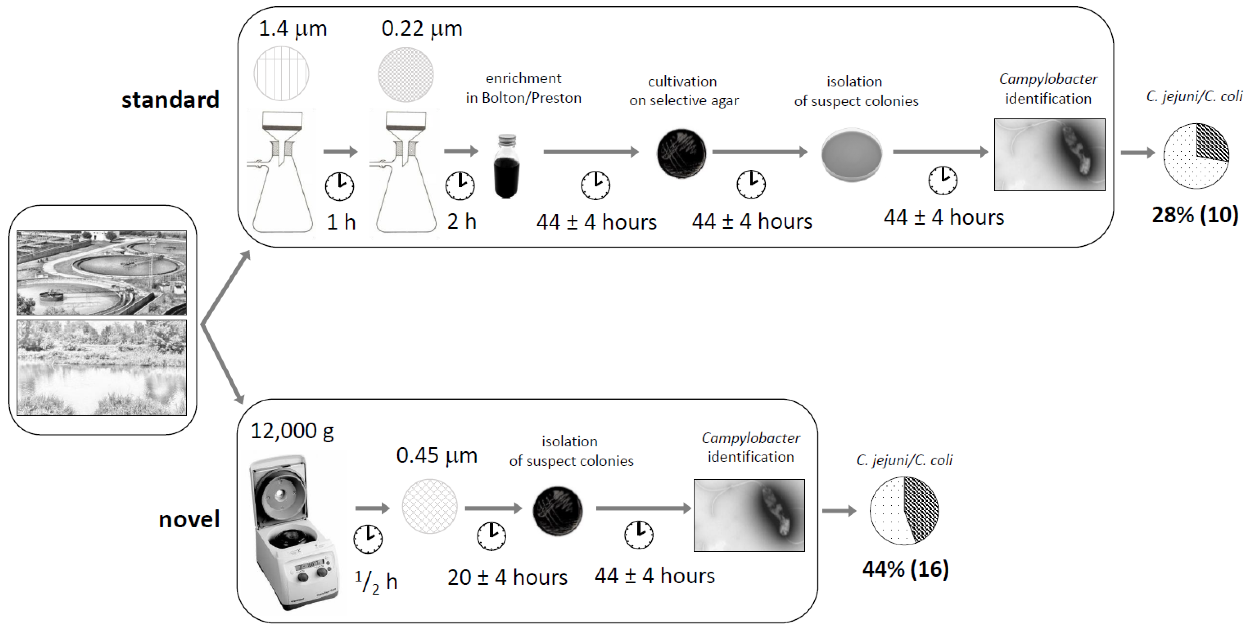
2.2. Isolation of Campylobacters from Poorly Filterable Water Samples by the Standard Cultivation Method

ISO 17995:2019 was used as a reference method for the isolation of campylobacters from wastewater and surface water samples [27]. Briefly, water samples (500 mL) were prefiltered (1.4 μm glass filter; Duren, Macherey Nagel, Germany) for quick removal of mechanical impurities and filtered (0.22 μm, mixed cellulose ester filter; Merk, Darmstadt, Germany); the filters were then transferred into 2 campylobacter selective broths (Preston and Bolton broth) for enrichment and incubated at 42 °C in an anaerostat (AnaeroJar, Oxoid, Basingstoke, UK) under a microaerobic atmosphere (CampyGen 3.5 L, Oxoid, Basingstoke, UK). After 44 ± 4 h of incubation, the inoculum was cultivated on Campylobacter blood-free selective agar (modified charcoal–cefoperazone–deoxycholate agar (mCCDA), Oxoid, Basingstoke, UK) and incubated for another 44 ± 4 h. After incubation under a microaerobic atmosphere at 42 °C, isolation of presumptive colonies on nonselective agar (blood agar) and mCCDA was performed under the same conditions. Finally, Campylobacter spp. were identified (see below).

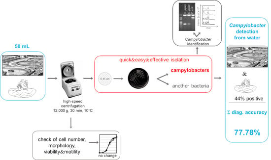
2.3. Novel Cultivation Method for Campylobacters from Poorly Filterable Water Samples

In parallel, Campylobacter spp. were isolated from water samples by a novel method without enrichment. In detail, 50 mL water samples were centrifuged at 12,000× g (acceleration time 300 s, deceleration time 40 s; HERMLE Z326K, Wehingen, Germany) for 30 min at 10 °C. Pellets were resuspended in 200 μL of sterile water and spread on a filter (0.45 μm, mixed cellulose ester filter; Merk, Darmstadt, Germany). Filters were placed face-up on top of mCCDA agar and incubated bottom-down for passive filtration at 42 °C under a microaerobic atmosphere. After overnight incubation, the filters were removed and cultivation continued at 42 °C under a microaerobic atmosphere for another 44 ± 4 h. Experiments were performed in triplicate.

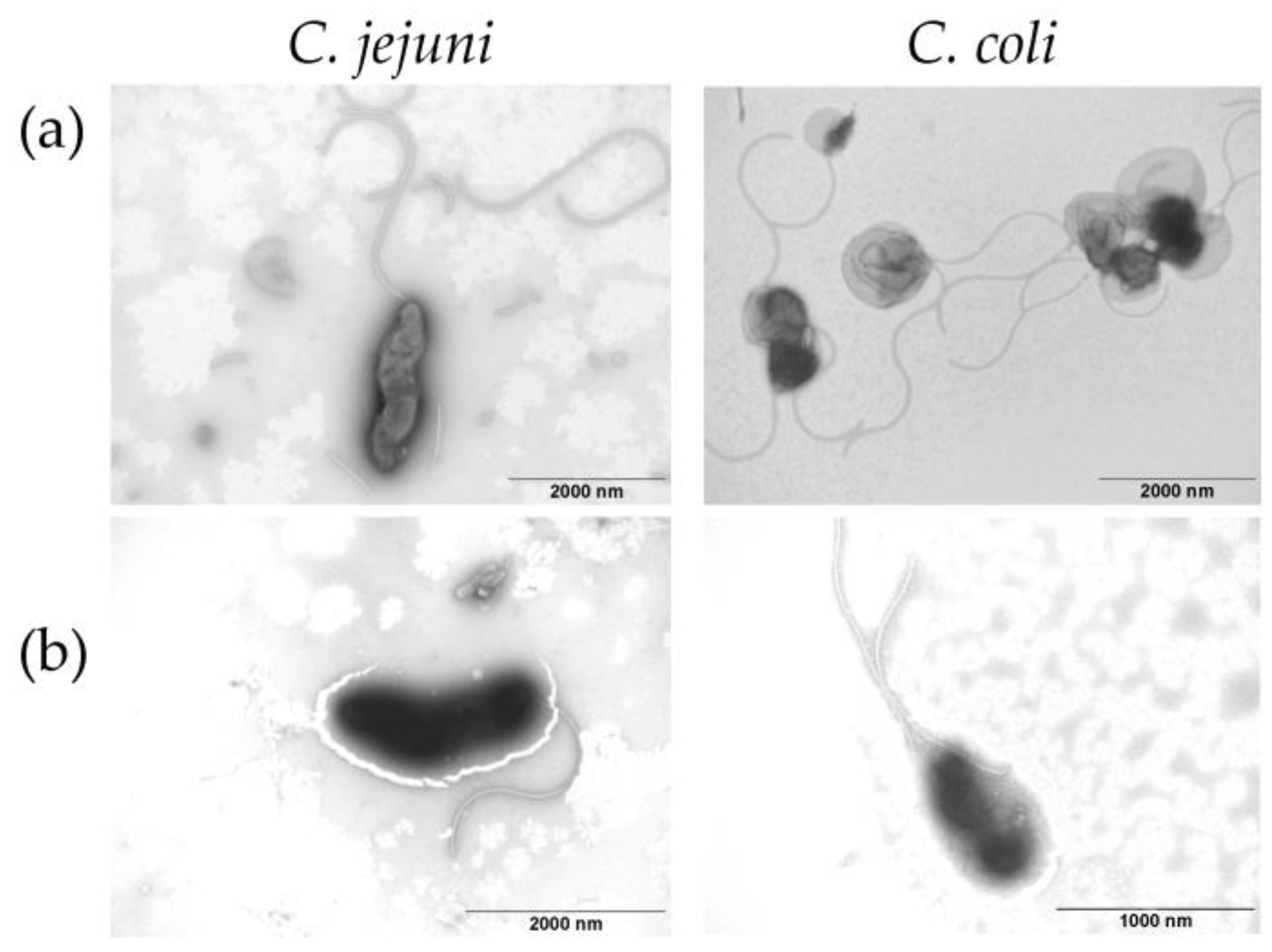
2.4. Campylobacter Species Identification

Suspected C. jejuni and C. coli colonies cultivated on mCCDA were plated on blood agar at 42 °C under a microaerobic atmosphere for 44 ± 4 h, and bacterial DNA was extracted by boiling. Multiplex PCR using PPP master mix (Top-Bio, Vestec, Czech Republic) was used to identify the genus Campylobacter and 2 species, C. jejuni and C. coli. The primers used (Generi-Biotech, Hradec Kralove, Czech Republic) [24,25,28] are shown in Table S1. Amplification was carried out with the following PCR conditions: 95 °C for 5 min, 35 cycles at 94 °C for 30 s, 55 °C for 30 s and 72 °C for 1 min, and 72 °C for 7 min.
Matrix-assisted laser desorption/ionisation time-of-flight mass spectrometry (MALDI-TOF/MS) was used for confirmation of Campylobacter species. In detail, when pure bacterial cultures were obtained, MALDI-TOF/MS with ethanol and formic acid extraction was used, and isolates were identified after comparing the bacterial spectrum in a Bruker database MBT 8468 by Biotyper software (version 3.1, Bruker Daltonics GmbH, Bremen, Germany).

2.5. Campylobacter Cell Numbers

Cell numbers of C. jejuni and C. coli were compared before and after centrifugation. Briefly, the bacterial suspension was prepared in 50 mL MilliQ water with optical density OD600nm 0.8–1.2. The suspension was centrifuged at 12,000× g for 30 min, the supernatant was removed, and the pellet was diluted in 50 mL MilliQ water. The cell number was evaluated by the optical density OD600nm before and after centrifugation at high speed.

2.6. Campylobacter Viability

The viability of campylobacters after centrifugation at high speed was tested on randomly selected C. jejuni and C. coli strains isolated from the water environment. The viability of bacterial cells was tested by colony enumeration of C. jejuni and C. coli after growth on agar plates. Ten-fold dilution series of bacterial suspension (100 μL) were applied on the mCCDA and chromogenic Brilliance CampyCount Agar (Oxoid, Perth, UK), and cultivation was performed for 24 ± 4 h at 42 °C under a microaerobic atmosphere. Finally, the numbers of colonies on the plates were evaluated before and after centrifugation.

2.7. Campylobacter Morphology

Transmission electron microscopy with negative staining of samples was performed for Campylobacter morphology testing. Briefly, randomly selected bacterial strains were suspended in MilliQ water. The suspension was covered by a grid-coated formvar film (Merk, Darmstadt, Germany) and carbon (Agar Scientific, Essex, UK) for 5 min, and the residual water was dried. Finally, 2% aqueous phosphotungstic acid was placed on the grid, and the sections were observed under a Philips 208 S Morgagni electron microscope (FEI, Dresden, Germany) with magnification of 14,000–36,000× and accelerating voltage of 80 kV.

2.8. Campylobacter Motility

Swarming soft agar motility assay was used to observe Campylobacter motility phenotypes [29,30]. The bacterial suspension (10 μL) was placed on 0.25% Lab Lemco agar (Oxoid, Basingstoke, UK). The cultivation was performed for 24 ± 4 h at 42 °C under a microaerobic atmosphere. Finally, the bacterial motility of C. jejuni and C. coli was compared before and after centrifugation. Non-motile Klebsiella pneumoniae were used as negative control.

2.9. Evaluation of Selectivity of the Novel Culture-Based Method

To check the selectivity of the novel culture-based method, non-motile Klebsiella pneumoniae were mixed with C. jejuni and C. coli. The tested bacterial suspensions (200 μL) were spread on filters (0.45 μm, mixed cellulose ester filter; Merk, Darmstadt, IN, USA) and incubated on mCCDA or Brilliance CampyCount Agar. Petri dishes were incubated bottom-down at 42 °C for 24 ± 4 h under a microaerobic atmosphere. After overnight incubation, the filters were removed, the dishes were inverted, and cultivation continued at 42 °C under a microaerobic atmosphere for another 44 ± 4 h. Finally, bacterial species were identified by PCR and MALDI-TOF/MS. Experiments were performed in triplicate.

2.10. Measurement of Diagnostic Accuracy of the Two Methods

A comparison of the modified detection method and the standard ISO method according to diagnostic accuracy was conducted according to EN ISO 16140–4:2020, commonly used as a validation method in food chain microbiology. This protocol is used for verification of reference methods and validated alternative methods in a single laboratory [31]. Briefly, the diagnostic accuracy, relative sensitivity, and relative specificity were calculated using the following formulas:
diagnostic accuracy (%) = (A + D)/(A + B + C + D) × 100
relative sensitivity (%) = A/(A + B) × 100
relative specificity (%) = D/(C + D) × 100
where A is the number of positive samples by both methods, B is the number of positive samples by the standard method, C is the number of positive samples by the modified method, and D is the number of negative samples by both methods.

3. Results

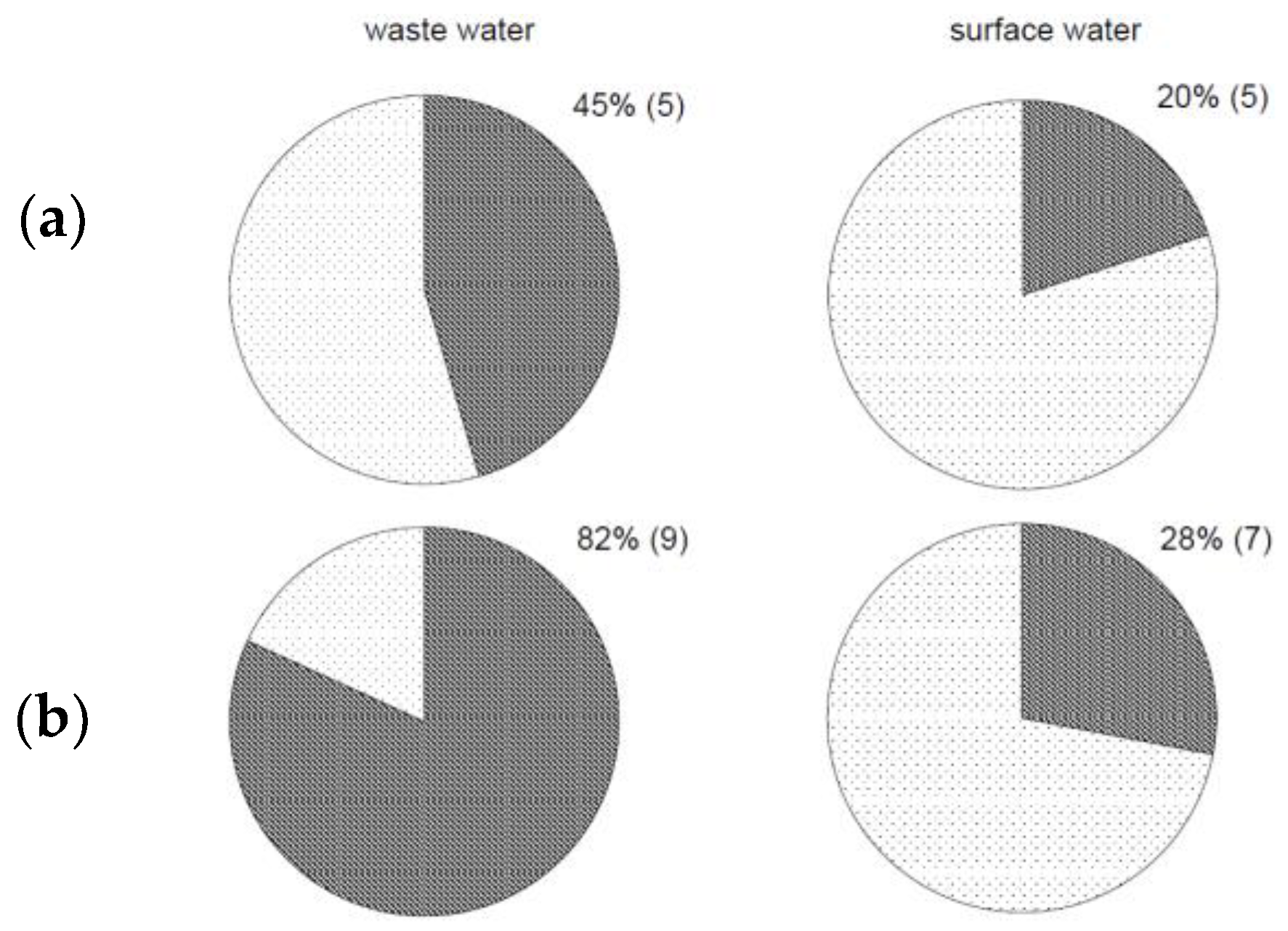
3.1. Detection of Campylobacter in Poorly Filterable Water Samples

Illustrations of the two methods for the detection of Campylobacter in water samples are shown in detail in Figure 1. The scheme of the standard method with slight modifications is shown in the upper part of the figure. The presented novel method (lower part of the figure) was based on the presumption that campylobacters are motile and can pass through filter pores. Campylobacter selective agar mCCDA was used to suppress the growth of redundant bacterial species in the water environment. The protocols are described above in detail. The rapid alternative method was easier to perform and took less time, saving approximately 72 h. Campylobacter identification was easier due to lower background microbiota levels of non-motile bacterial species. A water volume sample as small as 50 mL was a sufficient default volume for the isolation of Campylobacter by the modified method (Figure 1).

3.2. Comparison of Effectiveness between Standard and Modified Methods

The two methods used for the detection of thermotolerant campylobacters in water samples without distinguishing between water sources were compared (Figure 1). Of the 36 samples examined, 10 (28%) were positive for Campylobacter spp. according to the standard ISO method and 16 (44%) samples according to the modified method (Figure S1). In total, the effectiveness of thermotolerant Campylobacter strain detection from all water samples was 1.5 times higher with the modified method than the standard method (Figure 1). A list of all samples with detected and isolated Campylobacter strains is presented in Table S2.
Detection of campylobacters by the standard method was performed after enrichment in two selective broths, Bolton and Preston broth. Bolton broth was more efficient (22%; 8/36) for enrichment of Campylobacter from water samples than Preston broth (14%; 5/36). The presented novel culture-based method failed only in one sample compared with the standard method, which failed six times (Table S2). A statistical analysis of diagnostic accuracy was performed. The relative sensitivity and specificity were calculated. The relative sensitivity and specificity of the modified method were 90.0% and 73.08%, respectively. The diagnostic accuracy of the method was 77.78% (Table S3).
The detection of thermotolerant campylobacter strains was compared between the two methods based on water type (Figure 2); 45% of wastewater samples and 20% of surface water samples were positive (Figure 2a) according to the standard method with enrichment. According to the modified method, 82% of wastewater samples and 28% of surface water samples were positive for thermotolerant campylobacters (Figure 2b).
More details on the isolated campylobacter species are shown in Figure 3. From each plate, we identified more than one bacterial colony, so the number of Campylobacter isolates (Figure 3) was higher than the number of positive water samples (Figure 1). Surface water and, even more, wastewater contained a mixture of bacteria, and isolation of campylobacters from the water microbiota was difficult. However, we can summarise that the novel alternative method was more efficient at detecting and isolating thermotolerant Campylobacter strains in both wastewater and surface water. Our results show that campylobacters isolated by the culture-based method without enrichment were more easily obtained from agar media. Moreover, the method put neither C. jejuni nor C. coli at a disadvantage (Figure 3b).

3.3. Evaluation of the Novel Method

An evaluation of the novel method was performed by comparing the cell numbers, viability, morphology, and motility of Campylobacter spp. to confirm its suitability for Campylobacter spp. isolation. First, we checked whether high-speed centrifugation at 12,000× g would harm Campylobacter spp. and changed the cell numbers. Our results show that the cell numbers did not change, because OD600nm of bacterial suspensions was similar before and after centrifugation, at 0.38 and 0.34, respectively. Centrifugation has no effect on the viability of Campylobacter spp. Our results show that CFU/mL before (1.23 × 10⁶) and after (1.17 × 10⁶) centrifugation remained within the same logarithmic order on Brilliance CampyCount Agar (Figure 4) and on mCCDA (Figure S2, Table S4).
Transmission electron microscopy was used for the detection of cell morphology, e.g., flagellas of C. jejuni and C. coli before and after high-speed centrifugation (Figure 5). Our results show that 12,000× g did not damage flagellas in C. jejuni and C. coli; therefore, they stayed motile without morphologic changes despite high-speed centrifugation.
Finally, the motility assay confirmed that centrifugation had no effect on C. jejuni and C. coli cell motility; they were both motile before and after centrifugation (Figure 6 and Figure S3). The motility assay together with cell number analysis, cell viability, and cell morphology support the conclusion that centrifugation has no effect on C. jejuni and C. coli. In summary, our results confirm that high-speed centrifugation does not harm bacterial cells.
The selectivity of the novel method was confirmed by a model mixture of C. jejuni, C. coli, and K. pneumoniae. Non-motile K. pneumoniae (Figures S3 and S4) remained on the filter surface and were removed with the filter after overnight incubation. C. jejuni and C. coli passed through the pores of the filter and grew well on mCCDA. Species identification was confirmed by PCR and MALDI-TOF/MS (Figures S5 and S6). The Campylobacter selective agars contain antibiotics that suppressed the growth of K. pneumoniae. The growth of two randomly selected K. pneumoniae strains was checked on two Campylobacter selective agars, mCCDA and Brilliance CampyCount Agar. These agars allowed weak growth of K. pneumoniae as model bacteria of other contaminating bacteria of water samples (data not shown).
High-speed centrifugation together with the active motion of C. jejuni and C. coli through 0.45 μm filters did not harm the viability of these bacteria but increased the purity of the target bacteria grown.

4. Discussion

The natural environment, including surface waters, agricultural effluent, and marine environments, can be sources of campylobacters for humans [20,32,33,34]. Regarding a low infectious dose (<103 CFU) [35], human exposure can occur not only by the ingestion of food but also by the accidental ingestion of untreated surface water, e.g., during recreational swimming or leisure activities in natural lakes and rivers [36]. Surface water plays an important role in the spread of campylobacteriosis and, together with livestock, pet, and wild animal reservoirs, represents a significant risk to public health [26,37]. The aquatic environment thus may act as a reservoir of campylobacters that are generally considered sensitive to the external conditions of the water environment. Wastewater quality affects the estimation of the fate of pathogenic bacteria discharged from wastewater treatment plants and the risk they pose from the aquatic environment [38]. The next difficulty is determining Campylobacter’s sensitivity to stress during laboratory work requiring highly specific conditions for in vitro cultivation, including microaerobic conditions, temperature ranging from 37 to 42 °C, and high water activity.
Pathogens in urban water are commonly monitored by cultivation and non-cultivation methods [39]. ISO standard 17995 is a cultivation-based gold standard for detection of campylobacters. This standard protocol from 2005 [23] is only applicable to filterable waters. Water is not always suitable for filtration in the external environment because mechanical impurities can clog the pores in the filters, and microbiological analysis of such water becomes extremely difficult when using filtration. The novelisation of the ISO standard in 2019 extended the protocol for all water types by inoculation of non-filterable water sediments in a suitable diluent or directly into enrichment broth [27]. Despite the filtration of low contaminated water samples such as drinking water, which has no problem with clogging of pores, the ISO standard can be replaced by dead-end ultrafiltration because of the pathogen concentration in high water volumes [40].
The noncultivation methods are the second approach for the detection of bacteria [41]. The use of next-generation sequencing (NGS) methods in combination with amplification methods such as qPCR offers rapid and sensitive Campylobacter detection in environmental samples [42]. Full-length 16S rRNA gene sequencing can provide identification on the genus and species levels [19]. Unfortunately, noncultivation methods have several disadvantages compared with traditional culture methods. Detection of Campylobacter by qPCR can include viable but nonculturable (VBNC) cells or dead cells, or even free DNA, in the water samples. VBNC bacteria remain viable and express various degrees of metabolic activity. Campylobacter readily form VBNC cells in water under stress conditions, e.g., low temperature, high pH or high osmolality, and it has been shown that these cells can remain in water for weeks [43]. Other disadvantages include the cost involved, the specialised equipment required, and the expertise of the laboratory technical staff. The detection and isolation of C. jejuni and C. coli using culture-based methods are complex and time-consuming [44], as our also work confirmed. The presented cultivation method takes only 72 h, while the ISO standard method requires 144–192 h.
The ISO standard cultivation method uses filtration and Campylobacter enrichment in selective broths. This method and other modifications use filters with a pore size of 0.45 μm [27,45], which is larger than the campylobacter size (0.2–0.9 μm × 0.5–5 μm), and we supposed a loss of some Campylobacter by filtration. Compared to the ISO standard, the rapid culture method uses centrifugation of 50 mL samples and transition of the bacteria through filters. The volume can be increased for the isolation of Campylobacter from better quality water samples. Despite the increased volume, the loads stay very low. Our results show that centrifugation without Campylobacter enrichment is an effective approach for detection and isolation of C. jejuni and C. coli. This is in agreement with another study that used centrifugation for the detection of H. pylori from drinking water samples [46]. Moreover, high-speed centrifugation allows for smaller water volume and greater efficiency than were reported in that study. High-speed centrifugation can reduce the viability of pathogens [47], so verifications were added to confirm the robustness of the alternative method. It was verified that high-speed centrifugation as used in the modified method has no effect on the number, viability, morphology, or motility of C. jejuni and C. coli cells. Moreover, this approach can help to separate campylobacters from other bacteria and isolate them even from highly polluted water samples by the passage of C. jejuni and C. coli through filters. This method used filters with a pore size of 0.45 µm for the active transfer of motile bacteria. Filters with a smaller pore size (0.22 μm) are inappropriate, because campylobacters are bigger and not able to pass through. In general, our modified method can also be used for the detection and isolation of other bacterial species from water samples, but the viability has to be verified.
The probability of capturing Campylobacter depends on the sample volume; 100 mL of water for filtering is recommended to detect 10–100 CFU [27]. As it was supposed that a higher volume would increase the detection rate, the water volume was increased from 100 to 500 mL. Despite the low number of samples, our study clearly shows that the ISO standard discovered fewer positive samples (28%) than the modified method (44%). Moreover, the volume of the ISO standard method was increased. A comparison of the modified and standard ISO methods by diagnostic accuracy, relative sensitivity, and relative specificity was carried out according to EN ISO 16140 [31]. The diagnostic accuracy took into account target and nontarget microorganisms in the presence of a biological matrix, comprised sensitivity and specificity, and was 78%. Relative sensitivity was defined as the ability of the modified method to detect the agent compared to the reference method [27] in the presence of a biological matrix. The relative sensitivity of the modified method was 90%. This parameter can reveal false negative samples. The relative specificity was defined as the ability of the modified method to not detect the target organism when it was not detected by the reference method [48]. This parameter helped to reveal false positive samples, and the relative specificity of the modified method was 73%. Calculating it the other way round, the relative specificity of the standard ISO compared to the modified method is 95%.
Both methods were also compared on the basis of labour intensity and time consumption. The modified method is easy to perform and offers cleaner bacterial cultures compared to the standard method. When the standard method was used, Campylobacter enrichment and isolation on mCCDA agar allowed the survival of a wide spectrum of bacteria; therefore, another subcultivation was needed before the identification step. Our results show that suppression of water microbiota by Campylobacter selective agar is a very important task. This is in agreement with another study that used a method to support the growth of C. jejuni on Campylobacter selective agar, but unlike our approach, those authors filtrated the water samples [45].
In our study, we used water samples collected from different localities during all seasons to show the universality of the modified alternative protocol for samples from ponds, lakes, and wastewater treatment plants. The method was not only more sensitive for C. jejuni and C. coli detection, but also faster and simpler to perform than the ISO method.

5. Conclusions

Thermotolerant campylobacters C. jejuni and C. coli are present in aquatic environments, which may thus represent a source of Campylobacter infection. This paper presents a new methodological approach for the detection and cultivation of C. jejuni and C. coli from wastewater and surface water, without the influence of mechanical impurities of water samples. The modified method was more efficient in detecting thermotolerant campylobacters: 44% samples were Campylobacter positive with the modified method and 28% with the standard ISO method. We can conclude that the presented cultivated method was more efficient, faster, smarter, and easier than the ISO method. This method helped to increase the capture of C. jejuni and C. coli from a turbid aquatic environment; therefore, it can meet the ISO standard guideline.

Supplementary Materials

The following are available online at https://www.mdpi.com/article/10.3390/ijerph18116098/s1, Figure S1. Comparison of effective methods for isolation of thermotolerant campylobacters from water samples: (a) ISO standard method and (b) novel method. Level of Campylobacter detection in positive samples (n = 17) identified by both methods shown in dark colour. Figure S2. Viability testing of C. jejuni and C. coli on mCCDA agar (a) before and (b) after centrifugation. Figure S3. Campylobacter motility. C. coli and non-motile K. pneumoniae strains were cultivated on 0.25% agar (a) before and (b) after centrifugation. Figure S4. Effect of high-speed centrifugation on K. pneumoniae cell morphology. Representative figures of electron microscopy (a) before and (b) after centrifugation. Figure S5. Growth of mixture of thermotolerant campylobacters C. jejuni and C. coli with K. pneumoniae on mCCDA agar. Figure S6. Identification of C. jejuni and C. coli by PCR and MALDI-TOF/MS after their isolation from mixture of C. jejuni, C. coli, and K. pneumoniae. (a) PCR: size marker (lane 1); mixture of C. jejuni, C. coli, and K. pneumoniae (lane 2); C. jejuni-positive control (lane 3); C. coli-positive control (lane 4); negative control (lane 5). (b) MALDI-TOF/MS identification. Table S1. Primers used in this study for identification of thermotolerant C. jejuni and C. coli. Table S2. List of water samples. BW, wastewater; BP, pond/surface water. Localities of ponds where surface water was collected (1–25). Isolation of C. jejuni and C. coli strains from mCCDA agar. Table S3. Comparison of both methods by diagnostic accuracy (%), relative sensitivity (%), and relative specificity (%). Table S4. Colony formation of C. jejuni and C. coli before and after centrifugation at 12,000× g for 30 min.

Author Contributions

Conceptualisation, N.S. and R.K.; methodology, N.S. and R.K.; investigation, N.S., K.K. and P.K.; resources, N.S. and T.G.; data curation, N.S. and R.K.; writing–original draft preparation, N.S. and R.K.; writing–review and editing, N.S., R.K. and T.G.; visualisation, N.S., P.K. and R.K.; supervision, R.K.; project administration N.S.; funding acquisition, R.K. All authors have read and agreed to the published version of the manuscript.

Funding

This research was funded by the Czech Science Foundation, grant no. GA18-16549S, and the Czech Ministry of Education, Youth and Sport, grant no. CZ.1.05/2.1.00/19.0385.

Institutional Review Board Statement

Not applicable.

Informed Consent Statement

Not applicable.

Data Availability Statement

All treated data that support the findings of this study are included in the present article. Raw datasets can be obtained from the corresponding author upon request.

Acknowledgments

The authors thank Ludmila Faldikova for English proofreading.

Conflicts of Interest

The authors declare no conflict of interest.

References

  1. Campylobacter Sebald and Véron 1963 (Approved Lists 1980). Available online: https://lpsn.dsmz.de/genus/campylobacter (accessed on 3 October 2020).
  2. European Food Safety Authority; European Centre for Disease Prevention and Control. The European Union One Health 2018 Zoonoses Report. EFSA J. 2019, 17, e05926. [Google Scholar] [CrossRef] [Green Version]
  3. Igwaran, A.; Okoh, A.I. Human campylobacteriosis: A public health concern of global importance. Heliyon 2019, 5, e02814. [Google Scholar] [CrossRef]
  4. Ghosh, R.; Uppal, B.; Aggarwal, P.; Chakravarti, A.; Jha, A.K.; Dubey, A.P. A comparative study of conventional and molecular techniques in diagnosis of Campylobacter gastroenteritis in children. Ann. Clin. Lab. Sci. 2014, 44, 42–48. [Google Scholar] [PubMed]
  5. Koga, M.; Ang, C.W.; Yuki, N.; Jacobs, C.; Herbrink, P.; Van Der Meché, G.A.; Hirata, K.; Van Doorn, P.A. Comparative study of preceding Campylobacter jejuni infection in Guillain-Barré syndrome in Japan and The Netherlands. J. Neurol. Neurosurg. Psychiatry 2001, 70, 693–695. [Google Scholar] [CrossRef] [Green Version]
  6. Nyati, K.K.; Nyati, R. Role of Campylobacter jejuni infection in the pathogenesis of Guillain-Barré syndrome: An update. BioMed Res. Int. 2013, 2013, 852195. [Google Scholar] [CrossRef] [PubMed] [Green Version]
  7. Young, K.Y.; Davis, L.M.; DiRita, V.J. Campylobacter jejuni: Molecular biology and pathogenesis. Nat. Rev. Microbiol. 2007, 5, 665–679. [Google Scholar] [CrossRef]
  8. Burnham, P.M.; Hendrixson, D.R. Campylobacter jejuni: Collective components promoting a successful enteric lifestyle. Nat. Rev. Microbiol. 2018, 16, 551–565. [Google Scholar] [CrossRef] [PubMed]
  9. Pattis, I.; Moriarty, E.; Billington, C.; Gilpin, B.; Hodson, R.; Ward, N. Concentrations of Campylobacter spp., Escherichia coli, Enterococci, and Yersinia spp. in the Feces of Farmed Red Deer in New Zealand. J. Environ. Qual. 2017, 46, 819–827. [Google Scholar] [CrossRef]
  10. Hald, B.; Skov, M.N.; Nielsen, E.M.; Rahbek, C.; Madsen, J.J.; Wainø, M.; Chriél, M.; Nordentoft, S.; Baggesen, D.L.; Madsen, M. Campylobacter jejuni and Campylobacter coli in wild birds on Danish livestock farms. Acta Vet. Scand. 2016, 58, 11. [Google Scholar] [CrossRef] [Green Version]
  11. Moore, J.; Corcoran, D.; Dooley, J.; Fanning, S.; Lucey, B.; Matsuda, M.; McDowell, D.; Mégraud, F.; Millar, B.; O’Mahony, R.; et al. Campylobacter . Vet. Res. 2005, 36, 351–382. [Google Scholar] [CrossRef] [Green Version]
  12. Nag, R.; Whyte, P.; Markey, B.K.; O’Flaherty, V.; Bolton, D.; Fenton, O.; Richards, K.G.; Cummins, E. Ranking hazards pertaining to human health concerns from land application of anaerobic digestate. Sci. Total Environ. 2020, 710, 136297. [Google Scholar] [CrossRef]
  13. Ugarte-Ruiz, M.; Florez-Cuadrado, D.; Wassenaar, T.M.; Porrero, M.C.; Domínguez, L. Method Comparison for Enhanced Recovery, Isolation and Qualitative Detection of C. jejuni and C. coli from Wastewater Effluent Samples. Int. J. Environ. Res. Public Health 2015, 12, 2749–2764. [Google Scholar] [CrossRef] [PubMed] [Green Version]
  14. Koenraad, P.M.F.J.; Rombouts, F.M.; Notermans, S.H.W. Epidemiological aspects of thermophilic Campylobacter in water-related environments: A review. Water Environ. Res. 1997, 69, 52–63. [Google Scholar] [CrossRef]
  15. Jokinen, C.; Edge, T.A.; Ho, S.; Koning, W.; Laing, C.; Mauro, W.; Medeiros, D.; Miller, J.; Robertson, W.; Taboada, E.; et al. Molecular subtypes of Campylobacter spp., Salmonella enterica, and Escherichia coli O157:H7 isolated from faecal and surface water samples in the Oldman River watershed, Alberta, Canada. Water Res. 2011, 45, 1247–1257. [Google Scholar] [CrossRef]
  16. Pitkänen, T. Review of campylobacter spp. in drinking and environmental waters. J. Microbiol. Methods 2013, 95, 39–47. [Google Scholar] [CrossRef] [PubMed]
  17. Itoh, S.; Zhou, L. Effect of unboiled water consumption data on sensitivity analysis in quantitative microbial risk assessment. NPJ Clean Water 2018, 1, 18. [Google Scholar] [CrossRef]
  18. Arimi, S.M.; Fricker, C.R.; Park, R.W.A. Occurrence of “thermophilic” campylobacters in sewage and their removal by treatment processes. Epidemiol. Infect. 1988, 101, 279–286. [Google Scholar] [CrossRef] [Green Version]
  19. Marti, E.; Jofre, J.; Balcazar, J.L. Prevalence of Antibiotic Resistance Genes and Bacterial Community Composition in a River Influenced by a Wastewater Treatment Plant. PLoS ONE 2013, 8, e78906. [Google Scholar] [CrossRef]
  20. Schönberg-Norio, D.; Takkinen, J.; Hänninen, M.-L.; Katila, M.-L.; Kaukoranta, S.-S.; Mattila, L.; Rautelin, H. Swimming and Campylobacter Infections. Emerg. Infect. Dis. 2004, 10, 1474–1477. [Google Scholar] [CrossRef] [PubMed]
  21. Rechenburg, A.; Kistemann, T. Sewage effluent as a source of Campylobacter spp. in a surface water catchment. Int. J. Environ. Health Res. 2009, 19, 239–249. [Google Scholar] [CrossRef]
  22. Abulreesh, H.H.; Paget, T.A.; Goulder, R. A pre-enrichment step is essential for detection of Campylobacter spp. in turbid pond water. Trop. Biomed. 2014, 31, 320–326. [Google Scholar] [CrossRef] [PubMed]
  23. International Organization for Standardisation. Water Quality—Detection and Enumeration of Thermotolerant Campylobacter Species—ISO 17995; ISO: Geneva, Switzerland, 2005. [Google Scholar]
  24. Bang, D.D.; Wedderkopp, A.; Pedersen, K.; Madsen, M. Rapid PCR using nested primers of the 16s rRNA and the hippuricase (hipO) genes to detect Campylobacter jejuni and Campylobacter coli in environmental samples. Mol. Cell. Probes 2002, 16, 359–369. [Google Scholar] [CrossRef] [PubMed]
  25. Linton, D.; Lawson, A.J.; Owen, R.J.; Stanley, J. PCR detection, identification to species level, and fingerprinting of Campylobacter jejuni and Campylobacter coli direct from diarrheic samples. J. Clin. Microbiol. 1997, 35, 2568–2572. [Google Scholar] [CrossRef] [Green Version]
  26. Mughini-Gras, L.; Penny, C.; Ragimbeau, C.; Schets, F.M.; Blaak, H.; Duim, B.; Wagenaar, J.A.; de Boer, A.; Cauchie, H.M.; Mossong, J.; et al. Quantifying potential sources of surface water contamination with Campylobacter jejuni and Campylobacter coli. Water Res. 2016, 101, 36–45. [Google Scholar] [CrossRef]
  27. International Organization for Standardisation. Water Quality—Detection and Enumeration of Thermotolerant Campylobacter spp.—ISO 17995:2019; ISO: Geneva, Switzerland, 2019. [Google Scholar]
  28. Winters, D.K.; O’Leary, A.E.; Slavik, M.F. Polymerase chain reaction for rapid detection of Campylobacter jejuni in artificially contaminated foods. Lett. Appl. Microbiol. 1998, 27, 163–167. [Google Scholar] [CrossRef]
  29. Wolfe, A.J.; Berg, H.C. Migration of bacteria in semisolid agar. Proc. Natl. Acad. Sci. USA 1989, 86, 6973–6977. [Google Scholar] [CrossRef] [PubMed] [Green Version]
  30. Marini, E.; Magi, G.; Ferretti, G.; Bacchetti, T.; Giuliani, A.; Pugnaloni, A.; Rippo, M.R.; Facinelli, B. Attenuation of Listeria monocytogenes virulence by Cannabis sativa L. Essential oil. Front. Cell. Infect. Microbiol. 2018, 8, 1–11. [Google Scholar] [CrossRef] [Green Version]
  31. International Organization for Standardisation. ISO 16140-4:2020: Microbiology of the Food Chain—Method Validation—Part 4: Protocol for Method Validation in a Single Laboratory; ISO: Geneva, Switzerland, 2020. [Google Scholar]
  32. Hanninen, M.; Haajanen, H.; Pummi, T.; Wermundsen, K.; Katila, M.; Sarkkinen, H.; Miettinen, I.; Rautelin, H. Detection and Typing of Campylobacter jejuni and Campylobacter coli and Analysis of Indicator Organisms in Three Waterborne Outbreaks in Finland. Appl. Environ. Microbiol. 2003, 69, 1391–1396. [Google Scholar] [CrossRef] [Green Version]
  33. Nygård, K.; Andersson, Y.; Røttingen, J.A.; Svensson, Å.; Lindbäck, J.; Kistemann, T.; Giesecke, J. Association between environmental risk factors and Campylobacter infections in Sweden. Epidemiol. Infect. 2004, 132, 317–325. [Google Scholar] [CrossRef] [Green Version]
  34. Vereen, E.; Lowrance, R.R.; Cole, D.J.; Lipp, E.K. Distribution and Ecology of Campylobacters in Coastal Plain Streams (Georgia, United States of America). Appl. Environ. Microbiol. 2007, 73, 1395–1403. [Google Scholar] [CrossRef] [Green Version]
  35. Black, R.E.; Levine, M.M.; Clements, M.L.; Hughes, T.P.; Blaser, M.J. Experimental Campylobacter jejuni Infection in Humans. J. Infect. Dis. 1988, 157, 472–479. [Google Scholar] [CrossRef]
  36. Sales-Ortells, H.; Agostini, G.; Medema, G. Quantification of Waterborne Pathogens and Associated Health Risks in Urban Water. Environ. Sci. Technol. 2015, 49, 6943–6952. [Google Scholar] [CrossRef]
  37. Phiri, B.J.; French, N.P.; Biggs, P.J.; Stevenson, M.A.; Reynolds, A.D.; Garcia-R, J.C.; Hayman, D.T.S. Microbial contamination in drinking water at public outdoor recreation facilities in New Zealand. J. Appl. Microbiol. 2021, 130, 302–312. [Google Scholar] [CrossRef]
  38. Li, J.; Zhao, X.; Tian, X.; Li, J.; Sjollema, J.; Wang, A. Retention in treated wastewater affects survival and deposition of Staphylococcus aureus and Escherichia coli in sand columns. Appl. Environ. Microbiol. 2015, 81, 2199–2205. [Google Scholar] [CrossRef] [Green Version]
  39. Nachamkin, I.; Szymanski, C.; Blaser, M.J. Campylobacter, 3rd ed.; Nachamkin, I., Szymanski, C., Blaser, M.J., Eds.; ASM Press: Washington, DC, USA, 2008; ISBN 9781119738039. [Google Scholar]
  40. Ferrari, S.; Frosth, S.; Svensson, L.; Fernström, L.L.; Skarin, H.; Hansson, I. Detection of Campylobacter spp. in water by dead-end ultrafiltration and application at farm level. J. Appl. Microbiol. 2019, 127, 1270–1279. [Google Scholar] [CrossRef] [PubMed] [Green Version]
  41. De Beer, D.M.; Botes, M.; Cloete, T.E. The microbial community of a biofilm contact reactor for the treatment of winery wastewater. J. Appl. Microbiol. 2018, 124, 598–610. [Google Scholar] [CrossRef] [PubMed]
  42. Dong, P.; Cui, Q.; Fang, T.; Huang, Y.; Wang, H. Occurrence of antibiotic resistance genes and bacterial pathogens in water and sediment in urban recreational water. J. Environ. Sci. 2019, 77, 65–74. [Google Scholar] [CrossRef] [PubMed]
  43. Baffone, W.; Casaroli, A.; Citterio, B.; Pierfelici, L.; Campana, R.; Vittoria, E.; Guaglianone, E.; Donelli, G. Campylobacter jejuni loss of culturability in aqueous microcosms and ability to resuscitate in a mouse model. Int. J. Food Microbiol. 2006, 107, 83–91. [Google Scholar] [CrossRef] [PubMed]
  44. Williams, L.K.; Jørgensen, F.; Grogono-Thomas, R.; Humphrey, T.J. Enrichment culture for the isolation of Campylobacter spp.: Effects of incubation conditions and the inclusion of blood in selective broths. Int. J. Food Microbiol. 2009, 130, 131–134. [Google Scholar] [CrossRef] [PubMed]
  45. Blaser, M.J.; Cody, H.J. Methods for isolating Campylobacter jejuni from low-turbidity water. Appl. Environ. Microbiol. 1986, 51, 312–315. [Google Scholar] [CrossRef] [Green Version]
  46. Vesga, F.J.; Moreno, Y.; Ferrús, M.A.; Campos, C.; Trespalacios, A.A. Detection of Helicobacter pylori in drinking water treatment plants in Bogotá Colombia, using cultural and molecular techniques. Int. J. Hyg. Environ. Health 2018, 221, 595–601. [Google Scholar] [CrossRef] [PubMed]
  47. Liu, Z.; Carroll, Z.S.; Long, S.C.; Roa-Espinosa, A.; Runge, T. Centrifuge separation effect on bacterial indicator reduction in dairy manure. J. Environ. Manag. 2017, 191, 268–274. [Google Scholar] [CrossRef] [PubMed]
  48. Malorny, B.; Hoorfar, J.; Hugas, M.; Heuvelink, A.; Fach, P.; Ellerbroek, L.; Bunge, C.; Dorn, C.; Helmuth, R. Interlaboratory diagnostic accuracy of a Salmonella specific PCR-based method. Int. J. Food Microbiol. 2003, 89, 241–249. [Google Scholar] [CrossRef]
Figure 1. Scheme of standard and novel rapid method for detection of campylobacters, including timing and effectiveness. ISO standard method (above) and novel method (below) were used for isolation and detection of campylobacters from wastewater and surface water samples. Positive samples shown in dark colours.
Ijerph 18 06098 g001
Figure 2. Comparison of detection of thermotolerant campylobacters from water samples. Percentage (number) of Campylobacter-positive samples from wastewater (left charts) and surface water (right charts). Isolation and detection of C. jejuni and C. coli by (a) standard ISO 17995 method and (b) alternative method. Positive samples shown in dark colour.
Ijerph 18 06098 g002
Figure 3. Comparison of isolation of thermotolerant C. jejuni and C. coli from water samples. Isolation of C. jejuni by (a) standard ISO 17995 method and (b) culture-based method without enrichment. Positive samples shown in dark colour.
Ijerph 18 06098 g003
Figure 4. Campylobacter viability. C. jejuni strain was cultivated on Brilliance CampyCount Agar (a) before and (b) after centrifugation.
Ijerph 18 06098 g004
Figure 5. Effect of high-speed centrifugation on C. jejuni and C. coli cell morphology. Representative figures of electron microscopy (a) before and (b) after centrifugation. Magnification 14,000×.
Ijerph 18 06098 g005
Figure 6. Campylobacter motility. Motility of C. jejuni strain did not change (a) before and (b) after centrifugation.
Ijerph 18 06098 g006
Publisher’s Note: MDPI stays neutral with regard to jurisdictional claims in published maps and institutional affiliations.

