
The Mood-Improving Effect of Viewing Images of Nature and Its Neural Substrate

 ,
,  ,
,  and
and 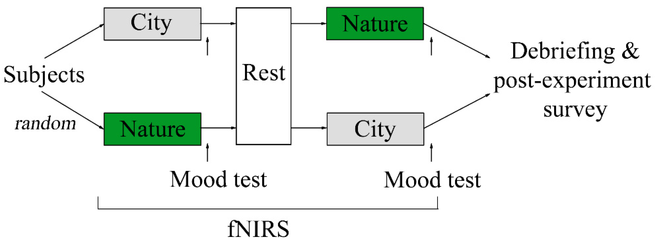
Abstract
1. Introduction
2. Materials and Methods
2.1. Participants
2.2. Design and Procedure
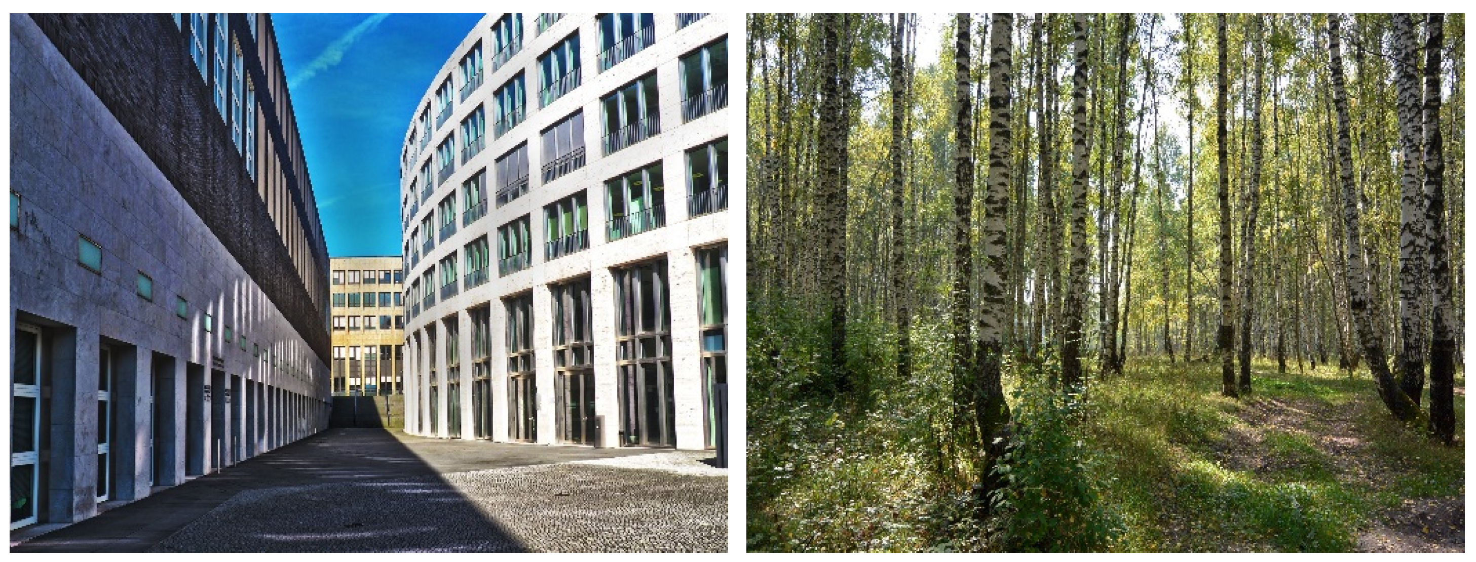
2.3. Stimuli
2.4. Mood Test
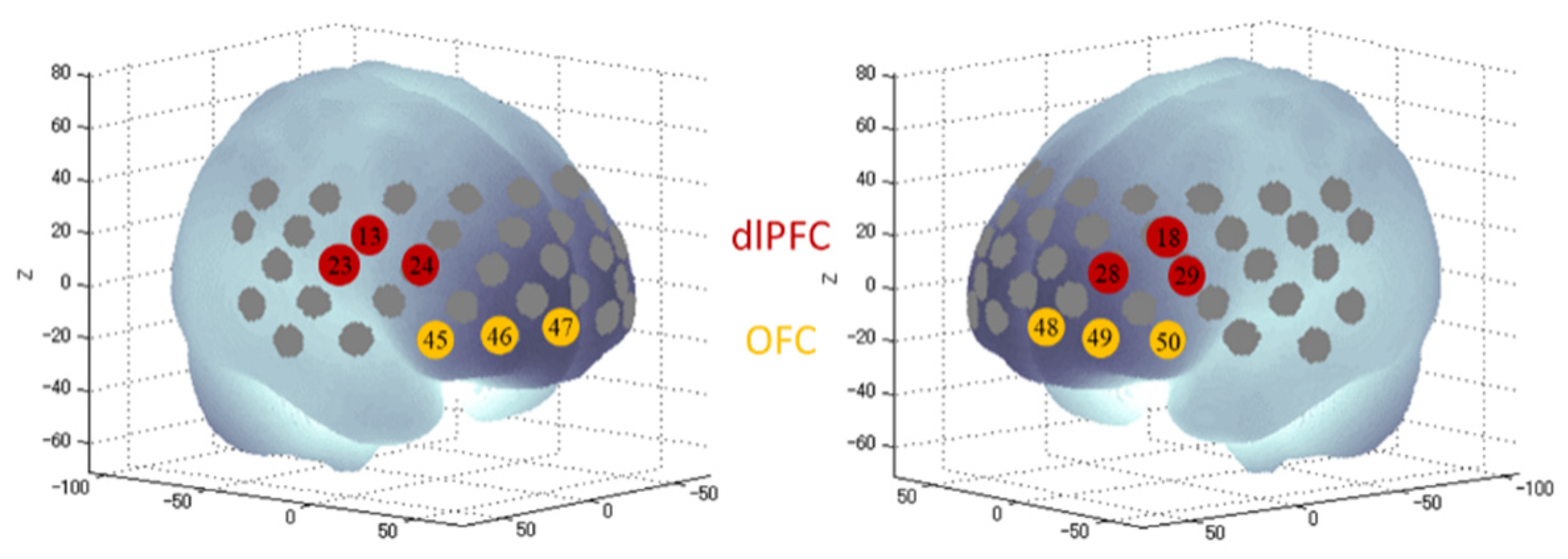
2.5. FNIRS
2.6. Blindness and Debriefing
2.7. Statistical Analysis
3. Results
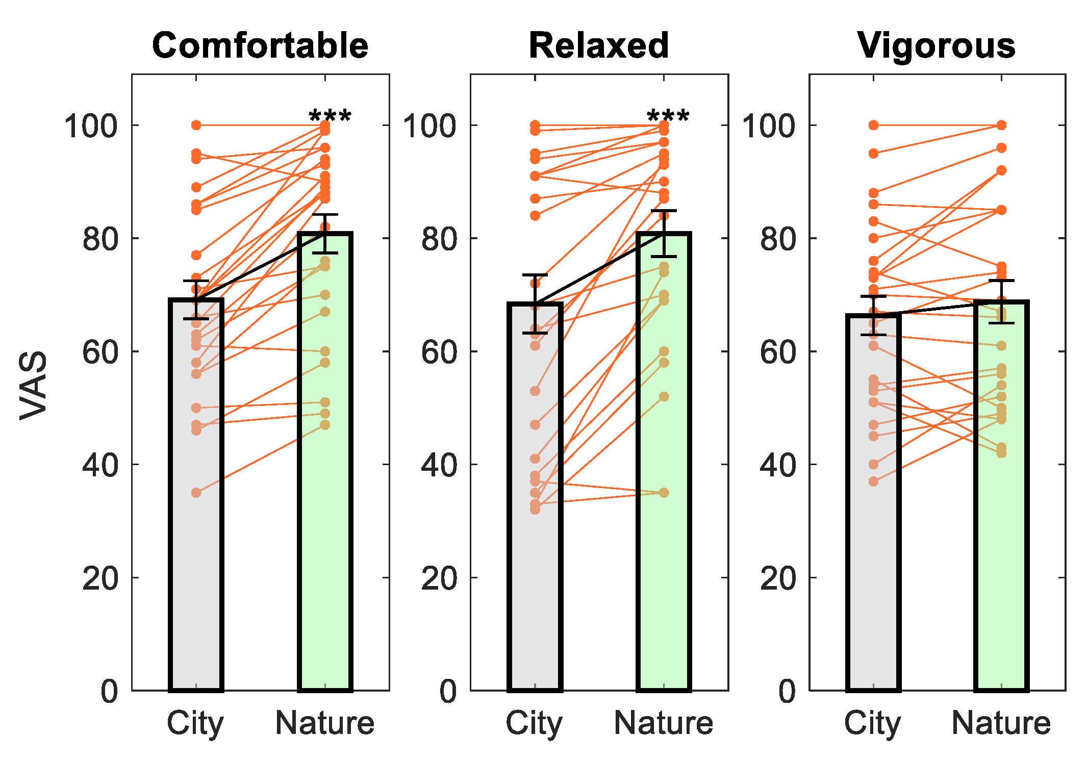
3.1. Mood Measures
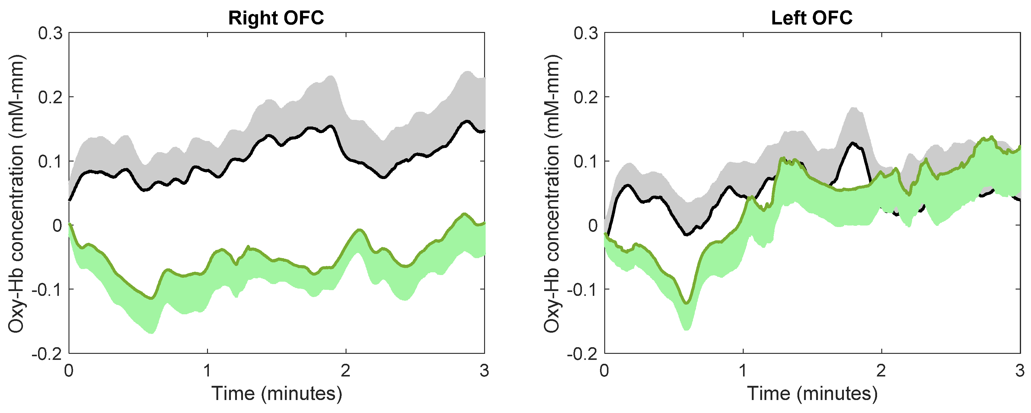
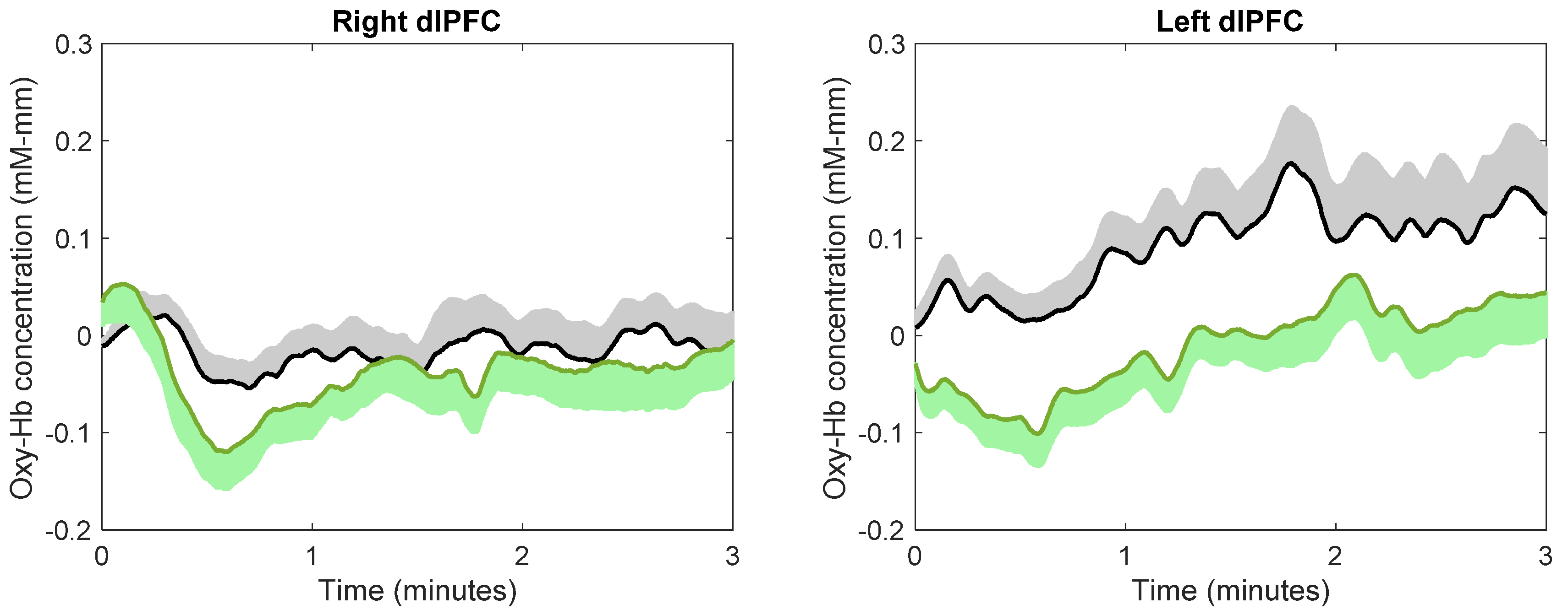
3.2. Brain Activity
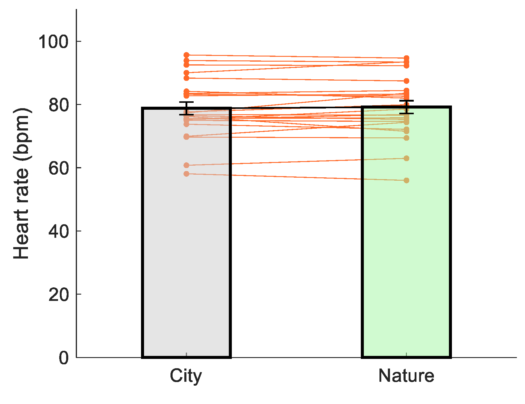
3.3. Heart Rate
3.4. Post-Experiment Survey
4. Discussion
5. Conclusions
Author Contributions
Funding
Institutional Review Board Statement
Informed Consent Statement
Data Availability Statement
Conflicts of Interest
Appendix A
References
- Bowler, D.E.; Buyung-Ali, L.M.; Knight, T.M.; Pullin, A.S. A systematic review of evidence for the added benefits to health of exposure to natural environments. BMC Public Health 2010, 10, 456. [Google Scholar] [CrossRef] [PubMed]
- McMahan, E.A.; Estes, D. The effect of contact with natural environments on positive and negative affect: A meta-analysis. J. Posit. Psychol. 2015, 10, 507–519. [Google Scholar] [CrossRef]
- Mantler, A.; Logan, A.C. Natural environments and mental health. Adv. Integr. Med. 2015, 2, 5–12. [Google Scholar] [CrossRef]
- Sandifer, P.A.; Sutton-Grier, A.E.; Ward, B.P. Exploring connections among nature, biodiversity, ecosystem ser-vices, and human health and wellbeing: Opportunities to enhance health and biodiversity conservation. Ecosyst. Serv. 2015, 12, 1–15. [Google Scholar] [CrossRef]
- Frumkin, H.; Bratman, G.N.; Breslow, S.J.; Cochran, B.; Kahn, P.H., Jr.; Lawler, J.J.; Wood, S.A. Nature contact and human health: A research agenda. Environ. Health Perspect. 2017, 125, 075001. [Google Scholar] [CrossRef]
- McSweeney, J.; Rainham, D.; Johnson, S.A.; Sherry, S.B.; Singleton, J. Indoor nature exposure (INE): A health-promotion framework. Heal. Promot. Int. 2014, 30, 126–139. [Google Scholar] [CrossRef]
- Franco, L.S.; Shanahan, D.F.; Fuller, R.A. A Review of the Benefits of Nature Experiences: More Than Meets the Eye. Int. J. Environ. Res. Public Health 2017, 14, 864. [Google Scholar] [CrossRef] [PubMed]
- Brooks, A.M.; Ottley, K.M.; Arbuthnott, K.D.; Sevigny, P. Nature-related mood effects: Season and type of nature contact. J. Environ. Psychol. 2017, 54, 91–102. [Google Scholar] [CrossRef]
- Song, C.; Ikei, H.; Miyazaki, Y. Physiological Effects of Nature Therapy: A Review of the Research in Japan. Int. J. Environ. Res. Public Health 2016, 13, 781. [Google Scholar] [CrossRef]
- Ikei, H.; Song, C.; Miyazaki, Y. Physiological effects of wood on humans: A review. J. Wood Sci. 2016, 63, 1–23. [Google Scholar] [CrossRef]
- Chen, C.; Nakagawa, S. Nature’s pathways on human health. In International Handbook of Forest Therapy; Kotte, D., Li, Q., Shin, W.S., Eds.; Cambridge Scholars Publishing: Newcastle upon Tyne, UK, 2020; pp. 12–31. [Google Scholar]
- Jo, H.; Song, C.; Miyazaki, Y. Physiological Benefits of Viewing Nature: A Systematic Review of Indoor Experiments. Int. J. Environ. Res. Public Health 2019, 16, 4739. [Google Scholar] [CrossRef] [PubMed]
- Norwood, M.F.; Lakhani, A.; Maujean, A.; Zeeman, H.; Creux, O.; Kendall, E. Brain activity, underlying mood and the environment: A systematic review. J. Environ. Psychol. 2019, 65, 101321. [Google Scholar] [CrossRef]
- Ferrari, M.; Quaresima, V. A brief review on the history of human functional near-infrared spectroscopy (fNIRS) development and fields of application. NeuroImage 2012, 63, 921–935. [Google Scholar] [CrossRef] [PubMed]
- Hoshi, Y. Hemodynamic signals in fNIRS. In Progress in Brain Research; Elsevier BV: Amsterdam, The Netherlands, 2016; Volume 225, pp. 153–179. [Google Scholar]
- Joung, D.; Kim, G.; Choi, Y.; Lim, H.; Park, S.; Woo, J.M.; Park, B.J. The prefrontal cortex activity and psychologi-cal effects of viewing forest landscapes in autumn season. Int. J. Environ. Res. Public Health 2015, 12, 7235–7243. [Google Scholar] [CrossRef]
- Park, S.A.; Song, C.; Choi, J.Y.; Son, K.C.; Miyazaki, Y. Foliage plants cause physiological and psychological re-laxation as evidenced by measurements of prefrontal cortex activity and profile of mood states. HortScience 2016, 51, 1308–1312. [Google Scholar] [CrossRef]
- Song, C.; Ikei, H.; Miyazaki, Y. Physiological Effects of Visual Stimulation with Forest Imagery. Int. J. Environ. Res. Public Health 2018, 15, 213. [Google Scholar] [CrossRef]
- Lee, J. Experimental Study on the Health Benefits of Garden Landscape. Int. J. Environ. Res. Public Health 2017, 14, 829. [Google Scholar] [CrossRef]
- Nakamura, M.; Ikei, H.; Miyazaki, Y. Physiological effects of visual stimulation with full-scale wall images com-posed of vertically and horizontally arranged wooden elements. J. Wood Sci. 2019, 65, 55. [Google Scholar] [CrossRef]
- Ikei, H.; Nakamura, M.; Miyazaki, Y. Physiological Effects of Visual Stimulation Using Knotty and Clear Wood Images among Young Women. Sustainability 2020, 12, 9898. [Google Scholar] [CrossRef]
- Koga, K.; Iwasaki, Y. Psychological and physiological effect in humans of touching plant foliage—Using the semantic differential method and cerebral activity as indicators. J. Physiol. Anthr. 2013, 32, 7. [Google Scholar] [CrossRef]
- Ikei, H.; Song, C.; Miyazaki, Y. Physiological effects of touching sugi (Cryptomeria japonica) with the palm of the hand. J. Wood Sci. 2019, 65, 1–7. [Google Scholar] [CrossRef]
- Ikei, H.; Miyazaki, Y. Positive physiological effects of touching sugi (Cryptomeria japonica) with the sole of the feet. J. Wood Sci. 2020, 66, 1–12. [Google Scholar] [CrossRef]
- Park, S.-A.; Song, C.; Oh, Y.-A.; Miyazaki, Y.; Son, K.-C. Comparison of Physiological and Psychological Relaxation Using Measurements of Heart Rate Variability, Prefrontal Cortex Activity, and Subjective Indexes after Completing Tasks with and without Foliage Plants. Int. J. Environ. Res. Public Health 2017, 14, 1087. [Google Scholar] [CrossRef] [PubMed]
- Park, B.-J.; Tsunetsugu, Y.; Kasetani, T.; Hirano, H.; Kagawa, T.; Sato, M.; Miyazaki, Y. Physiological Effects of Shinrin-yoku (Taking in the Atmosphere of the Forest)—Using Salivary Cortisol and Cerebral Activity as Indicators. J. Physiol. Anthr. 2007, 26, 123–128. [Google Scholar] [CrossRef]
- Bratman, G.N.; Hamilton, J.P.; Hahn, K.S.; Daily, G.C.; Gross, J.J. Nature experience reduces rumination and subgenual prefrontal cortex activation. Proc. Natl. Acad. Sci. USA 2015, 112, 8567–8572. [Google Scholar] [CrossRef]
- Okamoto, M.; Dan, H.; Sakamoto, K.; Takeo, K.; Shimizu, K.; Kohno, S.; Oda, I.; Isobe, S.; Suzuki, T.; Kohyama, K.; et al. Three-dimensional probabilistic anatomical cranio-cerebral correlation via the international 10–20 system oriented for transcranial functional brain mapping. NeuroImage 2004, 21, 99–111. [Google Scholar] [CrossRef]
- Matsubara, T.; Matsuo, K.; Nakashima, M.; Nakano, M.; Harada, K.; Watanuki, T.; Watanabe, Y. Prefrontal ac-tivation in response to emotional words in patients with bipolar disorder and major depressive disorder. Neuroimage 2014, 85, 489–497. [Google Scholar] [CrossRef]
- Hirata, K.; Egashira, K.; Harada, K.; Nakashima, M.; Hirotsu, M.; Isomura, S.; Matsuo, K. Differences in fronto-temporal dysfunction during social and non-social cognition tasks between patients with autism spectrum disorder and schizophrenia. Sci. Rep. 2018, 8, 1–10. [Google Scholar] [CrossRef]
- Takizawa, R.; Fukuda, M.; Kawasaki, S.; Kasai, K.; Mimura, M.; Pu, S.; Joint Project for Psychiatric Application of Near-Infrared Spectroscopy (JPSY-NIRS) Group. Neuroimaging-aided differential diagnosis of the depressive state. Neuroimage 2014, 85, 498–507. [Google Scholar] [CrossRef]
- Kringelbach, M.L. The human orbitofrontal cortex: Linking reward to hedonic experience. Nat. Rev. Neurosci. 2005, 6, 691–702. [Google Scholar] [CrossRef]
- Rolls, E.T.; Grabenhorst, F. The orbitofrontal cortex and beyond: From affect to decision-making. Prog. Neurobiol. 2008, 86, 216–244. [Google Scholar] [CrossRef]
- Herrington, J.D.; Mohanty, A.; Koven, N.S.; Fisher, J.E.; Stewart, J.L.; Banich, M.T.; Heller, W. Emotion-modulated performance and activity in left dorsolateral prefrontal cortex. Emotion 2005, 5, 200. [Google Scholar] [CrossRef] [PubMed]
- Heller, A.S.; Johnstone, T.; Peterson, M.J.; Kolden, G.G.; Kalin, N.H.; Davidson, R.J. Increases in prefrontal cortex activity when regulating negative emotion predicts symptom severity trajectory over six months in depression. JAMA Psychiatry 2013, 70, 1181–1189. [Google Scholar] [CrossRef] [PubMed]
- Wang, R.; Blackburn, G.; Desai, M.; Phelan, D.; Gillinov, L.; Houghtaling, P.; Gillinov, M. Accuracy of Wrist-Worn Heart Rate Monitors. JAMA Cardiol. 2017, 2, 104–106. [Google Scholar] [CrossRef] [PubMed]
- Kleiner, M.; Brainard, D.; Pelli, D. What’s New in Psychtoolbox-3? Perception 2007, 36, 1–16. [Google Scholar]
- Russell, J.A. Core affect and the psychological construction of emotion. Psychol. Rev. 2003, 110, 145. [Google Scholar] [CrossRef]
- Monk, T.H. A visual analogue scale technique to measure global vigor and affect. Psychiatry Res. 1989, 27, 89–99. [Google Scholar] [CrossRef]
- Pfennings, L.; Cohen, L.; Van Der Ploeg, H. Preconditions for Sensitivity in Measuring Change: Visual Analogue Scales Compared to Rating Scales in a Likert Format. Psychol. Rep. 1995, 77, 475–480. [Google Scholar] [CrossRef]
- Reips, U.-D.; Funke, F. Interval-level measurement with visual analogue scales in Internet-based research: VAS Generator. Behav. Res. Methods 2008, 40, 699–704. [Google Scholar] [CrossRef]
- Rossi, V.; Pourtois, G. Transient state-dependent fluctuations in anxiety measured using STAI, POMS, PANAS or VAS: A comparative review. Anxietystress. Coping 2012, 25, 603–645. [Google Scholar] [CrossRef]
- Fukuda, M. NIRS Hakei no Rinsyouhanntoku: Senshin Iryou Utsu Syoujyou no Hikari Topography Kensa Guidebook [Clinical Interpretation of NIRS Waves: A guidebook for the Advanced Medical Technique Optimal Topography for Depressive Symptoms]; Nakayama Shoten Co. Ltd.: Tokyo, Japan, 2011. [Google Scholar]
- Pfurtscheller, G.; Bauernfeind, G.; Wriessnegger, S.C.; Neuper, C. Focal frontal (de) oxyhemoglobin responses during simple arithmetic. Int. J. Psychophysiol. 2010, 76, 186–192. [Google Scholar] [CrossRef] [PubMed]
- Pu, S.; Nakagome, K.; Yamada, T.; Yokoyama, K.; Matsumura, H.; Mitani, H.; Kaneko, K. The relationship be-tween the prefrontal activation during a verbal fluency task and stress-coping style in major depressive disorder: A near-infrared spectroscopy study. J. Psychiatr. Res. 2012, 46, 1427–1434. [Google Scholar] [CrossRef] [PubMed]
- Singh, A.K.; Okamoto, M.; Dan, H.; Jurcak, V.; Dan, I. Spatial registration of multichannel multi-subject fNIRS data to MNI space without MRI. NeuroImage 2005, 27, 842–851. [Google Scholar] [CrossRef] [PubMed]
- Benowitz, L.I.; Bear, D.M.; Rosenthal, R.; Mesulam, M.M.; Zaidel, E.; Sperry, R.W. Hemispheric Specialization in Nonverbal Communication. Cortex 1983, 19, 5–11. [Google Scholar] [CrossRef]
- Semrud-Clikeman, M.; Hynd, G.W. Right hemisphere dysfunction in nonverbal learning disabilities: Social, aca-demic, and adaptive functioning in adults and children. Psychol. Bull. 1990, 107, 196. [Google Scholar] [CrossRef] [PubMed]
- Davis, M.; Whalen, P.J. The amygdala: Vigilance and emotion. Mol. Psychiatry 2001, 6, 13–34. [Google Scholar] [CrossRef]
- Drevets, W.C. Orbitofrontal Cortex Function and Structure in Depression. Ann. N. Y. Acad. Sci. 2007, 1121, 499–527. [Google Scholar] [CrossRef] [PubMed]
- Milad, M.R.; Rauch, S.L. The Role of the Orbitofrontal Cortex in Anxiety Disorders. Ann. N. Y. Acad. Sci. 2007, 1121, 546–561. [Google Scholar] [CrossRef] [PubMed]
- Funder, D.C.; Ozer, D.J. Evaluating Effect Size in Psychological Research: Sense and Nonsense. Adv. Methods Pr. Psychol. Sci. 2019, 2, 156–168. [Google Scholar] [CrossRef]
- Song, C.; Ikei, H.; Miyazaki, Y. Sustained effects of a forest therapy program on the blood pressure of office workers. Urban For. Urban Green. 2017, 27, 246–252. [Google Scholar] [CrossRef]
- Song, C.; Igarashi, M.; Ikei, H.; Miyazaki, Y. Physiological effects of viewing fresh red roses. Complement. Ther. Med. 2017, 35, 78–84. [Google Scholar] [CrossRef] [PubMed]
- White, M.; Smith, A.; Humphryes, K.; Pahl, S.; Snelling, D.; Depledge, M. Blue space: The importance of water for preference, affect, and restorativeness ratings of natural and built scenes. J. Environ. Psychol. 2010, 30, 482–493. [Google Scholar] [CrossRef]
- White, M.P.; Pahl, S.; Ashbullby, K.; Herbert, S.; Depledge, M.H. Feelings of restoration from recent nature visits. J. Environ. Psychol. 2013, 35, 40–51. [Google Scholar] [CrossRef]


Publisher’s Note: MDPI stays neutral with regard to jurisdictional claims in published maps and institutional affiliations. |
© 2021 by the authors. Licensee MDPI, Basel, Switzerland. This article is an open access article distributed under the terms and conditions of the Creative Commons Attribution (CC BY) license (https://creativecommons.org/licenses/by/4.0/).
Share and Cite
Yamashita, R.; Chen, C.; Matsubara, T.; Hagiwara, K.; Inamura, M.; Aga, K.; Hirotsu, M.; Seki, T.; Takao, A.; Nakagawa, E.; et al. The Mood-Improving Effect of Viewing Images of Nature and Its Neural Substrate. Int. J. Environ. Res. Public Health 2021, 18, 5500. https://doi.org/10.3390/ijerph18105500
Yamashita R, Chen C, Matsubara T, Hagiwara K, Inamura M, Aga K, Hirotsu M, Seki T, Takao A, Nakagawa E, et al. The Mood-Improving Effect of Viewing Images of Nature and Its Neural Substrate. International Journal of Environmental Research and Public Health. 2021; 18(10):5500. https://doi.org/10.3390/ijerph18105500
Chicago/Turabian StyleYamashita, Rikuto, Chong Chen, Toshio Matsubara, Kosuke Hagiwara, Masato Inamura, Kohei Aga, Masako Hirotsu, Tomoe Seki, Akiyo Takao, Erika Nakagawa, and et al. 2021. "The Mood-Improving Effect of Viewing Images of Nature and Its Neural Substrate" International Journal of Environmental Research and Public Health 18, no. 10: 5500. https://doi.org/10.3390/ijerph18105500
APA StyleYamashita, R., Chen, C., Matsubara, T., Hagiwara, K., Inamura, M., Aga, K., Hirotsu, M., Seki, T., Takao, A., Nakagawa, E., Kobayashi, A., Fujii, Y., Hirata, K., Ikei, H., Miyazaki, Y., & Nakagawa, S. (2021). The Mood-Improving Effect of Viewing Images of Nature and Its Neural Substrate. International Journal of Environmental Research and Public Health, 18(10), 5500. https://doi.org/10.3390/ijerph18105500