The Unexpected Holiday Souvenir: The Public Health Risk to UK Travellers from Ticks Acquired Overseas

 ,
,  ,
, 
Abstract
1. Introduction
2. Materials and Methods
3. Results
3.1. Tick Surveillance Scheme Imported Ticks
3.2. Literature Review of Pathogens Detected in Ticks in the Native Country
3.2.1. Europe—Ixodes ricinus
3.2.2. Europe: Dermacentor marginatus in France, Germany and Italy
3.2.3. Europe: Rhipicephalus sanguineus s.l. in Croatia and Italy
3.2.4. Europe: Hyalomma lusitanicum in Spain
3.2.5. North America: Amblyomma americanum in USA
3.2.6. North America: Dermacentor variabilis in USA
3.2.7. North America: Ixodes pacificus in USA
3.2.8. North America: Dermacentor andersoni in USA/Canada
3.2.9. North America: Amblyomma cajennense Sensu Lato in Mexico
3.2.10. Australia: Ixodes holocyclus
3.2.11. Africa: Hyalomma truncatum in Botswana
3.2.12. Africa: Rhipicephalus gertrudae in South Africa
3.2.13. Africa: Rhipicephalus appendiculatus in Eswatini
3.2.14. Asia: Haemaphysalis hystricis in Java, Indonesia
3.2.15. Asia: Dermacentor auratus in Nepal and Sri Lanka
4. Discussion
5. Conclusions
Author Contributions
Funding
Acknowledgments
Conflicts of Interest
References
- World Tourism Organization. UNWTO Annual Report 2017. UNWTO Madr. 2018, 6, 1–28. [Google Scholar]
- Office for National Statistics. Travel Trends: 2017. Available online: https://www.ons.gov.uk/peoplepopulationandcommunity/leisureandtourism/articles/traveltrends/2017 (accessed on 13 June 2019).
- Parola, P.; Raoult, D. Ticks and Tickborne Bacterial Diseases in Humans: An Emerging Infectious Threat. Clin. Infect. Dis. 2001, 32, 897–928. [Google Scholar] [CrossRef] [PubMed]
- Parola, P.; Paddock, C.D. Travel and tick-borne diseases: Lyme disease and beyond. Travel Med. Infect. Dis. 2018, 26, 1–2. [Google Scholar] [CrossRef] [PubMed]
- Pek, C.H.; Cheong, C.S.J.; Yap, Y.L.; Doggett, S.; Lim, T.C.; Ong, W.C.; Lim, J. Rare Cause of Facial Palsy: Case Report of Tick Paralysis by Ixodes holocyclus Imported by a Patient Travelling into Singapore from Australia. J. Emerg. Med. 2016, 51, e109–e114. [Google Scholar] [CrossRef]
- Pietzsch, M.E.; Hansford, K.; Medlock, J.M.; Doggett, S.L. Australian paralysis tick imported on a traveller returning to the UK. Travel Med. Infect. Dis. 2014, 12, 196–197. [Google Scholar] [CrossRef]
- Mathison, B.A.; Gerth, W.J.; Pritt, B.S.; Baugh, S. Introduction of the exotic tick Hyalomma truncatum on a human with travel to Ethiopia: A case report. Ticks Tick-Borne Dis. 2015, 6, 152–154. [Google Scholar] [CrossRef]
- Alderdice, J.M.; Burgess, I.F. The travels of a lone star tick. J. Clin. Pathol. 1998, 51, 403. [Google Scholar] [CrossRef]
- Molaei, G.; Andreadis, T.G.; Anderson, J.F.; Iii, K.C.S. An Exotic Hitchhiker: A Case Report of Importation into Connecticut from Africa of the Human Parasitizing Tick, Hyalomma truncatum (Acari: Ixodidae). J. Parasitol. 2018, 104, 302–305. [Google Scholar] [CrossRef]
- Burridge, M.J.; Simmons, L.A.; Simbi, B.H.; Mahan, S.M.; Fournier, P.-E.; Raoult, D. Introduction of the Exotic Tick Amblyomma hebraeum into Florida on a Human Host. J. Parasitol. 2002, 88, 800. [Google Scholar] [CrossRef]
- Heath, A.; Hardwick, S. The role of humans in the importation of ticks to New Zealand: A threat to public health and biosecurity. N. Z. Med. J. 2011, 124, 67–82. [Google Scholar]
- Anderson, J.F.; Magnarelli, L.A.; Burgdorfer, W.; Casper, E.A.; Philip, R.N. Importation Into the United States from Africa of Rhipicephalus simus on a Boutonneuse Fever Patient. Am. J. Trop. Med. Hyg. 1981, 30, 897–899. [Google Scholar] [CrossRef] [PubMed]
- McGarry, J.W. Travel and disease vector ticks. Travel Med. Infect. Dis. 2011, 9, 49–59. [Google Scholar] [CrossRef] [PubMed]
- Chmielewski, T.; Szymanek-Pasternak, A.; Mączka, I.; Fiecek, B.; Simon, K.; Tylewska-Wierzbanowska, S. Case report of African tick-bite fever from Poland. Advances in Dermatology and Allergology/Postȩpy Dermatol. Alergol. 2013, 30, 396–398. [Google Scholar] [CrossRef] [PubMed]
- Neal, S.P. African tick- bite fever among international travelers—Oregon, 1998. Morb. Mortal. Wkly. Rep. 1998, 47, 950–952. [Google Scholar]
- Martyn, K.P. Provisional Atlas of the Ticks (Ixodoidea) of the British Isles; Biological Records Centre Institute of Terrestrial Ecology: Huntingdon, UK, 1988. [Google Scholar]
- Jameson, L.J.; Medlock, J.M. Tick Surveillance in Great Britain. Vector-Borne Zoonotic Dis. 2011, 11, 403–412. [Google Scholar] [CrossRef] [PubMed]
- Cull, B.; Pietzsch, M.E.; Hansford, K.M.; Gillingham, E.L.; Medlock, J.M. Surveillance of British ticks: An overview of species records, host associations, and new records of Ixodes ricinus distribution. Ticks Tick-Borne Dis. 2018, 9, 605–614. [Google Scholar] [CrossRef]
- Clifford, C.M.; Arthur, D.R. British Ticks. J. Parasitol. 1964, 50, 285. [Google Scholar] [CrossRef]
- Hillyard, P.D. Ticks of North-West Europe (Synopses of the British Fauna); The Linnean Society of London: London, UK, 1996. [Google Scholar]
- Estrada-Peña, A.; Nava, S.; Petney, T. Description of all the stages of Ixodes inopinatus n. sp. (Acari: Ixodidae). Ticks Tick-Borne Dis. 2014, 5, 734–743. [Google Scholar] [CrossRef]
- Walker, A.; Keirans, J.E.; Horak, I.G. The Genus Rhipicephalus (Acari: Ixodidade): A Guide to the Brown Ticks of the World; Cambridge University Press: Cambridge, UK, 2005. [Google Scholar]
- Rubel, F.; Brugger, K.; Pfeffer, M.; Chitimia-Dobler, L.; Didyk, Y.M.; Leverenz, S.; Dautel, H.; Kahl, O. Geographical distribution of Dermacentor marginatus and Dermacentor reticulatus in Europe. Ticks Tick-Borne Dis. 2016, 7, 224–233. [Google Scholar] [CrossRef]
- Rizzoli, A.; Silaghi, C.; Obiegala, A.; Rudolf, I.; Hubálek, Z.; Földvári, G. Ixodes ricinus and Its Transmitted Pathogens in Urban and Peri-Urban Areas in Europe: New Hazards and Relevance for Public Health. Front. Public Health 2014, 2, 251. [Google Scholar] [CrossRef]
- Pato, F.J.; Panadero, R.; Vázquez, L.; López, C.M.; Díaz, P.; Vázquez, E.; Díez-Baños, P.; Morrondo, P.; Díaz, P. Seroprevalence of Borrelia burgdorferi sensu lato in roe deer (Capreolus capreolus) from northwestern Spain. J. Zoo Wildl. Med. 2013, 44, 660–665. [Google Scholar] [CrossRef] [PubMed]
- Hoby, S.; Mathis, A.; Doherr, M.G.; Robert, N.; Ryser-Degiorgis, M.-P. Babesia capreoli infections in Alpine chamois (Rupicapra r. rupicapra), roe deer (Capreolus c. capreolus) and red deer (Cervus elaphus) from Switzerland. J. Wildl. Dis. 2009, 45, 748–753. [Google Scholar] [CrossRef] [PubMed]
- Vázquez, L.; Panadero, R.; DaCal, V.; Pato, F.J.; López, C.; Díaz, P.; Arias, M.S.; Fernández, G.; Díez-Baños, P.; Morrondo, P.; et al. Tick infestation (Acari: Ixodidae) in roe deer (Capreolus capreolus) from northwestern Spain: Population dynamics and risk stratification. Exp. Appl. Acarol. 2010, 53, 399–409. [Google Scholar] [CrossRef] [PubMed]
- Pacilly, F.; Benning, M.; Jacobs, F.; Leidekker, J.; Sprong, H.; Van Wieren, S.; Takken, W. Blood feeding on large grazers affects the transmission of Borrelia burgdorferi sensu lato by Ixodes ricinus. Ticks Tick-Borne Dis. 2014, 5, 810–817. [Google Scholar] [CrossRef] [PubMed]
- Cull, B.; Vaux, A.G.C.; Ottowell, L.J.; Gillingham, E.L.; Medlock, J.M. Tick infestation of small mammals in an English woodland. J. Vector Ecol. 2017, 42, 74–83. [Google Scholar] [CrossRef] [PubMed]
- Hoodless, A.N.; Kurtenbach, K.; Peacey, M. The role of pheasants as hosts for ticks and Lyme disease spirochaetes in southern England. Game Wildl. 1998, 15, 477–490. [Google Scholar]
- Zdrazilova-Dubska, L.; Literak, I.; Kocianova, E.; Taragelova, V.; Sverakova, V.; Sychra, O.; Hromadko, M. Synanthropic Birds Influence the Distribution of Borrelia Species: Analysis of Ixodes ricinus Ticks Feeding on Passerine Birds. Appl. Environ. Microbiol. 2010, 77, 1115–1117. [Google Scholar] [CrossRef]
- Cayol, C.; Koskela, E.; Mappes, T.; Siukkola, A.; Kallio, E.R. Temporal dynamics of the tick Ixodes ricinus in northern Europe: Epidemiological implications. Parasites Vectors 2017, 10, 166. [Google Scholar] [CrossRef]
- Selmi, M.; Ballardini, M.; Salvato, L.; Ricci, E. Rickettsia spp. in Dermacentor marginatus ticks: Analysis of the host-vector-pathogen interactions in a northern Mediterranean area. Exp. Appl. Acarol. 2017, 72, 79–91. [Google Scholar] [CrossRef]
- Petney, T.N.; Pfäffle, M.; Skuballa, J.D. An annotated checklist of the ticks (Acari: Ixodida) of Germany. Syst. Appl. Acarol. 2012, 17, 115–170. [Google Scholar] [CrossRef]
- Walter, M.; Brugger, K.; Rubel, F. The ecological niche of Dermacentor marginatus in Germany. Parasitol. Res. 2016, 115, 2165–2174. [Google Scholar] [CrossRef] [PubMed]
- Selmi, M.; Tomassone, L.; Ceballos, L.A.; Crisci, A.; Ragagli, C.; Pintore, M.D.; Mignone, W.; Pautasso, A.; Ballardini, M.; Casalone, C.; et al. Analysis of the environmental and host-related factors affecting the distribution of the tick Dermacentor marginatus. Exp. Appl. Acarol. 2018, 75, 209–225. [Google Scholar] [CrossRef] [PubMed]
- Nosek, J. The ecology and public health importance of Dermacentor marginatus and D. reticulatus in Central Europe. Folia Parasitol. 1972, 19, 93–102. [Google Scholar]
- Hornok, S.; Farkas, R. Influence of biotope on the distribution and peak activity of questing ixodid ticks in Hungary. Med. Vet. Entomol. 2009, 23, 41–46. [Google Scholar] [CrossRef]
- Estrada-Peña, A.; Jongejan, F. Ticks Feeding on Humans: A Review of Records on Human-Biting Ixodoidea with Special Reference to Pathogen Transmission. Exp. Appl. Acarol. 1999, 23, 685–715. [Google Scholar] [CrossRef] [PubMed]
- Keirans, J.E.; Durden, L.A. Invasion: Exotic ticks (Acari: Argasidae, Ixodidae) imported into the United States. A review and new records. J. Med. Entomol. 2001, 38, 850–861. [Google Scholar] [CrossRef] [PubMed]
- Dantas-Torres, F.; Figueredo, L.A.; Brandão-Filho, S.P. Rhipicephalus sanguineus (Acari: Ixodidae), the brown dog tick, parasitizing humans in Brazil. Rev. Soc. Bras. Med. Trop. 2006, 39, 64–67. [Google Scholar] [CrossRef]
- Dantas-Torres, F. The brown dog tick, Rhipicephalus sanguineus (Latreille, 1806) (Acari: Ixodidae): From taxonomy to control. Vet. Parasitol. 2008, 152, 173–185. [Google Scholar] [CrossRef]
- Renvoisé, A.; Delaunay, P.; Blanchouin, E.; Cannavo, I.; Cua, E.; Socolovschi, C.; Parola, P.; Raoult, D. Urban family cluster of spotted fever rickettsiosis linked to Rhipicephalus sanguineus infected with Rickettsia conorii subsp. caspia and Rickettsia massiliae. Ticks Tick-Borne Dis. 2012, 3, 389–392. [Google Scholar] [CrossRef]
- Hansford, K.M.; Phipps, L.P.; Cull, B.; Pietzsch, M.E.; Medlock, J.M. Rhipicephalus sanguineus importation into the UK: Surveillance, risk, public health awareness and One Health response. Vet. Rec. 2016, 180, 119. [Google Scholar] [CrossRef]
- Estrada-Peña, A.; Palomar, A.M.; Santibáñez, P.; Sánchez, N.; Habela, M.A.; Portillo, A.; Romero, L.; Oteo, J.A. Crimean-Congo Hemorrhagic Fever Virus in Ticks, Southwestern Europe, 2010. Emerg. Infect. Dis. 2012, 18, 179–180. [Google Scholar] [CrossRef]
- Apanaskevich, D.A.; Santos-Silva, M.M.; Horak, I.G. The genus Hyalomma Koch, 1844. IV. Redescription of all parasitic stages of H. (Euhyalomma) lusitanicum Koch, 1844 and the adults of H. (E.) franchinii Tonelli Rondelli, 1932 (Acari: Ixodidae) with a first description of its immature stages. Folia Parasitol. 2008, 55, 61–74. [Google Scholar] [CrossRef]
- Valcárcel, F.; González, J.; Sánchez, J.L.P.; Jaime, J.M.T.; Olmeda, A.S. Long-Term Ecological Study of Host-Seeking Adults of Hyalomma lusitanicum (Acari: Ixodidae) in a Meso-Mediterranean Climate. J. Med. Entomol. 2015, 53, 221–224. [Google Scholar] [CrossRef]
- Ruiz-Fons, F.; Fernández-De-Mera, I.G.; Acevedo, P.; Höfle, U.; Vicente, J.; De La Fuente, J.; Gortazár, C. Ixodid ticks parasitizing Iberian red deer (Cervus elaphus hispanicus) and European wild boar (Sus scrofa) from Spain: Geographical and temporal distribution. Vet. Parasitol. 2006, 140, 133–142. [Google Scholar] [CrossRef] [PubMed]
- Santos-Silva, M.M.; Beati, L.; Santos-Silva, M.M.; De Sousa, R.; Núncio, M.S.; Melo, P.; Santos-Reis, M.; Fonseca, C.M.M.S.; Formosinho, P.; Vilela, C.; et al. The hard-tick fauna of mainland Portugal (Acari: Ixodidae): An update on geographical distribution and known associations with hosts and pathogens. Exp. Appl. Acarol. 2011, 55, 85–121. [Google Scholar] [CrossRef] [PubMed]
- Barandika, J.F.; Olmeda, S.A.; Casado-Nistal, M.A.; Hurtado, A.; Juste, R.A.; Valcárcel, F.; Anda, P.; Garcia-Perez, A.L. Differences in Questing Tick Species Distribution Between Atlantic and Continental Climate Regions in Spain. J. Med. Entomol. 2011, 48, 13–19. [Google Scholar] [CrossRef] [PubMed]
- Requena-García, F.; Cabrero-Sanudo, F.J.; Olmeda-García, S.; González, J.; Valcárcel, F. Influence of environmental temperature and humidity on questing ticks in central Spain. Exp. Appl. Acarol. 2017, 71, 277–290. [Google Scholar] [CrossRef]
- Childs, J.E.; Paddock, C.D. The ascendancy of Amblyomma americanum as a vector of pathogens affecting humans in the United States. Annu. Rev. Entomol. 2003, 48, 307–337. [Google Scholar] [CrossRef] [PubMed]
- Springer, Y.P.; Eisen, L.; Beati, L.; James, A.M.; Eisen, R.J. Spatial Distribution of Counties in the Continental United States With Records of Occurrence of Amblyomma americanum (Ixodida: Ixodidae). J. Med. Entomol. 2014, 51, 342–351. [Google Scholar] [CrossRef]
- A Merten, H.; Durden, L. A state-by-state survey of ticks recorded from humans in the United States. J. Vector Ecol. 2000, 25, 102–113. [Google Scholar]
- Stromdahl, E.Y.; Hickling, G.J. Beyond Lyme: Aetiology of Tick-borne Human Diseases with Emphasis on the South-Eastern United States. Zoonoses Public Health 2012, 59, 48–64. [Google Scholar] [CrossRef]
- Nadolny, R.M.; Wright, C.L.; Sonenshine, D.E.; Hynes, W.L.; Gaff, H.D. Ticks and spotted fever group rickettsiae of southeastern Virginia. Ticks Tick-Borne Dis. 2014, 5, 53–57. [Google Scholar] [CrossRef]
- Minigan, J.N.; Hager, H.A.; Peregrine, A.S.; Newman, J.A. Current and potential future distribution of the American dog tick (Dermacentor variabilis, Say) in North America. Ticks Tick-Borne Dis. 2018, 9, 354–362. [Google Scholar] [CrossRef]
- Burg, J.G. Seasonal activity and spatial distribution of host-seeking adults of the tick Dermacentor variabilis. Med. Vet. Entomol. 2001, 15, 413–421. [Google Scholar] [CrossRef]
- Stromdahl, E.Y.; Jiang, J.; Vince, M.; Richards, A.L. Infrequency of Rickettsia rickettsii in Dermacentor variabilis removed from humans, with comments on the role of other human-biting ticks associated with spotted fever group rickettsiae in the United States. Vector-Borne Zoonotic Dis. 2011, 11, 969–977. [Google Scholar] [CrossRef]
- James, A.M. Distribution, seasonality, and hosts of the Rocky Mountain wood tick in the United States. J. Med. Entomol. 2006, 43, 17–24. [Google Scholar] [CrossRef]
- Lane, R.S. Ecology of tick-borne agents in California. II. Further observations on rickettsiae. In Rickettsiae and Rickettsial Diseases; Burgdorfer, W., Ed.; Academic Press: Cambridge, MA, USA, 1981; pp. 575–584. [Google Scholar]
- Eads, R.B.; Smith, G.C. Seasonal activity and Colorado tick fever virus infection rates in Rocky Mountain wood ticks, Dermacentor andersoni (Acari: Ixodidae), in north-central Colorado, USA. J. Med. Entomol. 1983, 20, 49–55. [Google Scholar] [CrossRef]
- Eisen, R.J.; Eisen, L.; Beard, C.B. County-Scale Distribution of Ixodes scapularis and Ixodes pacificus (Acari: Ixodidae) in the Continental United States. J. Med. Entomol. 2016, 53, 349–386. [Google Scholar] [CrossRef]
- Eisen, R.J.; Eisen, L.; Castro, M.B.; Lane, R.S. Environmentally Related Variability in Risk of Exposure to Lyme Disease Spirochetes in Northern California: Effect of Climatic Conditions and Habitat Type. Environ. Entomol. 2003, 32, 1010–1018. [Google Scholar] [CrossRef]
- Padgett, K.; Lane, R.S. Life Cycle of Ixodes pacificus (Acari: Ixodidae): Timing of Developmental Processes Under Field and Laboratory Conditions. J. Med. Entomol. 2001, 38, 684–693. [Google Scholar] [CrossRef]
- Dingler, R.J.; Wright, S.A.; Donohue, A.M.; Macedo, P.A.; Foley, J. Surveillance for Ixodes pacificus and the tick-borne pathogens Anaplasma phagocytophilum and Borrelia burgdorferi in birds from California’s Inner Coast Range. Ticks Tick-Borne Dis. 2014, 5, 436–445. [Google Scholar] [CrossRef]
- Castro, M.B.; Wright, S.A. Vertebrate hosts of Ixodes pacificus (Acari: Ixodidae) in California. J. Vector Ecol. 2007, 32, 140–149. [Google Scholar] [CrossRef]
- Eisen, L.; Eisen, R.J.; Lane, R.S. The roles of birds, lizards, and rodents as hosts for the western black-legged tick Ixodes pacificus. J. Vector Ecol. 2004, 29, 295–308. [Google Scholar]
- Eisen, R.J. Prevalence and abundance of Ixodes pacificus immatures (Acari: Ixodidae) infesting western fence lizards (Sceloporus occidentalis) in northern California: Temporal trends and environmental correlates. J. Parasitol. 2001, 87, 1301–1307. [Google Scholar] [CrossRef]
- Eisen, L.; Eisen, R.J.; Lane, R.S. Seasonal activity patterns of Ixodes pacificus nymphs in relation to climatic conditions. Med. Vet. Entomol. 2002, 16, 235–244. [Google Scholar] [CrossRef]
- Clover, J.R.; Lane, R.S. Evidence Implicating Nymphal Ixodes pacificus (Acari: Ixodidae) in the Epidemiology of Lyme Disease in California. Am. J. Trop. Med. Hyg. 1995, 53, 237–240. [Google Scholar] [CrossRef]
- Salkeld, D.J.; Castro, M.B.; Bonilla, D.; Kjemtrup, A.; Kramer, V.L.; Lane, R.S.; Padgett, K.A. Seasonal activity patterns of the western black-legged tick, Ixodes pacificus, in relation to onset of human Lyme disease in northwestern California. Ticks Tick-Borne Dis. 2014, 5, 790–796. [Google Scholar] [CrossRef]
- Masina, S.; Broady, K. Tick paralysis: Development of a vaccine. Int. J. Parasitol. 1999, 29, 535–541. [Google Scholar] [CrossRef]
- Barker, S.C.; Walker, A.R. Ticks of domestic animals and humans in Australia. Zootaxa 2014, 3816, 1–144. [Google Scholar] [CrossRef]
- Hall-Mendelin, S.; Craig, S.B.; Hall, R.; O’Donoghue, P.; Tulsiani, S.M.; Graham, G.C. Tick paralysis in Australia caused by Ixodes holocyclus Neumann. Ann. Trop. Med. Parasitol. 2011, 105, 95–106. [Google Scholar] [CrossRef]
- Jahfari, S.; Ruyts, S.C.; Frazer-Mendelewska, E.; Jaarsma, R.I.; Verheyen, K.; Sprong, H. Melting pot of tick-borne zoonoses: The European hedgehog contributes to the maintenance of various tick-borne diseases in natural cycles urban and suburban areas. Parasites Vectors 2017, 10, 134. [Google Scholar] [CrossRef]
- Sándor, A.D.; D’Amico, G.; Gherman, C.M.; Dumitrache, M.O.; Domșa, C.; Mihalca, A.D. Mesocarnivores and macroparasites: Altitude and land use predict the ticks occurring on red foxes (Vulpes vulpes). Parasites Vectors 2017, 10, 173. [Google Scholar] [CrossRef][Green Version]
- Estrada-Peña, A.; Roura, X.; Sainz, A.; Miró, G.; Solano-Gallego, L. Species of ticks and carried pathogens in owned dogs in Spain: Results of a one-year national survey. Ticks Tick-Borne Dis. 2017, 8, 443–452. [Google Scholar] [CrossRef]
- Pfäffle, M.; Petney, T.; Skuballa, J.; Taraschewski, H. Comparative population dynamics of a generalist (Ixodes ricinus) and specialist tick (I. hexagonus) species from European hedgehogs. Exp. Appl. Acarol. 2011, 54, 151–164. [Google Scholar] [CrossRef]
- Beichel, E.; Petney, T.N.; Hassler, D.; Brückner, M.; Maiwald, M. Tick infestation patterns and prevalence of Borrelia burgdorferi in ticks collected at a veterinary clinic in Germany. Vet. Parasitol. 1996, 65, 147–155. [Google Scholar] [CrossRef]
- Meyer-Kayser, E.; Hoffmann, L.; Silaghi, C.; Pfister, K.; Mahling, M.; Passos, L.M.F. Dynamics of tick infestations in foxes in Thuringia, Germany. Ticks Tick-Borne Dis. 2012, 3, 232–239. [Google Scholar] [CrossRef]
- Faulde, M.K.; Rutenfranz, M.; Hepke, J.; Rogge, M.; Görner, A.; Keth, A. Human tick infestation pattern, tick-bite rate, and associated Borrelia burgdorferi s.l. infection risk during occupational tick exposure at the Seedorf military training area, northwestern Germany. Ticks Tick-Borne Dis. 2014, 5, 594–599. [Google Scholar] [CrossRef]
- Nijhof, A.M.; Bodaan, C.; Postigo, M.; Nieuwenhuijs, H.; Opsteegh, M.; Franssen, L.; Jebbink, F.; Jongejan, F. Ticks and Associated Pathogens Collected from Domestic Animals in the Netherlands. Vector-Borne Zoonotic Dis. 2007, 7, 585–596. [Google Scholar] [CrossRef]
- Sanogo, Y.O.; Parola, P.; Shpynov, S.; Camicas, J.L.; Brouqui, P.; Caruso, G.; Raoult, D. Genetic Diversity of Bacterial Agents Detected in Ticks Removed from Asymptomatic Patients in Northeastern Italy. Ann. N. Y. Acad. Sci. 2003, 990, 182–190. [Google Scholar] [CrossRef]
- Hubbard, M.J.; Baker, A.S.; Cann, K.J. Distribution of Borrelia burgdorferi s.l. spirochaete DNA in British ticks (Argasidae and Ixodidae) since the 19th Century, assessed by PCR. Med. Vet. Entomol. 1998, 12, 89–97. [Google Scholar] [CrossRef]
- Carter, W.I. A case of human parasitization by Ixodes hexagonus, Leach (hedgehog tick). Br. Med. J. 1955, 22, 1012. [Google Scholar] [CrossRef][Green Version]
- Estrada-Peña, A.; Guglielmone, A.; Mangold, A.J. The distribution and ecological ’preferences’ of the tick Amblyomma cajennense (Acari: Ixodidae), an ectoparasite of humans and other mammals in the Americas. Ann. Trop. Med. Parasitol. 2004, 98, 283–292. [Google Scholar] [CrossRef]
- Pires, M.S.; Dos Santos, T.M.; Santos, H.A.; Vilela, J.A.R.; Peixoto, M.P.; Roier, E.C.R.; Da Silva, C.B.; Barreira, J.D.; De Lemos, E.R.S.; Massard, C.L. Amblyomma cajennense infestation on horses in two microregions of the state of Rio de Janeiro, Brazil. Rev. Bras. Parasitol. Vet. 2013, 22, 235–242. [Google Scholar] [CrossRef]
- Oliveira, P.R.; Borges, L.M.F.; Leite, R.C.; Freitas, C.M.V. Seasonal dynamics of the Cayenne tick, Amblyomma cajennense on horses in Brazil. Med. Vet. Entomol. 2003, 17, 412–416. [Google Scholar] [CrossRef] [PubMed]
- Labruna, M.B.; Kasai, N.; Ferreira, F.; Faccini, J.L.; Gennari, S.M. Seasonal dynamics of ticks (Acari: Ixodidae) on horses in the state of São Paulo, Brazil. Vet. Parasitol. 2002, 105, 65–77. [Google Scholar] [CrossRef]
- Ramos, V.D.N.; Piovezan, U.; Franco, A.H.A.; Osava, C.F.; Herrera, G.P.; Szabó, M.P.J. Feral pigs as hosts for Amblyomma sculptum (Acari: Ixodidae) populations in the Pantanal, Mato Grosso do Sul, Brazil. Exp. Appl. Acarol. 2014, 64, 393–406. [Google Scholar] [CrossRef]
- Martins, T.F.; Barbieri, A.R.M.; Costa, F.B.; Terassini, F.A.; Camargo, L.M.A.; Peterka, C.R.L.; Pacheco, R.D.C.; Dias, R.A.; Nunes, P.H.; Marcili, A.; et al. Geographical distribution of Amblyomma cajennense (sensu lato) ticks (Parasitiformes: Ixodidae) in Brazil, with description of the nymph of A. cajennense (sensu stricto). Parasites Vectors 2016, 9, 1–14. [Google Scholar] [CrossRef]
- Saraiva, D.G.; Fournier, G.F.S.R.; Martins, T.F.; Leal, K.P.G.; Vieira, F.N.; Câmara, E.M.V.C.; Costa, C.G.; Onofrio, V.C.; Barros-Battesti, D.M.; Guglielmone, A.A.; et al. Ticks (Acari: Ixodidae) associated with small terrestrial mammals in the state of Minas Gerais, southeastern Brazil. Exp. Appl. Acarol. 2012, 58, 159–166. [Google Scholar] [CrossRef] [PubMed]
- Labruna, M.B.; Terassini, F.A.; Camargo, L.M.A. Notes on Population Dynamics of Amblyomma Ticks (Acari: Ixodidae) in Brazil. J. Parasitol. 2009, 95, 1016–1018. [Google Scholar] [CrossRef] [PubMed]
- Oliveira, P.R.; Borges, L.; Lopes, C.; Leite, R.C. Population dynamics of the free-living stages of Amblyomma cajennense (Fabricius, 1787) (Acari: Ixodidae) on pastures of Pedro Leopoldo, Minas Gerais State, Brazil. Vet. Parasitol. 2000, 92, 295–301. [Google Scholar] [CrossRef]
- Brites-Neto, J.; Nieri-Bastos, F.A.; Brasil, J.; Duarte, K.M.R.; Martins, T.F.; Veríssimo, C.J.; Barbieri, A.R.M.; Labruna, M.B. Environmental infestation and rickettsial infection in ticks in an area endemic for Brazilian spotted fever. Rev. Bras. Parasitol. Vet. 2013, 22, 367–372. [Google Scholar] [CrossRef]
- Beck, D.L.; Zavala, J.; Montalvo, E.O.; Quintana, F.G. Meteorological indicators for Amblyomma cajennense and population dynamics in the Tamaulipan Biotic Province in Texas. J. Vector Ecol. 2011, 36, 135–146. [Google Scholar] [CrossRef]
- Guglielmone, A.A.; Beati, L.; Barros-Battesti, D.M.; Labruna, M.B.; Nava, S.; Venzal, J.M.; Mangold, A.J.; Szabó, M.P.J.; Martins, J.R.; González-Acuña, D.; et al. Ticks (Ixodidae) on humans in South America. Exp. Appl. Acarol. 2006, 40, 83–100. [Google Scholar] [CrossRef] [PubMed]
- Labruna, M.B. Ecology of Rickettsia in South America. Ann. N. Y. Acad. Sci. 2009, 1166, 156–166. [Google Scholar] [CrossRef]
- Apanaskevich, D.A.; Horak, I.G. The genus Hyalomma. VI. Systematics of H. (Euhyalomma) truncatum and the closely related species, H. (E.) albiparmatum and H. (E.) nitidum (Acari: Ixodidae). Exp. Appl. Acarol. 2008, 44, 115–136. [Google Scholar] [CrossRef]
- Magano, S.R.; Els, D.; Chown, S. Feeding patterns of immature stages of Hyalomma truncatum and Hyalomma marginatum rufipes on different hosts. Exp. Appl. Acarol. 2000, 24, 301–313. [Google Scholar] [CrossRef]
- Apanaskevich, D.A.; Schuster, A.L.; Horak, I.G. The genus Hyalomma: VII. Redescription of all parasitic stages of Hy. (Euhyalomma) dromedarii and H. (E.) schulzei (Acari: Ixodidae). Morpho. System. Evo. 2008, 45, 817–831. [Google Scholar] [CrossRef]
- Dreyer, K.; Fourie, L.; Kok, D.J. Tick diversity, abundance and seasonal dynamics in a resource-poor urban environment in the Free State Province. Onderstepoort J. Vet. Res. 1998, 65, 305–316. [Google Scholar]
- Horak, I.G.; Fourie, L.; Braack, L. Small mammals as hosts of immature ixodid ticks. Onderstepoort J. Vet. Res. 2005, 72, 255–261. [Google Scholar] [CrossRef] [PubMed][Green Version]
- Horak, I.; Golezardy, H.; Uys, A. Ticks associated with the three largest wild ruminant species in Southern Africa. Onderstepoort J. Vet. Res. 2007, 74, 231–242. [Google Scholar] [CrossRef][Green Version]
- Walker, J.B. A review of the Ixodid ticks (Acari, Ixodidae) occurring in southern Africa. Onderstepoort J. Vet. Res. 1991, 58, 81–105. [Google Scholar]
- Fourie, L.; Kok, D.J.; Heyne, H. Adult ixodid ticks on two cattle breeds in the south-western Free State, and their seasonal dynamics. Onderstepoort J. Vet. Res. 1996, 63, 19–23. [Google Scholar]
- Horak, I.G.; Matthee, S. Parasites of domestic and wild animals in South Africa. XLIII. Ixodid ticks of domestic dogs and cats in the Western Cape Province. Onderstepoort J. Vet. Res. 2003, 70, 187–195. [Google Scholar]
- Brain, C.; Bohrmann, R. Tick infestation of baboons (Papio ursinus) in the Namib Desert. J. Wildl. Dis. 1992, 28, 188–191. [Google Scholar] [CrossRef]
- Horak, I.; Fourie, L. Parasites of domestic and wild animals in South Africa. XXXI. Adult ixodid ticks on sheep in the Cape Province and in the Orange Free State. Onderstepoort J. Vet. Res. 1992, 59, 275–283. [Google Scholar]
- Horak, I.; Fourie, L.; Heyne, H.; Walker, J.B.; Needham, G. Ixodid ticks feeding on humans in South Africa: With notes on preferred hosts, geographic distribution, seasonal occurrence and transmission of pathogens. Exp. Appl. Acarol. 2002, 27, 113–136. [Google Scholar] [CrossRef]
- Lessard, P.; L’Eplattenier, R.; Norval, R.; Kundert, K.; Dolan, T.T.; Croze, H.; Walker, J.B.; Irvin, A.D.; Perry, B.D. Geographical information systems for studying the epidemiology of cattle diseases caused by Theileria parva. Vet. Rec. 1990, 126, 255–262. [Google Scholar]
- Walker, A.R. Ticks of Domestic Animals in Africa: A Guide to Identification of Species; Bioscience Reports: Edinburgh, UK, 2003. [Google Scholar]
- Hoogstraal, H. Studies on southeast Asian Haemaphysalis ticks (Ixodoidea, Ixodidae). The identity, distribution, and hosts of H. (Kaiseriana) hystricis Supino. J. Parasitol. 1965, 51, 467–480. [Google Scholar] [CrossRef]
- Hou, J.; Ling, F.; Chai, C.; Lu, Y.; Yu, X.; Lin, J.; Sun, J.; Chang, Y.; Ye, X.; Gu, S.; et al. Prevalence of Borrelia burgdorferi sensu lato in ticks from eastern China. Am. J. Trop. Med. Hyg. 2014, 92, 262–266. [Google Scholar] [CrossRef]
- Khoo, J.J.; Lim, F.S.; Tan, K.-K.; Chen, F.S.; Phoon, W.H.; Khor, C.S.; Pike, B.L.; Chang, L.Y.; Abubakar, S. Detection in Malaysia of a Borrelia sp. From Haemaphysalis hystricis (Ixodida: Ixodidae). J. Med. Èntomol. 2017, 54, 1444–1448. [Google Scholar] [CrossRef]
- Shimada, Y.; Beppu, T.; Inokuma, H.; Okuda, M.; Onishi, T. Ixodid tick species recovered from domestic dogs in Japan. Med. Vet. Entomol. 2003, 17, 38–45. [Google Scholar] [CrossRef]
- Durden, L.A.; Merker, S.; Beati, L. The tick fauna of Sulawesi, Indonesia (Acari: Ixodoidea: Argasidae and Ixodidae). Exp. Appl. Acarol. 2008, 45, 85–110. [Google Scholar] [CrossRef]
- Grassman, L.I.; Sarataphan, N.; Tewes, M.E.; Silvy, N.J.; Nakanakrat, T. Ticks (Acari: Ixodidae) Parasitizing Wild Carnivores in Phu Khieo Wildlife Sanctuary, Thailand. J. Parasitol. 2004, 90, 657–659. [Google Scholar] [CrossRef]
- Yamauchi, T.; Yano, S.; Yamamoto, T.; Yamamoto, E.; Miyamoto, T. Ticks (Acari: Ixodidae) from medium-sized to large mammals in Ehime Prefecture, Japan. Exp. Appl. Acarol. 2012, 60, 263–270. [Google Scholar] [CrossRef]
- Tateno, M.; Sunahara, A.; Nakanishi, N.; Izawa, M.; Matsuo, T.; Setoguchi, A.; Endo, Y. Molecular survey of arthropod-borne pathogens in ticks obtained from Japanese wildcats. Ticks Tick-Borne Dis. 2015, 6, 281–289. [Google Scholar] [CrossRef]
- Ajithkumar, K.; Ravindran, R.; Ghosh, S. Dermacentor auratus Supino, 1897 (Acarina, Ixodidae) reported from Wayanad, Kerala. Indian J. Med. Res. 2012, 135, 435–436. [Google Scholar]
- Mariana, A.; Zuraidawati, Z.; Ho, T.M.; Kulaimi, B.M.; Saleh, I.; Shukor, M.N.; Shahrul-Anuar, M.S. Ticks (Ixodidae) and other ectoparasites in Ulu Muda Forest Reserve, Kedah, Malaysia. Southeast Asian J. Trop. Med. Public Health 2008, 39, 496–506. [Google Scholar]
- Vongphayloth, K.; Hertz, J.C.; Lakeomany, K.; Apanaskevich, D.; Robbins, R.G.; Sutherland, I.W.; Brey, P.T. The Genus Dermacentor (Acari: Ixodidae) in Laos: A Review and Update of Species Records. J. Med. Entomol. 2018, 55, 1047–1050. [Google Scholar] [CrossRef]
- Parola, P.; Cornet, J.-P.; Sanogo, Y.O.; Miller, R.S.; Van Thien, H.; Gonzalez, J.-P.; Raoult, D.; Telford, S.R., III; Wongsrichanalai, C. Detection of Ehrlichia spp., Anaplasma spp., Rickettsia spp., and Other Eubacteria in Ticks from the Thai-Myanmar Border and Vietnam. J. Clin. Microbiol. 2003, 41, 1600–1608. [Google Scholar] [CrossRef]
- Ariyarathne, S.; Dilrukshi, P.R.M.P.; Amarasinghe, P.H.; Rajakaruna, R.S. Intra-aural ecdysis of Dermacentor auratus Supino, 1897, in a human host. Ceylon Med. J. 2011, 56, 133. [Google Scholar] [CrossRef]
- Sumrandee, C.; Baimai, V.; Trinachartvanit, W.; Ahantarig, A. Hepatozoon and Theileria species detected in ticks collected from mammals and snakes in Thailand. Ticks Tick-Borne Dis. 2015, 6, 309–315. [Google Scholar] [CrossRef]
- Kirwan, E.O. A tick on the upper eye-lid. Br. J. Ophthalmol. 1935, 19, 659–661. [Google Scholar] [CrossRef][Green Version]
- Liyanaarachchi, D.; Rajakaruna, R.; Dikkumbura, A.; Rajapakse, R. Ticks infesting wild and domestic animals and humans of Sri Lanka with new host records. Acta Trop. 2015, 142, 64–70. [Google Scholar] [CrossRef]
- Ariyarathne, S.; Apanaskevich, D.A.; Amarasinghe, P.H.; Rajakaruna, R.S. Diversity and distribution of tick species (Acari: Ixodidae) associated with human otoacariasis and socio-ecological risk factors of tick infestations in Sri Lanka. Exp. Appl. Acarol. 2016, 70, 99–123. [Google Scholar] [CrossRef]
- Hoogstraal, H.; Wassef, H.Y. Dermacentor (Indocentor) auratus (Acari: Ixodoidea: Ixodidae): Hosts, Distribution, and Medical Importance in Tropical Asia. J. Med. Entomol. 1985, 22, 170–177. [Google Scholar] [CrossRef]
- Sumrandee, C.; Baimai, V.; Trinachartvanit, W.; Ahantarig, A. Molecular detection of Rickettsia, Anaplasma, Coxiella and Francisella bacteria in ticks collected from Artiodactyla in Thailand. Ticks Tick-Borne Dis. 2016, 7, 678–689. [Google Scholar] [CrossRef]
- Ghosh, S.; Bansal, G.C.; Gupta, S.C.; Ray, D.; Khan, M.Q.; Irshad, H.; Shahiduzzaman, M.; Seitzer, U.; Ahmed, J.S. Status of tick distribution in Bangladesh, India and Pakistan. Parasitol. Res. 2007, 101, 207–216. [Google Scholar] [CrossRef]
- Edussuriya, B.D.; Weilgama, D. Case reports: Intra-aural tick infestations in humans in Sri Lanka. Trans. R. Soc. Trop. Med. Hyg. 2004, 97, 412–413. [Google Scholar] [CrossRef]
- Strnad, M.; Hönig, V.; Růžek, D.; Grubhoffer, L.; Rego, R.O.M. Europe-Wide Meta-Analysis of Borrelia burgdorferi Sensu Lato Prevalence in Questing Ixodes ricinus Ticks. Appl. Environ. Microbiol. 2017, 83, e00609–e00617. [Google Scholar] [CrossRef]
- Gern, L.; Toutoungi, L.N.; Hu, C.M.; Aeschlimann, A. Ixodes (Pholeoixodes) hexagonus, an efficient vector of Borrelia burgdorferi in the laboratory. Med. Vet. Entomol. 1991, 5, 431–435. [Google Scholar] [CrossRef]
- Toutoungi, L.N.; Gern, L. Ability of transovarially and subsequent transstadially infected Ixodes hexagonus ticks to maintain and transmit Borrelia burgdorferi in the laboratory. Exp. Appl. Acarol. 1993, 17, 581–586. [Google Scholar] [CrossRef]
- Franke, J.; Hildebrandt, A.; Dorn, W. Exploring gaps in our knowledge on Lyme borreliosis spirochaetes – Updates on complex heterogeneity, ecology, and pathogenicity. Ticks Tick-Borne Dis. 2013, 4, 11–25. [Google Scholar] [CrossRef]
- Humair, P.-F.; Rais, O.; Gern, L. Transmission of Borrelia afzelii from Apodemus mice and Clethrionomys voles to Ixodes ricinus ticks: Differential transmission pattern and overwintering maintenance. Parasitology 1999, 118, 33–42. [Google Scholar] [CrossRef]
- Kurtenbach, K.; Peacey, M.; Rijpkema, S.G.T.; Hoodless, A.N.; Nuttall, P.A.; Randolph, S.E. Differential Transmission of the Genospecies of Borrelia burgdorferi Sensu Lato by Game Birds and Small Rodents in England. Appl. Environ. Microbiol. 1998, 64, 1169–1174. [Google Scholar] [CrossRef]
- Humair, P.-F.; Peter, O.; Wallich, R.; Gern, L. Strain Variation of Lyme Disease Spirochetes Isolated from Ixodes ricinus Ticks and Rodents Collected in Two Endemic Areas in Switzerland. J. Med. Entomol. 1995, 32, 433–438. [Google Scholar] [CrossRef]
- Kurtenbach, K.; Carey, D.; Hoodless, A.N.; Nuttall, P.A.; Randolph, S.E. Competence of Pheasants as Reservoirs for Lyme Disease Spirochetes. J. Med. Entomol. 1998, 35, 77–81. [Google Scholar] [CrossRef]
- Humair, P.-F.; Postic, D.; Wallich, R.; Gern, L. An Avian Reservoir (Turdus merula) of the Lyme Borreliosis Spirochetes. Zentralblatt Fur Bakteriol. 1998, 287, 521–538. [Google Scholar] [CrossRef]
- Kurtenbach, K.; De Michelis, S.; Etti, S.; Schäfer, S.M.; Sewell, H.-S.; Brade, V.; Kraiczy, P. Host association of Borrelia burgdorferi sensu lato—The key role of host complement. Trends Microbiol. 2002, 10, 74–79. [Google Scholar] [CrossRef]
- Baranton, G.; Postic, D.; Girons, I.S.; Boerlin, P.; Piffaretti, J.-C.; Assous, M.; Grimont, P.A.D. Delineation of Borrelia burgdorferi Sensu Stricto, Borrelia garinii sp. nov., and Group VS461 Associated with Lyme borreliosis. Int. J. Syst. Bacteriol. 1992, 42, 378–383. [Google Scholar] [CrossRef]
- Richter, D.; Schlee, D.B.; Matuschka, F.-R. Relationships of a Novel Lyme Disease Spirochete, Borrelia spielmani sp. nov., with Its Hosts in Central Europe. Appl. Environ. Microbiol. 2004, 70, 6414–6419. [Google Scholar] [CrossRef]
- Stanek, G.; Reiter, M. The expanding Lyme Borrelia complex—Clinical significance of genomic species? Clin. Microbiol. Infect. 2011, 17, 487–493. [Google Scholar] [CrossRef]
- Gritsun, T.S.; Lashkevich, V.A.; Gould, E.A. Tick-borne encephalitis. Antivir. Res. 2003, 57, 129–146. [Google Scholar] [CrossRef]
- Holzmann, H.; Aberle, S.W.; Stiasny, K.; Werner, P.; Mischak, A.; Zainer, B.; Netzer, M.; Koppi, S.; Bechter, E.; Heinz, F.X. Tick-borne Encephalitis from Eating Goat Cheese in a Mountain Region of Austria. Emerg. Infect. Dis. 2009, 15, 1671–1673. [Google Scholar] [CrossRef]
- Dobler, G.; Gniel, D.; Petermann, R.; Pfeffer, M. Epidemiology and distribution of tick-borne encephalitis. Wien. Med. Wochenschr. 2012, 162, 230–238. [Google Scholar] [CrossRef]
- Amicizia, D.; Domnich, A.; Panatto, D.; Lai, P.L.; Cristina, M.; Avio, U.; Gasparini, R. Epidemiology of tick-borne encephalitis (TBE) in Europe and its prevention by available vaccines. Hum. Vaccines Immunother. 2013, 9, 1163–1171. [Google Scholar] [CrossRef]
- Danielová, V.; Daniel, M.; Schwarzová, L.; Materna, J.; Rudenko, N.; Golovchenko, M.; Holubová, J.; Grubhoffer, L.; Kilian, P. Integration of a Tick-Borne Encephalitis Virus and Borrelia burgdorferi sensu lato into Mountain Ecosystems, Following a Shift in the Altitudinal Limit of Distribution of Their Vector, Ixodes ricinus (Krkonoše Mountains, Czech Republic). Vector-Borne Zoonotic Dis. 2010, 10, 223–230. [Google Scholar] [CrossRef]
- Arnez, M.; Avšič-Županc, T. Tick-borne encephalitis in children: An update on epidemiology and diagnosis. Expert Rev. Anti Infect. Ther. 2009, 7, 1251–1260. [Google Scholar] [CrossRef]
- Haglund, M.; Günther, G. Tick-borne encephalitis—pathogenesis, clinical course and long-term follow-up. Vaccine 2003, 21, S11–S18. [Google Scholar] [CrossRef]
- Lindquist, L.; Vapalahti, O. Tick-borne encephalitis. Lancet 2008, 371, 1861–1871. [Google Scholar] [CrossRef]
- Logar, M.; Bogovič, P.; Cerar, D.; Avšič-Županc, T.; Strle, F. Tick-borne encephalitis in Slovenia from 2000 to 2004: Comparison of the course in adult and elderly patients. Wien. Klin. Wochenschr. 2006, 118, 702–707. [Google Scholar] [CrossRef]
- Logar, M.; Arnez, M.; Kolbl, J.; Avšič-Županc, T.; Strle, F. Comparison of the epidemiological and clinical features of tick-borne encephalitis in children and adults. Infection 2000, 28, 74–77. [Google Scholar] [CrossRef]
- Steffen, R. Tick-borne encephalitis (TBE) in children in Europe: Epidemiology, clinical outcome and comparison of vaccination recommendations. Ticks Tick-Borne Dis. 2018, 10, 100–110. [Google Scholar] [CrossRef]
- Labuda, M.; Jones, L.D.; Williams, T.; Danielová, V.; Nuttall, P.A. Efficient Transmission of Tick-Borne Encephalitis Virus Between Cofeeding Ticks. J. Med. Entomol. 1993, 30, 295–299. [Google Scholar] [CrossRef]
- Labuda, M.; Kozuch, O.; Zuffová, E.; Elecková, E.; Hails, R.S.; Nuttall, P.A. Tick-Borne Encephalitis Virus Transmission between Ticks Cofeeding on Specific Immune Natural Rodent Hosts. Virology 1997, 235, 138–143. [Google Scholar] [CrossRef]
- Perkins, S.E.; Cattadori, I.M.; Tagliapietra, V.; Rizzoli, A.; Hudson, P.J. Empirical evidence for key hosts in persistence of a tick-borne disease. Int. J. Parasitol. 2003, 33, 909–917. [Google Scholar] [CrossRef]
- Kaiser, R. The clinical and epidemiological profile of tick-borne encephalitis in southern Germany 1994-98. A prospective study of 656 patients. Brain 1999, 122, 2067–2078. [Google Scholar] [CrossRef]
- Lesnicar, G.; Poljak, M.; Seme, K.; Lesnicar, J. Pediatric tick-borne encephalitis in 371 cases from an endemic region in Slovenia, 1959 to 2000. Pediatr. Infect. Dis. J. 2003, 22, 612–617. [Google Scholar] [CrossRef]
- Randolph, S.; Asokliene, L.; Avsic-Zupanc, T.; Bormane, A.; Burri, C.; Gern, L.; Golovljova, I.; Hubalek, Z.; Knap, N.; Kondrusik, M.; et al. Variable spikes in tick-borne encephalitis incidence in 2006 independent of variable tick abundance but related to weather. Parasites Vectors 2008, 1, 44. [Google Scholar] [CrossRef]
- Beauté, J.; Spiteri, G.; Warns-Petit, E.; Zeller, H. Tick-borne encephalitis in Europe, 2012 to 2016. Eurosurveillance 2018, 23, 1800201. [Google Scholar] [CrossRef]
- Süss, J. Epidemiology and ecology of TBE relevant to the production of effective vaccines. Vaccine 2003, 21, S19–S35. [Google Scholar] [CrossRef]
- Raoult, D.; Berbis, P.; Roux, V.; Xu, W.; Maurin, M. A new tick-transmitted disease due to Rickettsia slovaca. Lancet 1997, 350, 112–113. [Google Scholar] [CrossRef]
- Oteo, J.; Ibarra, V.; Blanco, J.; De Artola, V.M.; Márquez, F.J.; Portillo, A.; Raoult, D.; Anda, P. Dermacentor-borne necrosis erythema and lymphadenopathy: Clinical and epidemiological features of a new tick-borne disease. Clin. Microbiol. Infect. 2004, 10, 327–331. [Google Scholar] [CrossRef] [PubMed]
- Ibarra, V.; Portillo, A.; Santibáñez, S.; Blanco, J.; Pérez-Martínez, L.; Márquez, F.J.; Oteo, J.A. DEBONEL/TIBOLA: Is Rickettsia slovaca the Only Etiological Agent? Ann. N. Y. Acad. Sci. 2005, 1063, 346–348. [Google Scholar] [CrossRef] [PubMed]
- Portillo, A.; Ibarra, V.; Santibáñez, S.; Pérez-Martínez, L.; Blanco, J.; Oteo, J.A. Genetic characterisation of ompA, ompB and gltA genes from Candidatus Rickettsia rioja. Clin. Microbiol. Infect. 2009, 15, 307–308. [Google Scholar] [CrossRef] [PubMed]
- Mediannikov, O.; Matsumoto, K.; Samoylenko, I.; Drancourt, M.; Roux, V.; Rydkina, E.; Davoust, B.; Tarasevich, I.; Brouqui, P.; Fournier, P.-E. Rickettsia raoultii sp. nov., a spotted fever group rickettsia associated with Dermacentor ticks in Europe and Russia. Int. J. Syst. Evol. Microbiol. 2008, 58, 1635–1639. [Google Scholar] [CrossRef] [PubMed]
- Pérez-Pérez, L.; Portillo, A.; Allegue, F.; Zulaica, A.; Oteo, J.; Caeiro, J.; Fabeiro, J. Dermacentor-borne Necrosis Erythema and Lymphadenopathy (DEBONEL): A Case Associated with Rickettsia rioja. Acta Derm. Venereol. 2010, 90, 214–215. [Google Scholar] [CrossRef]
- Selmi, M.; Bertolotti, L.; Tomassone, L.; Mannelli, A. Rickettsia slovaca in Dermacentor marginatus and Tick-borne Lymphadenopathy, Tuscany, Italy. Emerg. Infect. Dis. 2008, 14, 817–820. [Google Scholar] [CrossRef]
- Lakos, A. Tick-borne lymphadenopathy–a new rickettsial disease? Lancet 1997, 350, 1006. [Google Scholar] [CrossRef]
- Oteo, J.A.; Portillo, A. Tick-borne rickettsioses in Europe. Ticks Tick-Borne Dis. 2012, 3, 271–278. [Google Scholar] [CrossRef]
- Komitova, R.; Lakos, A.; Aleksandrov, A.; Christova, I.; Murdjeva, M. A case of tick-transmitted lymphadenopathy in Bulgaria associated with Rickettsia slovaca. Scand. J. Infect. Dis. 2003, 35, 213. [Google Scholar] [CrossRef]
- Raoult, D.; Lakos, A.; Fenollar, F.; Beytout, J.; Brouqui, P.; Fournier, P.-E. Spotless Rickettsiosis Caused by Rickettsia slovaca and Associated with Dermacentor Ticks. Clin. Infect. Dis. 2002, 34, 1331–1336. [Google Scholar] [CrossRef] [PubMed]
- Parola, P.; Rovery, C.; Rolain, J.M.; Brouqui, P.; Davoust, B.; Raoult, D. Rickettsia slovaca and R. raoultii in Tick-borne Rickettsioses. Emerg. Infect. Dis. 2009, 15, 1105–1108. [Google Scholar] [CrossRef]
- Ibarra, V.; Oteo, J.A.; Portillo, A.; Santibáñez, S.; Blanco, J.; Metola, L.; Eiros, J.; Pérez-Martínez, L.; Sanz, M. Rickettsia slovaca Infection: DEBONEL/TIBOLA. Ann. N. Y. Acad. Sci. 2006, 1078, 206–214. [Google Scholar] [CrossRef] [PubMed]
- Földvári, G.; Rigó, K.; Lakos, A. Transmission of Rickettsia slovaca and Rickettsia raoultii by male Dermacentor marginatus and Dermacentor reticulatus ticks to humans. Diagn. Microbiol. Infect. Dis. 2013, 76, 387–389. [Google Scholar] [CrossRef] [PubMed]
- Sanogo, Y.; Davoust, B.; Parola, P.; Camicas, J.L.; Brouqui, P.; Raoult, D. Prevalence of Rickettsia spp. in Dermacentor marginatus ticks removed from game pigs (Sus scrofa) in southern France. Ann. N. Y. Acad. Sci. 2003, 990, 191–195. [Google Scholar] [CrossRef]
- Márquez, F.J.; Rojas, A.; Ibarra, V.; Cantero, A.; Rojas, J.; Oteo, J.A.; Muniain, M. Prevalence Data of Rickettsia slovaca and Other SFG Rickettsiae Species in Dermacentor marginatus in the Southeastern Iberian Peninsula. Ann. N. Y. Acad. Sci. 2006, 1078, 328–330. [Google Scholar] [CrossRef]
- Masala, G.; Chisu, V.; Satta, G.; Socolovschi, C.; Raoult, D.; Parola, P. Rickettsia slovaca from Dermacentor marginatus ticks in Sardinia, Italy. Ticks Tick-Borne Dis. 2012, 3, 393–395. [Google Scholar] [CrossRef]
- Špitalská, E.; Štefanidesová, K.; Kocianová, E.; Boldiš, V. Rickettsia slovaca and Rickettsia raoultii in Dermacentor marginatus and Dermacentor reticulatus ticks from Slovak Republic. Exp. Appl. Acarol. 2012, 57, 189–197. [Google Scholar] [CrossRef]
- Maioli, G.; Pistone, D.; Bonilauri, P.; Pajoro, M.; Barbieri, I.; Patrizia, M.; Vicari, N.; Dottori, M. Ethiological agents of rickettsiosis and anaplasmosis in ticks collected in Emilia-Romagna region (Italy) during 2008 and 2009. Exp. Appl. Acarol. 2012, 57, 199–208. [Google Scholar] [CrossRef]
- De Sousa, R.; Nóbrega, S.D.; Bacellar, F.; Torgal, J. Mediterranean spotted fever in Portugal: Risk factors for fatal outcome in 105 hospitalized patients. Ann. N. Y. Acad. Sci. 2003, 990, 285–294. [Google Scholar] [CrossRef]
- Fernández-Soto, P.; Pérez-Sánchez, R.; Álamo-Sanz, R.; Encinas-Grandes, A. Spotted Fever Group Rickettsiae in Ticks Feeding on Humans in Northwestern Spain. Ann. N. Y. Acad. Sci. 2006, 1078, 331–333. [Google Scholar] [CrossRef]
- Parola, P.; Paddock, C.D.; Raoult, D. Tick-Borne Rickettsioses around the World: Emerging Diseases Challenging Old Concepts. Clin. Microbiol. Rev. 2005, 18, 719–756. [Google Scholar] [CrossRef] [PubMed]
- Márquez, F.J.; Rodríguez-Liébana, J.J.; Soriguer, R.C.; Muniáin, M.A.; Bernabeu-Wittel, M.; Caruz, A.; Contreras-Chova, F. Spotted fever group Rickettsia in brown dog ticks Rhipicephalus sanguineus in southwestern Spain. Parasitol. Res. 2008, 103, 119–122. [Google Scholar] [CrossRef] [PubMed]
- Parola, P.; Socolovschi, C.; Jeanjean, L.; Bitam, I.; Fournier, P.-E.; Sotto, A.; Labauge, P.; Raoult, D. Warmer Weather Linked to Tick Attack and Emergence of Severe Rickettsioses. PLoS Negl. Trop. Dis. 2008, 2, e338. [Google Scholar] [CrossRef] [PubMed]
- Colomba, C.; Trizzino, M.; Giammanco, A.; Bonura, C.; Di Bona, D.; Tolomeo, M.; Cascio, A. Israeli Spotted Fever in Sicily. Description of two cases and minireview. Int. J. Infect. Dis. 2017, 61, 7–12. [Google Scholar] [CrossRef]
- de Sousa, R. Host and microbial risk factors and pathophysiology of fatal Rickettsia conorii infection in Portuguese patients. J. Infect. Dis. 2008, 198, 576–585. [Google Scholar] [CrossRef]
- Parola, P.; Paddock, C.D.; Socolovschi, C.; Labruna, M.B.; Mediannikov, O.; Kernif, T.; Abdad, M.Y.; Stenos, J.; Bitam, I.; Fournier, P.-E.; et al. Update on Tick-Borne Rickettsioses around the World: A Geographic Approach. Clin. Microbiol. Rev. 2013, 26, 657–702. [Google Scholar] [CrossRef]
- Vitale, G.; Mansueto, S.; Rolain, J.-M.; Raoult, D. Rickettsia massiliae Human Isolation. Emerg. Infect. Dis. 2006, 12, 174–175. [Google Scholar] [CrossRef]
- García-García, J.C. Case report: A patient from Argentina infected with Rickettsia massiliae. Am. J. Trop. Med. Hyg. 2010, 82, 691–692. [Google Scholar] [CrossRef]
- Cascio, A.; Torina, A.; Valenzise, M.; Blanda, V.; Camarda, N.; Bombaci, S.; Iaria, C.; De Luca, F.; Wasniewska, M. Scalp Eschar and Neck Lymphadenopathy Caused by Rickettsia massiliae. Emerg. Infect. Dis. 2013, 19, 836–837. [Google Scholar] [CrossRef]
- Önder, E. Crimean-Congo haemorrhagic fever. Lancet Infect. Dis. 2006, 6, 203–214. [Google Scholar] [CrossRef]
- Negredo, A.; Habela, M.Á.; De Arellano, E.R.; Diez, F.; LaSala, F.; López, P.; Sarriá, A.; Labiod, N.; Calero-Bernal, R.; Arenas, M.; et al. Survey of Crimean-Congo Hemorrhagic Fever Enzootic Focus, Spain, 2011-2015. Emerg. Infect. Dis. 2019, 25, 1177–1184. [Google Scholar] [CrossRef] [PubMed]
- Rapid Risk Assessment: Crimean-Congo Haemorrhagic Fever in Spain; European Centre for Disease Prevention and Control: Solna stad, Sweden, 9 September 2016.
- Communicable Disease Threats Report, Week 33; European Centre for Disease Prevention and Control: Solna stad, Sweden, 12–18 August 2018.
- Communicable Disease Threats Report, Week 33; European Centre for Disease Prevention and Control: Solna stad, Sweden, 9–15 August 2020.
- Communicable Disease Threats Report, Week 33; European Centre for Disease Prevention and Control: Solna stad, Sweden, 28 June–4 July 2020.
- Communicable Disease Threats Report, Week 33; European Centre for Disease Prevention and Control: Solna stad, Sweden, 7–13 June 2020.
- Stuart, J. Suspected case of Crimean-Congo haemorrhagic fever in British traveller returning from Zimbabwe. Eurosurveillance 1998, 2, 1256. [Google Scholar] [CrossRef]
- Atkinson, B.; Latham, J.; Chamberlain, J.; Logue, C.; O’Donoghue, L.; Osborne, J.; Carson, G.; Brooks, T.; Carroll, M.; Jacobs, M.; et al. Sequencing and phylogenetic characterisation of a fatal Crimean-Congo haemorrhagic fever case imported into the United Kingdom, October 2012. Eurosurveillance 2012, 17, 20327. [Google Scholar] [PubMed]
- Lumley, S.; Atkinson, B.; Dowall, S.D.; Pitman, J.K.; Staplehurst, S.; Busuttil, J.; Simpson, A.J.; Aarons, E.J.; Petridou, C.; Nijjar, M.; et al. Non-fatal case of Crimean-Congo haemorrhagic fever imported into the United Kingdom (ex Bulgaria), June 2014. Eurosurveillance 2014, 19, 20864. [Google Scholar] [CrossRef] [PubMed]
- Teltow, G.J.; Rawlings, J.A.; Fournier, P.V. Isolation of Borrelia burgdorferi from arthropods Collected in Texas. Am. J. Trop. Med. Hyg. 1991, 44, 469–474. [Google Scholar] [CrossRef]
- Feir, D.; Masters, E.; Weil, G.; Santanello, C.R.; Xie, C.-S.; Li, B.-W.; Marconi, R. Evidence Supporting the Presence of Borrelia burgdorferi in Missouri. Am. J. Trop. Med. Hyg. 1994, 51, 475–482. [Google Scholar] [CrossRef]
- Schulze, T.L.; Bowen, G.S.; Bosler, E.M.; Lakat, M.F.; E Parkin, W.; Altman, R.; Ormiston, B.G.; Shisler, J.K. Amblyomma americanum: A potential vector of Lyme disease in New Jersey. Science 1984, 224, 601–603. [Google Scholar] [CrossRef]
- Rudenko, N.; Golovchenko, M.; Clark, K.; Oliver, J.H.; Grubhoffer, L. Detection of Borrelia burgdorferi sensu stricto in Amblyomma americanum ticks in the southeastern United States: The case of selective compatibility. Emerg. Microbes Infect. 2016, 5, e48–e53. [Google Scholar] [CrossRef][Green Version]
- Oliver, J.H.; Chandler, F.W.; Luttrell, M.P.; James, A.M.; Stallknecht, D.E.; McGuire, B.S.; Hutcheson, H.J.; Cummins, G.A.; Lane, R.S. Isolation and transmission of the Lyme disease spirochete from the southeastern United States. Proc. Natl. Acad. Sci. USA 1993, 90, 7371–7375. [Google Scholar] [CrossRef]
- Piesman, J.; Sinsky, R.J. Ability of Ixodes scapularis, Dermacentor variabilis, and Amblyomma americanum (Acari: Ixodidae) to Acquire, Maintain, and Transmit Lyme Disease Spirochetes (Borrelia burgdorferi). J. Med. Entomol. 1988, 25, 336–339. [Google Scholar] [CrossRef] [PubMed]
- Sanders, F.H.; Oliver, H. Evaluation of Ixodes scapularis, Amblyomma americanum and Dermacentor variabilis (Acari: Ixodidae) from Georgia as vectors of a Florida strain of the Lyme disease spirochete, Borrelia burgdorferi. J. Med. Entomol. 1995, 32, 402–406. [Google Scholar] [CrossRef] [PubMed]
- Armstrong, P.M.; Brunet, L.R.; Spielman, A.; Telford, S.R. Risk of Lyme disease: Perceptions of residents of a Lone Star tick-infested community. Bull. World Health Organ. 2001, 79, 916–925. [Google Scholar]
- Kirkland, K.B. Erythema migrans-like rash illness at a camp in North Carolina: A new tick-borne disease? Arch. Intern. Med. 1997, 157, 2635–2641. [Google Scholar] [CrossRef] [PubMed]
- Barbour, A.G.; Maupin, G.O.; Teltow, G.J.; Carter, C.J.; Piesman, J. Identification of an Uncultivable Borrelia Species in the Hard Tick Amblyomma americanum: Possible Agent of a Lyme Disease-like Illness. J. Infect. Dis. 1996, 173, 403–409. [Google Scholar] [CrossRef] [PubMed]
- Killmaster, L.F.; Loftis, A.D.; Zemtsova, G.E.; Levin, M.L. Detection of Bacterial Agents in Amblyomma americanum (Acari: Ixodidae) From Georgia, USA, and the Use of a Multiplex Assay to Differentiate Ehrlichia chaffeensis and Ehrlichia ewingii. J. Med. Entomol. 2014, 51, 868–872. [Google Scholar] [CrossRef] [PubMed][Green Version]
- Mixson, T.R. Prevalence of Ehrlichia, Borrelia and Rickettsial agents in Amblyomma americanum (Acari: Ixodidae) collected from nine states. J. Med. Entomol. 2006, 43, 1261–1268. [Google Scholar] [CrossRef]
- Stegall-Faulk, T.; Clark, D.C.; Wright, S.M. Detection of Borrelia lonestari in Amblyomma americanum (Acari: Ixodidae) from Tennessee. J. Med. Entomol. 2003, 40, 100–102. [Google Scholar] [CrossRef]
- Hudman, D.; Sargentini, N. Detection of Borrelia, Ehrlichia, and Rickettsia spp. in ticks in northeast Missouri. Ticks Tick-Borne Dis. 2016, 7, 915–921. [Google Scholar] [CrossRef]
- A Hudman, D.; Sargentini, N.J. Prevalence of Tick-Borne Pathogens in Northeast Missouri. Mo. Med. 2018, 115, 162–168. [Google Scholar]
- Fritzen, C.M.; Huang, J.; Dunlap, B.; Moncayo, A.C.; Westby, K.; Dunn, J.R.; Yabsley, M.J.; Jones, T.F.; Schardein, M.; Freye, J.D. Infection Prevalences of Common Tick-borne Pathogens in Adult Lone Star Ticks (Amblyomma americanum) and American Dog Ticks (Dermacentor variabilis) in Kentucky. Am. J. Trop. Med. Hyg. 2011, 85, 718–723. [Google Scholar] [CrossRef] [PubMed]
- Schulze, T.L.; Jordan, R.A.; Schulze, C.J.; Mixson, T.; Papero, M. Relative Encounter Frequencies and Prevalence of Selected Borrelia, Ehrlichia, and Anaplasma Infections in Amblyomma americanum and Ixodes scapularis (Acari: Ixodidae) Ticks from Central New Jersey. J. Med. Entomol. 2005, 42, 450–456. [Google Scholar] [CrossRef]
- Schulze, T.L.; Jordan, R.A.; Healy, S.P.; Roegner, V.E.; Meddis, M.; Jahn, M.B.; Guthrie, D.L. Relative abundance and prevalence of selected Borrelia infections in Ixodes scapularis and Amblyomma americanum (Acari: Ixodidae) from publicly owned lands in Monmouth County, New Jersey. J. Med. Entomol. 2006, 43, 1269–1275. [Google Scholar] [CrossRef]
- Williamson, P.C.; Billingsley, P.M.; Teltow, G.J.; Seals, J.P.; Turnbough, M.A.; Atkinson, S.F. Borrelia, Ehrlichia, and Rickettsia spp. in Ticks Removed from Persons, Texas, USA. Emerg. Infect. Dis. 2010, 16, 441–446. [Google Scholar] [CrossRef] [PubMed]
- Mitchell, E.A.; Williamson, P.C.; Billingsley, P.M.; Seals, J.P.; Ferguson, E.E.; Allen, M.S. Frequency and Distribution of Rickettsiae, Borreliae, and Ehrlichiae Detected in Human-Parasitizing Ticks, Texas, USA. Emerg. Infect. Dis. 2016, 22, 312–315. [Google Scholar] [CrossRef]
- A Sayler, K.; Loftis, A.; Beatty, S.S.K.; Boyce, C.L.; Garrison, E.; Clemons, B.; Cunningham, M.; Alleman, A.R.; Barbet, A.F. Prevalence of Tick-Borne Pathogens in Host-Seeking Amblyomma americanum (Acari: Ixodidae) and Odocoileus virginianus (Artiodactyla: Cervidae) in Florida. J. Med. Entomol. 2016, 53, 949–956. [Google Scholar] [CrossRef]
- James, A.M.; Liveris, D.; Wormser, G.P.; Schwartz, I.; Montecalvo, M.A.; Johnson, B.J.B. Borrelia lonestari Infection after a Bite by an Amblyomma americanum Tick. J. Infect. Dis. 2001, 183, 1810–1814. [Google Scholar] [CrossRef] [PubMed]
- Anderson, B.; E Dawson, J.; Jones, D.C.; Wilson, K.H. Ehrlichia chaffeensis, a new species associated with human ehrlichiosis. J. Clin. Microbiol. 1991, 29, 2838–2842. [Google Scholar] [CrossRef] [PubMed]
- Buller, R.S. Ehrlichia ewingii, a newly recognised agent of human ehrlichiosis. N. Engl. J. Med. 1999, 341, 148–155. [Google Scholar] [CrossRef]
- Ismail, N.; Bloch, K.C.; McBride, J.W. Human Ehrlichiosis and Anaplasmosis. Clin. Lab. Med. 2010, 30, 261–292. [Google Scholar] [CrossRef]
- Little, S.E.; Barrett, A.W.; Nagamori, Y.; Herrin, B.H.; Normile, D.; Heaney, K.; Armstrong, R. Ticks from cats in the United States: Patterns of infestation and infection with pathogens. Vet. Parasitol. 2018, 257, 15–20. [Google Scholar] [CrossRef] [PubMed]
- Lee, S.; Kakumanu, M.L.; Ponnusamy, L.; Vaughn, M.F.; Funkhouser, S.W.; Thornton, H.; Meshnick, S.R.; Apperson, C.S. Prevalence of Rickettsiales in ticks removed from the skin of outdoor workers in North Carolina. Parasites Vectors 2014, 7, 1–14. [Google Scholar] [CrossRef]
- Maegli, A.; Loy, J.D.; Cortinas, R. Note on Ehrlichia chaffeensis, Ehrlichia ewingii, and “Borrelia lonestari” infection in lone star ticks (Acari: Ixodidae), Nebraska, USA. Ticks Tick-Borne Dis. 2016, 7, 154–158. [Google Scholar] [CrossRef] [PubMed]
- Wright, C.L.; Gaff, H.D.; Hynes, W.L. Prevalence of Ehrlichia chaffeensis and Ehrlichia ewingii in Amblyomma americanum and Dermacentor variabilis collected from southeastern Virginia, 2010–2011. Ticks Tick-Borne Dis. 2014, 5, 978–982. [Google Scholar] [CrossRef] [PubMed]
- Harmon, J.R.; Scott, M.C.; Baker, E.M.; Jones, C.J.; Hickling, G.J. Molecular identification of Ehrlichia species and host bloodmeal source in Amblyomma americanum L. from two locations in Tennessee, United States. Ticks Tick-Borne Dis. 2015, 6, 246–252. [Google Scholar] [CrossRef] [PubMed]
- Ijdo, J.W.; Wu, C.; Magnarelli, L.A.; Stafford, K.C.; Anderson, J.F.; Fikrig, E. Detection of Ehrlichia chaffeensis DNA in Amblyomma americanum Ticks in Connecticut and Rhode Island. J. Clin. Microbiol. 2000, 38, 4655–4656. [Google Scholar] [CrossRef] [PubMed]
- Steiert, J.G.; Gilfoy, F. Infection rates of Amblyomma americanum and Dermacentor variabilis by Ehrlichia chaffeensis and Ehrlichia ewingii in southwest Missouri. Vector-Borne Zoonotic Dis. 2002, 2, 53–60. [Google Scholar] [CrossRef]
- Simpson, D.T.; Teague, M.; Weeks, J.K.; Lewis, A.D.; D’Addio, P.M.; Moore, J.D.; Thompson, J.; Harris, A.C.; Cannella, R.T.; Kaup, B.Z.; et al. Broad, Multi-Year Sampling Effort Highlights Complex Dynamics of the Tick-Borne Pathogen Ehrlichia chaffeensis (Rickettsiales: Anaplasmatacae). J. Med. Entomol. 2018, 56, 162–168. [Google Scholar] [CrossRef]
- Yabsley, M.J. Natural History of Ehrlichia chaffeensis: Vertebrate hosts and tick vectors from the United States and evidence for endemic transmission in other countries. Vet. Parasitol. 2010, 167, 136–148. [Google Scholar] [CrossRef]
- Anziani, O.S.; A Ewing, S.; Barker, R.W. Experimental transmission of a granulocytic form of the tribe Ehrlichieae by Dermacentor variabilis and Amblyomma americanum to dogs. Am. J. Vet. Res. 1990, 51, 929–931. [Google Scholar]
- Loftis, A.D.; Reeves, W.K.; Spurlock, J.P.; Mahan, S.M.; Troughton, D.R.; A Dasch, G.; Levin, M.L. Infection of a goat with a tick-transmitted Ehrlichia from Georgia, USA., that is closely related to Ehrlichia ruminantium. J. Vector Ecol. 2006, 31, 213–223. [Google Scholar] [CrossRef]
- Reeves, W.K.; Loftis, A.; Nicholson, W.L.; Czarkowski, A.G. The first report of human illness associated with the Panola Mountain Ehrlichia species: A case report. J. Med. Case Rep. 2008, 2, 139–143. [Google Scholar] [CrossRef] [PubMed]
- Loftis, A.; Mixson, T.R.; Stromdahl, E.Y.; Yabsley, M.J.; E Garrison, L.; Williamson, P.C.; Fitak, R.R.; Fuerst, P.; Kelly, D.J.; Blount, K.W. Geographic distribution and genetic diversity of the Ehrlichia sp. from Panola Mountain in Amblyomma americanum. BMC Infect. Dis. 2008, 8, 54. [Google Scholar] [CrossRef] [PubMed]
- Burgdorfer, W. Review Article: A Review of Rocky Mountain Spotted Fever (Tick-Borne Typhus), its Agent, and its Tick Vectors in the United States. J. Med. Entomol. 1975, 12, 269–278. [Google Scholar] [CrossRef]
- McDade, J.E.; Newhouse, M.J. Natural history of Rickettsia rickettsii. Annu. Rev. Microbiol. 1986, 40, 287–309. [Google Scholar] [CrossRef]
- Dantas-Torres, F. Rocky Mountain spotted fever. Lancet Infect. Dis. 2007, 7, 724–732. [Google Scholar] [CrossRef]
- Kakumanu, M.L.; Ponnusamy, L.; Sutton, H.; Meshnick, S.R.; Nicholson, W.L.; Apperson, C.S. Prevalence of Rickettsia Species (Rickettsiales: Rickettsiaceae) in Dermacentor variabilis Ticks (Acari: Ixodidae) in North Carolina. J. Med. Entomol. 2018, 55, 1284–1291. [Google Scholar] [CrossRef]
- Hecht, J.A.; Allerdice, M.E.; Dykstra, E.A.; Mastel, L.; Eisen, R.J.; Johnson, T.L.; Gaff, H.D.; Varela-Stokes, A.S.; Goddard, J.; Pagac, B.B.; et al. Multistate Survey of American Dog Ticks (Dermacentor variabilis) for Rickettsia Species. Vector-Borne Zoonotic Dis. 2019, 19, 652–657. [Google Scholar] [CrossRef]
- Demma, L.J.; Traeger, M.S.; Nicholson, W.L.; Paddock, C.D.; Blau, D.M.; Eremeeva, M.E.; Dasch, G.A.; Levin, M.L.; Singleton, J.; Zaki, S.R.; et al. Rocky Mountain Spotted Fever from an Unexpected Tick Vector in Arizona. N. Engl. J. Med. 2005, 353, 587–594. [Google Scholar] [CrossRef]
- Goddard, J.; Norment, B.R. Spotted Fever Group Rickettsiae in the Lone Star Tick, Amblyomma americanum (Acari: Ixodidae). J. Med. Entomol. 1986, 23, 465–472. [Google Scholar] [CrossRef]
- Levin, M.L.; Zemtsova, G.E.; Killmaster, L.F.; Snellgrove, A.; Schumacher, L.B. Vector competence of Amblyomma americanum (Acari: Ixodidae) for Rickettsia rickettsii. Ticks Tick-Borne Dis. 2017, 8, 615–622. [Google Scholar] [CrossRef] [PubMed]
- Wood, H.; Dillon, L.; Patel, S.N.; Ralevski, F. Prevalence of Rickettsia species in Dermacentor variabilis ticks from Ontario, Canada. Ticks Tick-Borne Dis. 2016, 7, 1044–1046. [Google Scholar] [CrossRef]
- Dergousoff, S.J.; Gajadhar, A.J.A.; Chilton, N.B. Prevalence of Rickettsia Species in Canadian Populations of Dermacentor andersoni and D. variabilis. Appl. Environ. Microbiol. 2009, 75, 1786–1789. [Google Scholar] [CrossRef] [PubMed]
- Yunik, M.E.; Galloway, T.D.; Lindsay, L.R. Assessment of Prevalence and Distribution of Spotted Fever Group Rickettsiae in Manitoba, Canada, in the American Dog Tick, Dermacentor variabilis (Acari: Ixodidae). Vector-Borne Zoonotic Dis. 2015, 15, 103–108. [Google Scholar] [CrossRef] [PubMed]
- Ammerman, N.C. Spotted-fever group Rickettsia in Dermacentor variabilis, Maryland. Emerg. Infect. Dis. 2004, 10, 1478–1481. [Google Scholar] [CrossRef] [PubMed]
- Pagac, B.B.; Miller, M.K.; Mazzei, M.C.; Nielsen, D.H.; Jiang, J.; Richards, A.L. Rickettsia parkeri and Rickettsia montanensis, Kentucky and Tennessee, USA. Emerg. Infect. Dis. 2014, 20, 1750–1752. [Google Scholar] [CrossRef]
- Smith, M.P.; Ponnusamy, L.; Jiang, J.; Abu Ayyash, L.; Richards, A.L.; Apperson, C.S. Bacterial Pathogens in Ixodid Ticks from a Piedmont County in North Carolina: Prevalence of Rickettsial Organisms. Vector-Borne Zoonotic Dis. 2010, 10, 939–952. [Google Scholar] [CrossRef]
- Fryxell, R.T.T.; Hendricks, B.M.; Pompo, K.; Mays, S.E.; Paulsen, D.J.; Operario, D.J.; Houston, A.E. Investigating the Adult Ixodid Tick Populations and Their Associated Anaplasma, Ehrlichia, and Rickettsia Bacteria at a Rocky Mountain Spotted Fever Hotspot in Western Tennessee. Vector-Borne Zoonotic Dis. 2017, 17, 527–538. [Google Scholar] [CrossRef]
- McQuiston, J.H.; Zemtsova, G.; Perniciaro, J.; Hutson, M.; Singleton, J.; Nicholson, W.L.; Levin, M.L. Afebrile Spotted Fever Group Rickettsia Infection After a Bite from a Dermacentor variabilis Tick Infected with Rickettsia montanensis. Vector-Borne Zoonotic Dis. 2012, 12, 1059–1061. [Google Scholar] [CrossRef]
- Macdonald, A.J.; Hyon, D.W.; Brewington, J.B.; O’Connor, K.E.; Swei, A.; Briggs, C.J. Lyme disease risk in southern California: Abiotic and environmental drivers of Ixodes pacificus (Acari: Ixodidae) density and infection prevalence with Borrelia burgdorferi. Parasites Vectors 2017, 10, 7. [Google Scholar] [CrossRef]
- Crowder, C.D.; Carolan, H.E.; Rounds, M.A.; Hönig, V.; Mothes, B.; Haag, H.; Nolte, O.; Luft, B.J.; Grubhoffer, L.; Ecker, D.J.; et al. Prevalence of Borrelia miyamotoi in Ixodes Ticks in Europe and the United States. Emerg. Infect. Dis. 2014, 20, 1678–1682. [Google Scholar] [CrossRef] [PubMed]
- Cosson, J.-F.; Michelet, L.; Chotte, J.; Le Naour, E.; Cote, M.; Devillers, E.; Poulle, M.-L.; Huet, D.; Galan, M.; Geller, J.; et al. Genetic characterization of the human relapsing fever spirochete Borrelia miyamotoi in vectors and animal reservoirs of Lyme disease spirochetes in France. Parasites Vectors 2014, 7, 233. [Google Scholar] [CrossRef] [PubMed]
- Mukhacheva, T.; Salikhova, I.I.; Kovalev, S.Y. Multilocus spacer analysis revealed highly homogeneous genetic background of Asian type of Borrelia miyamotoi. Infect. Genet. Evol. 2015, 31, 257–262. [Google Scholar] [CrossRef] [PubMed]
- Siński, E.; Welc-Falęciak, R.; Zajkowska, J. Borrelia miyamotoi: A human tick-borne relapsing fever spirochete in Europe and its potential impact on public health. Adv. Med. Sci. 2016, 61, 255–260. [Google Scholar] [CrossRef]
- Scoles, G.A.; Papero, M.; Beati, L.; Fish, D. A Relapsing Fever Group Spirochete Transmitted by Ixodes scapularis Ticks. Vector-Borne Zoonotic Dis. 2001, 1, 21–34. [Google Scholar] [CrossRef]
- Barbour, A.G.; Fish, D.; Hoen, A.G.; Tsao, J.I.; Diuk-Wasser, M.A.; Bunikis, J.; Travinsky, B. Niche Partitioning of Borrelia burgdorferi and Borrelia miyamotoi in the Same Tick Vector and Mammalian Reservoir Species. Am. J. Trop. Med. Hyg. 2009, 81, 1120–1131. [Google Scholar] [CrossRef]
- Platonov, A.E.; Karan, L.S.; Kolyasnikova, N.M.; Makhneva, N.A.; Toporkova, M.G.; Maleev, V.V.; Fish, D.; Krause, P.J. Humans Infected with Relapsing Fever Spirochete Borrelia miyamotoi, Russia. Emerg. Infect. Dis. 2011, 17, 1816–1823. [Google Scholar] [CrossRef]
- Krause, P.J.; Narasimhan, S.; Wormser, G.P.; Rollend, L.; Fikrig, E.; Lepore, T.; Barbour, A.; Fish, D. Human Borrelia miyamotoi Infection in the United States. N. Engl. J. Med. 2013, 368, 291–293. [Google Scholar] [CrossRef]
- Gugliotta, J.L.; Goethert, H.K.; Berardi, V.P.; Telford, S.R. Meningoencephalitis from Borrelia miyamotoi in an Immunocompromised Patient. N. Engl. J. Med. 2013, 368, 240–245. [Google Scholar] [CrossRef]
- Hovius, J.W.R. A case of meningoencephalitis by the relapsing fever spirochaete Borrelia miyamotoi in Europe. Lancet 2013, 382, 658. [Google Scholar] [CrossRef]
- Aubry, C.; Socolovschi, C.; Raoult, D.; Parola, P. Bacterial agents in 248 ticks removed from people from 2002 to 2013. Ticks Tick-Borne Dis. 2016, 7, 475–481. [Google Scholar] [CrossRef] [PubMed]
- Sarksyan, D.S.; Platonov, A.E.; Karan, L.S.; Shipulin, G.; Sprong, H.; Hovius, J.W. Probability of Spirochete Borrelia miyamotoi Transmission from Ticks to Humans. Emerg. Infect. Dis. 2015, 21, 2273–2274. [Google Scholar] [CrossRef]
- Hofhuis, A.; Herremans, T.; Notermans, D.W.; Sprong, H.; Fonville, M.; van der Giessen, J.W.B.; Van Pelt, W. A Prospective Study among Patients Presenting at the General Practitioner with a Tick Bite or Erythema Migrans in the Netherlands. PLoS ONE 2013, 8, e64361. [Google Scholar] [CrossRef] [PubMed]
- Scott, M.C.; Rosen, M.E.; Hamer, S.A.; Baker, E.; Edwards, H.; Crowder, C.; Tsao, J.I.; Hickling, G.J. High-Prevalence Borrelia miyamotoi Infection Among Wild Turkeys (Meleagris gallopavo) in Tennessee. J. Med. Entomol. 2010, 47, 1238–1242. [Google Scholar] [CrossRef] [PubMed]
- Padgett, K.; Bonilla, D.; Kjemtrup, A.; Vilcins, I.-M.; Yoshimizu, M.H.; Hui, L.; Sola, M.; Quintana, M.; Kramer, V. Large Scale Spatial Risk and Comparative Prevalence of Borrelia miyamotoi and Borrelia burgdorferi Sensu Lato in Ixodes pacificus. PLoS ONE 2014, 9, e110853. [Google Scholar] [CrossRef]
- Eshoo, M.W.; Carolan, H.E.; Massire, C.; Chou, D.M.; Crowder, C.D.; Rounds, M.A.; Phillipson, C.A.; Schutzer, S.E.; Ecker, D.J. Survey of Ixodes pacificus Ticks in California Reveals a Diversity of Microorganisms and a Novel and Widespread Anaplasmataceae Species. PLoS ONE 2015, 10, e0135828. [Google Scholar] [CrossRef]
- Lynn, G.E.; Graham, C.B.; Horiuchi, K.; Eisen, L.; Johnson, T.L.; Lane, R.S.; Eisen, R.J. Prevalence and Geographic Distribution of Borrelia miyamotoi in Host-Seeking Ixodes pacificus (Acari: Ixodidae) Nymphs in Mendocino County, California. J. Med. Entomol. 2018, 55, 711–716. [Google Scholar] [CrossRef]
- Salkeld, D.J.; Cinkovich, S.; Nieto, N.C. Tick-borne Pathogens in Northwestern California, USA. Emerg. Infect. Dis. 2014, 20, 493–494. [Google Scholar] [CrossRef]
- Mun, J.; Eisen, R.J.; Eisen, L.; Lane, R.S. Detection of a Borrelia miyamotoi Sensu Lato Relapsing-Fever Group Spirochete from Ixodes pacificus in California. J. Med. Entomol. 2006, 43, 120–123. [Google Scholar] [CrossRef]
- Fedorova, N.; Kleinjan, J.E.; James, D.; Hui, L.T.; Peeters, H.; Lane, R.S. Remarkable diversity of tick or mammalian-associated Borreliae in the metropolitan San Francisco Bay Area, California. Ticks Tick-Borne Dis. 2014, 5, 951–961. [Google Scholar] [CrossRef]
- Cutler, S.J.; Vayssier-Taussat, M.; Estrada-Peña, A.; Potkonjak, A.; Mihalca, A.D.; Zeller, H. A new Borrelia on the block: Borrelia miyamotoi–A human health risk? Eurosurveillance 2019, 24, 1800170. [Google Scholar] [CrossRef] [PubMed]
- Lane, R.S.; Steinlein, D.B.; Mun, J. Human Behaviors Elevating Exposure to Ixodes pacificus (Acari: Ixodidae) Nymphs and Their Associated Bacterial Zoonotic Agents in a Hardwood Forest. J. Med. Entomol. 2004, 41, 239–248. [Google Scholar] [CrossRef] [PubMed]
- Newman, E.A.; Eisen, L.; Eisen, R.J.; Fedorova, N.; Hasty, J.M.; Vaughn, C.; Lane, R.S. Borrelia burgdorferi Sensu Lato Spirochetes in Wild Birds in Northwestern California: Associations with Ecological Factors, Bird Behavior and Tick Infestation. PLoS ONE 2015, 10, e0118146. [Google Scholar] [CrossRef] [PubMed]
- Holden, K.; Boothby, J.T.; Kasten, R.W.; Chomel, B.B. Co-detection of Bartonella henselae, Borrelia burgdorferi, and Anaplasma phagocytophilum in Ixodes pacificus Ticks from California, USA. Vector-Borne Zoonotic Dis. 2006, 6, 99–102. [Google Scholar] [CrossRef] [PubMed]
- Rose, I.; Yoshimizu, M.H.; Bonilla, D.L.; Fedorova, N.; Lane, R.S.; Padgett, K. Phylogeography of Borrelia spirochetes in Ixodes pacificus and Ixodes spinipalpis ticks highlights differential acarological risk of tick-borne disease transmission in northern versus southern California. PLoS ONE 2019, 14, e0214726. [Google Scholar] [CrossRef]
- Eisen, R.J.; Mun, J.; Eisen, L.; Lane, R.S. Life Stage-Related Differences in Density of Questing Ticks and Infection with Borrelia burgdorferi sensu lato Within a Single Cohort of Ixodes pacificus(Acari: Ixodidae). J. Med. Entomol. 2004, 41, 768–773. [Google Scholar] [CrossRef]
- Lane, R.S. Acarologic risk of exposure to emerging tick-borne bacterial pathogens in a semi-rural community in northern California. Vector-Borne Zoonotic Dis. 2001, 1, 197–210. [Google Scholar] [CrossRef]
- Wright, S.A.; Thompson, M.A.; Miller, M.J.; Knerl, K.M.; Elms, S.L.; Karpowicz, J.C.; Young, J.F.; Kramer, V.L. Ecology of Borrelia burgdorferi in Ticks (Acari: Ixodidae), Rodents, and Birds in the Sierra Nevada Foothills, Placer County, California. J. Med. Entomol. 2000, 37, 909–918. [Google Scholar] [CrossRef]
- Lane, R.S.; Fedorova, N.; Kleinjan, J.E.; Maxwell, M. Eco-epidemiological factors contributing to the low risk of human exposure to ixodid tick-borne borreliae in southern California, USA. Ticks Tick-Borne Dis. 2013, 4, 377–385. [Google Scholar] [CrossRef]
- Salkeld, D.J.; Nieto, N.C.; Carbajales-Dale, P.; Dale, M.; Cinkovich, S.S.; Lambin, E.F. Disease Risk & Landscape Attributes of Tick-Borne Borrelia Pathogens in the San Francisco Bay Area, California. PLoS ONE 2015, 10, e0134812. [Google Scholar] [CrossRef]
- Davis, R.S.; Ramirez, R.A.; Anderson, J.L.; Bernhardt, S. Distribution and Habitat of Ixodes pacificus (Acari: Ixodidae) and Prevalence of Borrelia burgdorferi in Utah. J. Med. Entomol. 2015, 52, 1361–1367. [Google Scholar] [CrossRef] [PubMed]
- Eisen, R.J.; Eisen, L.; Girard, Y.A.; Fedorova, N.; Mun, J.; Slikas, B.; Leonhard, S.; Kitron, U.; Lane, R.S. A spatially-explicit model of acarological risk of exposure to Borrelia burgdorferi-infected Ixodes pacificus nymphs in northwestern California based on woodland type, temperature, and water vapor. Ticks Tick-Borne Dis. 2010, 1, 35–43. [Google Scholar] [CrossRef]
- Swei, A.; Meentemeyer, R.; Briggs, C.J. Influence of abiotic and environmental factors on the density and infection prevalence of Ixodes pacificus (Acari:Ixodidae) with Borrelia burgdorferi. J. Med. Entomol. 2011, 48, 20–28. [Google Scholar] [CrossRef] [PubMed]
- Xu, G.; Pearson, P.; Dykstra, E.; Andrews, E.S.; Rich, S.M. Human-Biting Ixodes Ticks and Pathogen Prevalence from California, Oregon, and Washington. Vector-Borne Zoonotic Dis. 2019, 19, 106–114. [Google Scholar] [CrossRef] [PubMed]
- Burri, C.; Schumann, O.; Gern, L.; Schümann, C. Are Apodemus spp. mice and Myodes glareolus reservoirs for Borrelia miyamotoi, Candidatus Neoehrlichia mikurensis, Rickettsia helvetica, R. monacensis and Anaplasma phagocytophilum? Ticks Tick-Borne Dis. 2014, 5, 245–251. [Google Scholar] [CrossRef] [PubMed]
- Szekeres, S.; Coipan, E.C.; Rigó, K.; Majoros, G.; Jahfari, S.; Sprong, H.; Földvári, G. Eco-epidemiology of Borrelia miyamotoi and Lyme borreliosis spirochetes in a popular hunting and recreational forest area in Hungary. Parasites Vectors 2015, 8, 309. [Google Scholar] [CrossRef] [PubMed]
- Wodecka, B.; Rymaszewska, A.; Skotarczak, B. Host and pathogen DNA identification in blood meals of nymphal Ixodes ricinus ticks from forest parks and rural forests of Poland. Exp. Appl. Acarol. 2013, 62, 543–555. [Google Scholar] [CrossRef]
- Stuen, S.; Granquist, E.G.; Silaghi, C. Anaplasma phagocytophilum—A widespread multi-host pathogen with highly adaptive strategies. Front. Cell. Infect. Microbiol. 2013, 3, 31. [Google Scholar] [CrossRef]
- Foley, J.; Piovia-Scott, J. Vector biodiversity did not associate with tick-borne pathogen prevalence in small mammal communities in northern and central California. Ticks Tick-Borne Dis. 2014, 5, 299–304. [Google Scholar] [CrossRef]
- Telford, S.R.; Dawson, J.E.; Katavolos, P.; Warner, C.K.; Kolbert, C.P.; Persing, D.H. Perpetuation of the agent of human granulocytic ehrlichiosis in a deer tick-rodent cycle. Proc. Natl. Acad. Sci. USA 1996, 93, 6209–6214. [Google Scholar] [CrossRef]
- Walls, J.J.; Greig, B.; Neitzel, D.F.; Dumler, J.S. Natural infection of small mammal species in Minnesota with the agent of human granulocytic ehrlichiosis. J. Clin. Microbiol. 1997, 35, 853–855. [Google Scholar] [CrossRef] [PubMed]
- Foley, J.; Foley, P.; Brown, R.N.; Lane, R.S.; Dumlers, J.S.; E Madigan, J. Ecology of Anaplasma phagocytophilum and Borrelia burgdorferi in the western United States. J. Vector Ecol. 2004, 29, 41–50. [Google Scholar] [PubMed]
- Woldehiwet, Z. Anaplasma phagocytophilum in Ruminants in Europe. Ann. N. Y. Acad. Sci. 2006, 1078, 446–460. [Google Scholar] [CrossRef] [PubMed]
- Daniels, T.J.; Falco, R.C.; Schwartz, I.; Varde, S.; Robbins, R.G. Deer ticks (Ixodes scapularis) and the agents of Lyme disease and human granulocytic ehrlichiosis in a New York City park. Emerg. Infect. Dis. 1997, 3, 353–355. [Google Scholar] [CrossRef]
- A Magnarelli, L.; Stafford, K.C.; Mather, T.N.; Yeh, M.T.; Horn, K.D.; Dumler, J.S. Hemocytic rickettsia-like organisms in ticks: Serologic reactivity with antisera to Ehrlichiae and detection of DNA of agent of human granulocytic ehrlichiosis by PCR. J. Clin. Microbiol. 1995, 33, 2710–2714. [Google Scholar] [CrossRef]
- Fritz, C.L.; Bronson, L.R.; Smith, C.R.; Crawford-Miksza, L.; Yeh, E.; Schnurr, D. Clinical, epidemiologic, and environmental surveillance for ehrlichiosis and anaplasmosis in an endemic area of northern California. J. Vector Ecol. 2005, 30, 4–10. [Google Scholar]
- Burgdorfer, W.; Lackman, D. Identification of Rickettsia rckettsii in the Wood Tick, Dermacentor andersoni, by Means of Fluorescent Antibody. J. Infect. Dis. 1960, 107, 241–244. [Google Scholar] [CrossRef]
- Johnson, A.J.; Karabatsos, N.; Lanciotti, R.S. Detection of Colorado tick fever virus by using reverse transcriptase PCR and application of the technique in laboratory diagnosis. J. Clin. Microbiol. 1997, 35, 1203–1208. [Google Scholar] [CrossRef]
- Burgdorfer, W.; Eklund, C.M. Studies on the ecology of Colorado tick fever virus in Western Montana. Am. J. Epidemiol. 1959, 69, 127–137. [Google Scholar] [CrossRef]
- Burgdorfer, W.; Eklund, C.M. Colorado Tick Fever I. Further Ecological Studies in Western Montana. J. Infect. Dis. 1960, 107, 379–383. [Google Scholar] [CrossRef]
- Geissler, A.L.; Thorp, E.; Van Houten, C.; Lanciotti, R.S.; Panella, N.; Cadwell, B.L.; Murphy, T.; Staples, J.E. Infection with Colorado Tick Fever Virus Among Humans and Ticks in a National Park and Forest, Wyoming. Vector-Borne Zoonotic Dis. 2014, 14, 675–680. [Google Scholar] [CrossRef] [PubMed]
- Williamson, B.N.; Fischer, R.J.; Lopez, J.E.; Ebihara, H.; Schwan, T.G. Prevalence and Strains of Colorado Tick Fever Virus in Rocky Mountain Wood Ticks in the Bitterroot Valley, Montana. Vector-Borne Zoonotic Dis. 2019, 19, 694–702. [Google Scholar] [CrossRef] [PubMed]
- Brown, S.E.; Miller, B.R.; McLean, R.G.; Knudson, D.L. Co-Circulation of Multiple Colorado Tick Fever Virus Genotypes. Am. J. Trop. Med. Hyg. 1989, 40, 94–101. [Google Scholar] [CrossRef] [PubMed]
- Elston, D.M.; Hivnor, C. What’s eating you? Dermacentor andersoni. Cutis 2001, 67, 113–115. [Google Scholar]
- Dworkin, M.S.; Shoemaker, P.C.; Anderson, J.D.E. Tick Paralysis: 33 Human Cases in Washington State, 1946–1996. Clin. Infect. Dis. 1999, 29, 1435–1439. [Google Scholar] [CrossRef]
- Krawczak, F.S.; Nieri-Bastos, F.A.; Nunes, F.P.; Soares, J.F.; Moraes-Filho, J.; Labruna, M.B. Rickettsial infection in Amblyomma cajennense ticks and capybaras (Hydrochoerus hydrochaeris) in a Brazilian spotted fever-endemic area. Parasites Vectors 2014, 7, 7. [Google Scholar] [CrossRef]
- Biggs, H.M.; Behravesh, C.B.; Bradley, K.K.; Dahlgren, F.S.; Drexler, N.A.; Dumler, J.S.; Folk, S.M.; Kato, C.Y.; Lash, R.R.; Levin, M.L.; et al. Diagnosis and Management of Tickborne Rickettsial Diseases: Rocky Mountain Spotted Fever and Other Spotted Fever Group Rickettsioses, Ehrlichioses, and Anaplasmosis—United States. MMWR Recomm. Rep. 2016, 65, 1–44. [Google Scholar] [CrossRef]
- Soares, J.F.; Soares, H.S.; Barbieri, A.M.; Labruna, M.B. Experimental infection of the tick Amblyomma cajennense, Cayenne tick, with Rickettsia rickettsii, the agent of Rocky Mountain spotted fever. Med. Vet. Entomol. 2011, 26, 139–151. [Google Scholar] [CrossRef]
- Labruna, M.B. Comparative susceptibility of larval stages of Amblyomma aureolatum, Amblyomma cajennense, and Rhipicephalus sanguineus to infection by Rickettsia rickettsii. J. Med. Entomol. 2008, 45, 1156–1159. [Google Scholar] [CrossRef]
- Grattan-Smith, P.J.; Morris, J.G.; Johnston, H.M.; Yiannikas, C.; Malik, R.; Russell, R.; Ouvrier, R. Clinical and neurophysiological features of tick paralysis. Brain 1997, 120, 1975–1987. [Google Scholar] [CrossRef]
- Goodrich, B.; Murray, M. Factors influencing the toxicity of salivary gland extracts of Ixodes holocyclus Neumann. Int. J. Parasitol. 1978, 8, 313–320. [Google Scholar] [CrossRef]
- Eppleston, K.; Kelman, M.; Ward, M.P. Distribution, seasonality and risk factors for tick paralysis in Australian dogs and cats. Vet. Parasitol. 2013, 196, 460–468. [Google Scholar] [CrossRef] [PubMed]
- Edlow, J.A.; McGillicuddy, D.C. Tick Paralysis. Infect. Dis. Clin. North. Am. 2008, 22, 397–413. [Google Scholar] [CrossRef] [PubMed]
- Barker, S.C.; Walker, A.R.; Campelo, D. A list of the 70 species of Australian ticks; diagnostic guides to and species accounts of Ixodes holocyclus (paralysis tick), Ixodes cornuatus (southern paralysis tick) and Rhipicephalus australis (Australian cattle tick); and consideration of the place of Australia in the evolution of ticks with comments on four controversial ideas. Int. J. Parasitol. 2014, 44, 941–953. [Google Scholar] [CrossRef] [PubMed]
- Logan, T.M.; Watts, D.M.; Linthicum, K.J.; Moulton, J.R.; Bailey, C.L. Experimental Transmission of Crimean-Congo Hemorrhagic Fever Virus by Hyalomma truncatum Koch. Am. J. Trop. Med. Hyg. 1989, 40, 207–212. [Google Scholar] [CrossRef] [PubMed]
- Gonzalez, J.; Camicas, J.; Cornet, J.; Faye, O.; Wilson, M. Sexual and transovarian transmission of Crimean-Congo haemorrhagic fever virus in Hyalomma truncatum ticks. Res. Virol. 1992, 143, 23–28. [Google Scholar] [CrossRef]
- Wilson, M.; Gonzalez, J.-P.; Cornet, J.-P.; Camicas, J.-L. Transmission of Crimean-Congo haemorrhagic fever virus from experimentally infected sheep to Hyalomma truncatum ticks. Res. Virol. 1991, 142, 395–404. [Google Scholar] [CrossRef]
- Linthicum, K.J.; Logan, T.M.; Bailey, C.L.; Dohm, D.J.; Moulton, J.R. Transstadial and Horizontal Transmission of Rift Valley Fever Virus in Hyalomma truncatum. Am. J. Trop. Med. Hyg. 1989, 41, 491–496. [Google Scholar] [CrossRef]
- Nchu, F.; Rand, A. Rift Valley fever outbreaks: Possible implication of Hyalomma truncatum (Acari: Ixodidae). Afr. J. Microbiol. Res. 2013, 7, 3891–3894. [Google Scholar]
- Kumsa, B.; Socolovschi, C.; Raoult, D.; Parola, P. Spotted fever group rickettsiae in ixodid ticks in Oromia, Ethiopia. Ticks Tick-Borne Dis. 2015, 6, 8–15. [Google Scholar] [CrossRef]
- Mediannikov, O.; Diatta, G.; Fenollar, F.; Sokhna, C.; Trape, J.-F.; Raoult, D. Tick-Borne Rickettsioses, Neglected Emerging Diseases in Rural Senegal. PLoS Neglected Trop. Dis. 2010, 4, e821. [Google Scholar] [CrossRef] [PubMed]
- Morita, C.; El Hussein, A.R.M.; Matsuda, E.; Gabbar, K.M.A.A.; Muramatsu, Y.; Rahman, M.B.A.; Eleragi, A.M.H.; Hassan, S.M.; Chitambo, A.M.; Ueno, H. Spotted fever group rickettsiae from ticks captured in Sudan. Jpn. J. Infect. Dis. 2004, 57, 107–109. [Google Scholar]
- Mutai, B.K.; Wainaina, J.M.; Magiri, C.G.; Nganga, J.K.; Ithondeka, P.M.; Njagi, O.N.; Jiang, J.; Richards, A.L.; Waitumbi, J.N. Zoonotic Surveillance for Rickettsiae in Domestic Animals in Kenya. Vector-Borne Zoonotic Dis. 2013, 13, 360–366. [Google Scholar] [CrossRef] [PubMed]
- Tomassone, L.; De Meneghi, D.; Adakal, H.; Rodighiero, P.; Pressi, G.; Grego, E. Detection of Rickettsia aeschlimannii and Rickettsia africae in ixodid ticks from Burkina Faso and Somali Region of Ethiopia by new real-time PCR assays. Ticks Tick-Borne Dis. 2016, 7, 1082–1088. [Google Scholar] [CrossRef]
- Perry, B.; Kruska, R.; Lessard, P.; Norval, R.; Kundert, K. Estimating the distribution and abundance of Rhipicephalus appendiculatus in Africa. Prev. Vet. Med. 1991, 11, 261–268. [Google Scholar] [CrossRef]
- Mahara, F. Japanese Spotted Fever: Report of 31 Cases and Review of the Literature. Emerg. Infect. Dis. 1997, 3, 105–111. [Google Scholar] [CrossRef] [PubMed]
- Lu, M.; Tian, J.-H.; Yu, B.; Guo, W.-P.; Holmes, E.C.; Zhang, Y.-Z. Extensive diversity of rickettsiales bacteria in ticks from Wuhan, China. Ticks Tick-Borne Dis. 2017, 8, 574–580. [Google Scholar] [CrossRef]
- Seki, M.; Ikari, N.; Yamamoto, S.; Yamagata, Y.; Kosai, K.; Yanagihara, K.; Kakugawa, T.; Kurihara, S.; Izumikawa, K.; Miyazaki, Y.; et al. Severe Japanese Spotted Fever Successfully Treated with Fluoroquinolone. Intern. Med. 2006, 45, 1323–1326. [Google Scholar] [CrossRef][Green Version]
- Arthan, W.; Sumrandee, C.; Hirunkanokpun, S.; Kitthawee, S.; Baimai, V.; Trinachartvanit, W.; Ahantarig, A. Detection of Coxiella-like endosymbiont in Haemaphysalis tick in Thailand. Ticks Tick-Borne Dis. 2015, 6, 63–68. [Google Scholar] [CrossRef]
- Khoo, J.-J.; Lim, F.-S.; Chen, F.; Phoon, W.-H.; Khor, C.-S.; Pike, B.L.; Chang, L.-Y.; Abubakar, S. Coxiella Detection in Ticks from Wildlife and Livestock in Malaysia. Vector-Borne Zoonotic Dis. 2016, 16, 744–751. [Google Scholar] [CrossRef]
- Tan, D.S.K.; Smith, C.E.G.; McMahon, D.A.; Bowen, E.T.W. Lanjan Virus, a New Agent isolated from Dermacentor auratus in Malaya. Nat. Cell Biol. 1967, 214, 1154–1155. [Google Scholar] [CrossRef]
- Adamantos, S. Australian tick paralysis in a dog imported into the UK. Vet. Rec. 2005, 83, 327. [Google Scholar] [CrossRef]
- Attipa, C.; Maguire, D.; Solano-Gallego, L.; Szladovits, B.; Barker, E.N.; Farr, A.; Baneth, G.; Tasker, S. Hepatozoon canis in three imported dogs: A new tickborne disease reaching the United Kingdom. Vet. Rec. 2018, 183, 716. [Google Scholar] [CrossRef] [PubMed]
- Hansford, K.M.; Pietzsch, M.; Cull, B.; Medlock, J.M.; Wall, R. Overwintering of the brown dog tick in residential properties in England–raising awareness. Vet. Rec. 2015, 177, 156. [Google Scholar] [CrossRef]
- Hansford, K.M.; Medlock, J.M.; Atkinson, B.; Santos-Silva, M.M. Importation of a Hyalomma lusitanicum tick into the UK on a dog. Vet. Rec. 2016, 179, 415. [Google Scholar] [CrossRef] [PubMed]
- Abdullah, S.; Helps, C.; Tasker, S.; Newbury, H.; Wall, R. Ticks infesting domestic dogs in the UK: A large-scale surveillance programme. Parasites Vectors 2016, 9, 1–9. [Google Scholar] [CrossRef]
- Hansford, K.M.; Pietzsch, M.; Cull, B.; Medlock, J.M. Brown dog tick infestation of a home in England. Vet. Rec. 2015, 176, 129–130. [Google Scholar] [CrossRef] [PubMed]
- Hansford, K.M.; E Pietzsch, M.; Cull, B.; Medlock, J.M. Importation of R sanguineus into the UK via dogs: Tickborne diseases. Vet. Rec. 2014, 175, 385–386. [Google Scholar] [CrossRef]
- Featherstone, C.; Phipps, P.; Pietzsch, M.; Hansford, K.; Medlock, J. Tick surveillance in the UK. Vet. Rec. 2012, 171. [Google Scholar] [CrossRef]
- Hansford, K.M.; E Pietzsch, M.; Cull, B.; Gillingham, E.L.; Medlock, J.M. Potential risk posed by the importation of ticks into the UK on animals: Records from the Tick Surveillance Scheme. Vet. Rec. 2017, 182, 107. [Google Scholar] [CrossRef]
- Bates, P.; Rankin, M.; Shickle, L. Importation of the brown dog or kennel tick (Rhipicephalus sanguineus) into the UK. Vet. Rec. 2002, 150, 224. [Google Scholar]
- Hansford, K.M.; Carter, D.; Gillingham, E.L.; Hernandez-Triana, L.M.; Chamberlain, J.; Cull, B.; McGinley, L.; Phipps, L.P.; Medlock, J.M. Hyalomma rufipes on an untraveled horse: Is this the first evidence of Hyalomma nymphs successfully moulting in the United Kingdom? Ticks Tick-Borne Dis. 2019, 10, 704–708. [Google Scholar] [CrossRef] [PubMed]
- Jameson, L.J.; Morgan, P.J.; Medlock, J.M.; Watola, G.; Vaux, A.G. Importation of Hyalomma marginatum, vector of Crimean-Congo haemorrhagic fever virus, into the United Kingdom by migratory birds. Ticks Tick-Borne Dis. 2012, 3, 95–99. [Google Scholar] [CrossRef]
- Pietzsch, M.; Mitchell, R.; Jameson, L.J.; Morgan, C.; Medlock, J.; Collins, D.; Chamberlain, J.; Gould, E.; Hewson, R.; Taylor, M.; et al. Preliminary evaluation of exotic tick species and exotic pathogens imported on migratory birds into the British Isles. Vet. Parasitol. 2008, 155, 328–332. [Google Scholar] [CrossRef]
- Mihalca, A.D. Ticks imported to Europe with exotic reptiles. Vet. Parasitol. 2015, 213, 67–71. [Google Scholar] [CrossRef] [PubMed]
- Pietzsch, M.; Quest, R.; Hillyard, P.D.; Medlock, J.M.; Leach, S. Importation of Exotic Ticks into the United Kingdom via the International Trade in Reptiles. Exp. Appl. Acarol. 2006, 38, 59–65. [Google Scholar] [CrossRef]
- Kenny, M.J.; Shaw, S.E.; Hillyard, P.D.; Forbes, A.B. Ectoparasite and haemoparasite risks associated with imported exotic reptiles. Vet. Rec. 2004, 154, 434–435. [Google Scholar] [CrossRef] [PubMed]
- Pietzsch, M.E. Detection of Dermacentor marginatus and a possible Rickettsia slovaca case in the United Kingdom—The risk of the visiting traveller. Travel Med. Infect. Dis. 2015, 13, 200–201. [Google Scholar] [CrossRef] [PubMed]
- Office for National Statistics. Overseas Travel and Tourism, Monthly. 2019. Available online: https://www.ons.gov.uk/peoplepopulationandcommunity/leisureandtourism/datasets/monthlyoverseastravelandtourismreferencetables (accessed on 1 September 2020).
- Dobson, A.D.M.; Taylor, J.L.; Randolph, S.E. Tick (Ixodes ricinus) abundance and seasonality at recreational sites in the UK: Hazards in relation to fine-scale habitat types revealed by complementary sampling methods. Ticks Tick-Borne Dis. 2011, 2, 67–74. [Google Scholar] [CrossRef]
- Pérez, D.; Kneubühler, Y.; Rais, O.; Gern, L. Seasonality of Ixodes ricinus Ticks on Vegetation and on Rodents and Borrelia burgdorferi sensu lato Genospecies Diversity in Two Lyme Borreliosis–Endemic Areas in Switzerland. Vector-Borne Zoonotic Dis. 2012, 12, 633–644. [Google Scholar] [CrossRef]
- Schulz, M.; Mahling, M.; Pfister, K. Abundance and seasonal activity of questing Ixodes ricinus ticks in their natural habitats in southern Germany in 2011. J. Vector Ecol. 2014, 39, 56–65. [Google Scholar] [CrossRef] [PubMed]
- Hubálek, Z. Epidemiology of Lyme Borreliosis. Curr. Probl. Dermatol. 2009, 37, 31–50. [Google Scholar] [CrossRef] [PubMed]
- Rizzoli, A.; Hauffe, H.; Carpi, G.; Neteler, M.; Rosa, R. Lyme borreliosis in Europe. Eurosurveillance 2011, 16, 1–8. [Google Scholar]
- Stanek, G. Lyme borreliosis. Lancet 2012, 379, 461–473. [Google Scholar] [CrossRef]
- Hofhuis, A. Lyme borreliosis in the Netherlands: Strong increase in GP consultations and hospital admissions in the past ten years. Eurosurveillance 2006, 11, e060622.2. [Google Scholar] [CrossRef]
- Vanthomme, K.; Bossuyt, N.; Boffin, N.; van Casteren, V. Incidence and management of presumption of Lyme borreliosis in Belgium: Recent data from the sentinel network of general practitioners. Eur. J. Clin. Microbiol. Infect. Dis. 2012, 31, 2385–2390. [Google Scholar] [CrossRef]
- Letrilliart, L.; Ragon, B.; Hanslik, T.; Flahault, A. Lyme disease in France: A primary care-based prospective study. Epidemiol. Infect. 2005, 133, 935–942. [Google Scholar] [CrossRef]
- Smith, R.; Takkinen, J.; Collective Editorial Team. Lyme borreliosis: Europe-wide coordinated surveillance and action needed? Eurosurveillance 2006, 11, 2977. [Google Scholar] [CrossRef]
- Sykes, R.A.; Makiello, P. An estimate of Lyme borreliosis incidence in Western Europe. J. Public Health 2016, 39, 76–87. [Google Scholar] [CrossRef]
- Granger, D.M. Tick-borne encephalitis among US travelers to Europe and Asia 2000–2009. Morb. Mortal. Wkly. Rep. 2010, 59, 335–338. [Google Scholar]
- Reusken, C.B.; Reimerink, J.; Verduin, C.; Sabbe, L.; Cleton, N.; Koopmans, M. Case report: Tick-borne encephalitis in two Dutch travellers returning from Austria, Netherlands, July and August 2011. Eurosurveillance 2011, 16, 20003. [Google Scholar] [PubMed]
- Rendi-Wagner, P. Risk and Prevention of Tick-borne Encephalitis in Travelers. J. Travel Med. 2006, 11, 307–312. [Google Scholar] [CrossRef]
- Haditsch, M.; Kunze, U. Tick-borne encephalitis: A disease neglected by travel medicine. Travel Med. Infect. Dis. 2013, 11, 295–300. [Google Scholar] [CrossRef]
- CDC. Anaplasmosis. 2019. Available online: https://www.cdc.gov/anaplasmosis/stats/index.html (accessed on 8 August 2019).
- Dumler, J.S.; Choi, K.-S.; Garcia-Garcia, J.C.; Barat, N.S.; Scorpio, D.G.; Garyu, J.W.; Grab, D.J.; Bakken, J.S. Human Granulocytic Anaplasmosis and Anaplasma phagocytophilum. Emerg. Infect. Dis. 2005, 11, 1828–1834. [Google Scholar] [CrossRef] [PubMed]
- Blanco, J.; Oteo, J.A. Human granulocytic ehrlichiosis in Europe. Clin. Microbiol. Infect. 2002, 8, 763–772. [Google Scholar] [CrossRef] [PubMed]


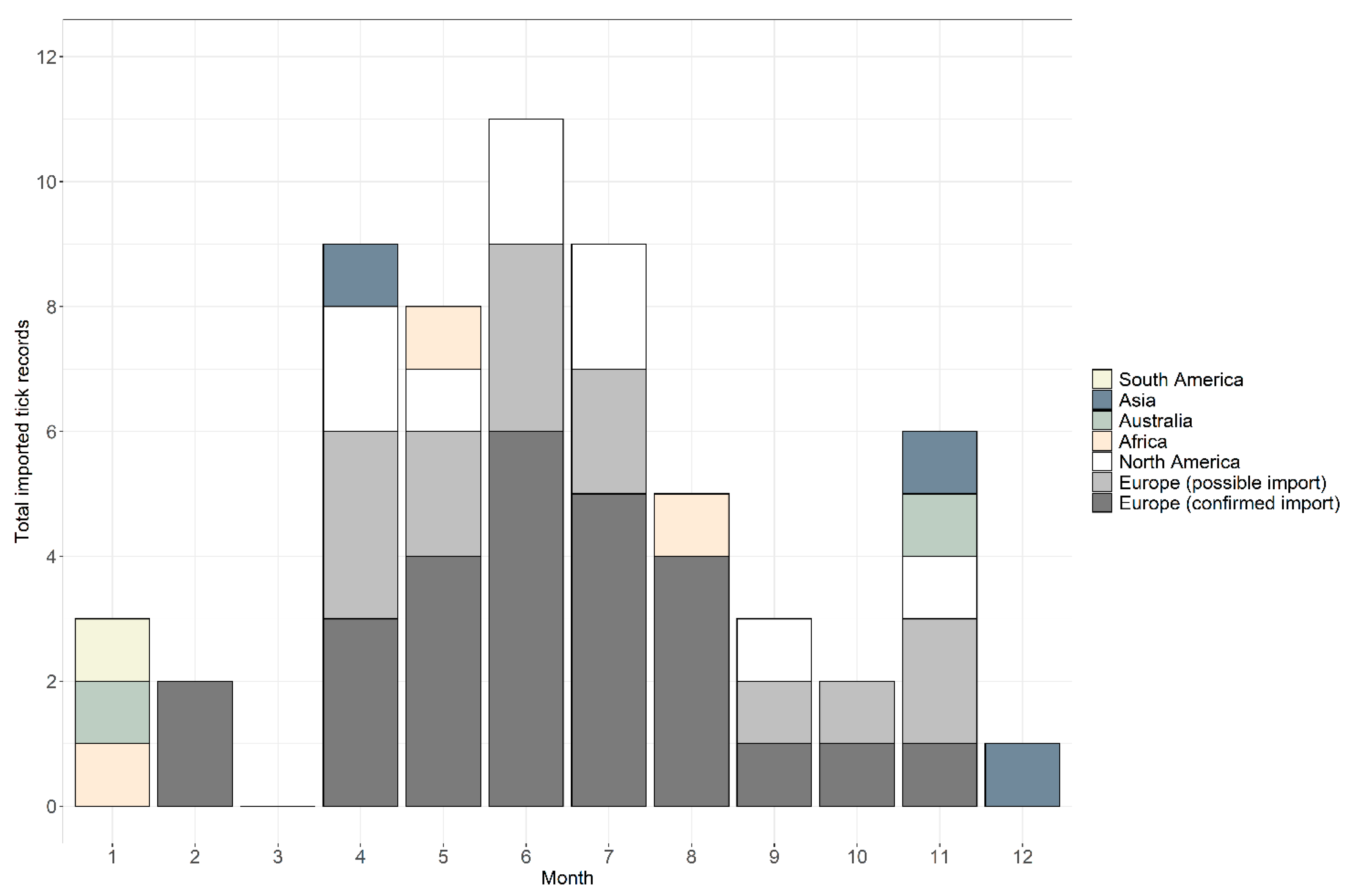
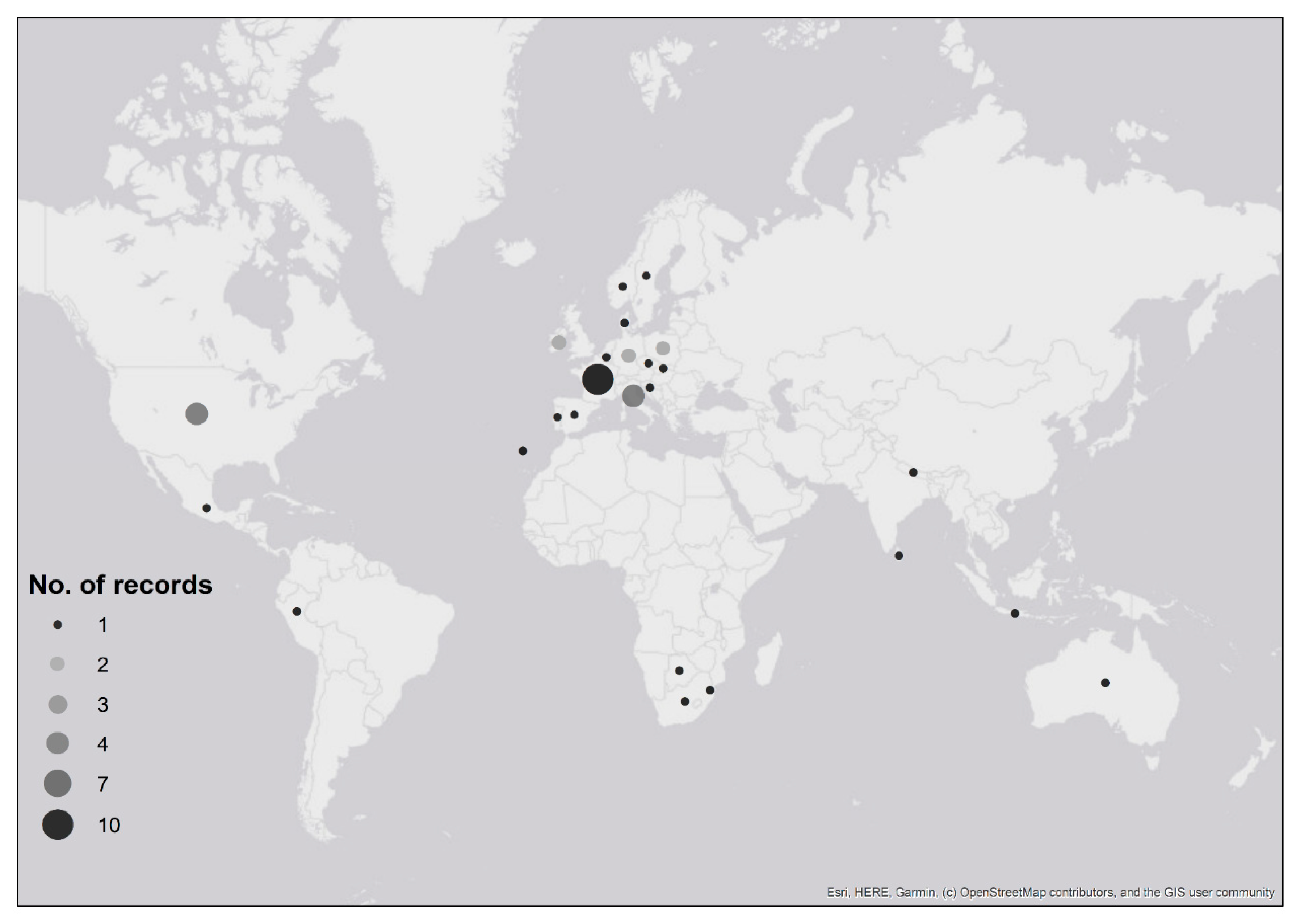
| Tick Species | Continent | History of Travel | No. Records | No. Confirmed/Possible Imported Records | Males | Females | Nymphs | Total Ticks |
|---|---|---|---|---|---|---|---|---|
| Amblyomma americanum | North America | USA | 5 | 5/0 | - | 3 | 3 | 6 |
| Amblyomma cajennense s.l. | North America | Mexico | 1 | 1/0 | - | 1 | - | 1 |
| Amblyomma species * | South America | Peru | 1 | 1/0 | - | - | 1 | 1 |
| Dermacentor andersoni | North America | USA/Canada | 1 | 1/0 | 1 | - | - | 1 |
| Dermacentor auratus | Asia | Nepal | 1 | 1/0 | - | - | 1 | 1 |
| Sri Lanka | 1 | 1/0 | - | - | 1 | 1 | ||
| Dermacentor marginatus | Europe | Holland/Germany/France | 1 | 1/0 | - | 1 | - | 1 |
| Italy | 2 | 2/0 | 2 | - | - | 2 | ||
| Dermacentor variablis | North America | USA | 1 | 1/0 | 1 | - | - | 1 |
| Haemaphysalis hystricis | Asia | Java, Indonesia | 1 | 1/0 | - | 1 | - | 1 |
| Hyalomma lusitanicum | Europe | Spain | 1 | 1/0 | 1 | - | - | 1 |
| Hyalomma truncatum | Africa | Botswana | 1 | 1/0 | 1 | - | - | 1 |
| Ixodes hexagonus | Europe | Madeira | 1 | 0/1 | - | 1 | - | 1 |
| Portugal | 1 | 1/0 | - | 1 | - | 1 | ||
| Ixodes holocyclus | Australia | Australia | 2 | 2/0 | - | 1 | 1 | 2 |
| Ixodes pacificus | North America | USA | 1 | 1/0 | - | - | 1 | 1 |
| Ixodes ricinus | Europe | Czechia | 1 | 1/0 | - | - | 1 | 1 |
| Denmark | 1 | 0/1 | - | 1 | - | 1 | ||
| France | 10 | 6/4 | - | 3 | 10 | 13 | ||
| Germany | 4 | 3/1 | - | 1 | 4 | 5 | ||
| Ireland | 3 | 1/2 | - | - | 4 | 4 | ||
| Italy | 4 | 2/2 | - | 1 | 3 | 4 | ||
| Norway | 2 | 2/0 | 3 | 1 | 1 | 5 | ||
| Poland | 3 | 1/2 | - | 2 | 1 | 3 | ||
| Slovenia | 1 | 1/0 | - | - | 1 | 1 | ||
| Spain | 1 | 1/0 | - | 1 | - | 1 | ||
| Sweden | 2 | 2/0 | - | 1 | 1 | 2 | ||
| Ixodes species * | Europe | Belgium | 1 | 0/1 | - | 1 | - | 1 |
| Rhipicephalus appendiculatus | Africa | Eswatini | 1 | 1/0 | - | - | 1 | 1 |
| Rhipicephalus gertrudae | Africa | South Africa | 1 | 1/0 | 1 | - | - | 1 |
| Rhipicephalus sanguineus s.l. | Europe | Croatia | 1 | 1/0 | 1 | - | - | 1 |
| Italy | 1 | 1/0 | - | - | 1 | 1 | ||
| Total | 59 | 45/14 | 11 | 21 | 36 | 68 | ||
| Tick Species | Distribution | Habitat and Climate Requirements | Hosts | Seasonality in Countries of Interest | Commonality of Human Biting |
|---|---|---|---|---|---|
| Ixodes ricinus (sheep or castor bean tick) | Most widespread tick across Europe, also present in parts of North Africa and Asia [24] | Deciduous and mixed forests, scrub, parks | Larvae: small mammals
Nymphs: woodland and game birds Adults: large mammals, particularly deer [25,26,27,28,29,30,31] | Active year-round; peaks in larvae during June. Nymphs and females peak in May-June, with smaller peak in September [32] | Most common tick species found biting humans in Europe |
| Dermacentor marginatus (ornate sheep tick) | Found south of the Alps, stretching from Portugal across Southern Europe, into northern Africa and mountain region of central Asia [23,33] | Dry habitats with sparse vegetation, e.g., alpine and forest steppes and semi-desert regions; tolerant to a wide range of environmental conditions [34,35,36] | Larvae: small mammals
Nymphs: small mammals Adults: larger mammals including horses, cattle, dogs, deer, sheep [34] | Larvae: June and July
Nymphs: July and August Adults: March–May, recommences in September & October [37,38] | Adults are known to bite humans [23,39] |
| Rhipicephalus sanguineus s.l. (brown dog tick) | Most widely distributed tick species in the world; more commonly associated with Mediterranean areas in Europe [40] | Capable of surviving in temperate regions. Can survive inside buildings, particularly those housing dogs but also residential homes | Dogs are the primary host of all stages, although immature stages can feed on rodents and small mammals | Active in temperate regions between spring and autumn [40]. Can be active year-round inside properties | Low probability of biting humans, but can be a problem in infested residential homes [39,41,42,43,44] |
| Hyalomma lusitanicum | Limited to a narrow strip from Sicily to Portugal and North Africa [45,46] | Adapted to meso-Mediterranean climate summer conditions [47] | Larvae and nymphs: Mediterranean rabbits (Oryctolagus cuniculus huxleyi)
Adults: ungulates, insectivores, carnivores [45,48,49] | Adult questing begins in March, peaks May–July then declines, with a small increase in September–October [47,50,51] | Human biting considered rare and accidental [49] |
| Amblyomma americanum (lone star tick) | Found in 39 states plus the District of Columbia in USA, stretches across eastern, south-eastern, mid-western and north-eastern USA [52,53] | Wooded areas with thick underbrush, as well as scrub and meadows, where the primary host (white-tailed deer, Odocoileus virginianus) resides | Larvae and nymphs feed on small- and medium-sized hosts, adults feed on medium- and large-sized mammals. Host range is vast, but white-tailed deer are the most important host and can feed all life stages—recent expansions in lone star tick populations due to increase in white-tailed deer populations [52] | Larval activity is highest during the summer, nymph and adult activity peaks April–June and then declines [52] | One of the most aggressive human-biting tick species, often the most abundant human biting species in many studies [52,53,54,55,56] |
| Dermacentor variabilis (American dog tick or wood tick) | Found in USA from Gulf of Mexico to New England and through the mid-western states to the east of the Rocky Mountains, with an independent population also found in California. Also present in parts of Mexico | Found in areas with limited tree cover, like brushy field habitat and scrubland [57] | Larvae and nymphs: small mammals
Adults: dogs, cervids, ruminants | Two peaks in adult questing: mid-April to late May; July [58] | Adults are the only stage known to bite humans [59] |
| Dermacentor andersoni (Rocky Mountain wood tick) | Found throughout the western United States and is established in at least 14 states [60] | Found in semiarid and mountainous areas with woodland, scrub and grassy areas [60,61,62] | Larvae and nymphs: small mammals, e.g., rats, squirrels, chipmunks
Adults: larger hosts such as cattle, deer, horses and elk [60]. | Larvae and nymphs occur between March and October: larval peaks occur from June to July whilst nymphs peak from May to June [60]. Adults are active between February and November, with greatest abundance recorded from March to April [60] | Larvae and nymphs rarely bite humans |
| Ixodes pacificus (western blacklegged tick) | Found in western parts of the United States, particularly Washington, Oregon, California, Arizona, Nevada and Utah [63] | More nymphs found in warmer and drier hardwood-dominated woodlands compared with cooler and more humid woodlands dominated by redwoods [64]. Survival is limited by summer drought conditions; larvae that hatch in the early summer remain in behavioral diapause until the following spring to avoid desiccation [64,65] | Larvae and nymphs: birds (migratory and non-migratory), rodents and lizards
Adults: birds and lizards, as well as larger mammals such as cats, dogs and ruminants [66,67,68] | Larval and nymphal infestation of birds and lizards peaks in April and May [66,69,70]. Questing nymphs active March–September, peaking in May and June, whilst adults are active October–June with peaks in November–January and March [71,72] | Primary human-biting species in California [71] |
| Ixodes holocyclus (paralysis/scrub typhus tick) | Found along the East Coast of Australia, from northern Queensland down to Victoria [73] | The limited distribution is thought to be driven by climatic conditions, particularly humidity, and it is often found in wet forested (scrub) areas [73] | Generalist feeder, has been recorded infesting mammals and birds [74]. In south-eastern Queensland, northern brown bandicoots (Isoodon macrourus) and long-nosed bandicoots (Perameles nasuta) are required for I. holocyclus populations to persist from one season to the next [74] | Nymphs most abundant between April and September; female adults peak between October and December [74] | Known to frequently bite humans [75] |
| Ixodes hexagonus (hedgehog tick) | Widespread distribution across temperate Europe and North Africa [34] | Nidicolous, found in dark humid places e.g., nests/burrows [76,77]; distribution is not limited by or dependent upon ambient climatic conditions | Hedgehogs are the most common hosts, but also found on a range of mammals [34] | Infestations occur throughout the year [78,79,80,81] | Human biting has been reported [17,82,83,84,85,86] |
| Amblyomma cajennense s.l. | Range spans from parts of southern USA, across Central America, the Caribbean, extending into South America to parts of northern Argentina [87] | Found in a wide range of habitats including dry grasslands, tropical forests and highlands of the Peruvian Andes [87] | Giant anteaters, wild pigs, tapir, water buffalo, dogs, horses, capybara and small mammals are common hosts [88,89,90,91,92,93] | Larvae most active between August and February, peak nymph activity between December and March, adult activity increases when daily mean temperature reaches 20 °C, peaking between February and September [89,90,94,95,96,97] | All life stages bite humans, and in South America it bites humans almost twice as frequently as all other tick species combined [98,99] |
| Hyalomma truncatum | Widely distributed throughout sub-Saharan Africa | Unknown | Parasitises a wide range of mammals, including rodents, lagomorphs, canids, ungulates and carnivores [9,100,101] | Unknown | Human infestation is rare [102], but there are at least two reports of this tick species found attached to returned travellers [7,9] |
| Rhipicephalus gertrudae | Found extensively across South Africa, with its distribution reaching the southern province of Western Cape, as well as northwards into Namibia and eastwards to the Free State province [103] | Unknown | Larvae and nymphs: small mammals [104,105]
Adults: cattle, goats, sheep, horses, zebra, antelopes, primates, dogs, cats [22,106,107,108,109,110] | Highest numbers of biting ticks occur between September and February [103,107,111] | Adults most commonly bite humans [111] |
| Rhipicephalus appendiculatus (brown ear tick) | Widely distributed between southern Sudan and the east coast of South Africa, with a noticeable absence in the Horn of Africa [112] | Unable to survive on open plains, and predominantly found in woodland and savannah with good vegetation cover, but can disappear following overgrazing [111] | Larvae and nymphs: small antelopes and hares
Adults: large ungulates including cattle, buffalo, wildebeest [113] | Larvae active late summer to winter (April–August)
Nymphs active in winter and early spring (July to October) Adults active in rainy season (December to March) [113] | All stages are known to bite humans [111] |
| Haemaphysalis hystricis | Distributed from the eastern Himalayas to India, through the coasts of Vietnam and China to Taiwan and Japan, and further south including Thailand, Malaysia and Indonesia [114,115,116,117,118,119] | Unknown | Commonly infests many hosts including cats, dogs, Japanese marten, wild boar, wildcats [115,117,120,121] | Unknown | Occasionally bite humans |
| Dermacentor auratus | Widespread distribution across much of Asia, including Borneo, China, India, Java, Laos, Malaysia, Myanmar, Nepal, Sri Lanka, Sumatra, Thailand and Vietnam [39,122,123,124,125,126,127,128,129,130,131,132] | It is a common tick species found in forests below 400 m altitude, although there are data of tick bites occurring at higher altitudes [131] | Larvae infest rodents; adults found on wild pigs, cattle, buffalo, deer, dogs and birds [125,126,129,131,132,133] | Unknown | Intra-aural infestation in particular has been reported [122,125,126,128,129,131,134] |
Publisher’s Note: MDPI stays neutral with regard to jurisdictional claims in published maps and institutional affiliations. |
© 2020 by the authors. Licensee MDPI, Basel, Switzerland. This article is an open access article distributed under the terms and conditions of the Creative Commons Attribution (CC BY) license (http://creativecommons.org/licenses/by/4.0/).
Share and Cite
Gillingham, E.L.; Cull, B.; Pietzsch, M.E.; Phipps, L.P.; Medlock, J.M.; Hansford, K. The Unexpected Holiday Souvenir: The Public Health Risk to UK Travellers from Ticks Acquired Overseas. Int. J. Environ. Res. Public Health 2020, 17, 7957. https://doi.org/10.3390/ijerph17217957
Gillingham EL, Cull B, Pietzsch ME, Phipps LP, Medlock JM, Hansford K. The Unexpected Holiday Souvenir: The Public Health Risk to UK Travellers from Ticks Acquired Overseas. International Journal of Environmental Research and Public Health. 2020; 17(21):7957. https://doi.org/10.3390/ijerph17217957
Chicago/Turabian StyleGillingham, Emma L., Benjamin Cull, Maaike E. Pietzsch, L. Paul Phipps, Jolyon M. Medlock, and Kayleigh Hansford. 2020. "The Unexpected Holiday Souvenir: The Public Health Risk to UK Travellers from Ticks Acquired Overseas" International Journal of Environmental Research and Public Health 17, no. 21: 7957. https://doi.org/10.3390/ijerph17217957
APA StyleGillingham, E. L., Cull, B., Pietzsch, M. E., Phipps, L. P., Medlock, J. M., & Hansford, K. (2020). The Unexpected Holiday Souvenir: The Public Health Risk to UK Travellers from Ticks Acquired Overseas. International Journal of Environmental Research and Public Health, 17(21), 7957. https://doi.org/10.3390/ijerph17217957