Evaluation of the Oxidative Stress Status in Zebrafish (Danio rerio) Liver Induced by Three Typical Organic UV Filters (BP-4, PABA and PBSA)


Abstract
1. Introduction
2. Materials and Methods
2.1. Reagents and Materials
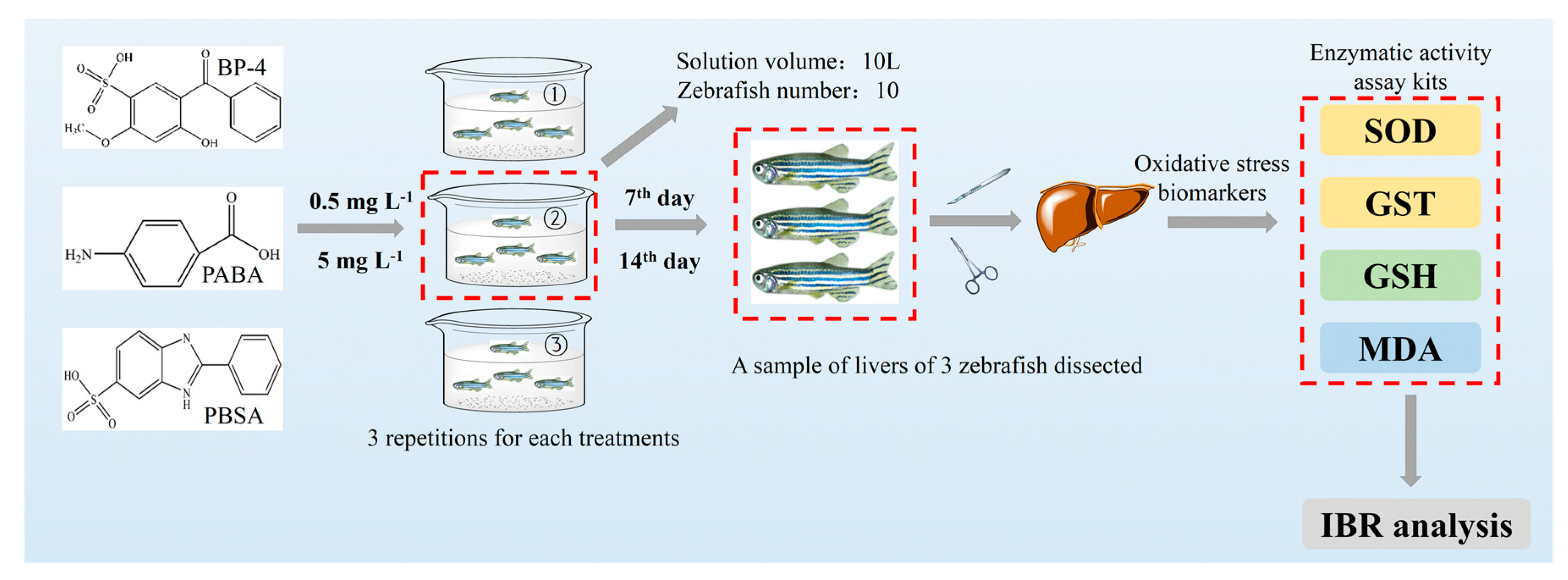
2.2. Fish Culture and Experimental Design
2.3. Sample Preparation and Biochemical Analysis
2.4. Determination of the Concentrations of Three Compounds in Exposure Medium
2.5. Integrated Biomarker Response (IBR)
2.6. Statistical Analysis
3. Results
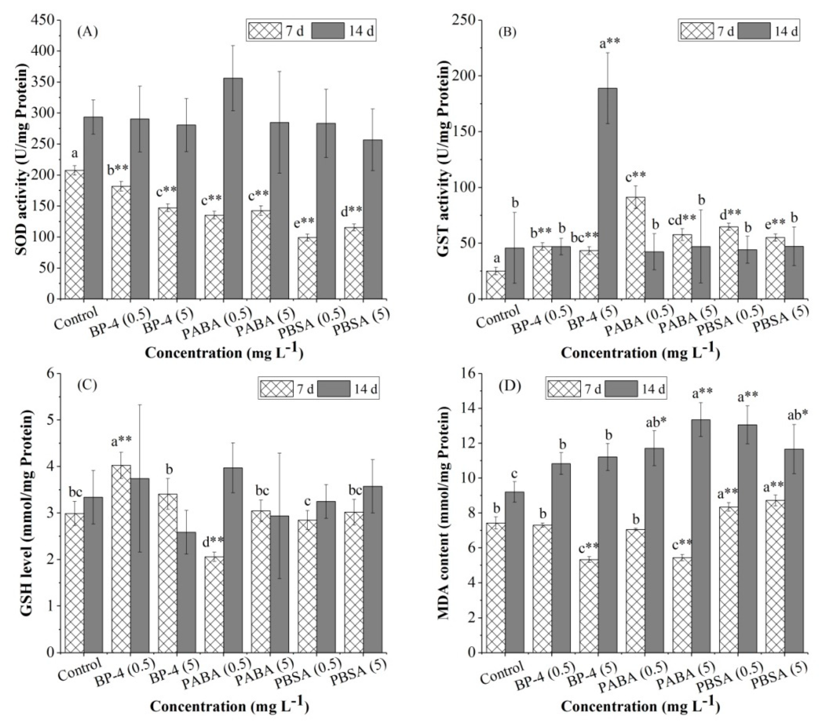
3.1. Antioxidant Enzyme Activities
3.2. GSH Levels
3.3. MDA Contents
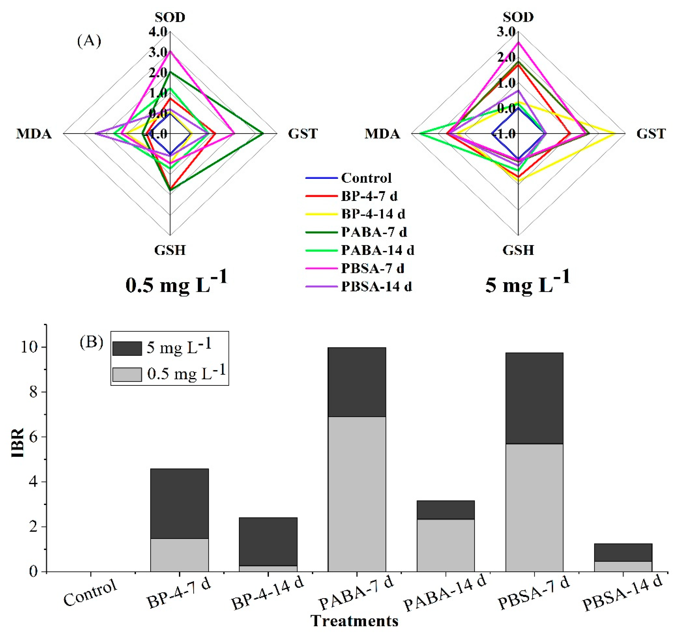
3.4. IBR Index
4. Discussion
4.1. Antioxidative Responses
4.2. Estimation of Lipid Peroxidation
4.3. Comparison of the Oxidative Stress-Inducing Potentials of Three Organic UV Filters in Zebrafish Liver
4.4. Limitations of the Present Study
5. Conclusions
Author Contributions
Funding
Conflicts of Interest
References
- Díaz-Cruz, M.S.; Llorca, M.; Barcelo, D. Organic UV filters and their photodegradates, metabolites and disinfection by-products in the aquatic environment. TrAC Trends Anal. Chem. 2008, 27, 873–887. [Google Scholar] [CrossRef]
- Gao, L.; Yuan, T.; Zhou, C.; Cheng, P.; Bai, Q.; Ao, J.; Wang, W.; Zhang, H. Effects of four commonly used UV filters on the growth, cell viability and oxidative stress responses of the Tetrahymena thermophila. Chemosphere 2013, 93, 2507–2513. [Google Scholar] [CrossRef]
- Chisvert, A.; León-González, Z.; Tarazona, I.; Salvador, A.; Giokas, D. An overview of the analytical methods for the determination of organic ultraviolet filters in biological fluids and tissues. Anal. Chim. Acta 2012, 752, 11–29. [Google Scholar] [CrossRef]
- Rodil, R.; Moeder, M. Development of a method for the determination of UV filters in water samples using stir bar sorptive extraction and thermal desorption–gas chromatography–mass spectrometry. J. Chromatogr. A 2008, 1179, 81–88. [Google Scholar] [CrossRef] [PubMed]
- Gao, L.; Yuan, T.; Wang, W.H. Ecological risk assessment of organic UV filters in aquatic environment. J. Environ. Health 2015, 32, 332–336. [Google Scholar]
- Kaiser, D.; Wappelhorst, O.; Oetken, M.; Oehlmann, J. Occurrence of widely used organic UV filters in lake and river sediments. Environ. Chem. 2012, 9, 139–147. [Google Scholar] [CrossRef]
- Buser, H.R.; Balmer, M.E.; Schmid, P.; Kohler, M. Occurrence of UV filters 4-methybenzylidene camphor and octocrylene in fish from various Swiss rivers with inputs from wastewater treatment plants. Environ. Sci. Technol. 2006, 40, 1427–1431. [Google Scholar] [CrossRef]
- Jurado, A.; Gago-Ferrero, P.; Vàzquez-Suñé, E.; Carrera, J.; Pujades, E.; Díaz-Cruz, M.S.; Barceló, B. Urban groundwater contamination by residues of UV filters. J. Hazard. Mater. 2014, 271, 141–149. [Google Scholar] [CrossRef]
- Balmer, M.E.; Buser, H.R.; Müller, M.D.; Poiger, T. Occurrence of some organic UV filters in wastewater, in surface waters, and in fish from Swiss Lakes. Environ. Sci. Technol. 2005, 39, 953–962. [Google Scholar] [CrossRef]
- Fent, K.; Kunz, P.Y.; Zenker, A.; Rapp, M. A tentative environmental risk assessment of the UV-filters 3-(4-methylbenzylidene-camphor), 2-ethyl-hexyltrime-thoxycinnamate, benzophenone-3, benzophenone-4 and 3-benzylidene camphor. Mar. Environ. Res. 2010, 69, S1–S6. [Google Scholar] [CrossRef]
- Lemaire, P.; Matthews, A.; Förlin, L.; Livingstone, D.R. Stimulation of oxyradical production of hepatic microsomes of flounder (Plafichthys flesus) and perch (Perca fluviatilis) by model and pollutant xenobiotics. Arch. Environ. Contam. Toxicol. 1994, 26, 191–200. [Google Scholar] [CrossRef] [PubMed]
- Canales-Aguirre, A.; Padilla-Camberos, E.; Gómez-Pinedo, U.; Salado-Ponce, H.; Feria-Velasco, A.; De Celis, R. Genotoxic effect of chronic exposure to DDT on lymphocytes, oral mucosa and breast cells of female rats. Int. J. Environ. Res. Public Health 2011, 8, 540–553. [Google Scholar] [CrossRef]
- Van der Oost, R.; Beyer, J.; Vermeulen, N.P. Fish bioaccumulation and biomarkers in environmental risk assessment: A review. Environ. Toxicol. Pharmacol. 2003, 13, 57–149. [Google Scholar] [CrossRef]
- Miller, L.L.; Wang, F.; Palace, V.P.; Hontela, A. Effects of acute and subchronic exposures to waterborne selenite on the physiological stress response and oxidative stress indicators in juvenile rainbow trout. Aquat. Toxicol. 2007, 83, 263–271. [Google Scholar] [CrossRef] [PubMed]
- Choudhary, M.; Jetley, U.K.; Khan, M.A.; Zutshi, S.; Fatma, T. Effect of heavy metal stress on proline, malondialdehyde, and superoxide dismutase activity in the cyanobacterium Spirulina platensis-S5. Ecotoxicol. Environ. Saf. 2007, 66, 204–209. [Google Scholar] [CrossRef] [PubMed]
- Campos, D.; Gravato, C.; Quintaneiro, C.; Golovko, O.; Žlábek, V.; Soares, A.M.V.M.; Pestana, J.L.T. Toxicity of organic UV-filters to the aquatic midge Chironomus riparius. Ecotoxicol. Environ. Saf. 2017, 143, 210–216. [Google Scholar] [CrossRef] [PubMed]
- Quintaneiro, C.; Teixeira, B.; Benedé, J.L.; Chisvert, A.; Soares, A.M.V.M.; Monteiro, M.S. Toxicity effects of the organic UV-filter 4-methylbenzylidene camphor in zebrafish embryos. Chemosphere 2019, 218, 273–281. [Google Scholar] [CrossRef]
- Mayden, R.L.; Tang, K.L.; Conway, K.W.; Freyhof, J.; Chamberlain, S.; Haskins, M.; Schneider, L.; Sudkamp, M.; Wood, R.M.; Agnew, M.; et al. Phylogenetic relationships of Danio within the order cypriniformes: A framework for comparative and evolutionary studies of a model species. J. Exp. Zool. Part B Mol. Dev. Evol. 2007, 308, 642–654. [Google Scholar] [CrossRef]
- Alzualde, A.; Behl, M.; Sipes, N.S.; Hsieh, J.H.; Alday, A.; Tice, R.R.; Paules, R.S.; Muriana, A.; Quevedo, C. Toxicity profiling of flame retardants in zebrafish embryos using a battery of assays for developmental toxicity, neurotoxicity, cardiotoxicity and hepatotoxicity toward human relevance. Neurotoxicol. Teratol. 2018, 70, 40–50. [Google Scholar] [CrossRef]
- McCord, J.M.; Fridovich, I. Superoxide dismutase an enzymic function for erythrocuprein (hemocuprein). J. Biol. Chem. 1969, 244, 6049–6055. [Google Scholar]
- Han, J.; Won, E.J.; Hwang, D.S.; Rhee, J.S.; Kim, I.C.; Lee, J.S. Effect of copper exposure on GST activity and on the expression of four GSTs under oxidative stress condition in the monogonont rotifer, Brachionus koreanus. Comp. Biochem. Phys. C 2013, 158, 91–100. [Google Scholar] [CrossRef]
- Devasagayam, T.P.A. Lipid peroxidation in rat uterus. BBA- Lipids Lipid Metab. 1986, 876, 507–514. [Google Scholar] [CrossRef]
- Jollow, D.; Mitchell, J.; Zampaglione, N.A.; Gillette, J. Bromobenzene-induced liver necrosis. Protective role of glutathione and evidence for 3, 4-bromobenzene oxide as the hepatotoxic metabolite. Pharmacology 1974, 11, 151–169. [Google Scholar] [CrossRef]
- Bradford, M.M. A rapid and sensitive method for quantitation of microgram quantities of protein utilizing the principle of protein-dye-binding. Anal. Biochem. 1976, 7, 248–254. [Google Scholar] [CrossRef]
- Beliaeff, B.; Burgeot, T. Integrated biomarker response: A useful tool for ecological risk assessment. Environ. Toxicol. Chem. 2002, 21, 1316–1322. [Google Scholar] [CrossRef] [PubMed]
- Broeg, K.; Lehtonen, K.K. Indices for the assessment of environmental pollution of the Baltic Sea coasts: Integrated assessment of a multi-biomarker approach. Mar. Pollut. Bull. 2006, 53, 508–522. [Google Scholar] [CrossRef] [PubMed]
- Zucchi, S.; Bluthgen, N.; Ieronimo, A. The UV-absorder benzophenone-4 alterstranscripts of genes involved in hormonal pathways in zebrafish (Danio rerio) eleuthero-embryos and adult males. Toxicol. Appl. Pharmacol. 2011, 250, 137–146. [Google Scholar] [CrossRef]
- Kimisue, T.; Chen, Z.; Louis, G.M.B.; Sundaram, R.; Hediger, M.L.; Kannan, K. Urinary concentrations of benzophenone-type UV filters in U.S. women and their association with endometriosis. Environ. Sci. Technol. 2012, 46, 4624–4632. [Google Scholar] [CrossRef]
- Kyrtopoulos, S.A. Biomarkers in environmental carcinogenesis research: Striving for a new momentum. Toxicol. Lett. 2006, 162, 3–15. [Google Scholar] [CrossRef]
- Tao, Y.X.; Pan, L.Q.; Zhang, H.; Tian, S.M. Assessment of the toxicity of organochlorine pesticide endosulfan in clams Ruditapes philippinarum. Ecotoxicol. Environ. Saf. 2013, 93, 22–30. [Google Scholar] [CrossRef]
- Puerto, M.; Pichardo, S.; Jos, A.; Prieto, A.I.; Sevilla, E.; Frías, J.E.; Cameán, A.M. Differential oxidative stress responses to pure microcystin-LR and microcystin-containing and non-containing cyanobacterial crude extracts on Caco-2 cells. Toxicon 2010, 55, 514–522. [Google Scholar] [CrossRef] [PubMed]
- Li, C.G.; Qin, L.; Qu, R.J.; Sun, P.; Wang, Z.Y. Responses of antioxidant defense system to polyfluorinated dibenzo-p-dioxins (PFDDs) exposure in liver of freshwater fish Carassius auratus. Ecotoxicol. Environ. Saf. 2016, 126, 170–176. [Google Scholar] [CrossRef] [PubMed]
- Falfushynska, H.; Horyn, O.; Fedoruk, O.; Khoma, V.; Rzymski, P. Difference in biochemical markers in the gibel carp (Carassius auratus gibelio) upstream and downstream of the hydropower plant. Environ. Pollut. 2019, 255, 113213. [Google Scholar] [CrossRef] [PubMed]
- Aytekin, T.; Kargın, F.; Çoğun, H.Y. The effect of copper on superoxide dismutase (SOD) activity in tissues of nile fish (Oreochromis niloticus). Toxicol. Lett. 2017, 280, S315. [Google Scholar] [CrossRef]
- Hellou, J.; Ross, N.W.; Moon, T.W. Glutathione, glutathione S-transferase, and glutathione conjugates, complementary markers of oxidative stress in aquatic biota. Environ. Sci. Pollut. Res. 2012, 19, 2007–2023. [Google Scholar] [CrossRef] [PubMed]
- Camara, A.Y.; Wan, Y.; Yu, Y.; Wang, Q.; Wang, K.; Li, H. Effect of endogenous selenium on arsenic uptake and antioxidative enzymes in as-exposed rice seedlings. Int. J. Environ. Res. Public Health 2019, 16, 3350. [Google Scholar] [CrossRef]
- Ali, M.; Parvez, S.; Pandey, S.; Atif, F.; Kaur, M.; Rehman, H.; Raisuddin, S. Fly ash leachate induces oxidative stress in freshwater fish Channa punctata (Bloch). Environ. Int. 2004, 30, 933–938. [Google Scholar] [CrossRef]
- Cheng, D.R.; Cao, K.; Wang, T.T.; Zhang, X.S.; Feng, M.B.; Liu, H. Evaluation of the oxidative stress in liver of crucian carp (Carassius auratus) induced by 3,4,4′-tri-CDE, 2-MeO-3′,4,4′-tri-CDE and 2-HO-3′,4,4′-tri-CDE. Environ. Sci. Pollut. Res. 2019, 26, 5164–5175. [Google Scholar] [CrossRef]
- Zhang, J.F.; Bai, K.W.; Su, W.P.; Wang, A.A.; Zhang, L.L.; Huang, K.H.; Wang, T. Curcumin attenuates heat-stress-induced oxidant damage by simultaneous activation of GSH-related antioxidant enzymes and Nrf2-mediated phase ii detoxifying enzyme systems in broiler chickens. Poult. Sci. 2018, 97, 1209–1219. [Google Scholar] [CrossRef]
- Woo, J.S.; Chung, J.K. Effects of trichlorfon on oxidative stress, neurotoxicity, and cortisol levels in common carp, Cyprinus carpio L., at different temperatures. Comp. Biochem. Physiol. Part. C Toxicol. Pharmacol. 2019. [Google Scholar] [CrossRef]
- Zhang, J.F.; Shen, H.; Wang, X.R.; Wu, J.C.; Xue, Y.Q. Effects of chronic exposure of 2,4-dichlorophenol on the antioxidant system in liver of freshwater fish Carassius auratus. Chemosphere 2004, 55, 167–174. [Google Scholar] [CrossRef] [PubMed]
- Ming, J.H.; Ye, J.Y.; Zhang, Y.X.; Xu, P.; Xie, J. Effects of dietary reduced glutathione on growth performance, non-specific immunity, antioxidant capacity and expression levels of IGF-I and HSP70 mRNA of grass carp (Ctenopharyngodon idella). Aquaculture 2015, 438, 39–46. [Google Scholar] [CrossRef]
- Chen, H.L.; Hsu, C.H.; Hung, D.Z.; Hu, M.L. Lipid peroxidation and antioxidant status in workers exposed to PCDD/Fs of metal recovery plants. Sci. Total Environ. 2006, 372, 12–19. [Google Scholar] [CrossRef]
- Zhao, F.; Xiang, Q.; Zhou, Y.; Xu, X.; Qiu, X.; Yu, Y. Evaluation of the toxicity of herbicide topramezone to, chlorella vulgaris: Oxidative stress, cell morphology and photosynthetic activity. Ecotoxicol. Environ. Saf. 2017, 143, 129–135. [Google Scholar] [CrossRef] [PubMed]
- Dong, W.Q.; Sun, H.J.; Zhang, Y.; Lin, H.J.; Chen, J.R.; Hong, H.C. Impact on growth, oxidative stress, and apoptosis-related gene transcription of zebrafish after exposure to low concentration of arsenite. Chemosphere 2018, 211, 648–652. [Google Scholar] [CrossRef] [PubMed]
- Souid, G.; Souayed, N.; Yaktiti, F.; Maaroufi, K. Effect of acute cadmium exposure on metal accumulation and oxidative stress biomarkers of Sparus aurata. Ecotoxicol. Environ. Saf. 2013, 89, 1–7. [Google Scholar] [CrossRef]
- Wang, G.H.; Xiong, D.M.; Wu, M.N.; Wang, L.X.; Yang, J. Induction of time- and dose-dependent oxidative stress of triazophos to brain and liver in zebrafish (Danio rerio). Comp. Biochem. Physiol. Part. C Toxicol. Pharmacol. 2020, 228, 108640. [Google Scholar] [CrossRef]
- Tsikas, D. Assessment of lipid peroxidation by measuring malondialdehyde (MDA) and relatives in biological samples: Analytical and biological challenges. Anal. Biochem. 2017, 524, 13–30. [Google Scholar] [CrossRef]
- Li, Y.; Li, M.; Shi, J.Q.; Yang, X.; Wang, Z.Y. Hepatic antioxidative responses to PCDPSs and estimated short-term biotoxicity in freshwater fish. Aquat. Toxicol. 2012, 120–121, 90–98. [Google Scholar] [CrossRef]
- Yonar, M.E. Chlorpyrifos-induced biochemical changes in Cyprinus carpio: Ameliorative effect of curcumin. Ecotoxicol. Environ. Saf. 2018, 151, 49–54. [Google Scholar] [CrossRef]
- Qu, R.J.; Feng, M.B.; Wang, X.H.; Qin, L.; Wang, C.; Wang, Z.Y.; Wang, L.S. Metal accumulation and oxidative stress biomarkers in liver of freshwater fish Carassius auratus following in vivo exposure to waterborne zinc under different pH values. Aquatic Toxicol. 2014, 150, 9–16. [Google Scholar] [CrossRef] [PubMed]


| Compounds | Chemical Structure | Log Kow a | Solubility (g L−1) b |
|---|---|---|---|
| BP-4 |  | 0.37 | 250 |
| PABA |  | 0.83 | 6.11 |
| PBSA |  | −0.16 | 23.6 |
| Compounds | Nominal Concentration | Measured Concentration |
|---|---|---|
| (mg L−1) | (mg L−1) a | |
| BP-4 | 0.5 | 0.47 ± 0.03 |
| 5 | 4.58 ± 0.23 | |
| PABA | 0.5 | 0.46 ± 0.04 |
| 5 | 4.79 ± 0.66 | |
| PBSA | 0.5 | 0.53 ± 0.06 |
| 5 | 5.38 ± 0.83 |
© 2020 by the authors. Licensee MDPI, Basel, Switzerland. This article is an open access article distributed under the terms and conditions of the Creative Commons Attribution (CC BY) license (http://creativecommons.org/licenses/by/4.0/).
Share and Cite
Huang, X.; Li, Y.; Wang, T.; Liu, H.; Shi, J.; Zhang, X. Evaluation of the Oxidative Stress Status in Zebrafish (Danio rerio) Liver Induced by Three Typical Organic UV Filters (BP-4, PABA and PBSA). Int. J. Environ. Res. Public Health 2020, 17, 651. https://doi.org/10.3390/ijerph17020651
Huang X, Li Y, Wang T, Liu H, Shi J, Zhang X. Evaluation of the Oxidative Stress Status in Zebrafish (Danio rerio) Liver Induced by Three Typical Organic UV Filters (BP-4, PABA and PBSA). International Journal of Environmental Research and Public Health. 2020; 17(2):651. https://doi.org/10.3390/ijerph17020651
Chicago/Turabian StyleHuang, Xinxin, Yuanyuan Li, Tantan Wang, Hui Liu, Jiaqi Shi, and Xuesheng Zhang. 2020. "Evaluation of the Oxidative Stress Status in Zebrafish (Danio rerio) Liver Induced by Three Typical Organic UV Filters (BP-4, PABA and PBSA)" International Journal of Environmental Research and Public Health 17, no. 2: 651. https://doi.org/10.3390/ijerph17020651
APA StyleHuang, X., Li, Y., Wang, T., Liu, H., Shi, J., & Zhang, X. (2020). Evaluation of the Oxidative Stress Status in Zebrafish (Danio rerio) Liver Induced by Three Typical Organic UV Filters (BP-4, PABA and PBSA). International Journal of Environmental Research and Public Health, 17(2), 651. https://doi.org/10.3390/ijerph17020651