Abstract
Pseudonocardians A–C (2–4), three new diazaanthraquinone derivatives, along with a previously synthesized compound deoxynyboquinone (1), were produced by the strain SCSIO 01299, a marine actinomycete member of the genus Pseudonocardia, isolated from deep-sea sediment of the South China Sea. The structures of compounds 1–4 were determined by mass spectrometry and NMR experiments (1H, 13C, HSQC, and HMBC). The structure of compound 1, which was obtained for the first time from a natural source, was confirmed by X-ray analysis. Compounds 1–3 exhibited potent cytotoxic activities against three tumor cell lines of SF-268, MCF-7 and NCI-H460 with IC50 values between 0.01 and 0.21 μm, and also showed antibacterial activities on Staphylococcus aureus ATCC 29213, Enterococcus faecalis ATCC 29212 and Bacillus thuringensis SCSIO BT01, with MIC values of 1–4 μg mL−1.
1. Introduction
Marine microorganisms, especially marine actinomycetes, are continuing to be rich sources of bioactive metabolites, with 273 new compounds reported from marine microbes in 2009 [1]. Natural products derived from marine actinomycetes displayed a wide range of bioactivities, such as antitumor [2,3], antiinfective [4], and antimalarial [5]. In recent years, South China Sea has been emerging as a potentially abundant source of novel species/genera of marine actinomycetes [6–10]. Some new bioactive compounds, such as marinactinones A–C [10], lobophorins E and F [11], were reported from marine actinomycetes isolated from the South China Sea.
In our continuous search for bioactive secondary metabolites from South China Sea-derived actinomycetes, crude extracts of the strain SCSIO 01299 were found to exhibit significant cytotoxic and antibacterial activities. Upon large fermentation of the strain SCSIO 01299, four products (1–4) were isolated and the main product 1 showed UV absorptions at 276, 354, 461 nm, characteristic of a highly conjugated chromophore related to anthraquinone compounds [12]. Herein we report the preliminary characterization of the strain SCSIO 01299 as a member of the genus Pseudonocardia by 16S rRNA gene sequence analysis, the isolation and structural elucidation of four diazaanthraquinone derivatives including a previously synthesized compound deoxynyboquinone (DNQ, 1) and three new analogues, pseudonocardians A–C (2–4). In addition, antibacterial and cytotoxic activities for compounds 1–4 were investigated.
2. Results and Discussion
2.1. Taxonomy of the Producing Strain
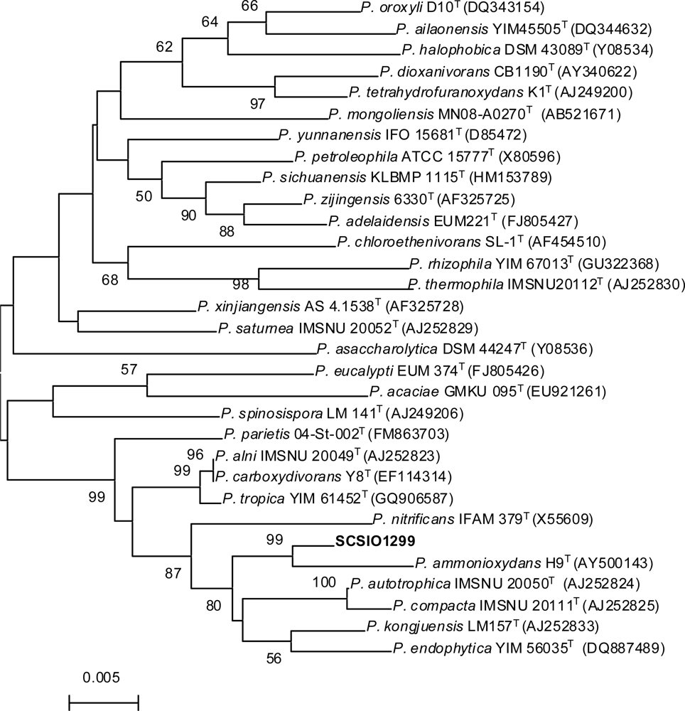
The strain SCSIO 01299 was isolated from deep-sea sediment (−3258 m) of the South China Sea, and was preliminarily identified as an actinomycetal species based on morphology observation. The 16S rRNA gene of the strain SCSIO 01299 was PCR amplified, sequenced and submitted to GenBank (accession number is JN204514). BLAST results showed that the new isolate had the highest similarity (98%) with Pseudonocardia autotrophica IMSNU 20050T [13]. The phylogenetic tree generated by a neighbor-joining method clearly revealed the evolutionary relationship of the strain SCSIO 01299 to a group of Pseudonocardia species (Figure 1). Thus, this strain was designated Pseudonocardia sp. SCSIO 01299.

Figure 1.
Phylogenetic dendrogram of the strain SCSIO 01299 and its closest relatives reconstructed by the neighbor-joining method based on 16S rRNA gene sequences.
2.2. Structural Elucidation
Compound 1 was obtained as red needles. It gave a [M + H]+ at m/z 285.0 and a [M − H]− at m/z 283.2 in the ESI-MS, indicating a molecular weight of 284.0. The 1H and 13C NMR spectra of 1 displayed 15 carbon signals, including two methyl doublets [δH 2.55 (3H, d, J = 1.0 Hz, Me-17), 2.59 (3H, d, J = 1.0 Hz, Me-16); δC 22.1 (q, Me-17), 23.0 (q, Me-16)] and one methyl singlet [δH 4.01 (3H, s, Me-15); δC 33.9 (q, Me-15)], two sp2 methines [δH 6.78 (1H, d, J = 1.0 Hz, H-7), 6.82 (1H, d, J = 1.0 Hz, H-3); δC 126.8 (d, C-3) and 127.1 (d, C-7)], and 10 sp2 quarternary carbons with four from carbonyls [δC 161.4 (s), 162.4 (s), 182.4 (s), and 176.9 (s)] (Table 1). The structure of ring A in 1 was constructed based on HMBC correlations of the methyl H3-15 to C-2/C-11, the methyl H3-16 to C-3/C-4/C-12, and H-3 to C-2/C-12/C-16. The structure of ring B was deduced from HMBC correlations of H-7 to C-8/C-13/C-17 and H3-17 to C-6/C-7/C-13. These two moieties were then connected through two carbonyls [δC 182.4 and 176.9]. Finally, 1 was unambiguously identified to be deoxynyboquinone (DNQ, Figures 2 and 3), a chemically synthesized compound [14,15], by X-ray crystallographic analysis.

Table 1.
1H and 13C NMR spectroscopic data of compounds 1–3.
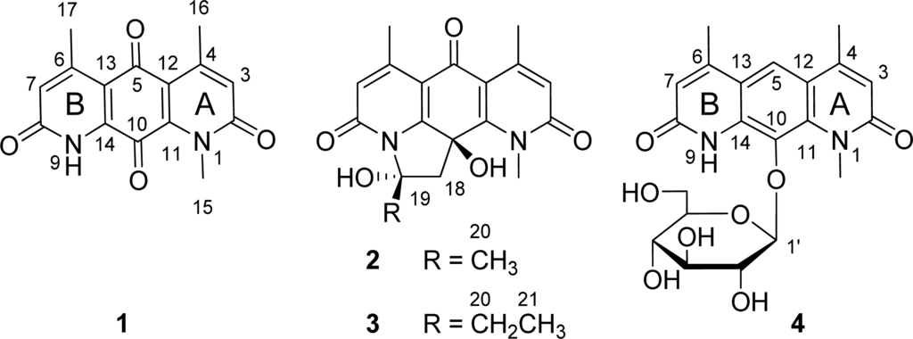
Figure 2.
Chemical structures of compounds 1–4.
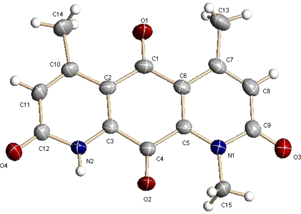
Figure 3.
X-ray analysis of compound 1.
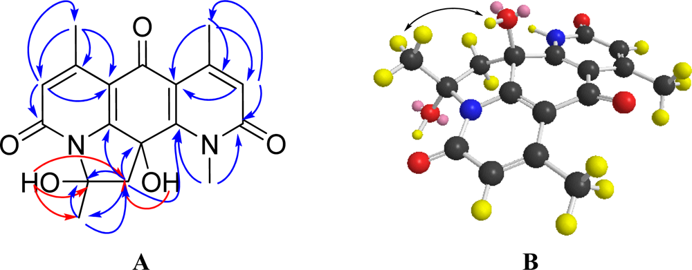
Compound 2, designated pseudonocardian A, was isolated as a white solid. The molecular formula of compound 2 was established as C18H18N2O5 (m/z 343.1305, calculated for 343.1294 [M + H]+), indicating 11 degrees of unsaturation. The 1H and 13C NMR spectra of compound 2 were similar to those of 1, except that the C-10 carbonyl in 1 was absent in 2. Instead, more modifications on C-10 were found in 2, including two oxygenated quaternary carbons [δC 72.7 (s, C-10), 97.6 (s, C-19)], a methylene [δH 2.71 (d, J = 13.5 Hz, H-18), 3.30 (d, J = 13.5 Hz, H-18), δC 52.4 (t, C-18)] and a methyl singlet [δH 2.19 (s, Me-20), δC 26.5 (q, Me-20)]. Taking the unsaturation degrees into consideration, there should be an additional ring in 2. Based on the HMBC correlations of H-18 to C-10/C-11/C-14/C-19, and of H-20 to C-18/C-19 (Figure 4A), C-19 was supposed to be connected to N-9. The assumption was confirmed by the downfield shift of C-19 at δC 97.6. After careful analysis of HMBC correlations, the planar structure of 2 was established (Figure 2). In order to assign the relative configuration of 2, a NOESY experiment was carried out in DMSO-d6. NOESY correlation of OH-10 to H3-20 was found (Figure 4B), indicating the two hydroxyls (OH-10 and OH-19) were on the opposite sides. This was consistent with the mimic configuration using the MM2 minimum energy calculation by ChemBio3D Ultra 11.0 (Figure 4B). Therefore, the relative configuration of compound 2 was shown in Figure 2.
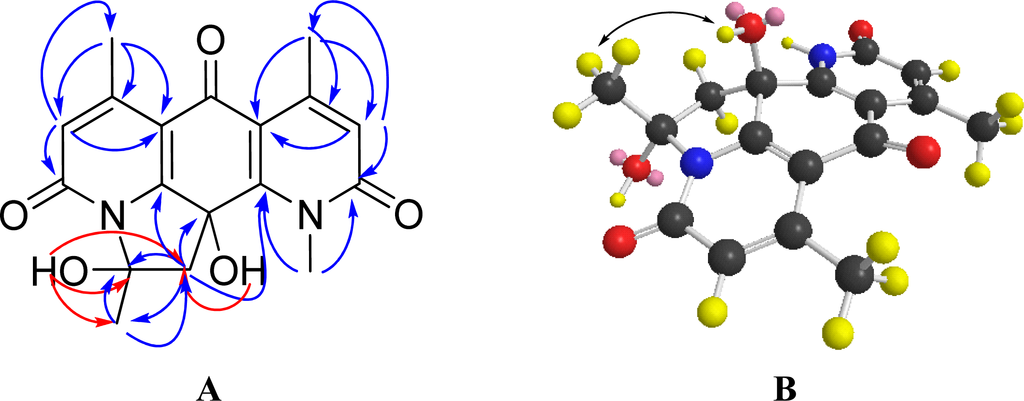
Figure 4.
Two dimensional NMR characterizations of compound 2: (A) Selected HMBC correlations; (B) Key NOESY correlation and mimic structure of 2, using the MM2 minimum energy calculation by ChemBio3D Ultra 11.0.
Compound 3, designated pseudonocardian B, was obtained as white solid. Its molecular formula was assigned as C19H20N2O5 based on the HR-MS data (m/z 357.1456, calculated for 357.1450, [M + H]+). Comparing the 1H and 13C NMR spectroscopic data of 3 with 2, the only difference was that the C-19 methyl singlet in 2 was substituted by an ethyl group [δH 1.22 (t, J = 7.5 Hz, Me-21), 2.41 (dd, J = 7.5, 14.0 Hz, H-20a), 2.84 (dd, J = 7.5, 14.0 Hz, H-20b), δC 9.3 (q, Me-21), 31.6 (t, C-20)] in 3 (Table 1). This substitution was confirmed by HMBC correlations of H-21 to C-19/C-20 and of H-20 to C-18/C-19/C-21. Therefore, the structure of 3 was established as shown in Figure 2.
Compound 4, designated pseudonocardian C, was isolated as a red brown powder. The molecular formula of 4 was established as C21H24N2O8 by HR-MS (m/z 433.1609, calculated for 433.1611 [M + H]+). In comparison of the 1H and 13C NMR spectroscopic data of 4 and 1, signals for both ring A [δH 6.57 (s, H-3), δC 166.0 (s, C-2), 151.4 (s, C-2), 137.3 (s, C-11), 120.8 (d, C-3), 118.7 (s, C-12)] and ring B [δH 6.54 (s, H-7), δC 164.9 (s, C-8), 149.4 (s, C-6), 135.1 (s, C-14), 120.4 (d, C-7), 120.4 (s, C-13)] were found in 4. However, the two ketone groups (C-5 and C-10) in 1 were displaced in 4 by a sp2 methine singlet [δH 8.01 (s, H-5), δC 119.7 (d, C-5)], an oxygenated sp2 quaternary carbon signals [δC 132.1 (s, C-10)], and a β-glucose moiety with coupling constant J1′,2′ of 8.0 Hz [16]: [δH 4.57 (1H, d, J = 8.0 Hz, H-1′), 3.66 (1H, m, H-2′), 3.44 (1H, m, H-3′), 3.46 (1H, m, H-4′), 3.13 (m, H-5′), 3.73 (1H, dd, J = 2.3, 11.8 Hz, H-6a′), 3.68 (1H, dd, J = 5.0, 11.8 Hz, H-6b′); δC 107.8 (d, C-1′), 75.5 (d, C-2′), 78.0 (d, C-3′), 70.9 (d, C-4′), 78.7 (d, C-5′), 62.3 (t, C-6′)]. The HMBC correlation from H-5 to C-4/C-6/C-11/C-14 indicated that moiety A and moiety B were connected by a benzene ring. And the HMBC correlation from H-1 to C-10 showed that the glucose was located at C-10. On these bases of these cumulative evidences, the structure of 4 was established as shown in Figure 2.
2.3. Biological Activities
Compounds 1–4 were evaluated for their antibacterial activities against Staphylococcus aureus ATCC 29213, Enterococcus faecalis ATCC 29212 and Bacillus thuringensis SCSIO BT01, and in vitro cytotoxic activities against three human tumor cell lines, including SF-268 (human glioma cell line), MCF-7 (human breast adenocarcinoma cell line) and NCI-H460 (human non-small cell lung cancer cell line) (Table 2). Compounds 1–3 showed strong antibacterial activities towards all three assayed indicators with MIC values ranging from 1–4 μg mL−1. Pseudonocardian A (2), bearing a methyl group at C-19, displayed one-fold less antibacterial activities than pseudonocardian B (3), which contained an ethyl group at C-19. DNQ (1), pseudonocardians A (2) and B (3) exhibited nearly the same potency against tumor cell lines SF-268 and MCF-7 with IC50 values in the range of 15–28 nM (Table 2). DNQ (1) showed a slightly better activity than pseudonocardians A (2) and B (3) against NCI-H460 (Table 2). Pseudonocardian C (4) showed no antibacterial activity, while preserved certain in vitro cytotoxic activities comparable to those of the control compound cisplatin. However, its cytotoxicities were largely reduced when compared to 1–3 (Table 2).
Table 2.
Antibacterial and cytotoxic activities of compounds 1–4.
2.4. Discussion
Diazaanthraquinones comprise a class of natural products, either naturally-occurring [17–22], or chemically synthesized [23–30], both of them exhibited good antitumor or antibacterial activities, highlighting their potential for drug development. Deoxynyboquinone (DNQ, 1), a diazaanthraquinone, was originally synthesized during nybomycin structural studies [14]. Recently, a facile synthetic route for DNQ (1) was reported [15]. Studies on biological activity and mechanism of action revealed that DNQ (1) rivaled doxorubicin for the potency in tumor cell culture and was even effective against a doxorubincin-resistant cell line, with a mechanism of inducing cell death through generation of reactive oxygen species (ROS) [15].
In this study, we reported the isolation of DNQ (1) for the first time from a natural source, the marine actinomycete Pseudonocardia sp. SCSIO 01299. In addition, we confirmed the structure of DNQ (1) by X-ray crystallographic analysis for the first time. More interestingly, we isolated 3 novel DNQ (1) analogues, pseudonocardians A–C (2–4), from the same strain. Consistent with previous report [15], DNQ (1) exhibited potent in vitro cytotoxic activities against three tumor cell lines of SF-268, MCF-7 and NCI-H460, with IC50 values of 22, 15, and 80 nM, respectively. Pesudonocardians A (2) and B (3) had almost the same in vitro cytotoxic activities as those of DNQ (1), with the added benefit of being ∼10-fold more aqueously soluble than 1. Pseudonocardian C (4), although being >300-fold less potent than 1–3, showed comparable cytotoxicity to the positive control cisplatin (Table 2). Unlike 1–3, pseudonocardian C (4) displayed no observable antibacterial activities, probably due to the glucosylation at C-10, which resembled the inactivation of macrolide antibiotics by glycosylation as a resistance mechanism [31].
3. Experimental Section
3.1. General Experimental Procedures
Materials for column chromatography (CC) were silica gel (100–200 mesh; 300–400 mesh; Jiangyou Silica Gel Development, Inc., Yantai, China), Sephadex LH-20 (40–70 μm; Amersham Pharmacia Biotech AB, Uppsala, Sweden), and YMC-GEL ODS-A (12 nm S-50 μm; Japan). Thin layer chromatography (TLC, 0.1–0.2 mm or 0.3–0.4 mm) was conducted with precoated glass plates (silica gel GF254, 10–40 nm, Jiangyou Silica Gel Development, Inc., Yantai, China). Medium pressure liquid chromatography (MPLC) was performed on automatic flash chromatography (EZ Purifier III, Leisure Science Co., Ltd. Shanghai, China) with monitor wavelength of 234 nm and collector wavelength of 300 nm. Mass spectral data were obtained on a quadrupole-time-of-flight mass spectrometry (Waters, Milford, MA, USA) for high resolution fast atom bombardment mass spectrometry (HRFABMS). The optical rotation was recorded on a 341 polarimeter (Perkin Elmer, Inc., Norwalk, CT, USA). 1H, 13C NMR and 2D NMR spectra were recorded on a Bruker AV-500 MHz NMR spectrometer (Bruker Biospin GmbH, Rheinstetten, Germany) with tetramethylsilane (TMS, δ 0.0 ppm) as the internal standard. 1H NMR data were reported as chemical shift (multiplicity [singlet (s), doublet (d), triplet (t), and multiplet (m)], and coupling constants (Hz); 13C NMR data were reported as follows: chemical shift [quaternary carbon (s), methine (d), methylene (t), methyl (q)]. Deuterated NMR solvents were purchased from Cambridge Isotopes (Andover, MA, USA).
3.2. Microbiological Material
The strain SCSIO 01299 was isolated from a sediment sample (E 120°0.975′, N 19°0.664′) at the depth of 3258 m collected from an open voyage to the South China Sea in August 2007, and was deposited in the type culture collection of Center for Marine Microbiology, Research Network of Applied Microbiology, South China Sea Institute of Oceanology, Chinese Academy of Sciences, Guangzhou, China. Genomic DNA isolation, PCR amplification of 16S rRNA gene, sequence alignment, and phylogenetic tree construction of the strain SCSIO 01299 were performed as described previously [6]. A single colony of SCSIO 01299 was inoculated into 50 mL seed medium (soybean meal 0.5%, soluble starch 1.5%, peptone bacteriological 1.5%, glycerol 1.5%, CaCO3 0.2%, sea salt 3%, pH 7.4, adjusted before sterilization) in 250 mL Erlenmeyer flasks, and was cultured on a rotary shaker at 200 r.p.m. and 28 °C for 2 days. 10% inoculums were transferred into 50 mL production medium (the same as the seed medium) in 250 mL Erlenmeyer flasks, and were subsequently incubated on a rotary shaker at 200 r.p.m., 28 °C for 5 days.
3.3. Extraction and Isolation
The fermentation broth (9 L) was extracted with equal volume of butanone for 4 times to afford the residue A after evaporation. The mycelia cake was extracted 3 times with 6 L acetone. After removing acetone, the residue was re-extracted by 6 L butanone to afford the residue B upon removal of the solvent under vacuum. Residues A and B were combined and subjected to column chromatography (CC) over silica gel (300–400 mesh, 150 g), eluting with a gradient of CHCl3/CH3OH (100:0→0:100) to give three fractions (Fr.1–Fr.3). Compound 1 (150.3 mg, 16.7 mg L−1) was obtained from fraction Fr.1 after elution with CHCl3/CH3OH (1:1) on Sephadex LH-20 and repeated recrystallization in CHCl3/CH3OH (10:1). Fraction Fr.2 was subjected to Sephadex LH-20 column chromatography, eluting with CHCl3/CH3OH (1:1), and then further chromatographed on silica gel (300–400 mesh, 40 g), eluting with CHCl3/CH3OH (20:1). Final purification was conducted by C18 reverse phase MPLC (20 × 2.5 cm ID), eluting with a linear gradient of CH3OH/H2O (0%→55%, 15 mL min−1, 100 min), to afford 2 (15.8 mg, 1.76 mg L−1) and 3 (12.5 mg, 1.39 mg L−1). Similarly, 4 (6.6 mg, 0.73 mg L−1) was obtained from Fr.4 after column chromatography on Sephadex LH-20 (CHCl3/CH3OH, 1:1) and further purification by RP-MPLC.
3.4. Characterization Data
Deoxynyboquinone (1). Red needles, UV (CH3CN/H2O/TFA) λmax: 276, 354, 461 nm. 1H and 13C NMR data, see Table 1; ESIMS m/z 285.0 [M + H]+, 569.7 [2M + H]+, 283.2 [M − H]−, 567.4 [2M − H]−.
Crystal data for deoxynyboquinone (1) [32]: Red triclinic crystal of C15H12N2O4. Space group P-1, a = 4.8353 (10) Å, α = 85.195(4)°; b = 9.373(2) Å, β = 89.350(5)°; c = 13.780 (3) Å, γ = 82.094(4)°; V = 616.5 (2) Å3, Z = 2; crystal size 0.301 × 0.126 × 0.057 mm3. A total of 3289 unique reflections (θ = 2.20–25.49°) were collected using graphite monochromated Mo Kα (λ = 0.71073 Å) on a CCD area detector diffractometer. The structure was solved by direct methods (SHELXS-97) and expanded using Fourier techniques (SHELXS-97). The final cycle of full-matrix least-squares refinement was based on 2273 data, 0 restraints and 198 variable parameters. Final R indicates R1 = 0.0690, wR2 = 0.1725 [I > 2σ(I)].
Pseudonocardian A (2). White solid,
+1.52° (c 0.46, MeOH), UV (CH3CN/H2O/TFA) λmax: 235, 300 nm. 1H and 13C NMR data, see Table 1. HRESI-MS [M + H]+ m/z 343.1305 (calcd. for C18H19N2O5 343.1294), ESIMS: m/z 343.2 [M + H]+, 685.4 [2M + Na]+, 341.1 [M − H]−, 683.3 [2M − H]−.
Pseudonocardian B (3). White solid,
−1.56° (c 0.90, MeOH), UV (CH3CN/H2O/TFA) λmax: 235, 300 nm. 1H and 13C NMR data, see Table 1. HRESI-MS: m/z [M + H]+ 357.1456 (calcd. for C19H21N2O5, 357.1450); ESIMS: m/z 357.2 [M + H]+, 379.3 [M + Na]+, 735.3 [2M + Na]+, 355.2 [M − H]−, 711.0 [2M − H]−.
Pseudonocardian C (4). Red brown powder,
−25.6° (c 0.16, MeOH), UV (CH3CN/H2O/TFA) λmax: 236, 264, 352, 368 nm. 1H NMR (500 MHz, CD3OD): 8.01 (1H, s, H-5), 6.57 (1H, s, H-3), 6.54 (1H, s, H-7), 4.57 (1H, d, J = 8.0 Hz, H-1′), 3.96 (1H, s, H-15), 3.73 (1H, dd, J = 2.3, 11.8 Hz, H-6′a), 3.68 (1H, dd, J = 5.0, 11.8 Hz, H-6′b), 3.66 (1H, m, H-2′), 3.46 (1H, m, H-4′), 3.44 (1H, m, H-3′), 3.13 (1H, m, H-5′), 2.63 (1H, s, H-16), 2.60 (1H, s, H-17); and 13C NMR (125 MHz, CD3OD): 166.0 (s, C-2), 164.9 (s, C-8), 151.4 (s, C-6), 149.4 (s, C-4), 137.3 (s, C-11), 135.1 (s, C-14), 132.1 (s, C-10), 120.8 (d, C-3), 120.4 (d, C-7), 120.4 (s, C-13), 119.7 (d, C-5), 118.7 (s, C-12), 107.8 (d, C-1′), 78.7 (d, C-5′), 78.0 (d, C-3′), 75.5 (d, C-2′), 70.9 (d, C-4′), 62.3 (t, C-6′), 37.4 (q, Me-15), 19.3 (q, Me-17), 19.0 (q, Me-16). HRESI-MS m/z 433.1609 [M + H]+ (calcd. for C21H25N2O8, [M + H]+ 433.1611); ESIMS m/z 433.2 [M + H]+, 455.4 [M + Na]+, 887.2 [2M + Na]+, 431.3 [M − H]−, 467.2 [M + Cl]−, 863.3 [2M − H]−.
3.5. Antibacterial and Cytotoxic Assay
Minimal Inhibition Concentration (MIC) values of deoxynyboquinone (1) and pseudonocardians A–C (2–4) were determined against 3 indicators (Staphylococcus aureus ATCC 29213, Enterococcus faecalis ATCC29212 and Bacillus thuringiensis SCSIO BT01), according to previously described methods [33]. Cytotoxicities of the four compounds were assayed against 3 tumor cell lines, including MCF-7 (human breast adenocarcinoma cell line), NCI-H460 (human non-small cell lung cancer cell line), and SF-268 (human glioma cell line). Assays were performed as described previously [34].
4. Conclusions
We have found that deep-sea actinomycete Pseudonocardia sp. SCSIO 01299 is a natural producer of the promising anticancer drug candidate deoxynyboquinone (1) and three diazaanthraquinone derivatives pseudonocardians A–C (2–4). In comparison with 1, pseudonocardians A (2) and B (3) showed the same anticancer potency but had enhanced aqueous solubility. These findings once again highlighted the potential of marine actinomycetes for novel drug discovery.
Acknowledgments
This study was supported in part by the Funds of the Chinese Academy of Sciences for Key Topics in Innovation Engineering (KSCX2-YW-G-065, KZCX2-YW-JC202, KSCX2-EW-G-12, LYQY200805), National Science Foundation for Young Scientists of China (41006089, 40906075), the 973 program (2010CB833805), Science and Technology planning project of Guangdong Province (2010B030600010), Natural Science Funds of South China Sea Institute of Oceanology for Young Scholar (SQ200903), and Open Project Program of Key Laboratory of Marine Bio-resources Sustainable Utilization (LMB091013). C. Z. is a scholar of the “100 Talents Project” of the Chinese Academy of Sciences (08SL111002). We are grateful to the analytical facilities in the South China Sea Institute of Oceanology, CAS for recording NMR data, South China Botanical Garden, CAS for MS data, and Guangdong Pharmaceutical University for HR-MS data.
References and Notes
- Blunt, JW; Copp, BR; Munro, MH; Northcote, PT; Prinsep, MR. Marine natural products. Nat. Prod. Rep. 2011, 28, 196–268. [Google Scholar]
- Bhatnagar, I; Kim, SK. Marine antitumor drugs: status, shortfalls and strategies. Mar. Drugs 2010, 8, 2702–2720. [Google Scholar]
- Olano, C; Mendez, C; Salas, JA. Antitumor compounds from marine actinomycetes. Mar. Drugs 2009, 7, 210–248. [Google Scholar]
- Rahman, H; Austin, B; Mitchell, WJ; Morris, PC; Jamieson, DJ; Adams, DR; Spragg, AM; Schweizer, M. Novel anti-infective compounds from marine bacteria. Mar. Drugs 2010, 8, 498–518. [Google Scholar]
- Fattorusso, E; Taglialatela-Scafati, O. Marine antimalarials. Mar. Drugs 2009, 7, 130–152. [Google Scholar]
- Tian, XP; Zhi, XY; Qiu, YQ; Zhang, YQ; Tang, SK; Xu, LH; Zhang, S; Li, WJ. Sciscionella marina gen. nov., sp. nov., a marine actinomycete isolated from a sediment in the northern South China Sea. Int. J. Syst. Evol. Microbiol. 2009, 59, 222–228. [Google Scholar]
- Tian, XP; Zhang, YQ; Li, QX; Zhi, XY; Tang, SK; Zhang, S; Li, WJ. Streptomyces nanshensis sp. nov., isolated from the Nansha Islands in the South China Sea. Int. J. Syst. Evol. Microbiol. 2009, 59, 745–749. [Google Scholar]
- Tian, XP; Tang, SK; Dong, JD; Zhang, YQ; Xu, LH; Zhang, S; Li, WJ. Marinactinospora thermotolerans gen. nov., sp. nov., a marine actinomycete isolated from a sediment in the northern South China Sea. Int. J. Syst. Evol. Microbiol. 2009, 59, 948–952. [Google Scholar]
- Dai, HQ; Wang, J; Xin, YH; Pei, G; Tang, SK; Ren, B; Ward, A; Ruan, JS; Li, WJ; Zhang, LX. Verrucosispora sediminis sp. nov., a cyclodipeptide-producing actinomycete from deep-sea sediment. Int. J. Syst. Evol. Microbiol. 2010, 60, 1807–1812. [Google Scholar]
- Tian, XP; Long, LJ; Wang, FZ; Xu, Y; Li, J; Zhang, J; Zhang, CS; Zhang, S; Li, WJ. Streptomyces nanhaiensis sp. nov., a novel marine streptomycete isolated from a deep sea sediment in Southern China Sea. Int. J. Syst. Evol. Microbiol. 2011. [Google Scholar] [CrossRef]
- Niu, S; Li, S; Chen, Y; Tian, X; Zhang, H; Zhang, G; Zhang, W; Yang, X; Zhang, S; Ju, J; Zhang, C. Lobophorins E and F, new spirotetranate antibiotics from a South China Sea-derived Streptomyces sp. SCSIO 01127. J. Antibiot. 2011, in press.. [Google Scholar]
- Chu, M; Mierzwa, R; Xu, L; Yang, SW; He, L; Patel, M; Stafford, J; Macinga, D; Black, T; Chan, TM; Gullo, V. Structure elucidation of Sch 538415, a novel acyl carrier protein synthase inhibitor from a microorganism. Bioorg. Med. Chem. Lett. 2003, 13, 3827–3829. [Google Scholar]
- Lee, SD; Kim, ES; Hah, YC. Phylogenetic analysis of the genera Pseudonocardia and Actinobispora based on 16S ribosomal DNA sequences. FEMS Microbiol. Lett. 2000, 182, 125–129. [Google Scholar]
- Rinehart, KL; Renefroe, HB. The structure of nybomycin. J. Am. Chem. Soc. 1961, 83, 3729–3731. [Google Scholar]
- Bair, JS; Palchaudhuri, R; Hergenrother, PJ. Chemistry and biology of deoxynyboquinone, a potent inducer of cancer cell death. J. Am. Chem. Soc. 2010, 132, 5469–5478. [Google Scholar]
- Agrawal, PK. NMR spectroscopy in the structureal elucidation of oligosaccharides and glycosides. Phytochemistry 1992, 31, 3307–3330. [Google Scholar]
- Maskey, RP; Grun-Wollny, I; Laatsch, H. Isolation and structure elucidation of diazaquinomycin C from a terrestrial Streptomyces sp. and confirmation of the akashin structure. Nat. Prod. Res. 2005, 19, 137–142. [Google Scholar]
- Vicent, MJ; Manzanaro, S; de la Fuente, JA; Duncan, R. HPMA copolymer-1,5-diazaanthraquinone conjugates as novel anticancer therapeutics. J. Drug Target. 2004, 12, 503–515. [Google Scholar]
- Egawa, K; Yamori, T; Nosaka, C; Kunimoto, S; Takeuchi, T; Nos, K. Deoxynybomycin is a selective anti-tumor agent inducing apoptosis and inhibiting topoisomerase I. Biol. Pharm. Bull. 2000, 23, 1036–1040. [Google Scholar]
- Murata, M; Miyasaka, T; Tanaka, H; Omura, S. Diazaquinomycin A, a new antifolate antibiotic, inhibits thymidylate synthase. J. Antibiot. 1985, 38, 1025–1033. [Google Scholar]
- Nadzan, AM; Rinehart, KL, Jr; Sokolski, WT. Hydroxynybomycin: isolation, structure and bioactivity. J. Antibiot. 1977, 30, 523–524. [Google Scholar]
- Naganawa, H; Wakashiro, T; Yagi, A; Kondo, S; Takita, T. Deoxynybomycin from a Streptomyces. J. Antibiot. 1970, 23, 365–368. [Google Scholar]
- Manzanaro, S; Vicent, MJ; Martin, MJ; Salvador-Tormo, N; Perez, JM; del Mar Blanco, M; Avendano, C; Menendez, JC; de la Fuente, JA. Synthesis and biological evaluation of new 1,5-diazaanthraquinones with cytotoxic activity. Bioorg. Med. Chem. 2004, 12, 6505–6515. [Google Scholar]
- Avendano, C; Perez, JM; Blanco Mdel, M; de la Fuente, JA; Manzanaro, S; Vicent, MJ; Martin, MJ; Salvador-Tormo, N; Menendez, JC. Synthesis and structure-activity relationships of 1,5-diazaanthraquinones as antitumour compounds. Bioorg. Med. Chem. Lett. 2004, 14, 3929–3932. [Google Scholar]
- Lee, H; Lee, SI; Cho, J; Choi, SU; Yang, SI. Synthesis and in vitro evaluation of 1,8-diazaanthraquinones bearing 3-dialkylaminomethyl or 3-(N-alkyl- or N-aryl)carbamoyloxymethyl substituent. Eur. J. Med. Chem. 2003, 38, 695–702. [Google Scholar]
- Lee, H; Lee, SI; Yang, SI. Synthesis and in vitro cytotoxicity of 3-substituted-1, 8-diazaanthraquinones produced by Lewis-acid catalyzed hetero Diels-Alder reaction. Bioorg. Med. Chem. Lett. 1998, 8, 2991–2994. [Google Scholar]
- Ramos, MT; Diaz-Guerra, LM; Garcia-Copin, S; Avendano, C; Garcia-Gravalos, D; Garcia de Quesada, T. Synthesis and antitumour activity of fluorinated 1-aza and 1,8-diazaanthraquinones. Farmaco 1996, 51, 375–379. [Google Scholar]
- Nebois, P; do Nascimento, SC; Boitard, M; Bartoli, MH; Fillion, H. Synthesis and in vitro cytotoxic activity of aza- and diazaanthraquinone derivatives. Pharmazie 1994, 49, 819–821. [Google Scholar]
- Gesto, C; de la Cuesta, E; Avendano, C; Emling, F. Synthesis and biological activity of new 1,8-diaza-2,9,10-anthracenetrione derivatives. J. Pharm. Sci. 1992, 81, 815–816. [Google Scholar]
- Tsuzuki, K; Yokozuka, T; Murata, M; Tanaka, H; Omura, S. Synthesis and biological activity of analogues of diazaquinomycin A, a new thymidylate synthase inhibitor. J. Antibiot. 1989, 42, 727–737. [Google Scholar]
- Salas, JA; Hernandez, C; Mendez, C; Olano, C; Quiros, LM; Rodriguez, AM; Vilches, C. Intracellular glycosylation and active efflux as mechanisms for resistance to oleandomycin in Streptomyces antibioticus, the producer organism. Microbiologia 1994, 10, 37–48. [Google Scholar]
- Crystallographic data (excluding structure factors) for the structure of DNQ (1) in this paper have been deposited in the Cambridge Crystallographic Data Centre as supplementary publication numbers CCDC 834052. Copies of the data can be obtained, free of charge, on application to CCDC, 12 Union Road, Cambridge CB2 1EZ, UK. Fax: +44(0)-1223-336033 or E-Mail: deposit@ccdc.cam.ac.uk.
- Xiao, Y; Li, S; Niu, S; Ma, L; Zhang, G; Zhang, H; Zhang, G; Ju, J; Zhang, C. Characterization of tiacumicin B biosynthetic gene cluster affording diversified tiacumicin analogs and revealing a tailoring Di-halogenase. J. Am. Chem. Soc. 2011, 133, 1092–1105. [Google Scholar]
- Wu, ZC; Li, DL; Chen, YC; Zhang, WM. A new isofuranonaphthalenone and benzopyrans from the endophytic fungus Nodulisporium sp. A4 from Aquilaria sinensis. Helv. Chim. Acta 2010, 93, 920–924. [Google Scholar]


© 2011 by the authors; licensee MDPI, Basel, Switzerland. This article is an open access article distributed under the terms and conditions of the Creative Commons Attribution license (http://creativecommons.org/licenses/by/3.0/).