Article

Luteophanol D, New Polyhydroxyl Metabolite from Marine Dinoflagellate Amphidinium sp.

by
Takaaki Kubota
,
Ayako Takahashi
,
Masashi Tsuda
and
Jun\'ichi Kobayashi
*
Graduate School of Pharmaceutical Sciences, Hokkaido University, Sapporo 060-0812, Japan
*
Author to whom correspondence should be addressed.
Mar. Drugs 2005, 3(4), 113-118; https://doi.org/10.3390/md304113
Submission received: 20 October 2005 / Accepted: 2 November 2005 / Published: 2 November 2005

Abstract

:
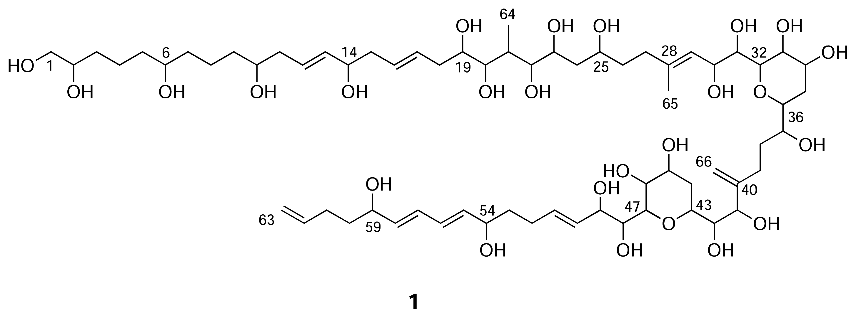
Luteophanol D (1), a new polyhydroxyl linear carbon-chain metabolite, has been isolated from the cultured marine dinoflagellate Amphidinium sp., which was isolated from Okinawan marine acoel flatworm Pseudaphanostoma luteocoloris. The structure of 1 was elucidated by detailed analyses of 2D NMR spectra. Luteophanol D (1) possesses two tetrahydropyran rings and twenty-three hydroxyl groups on C63-linear aliphatic chain with one exo-methylene and two methyl branches.

Introduction

During our continuing search for structurally unique secondary metabolites from marine dinoflagellates, we have isolated a series of cytotoxic macrolides, amphidinolides, as well as long chain polyhydroxyl metabolites from dinoflagellates Amphidinium sp. [1]. We previously investigated a strain of Amphidinium sp. (strain number Y-52), which was isolated from the inside cells of the Okinawan marine acoel flatworm Pseudaphanostoma luteocoloris, and isolated polyhydroxyl metabolites, luteophanols A ~ C [23]. Further investigation of extracts of the cultured dinoflagellate (Y-52) led to the isolation of a new polyhydroxyl metabolite, luteophanol D (1), possessing two tetrahydropyran rings and twenty three hydroxy groups on C63-linear aliphatic chain. In this paper we describe the isolation and structure elucidation of 1.
Marinedrugs 03 00113f3

Results and Discussion

The dinoflagellate was unialgally cultured at 25 °C for two weeks in seawater medium enriched with 1% ES supplement. The cultured algal cells were harvested by centrifugation and extracted with MeOH. The extract was partitioned between hexane and 1M NaCl aq, and the aqueous phase was successively extracted with CHCl3 and then n-BuOH. The n-BuOH soluble materials were subjected to gel filtration on Sephadex LH-20 (MeOH and then MeOH/H2O, 1:1) followed by purification with reversed-phase HPLC (MeOH/H2O) to afford luteophanol D (1, 0.00023 %, wet weight).
ESIMS of luteophanol D { 1, [α]D23 +4.7° (c 0.25, MeOH)} showed the pseudomolecular ion peak at m/z 1329 (M+Na)+, and its molecular formula, C66H114O25, was established by HRESIMS [m/z 1329.7522 (M+Na)+, Δ-2.5 mmu]. The UV and IR spectra indicated the presence of conjugated diene chromophore (λmax 232 nm) and hydroxy group (υmax 3430 cm−1), respectively. The 1H and 13C NMR data revealed that 1 contained two sp2 quaternary carbons, twelve sp2 methines, two sp2 methylenes, twenty-seven sp3 methines, of which twenty-six were oxymethines, twenty-one sp3 methylenes including one oxymethylene, and two methyl groups. Since eight out of ten elements of unsaturation implied by the molecular formula were accounted for, 1 was inferred to possess two rings. This suggested that luteophanol D (1) was a congener of luteophanol B or C.
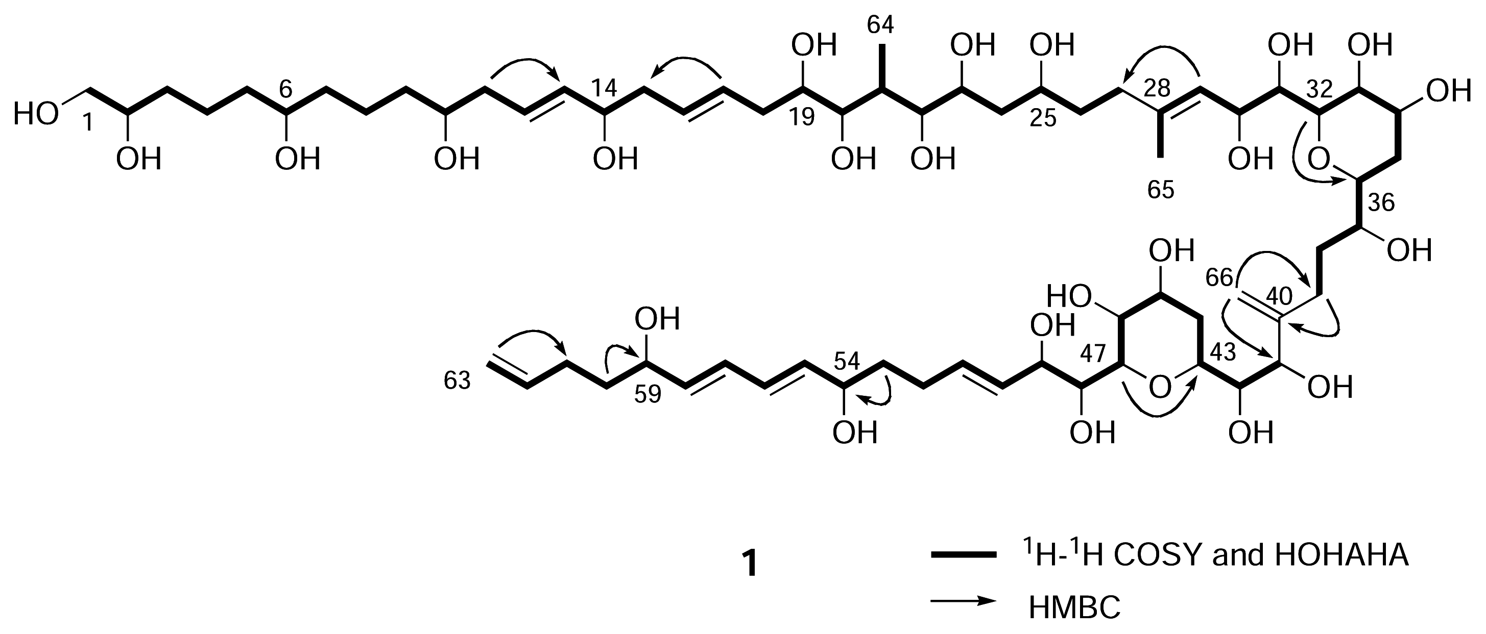
The structure of luteophanol D (1) was elucidated by extensive 2D NMR experiments including 1H-1H COSY, HOHAHA, ROESY, HSQC, and HMBC. Detailed analyses of 1H-1H COSY, HOHAHA, and HSQC spectra revealed the proton-proton connectivities from H2-1 to H2-27, from H3-65 to H2-39, and from H-41 to H2-63. The HMBC spectrum of 1 showed cross-peaks for H-29/C-27, H2-66/C-39, H2-66/C-41, and H2-39/C-40, indicating connecitivities of C-27 to C-28, C-39 to C-40, and C-40 to C-41. Two tetrahydropyran rings (C-32 to C-36 and C-43 to C-47) were assigned by HMBC cross-peaks for H-32/C-36 and H-47/C-43.
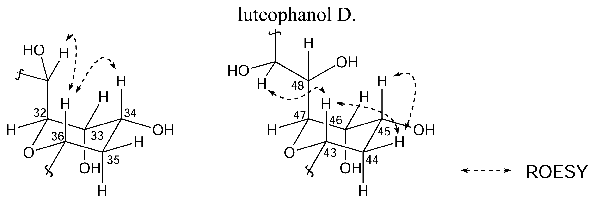
Five di- and one tri-substituted double bonds were indicated to have all E geometries by ROESY data (H-30/H3-65, H-54/H-56, H-55/H-57, H-56/H-58, and H-57/H-59) and 1H-1H coupling constants (J12,13 = 16 Hz, J16,17 = 15 Hz, and J51,52 = 15 Hz). Relative stereochemistry of the two tetrahydropyran rings (C-32 to C-36 and C-43 to C-47) was elucidated on the basis of ROESY data and 1H-1H coupling constants (J32/33 = ~0 Hz, J33/34 = ~0 Hz, J34/35a = 10 Hz, J34/35b = 2 Hz, J35a/36 = 10 Hz, J35b/36 = 2 Hz, J43/44a = 10 Hz, J43/44b = 2 Hz, J44a/45 = 9 Hz, J44b/45 = 2 Hz, J45/46 = ~0 Hz, and J46/47 = ~0 Hz) of 1 (Figure. 2). Thus, luteophanol D (1) was assigned as a luteophanol B congener lacking one methylene between C-52 and C-54 of luteophanol B.
Luteophanol D (1) possesses two tetrahydropyran rings and twenty-three hydroxyl groups on a C63-1inear carbon chain with one exo-methylene and two methyl branches. Luteophanol D (1) contains a hydrophilic diene portion at C-54 ~ C-59, whereas known polyhydroxyl metabolites, amphidinols [48] and lingshuiols [910], previously isolated from dinoflagellates Amphidinium sp. comprise a hydrophobic moiety in this portion. The biosynthesis of amphidinols has been studied by acetate incorporation experiments [11]. Since the polyketide chain of 1 was shorter than those of luteophanols B and C by only one carbon, this truncation of the carbon chain might be occurred by unusual dinoflagellate polyketide biosynthesis. Luteophanol D (1) exhibited antibacterial activity against Micrococcus luteus (MIC, 33 μg/ml).

Experimental

General

IR and UV spectra were recorded on JASCO FT/IR-5300 and JASCO Ubest-35 spectrophotometers, respectively. 1H and 13C NMR spectra were recorded on a Bruker AMX-600 spectrometer. The 3.35 and 49.8 ppm resonances of residual CD3OD were used as internal references for 1H and 13C NMR spectra, respectively. ESI mass spectra were measured on JEOL JMX-SX102A spectrometer.

Cultivation and Isolation

The dinoflagellate Amphidinium sp. (strain number Y-52) was unialgally cultured at 25 °C for two weeks in seawater medium enriched with 1% ES supplement. The harvested cells of the cultured dinoflagellate (385 g wet weight, from 725 L of culture) were extracted with MeOH (1 L x 3). The MeOH extract (15.19 g) was partitioned between hexane (500 mL x 3) and 1M NaCl aq., and the aqueous phase was successively extracted with CHCl3 (500 mL x 3) and then n-BuOH (500 mL x 3). The n-BuOH soluble fraction (4.47 g) was subjected to a Sephadex LH-20 column [MeOH and then MeOH/H2O, (1:1)] followed by reversed-phase HPLC (Develosil ODS-5, 10 x 250 mm; eluent: 55% MeOH/H2O then 45% MeOH/H2O; flow rate: 2.5 mL/min; UV detection at 226 nm) to afford luteophanol D (1, 0.9 mg, 0.00023 % wet weight).
Luteophanol D (1). Colorless amorphous solid; [α]D23 +4.7° (c 0.25, MeOH); UV (MeOH) λmax 232 nm (ɛ 22800); IR υmax 3430, 2920, 1640, and 1050 cm−1; ESIMS m/z 1329 (M + Na)+; HRESIMS m/z 1329.7522 (M + Na)+. Calcd. for C66H114O25Na, 1329.7547; 1H NMR (CD3OD/C5D5N 2:1) δ 3.62 (2H, H2-1); 3.75 (m, 1H, H-2); 1.50 (1H, H-3a); 1.65 (1H, H-3b); 1.47 (1H, H-4a); 1.75 (1H, H-4b); 1.50 (2H, H2-5); 3.63 (1H,H-6); 1.50 (2H, H2-7); 1.47 (1H, H-8a); 1.75 (1H, H-8b); 1.47 (1H, H-9a); 1.58 (1H, H-9b); 3.68 (1H, H-10); 2.25 (2H, H2-11); 5.80 (1H, H-12); 5.63 (1H, H-13); 4.17 (1H,H-14); 2.35 (2H, H2-15); 5.67 (1H, H-16); 5.76 (1H, H-17); 2.30 (1H, H-18a ); 2.70 (1H, H-18b); 3.75 (1H, H-19); 3.77 (1H, H-20); 2.67 (1H, H-21); 3.77 (1H,H-22); 3.95 (1H, H-23); 1.70 (1H, H-24a); 2.13 (1H, H-24b); 4.02 (1H, H-25); 1.70 (2H, H2-26); 2.10 (1H, H-27a); 2.32 (1H, H-27b); 5.70 (1H, H-29); 4.80 (1H, H-30); 3.90 (1H,H-31); 4.33 (1H, H-32); 4.40 (1H,H-33); 4.20 (1H, H-34); 2.02 (1H, H-35a); 2.11 (1H, H-35b); 3.68 (1H, H-36); 3.77 (1H, H-37); 1.75 (1H, H-38a); 2.19 (1H, H-38b); 2.32 (1H, H-39a); 2.69 (1H, H-39b); 4.45 (1H, H-41); 3.58 (1H, H-42); 4.30 (1H, H-43); 1.70 (1H,H-44a); 2.40 (1H, H-44b); 4.26 (1H, H-45); 4.39 (1H, H-46); 4.08 (1H, H-47); 4.28 (1H, H-48); 4.70 (1H, H-49); 5.90 (1H, H-50); 5.86 (1H, H-51); 2.18 (2H, H2-52); 1.65 (1H, H-53a); 1.70 (1H, H-53b); 4.18 (1H, H-54), 5.73 (1H, H-55); 6.30 (1H, H-56), 6.30 (1H, H-57), 5.78 (1H, H-58), 4.18 (1H, H-59), 1.60 (1H, H-60a); 1.65 (1H, H-60b); 2.15 (2H, H2-61); 5.83 (1H, H-62); 4.93 (1H, H-63a); 5.01 (1H, H-63b); 1.18 (d3H, H3-64); 1.75 (3H, H3-65); 5.03 (1H, H-66a); 5.18 (1H, H-66b); 13C NMR (CD3OD/C5D5N 2:1) δ C-1, 68.3; C-2, 74.0; C-3, 35.5; C-4, 23.8; C-5, 39.4; C-6, 72.8; C-7, 39.4; C-8, 23.8; C-9, 38.8; C-10, 72.8; C-11, 42.3; C-12, 129.4; C-13, 137.7; C-14, 74.0; C-15, 43.0; C-16, 131.2; C-17, 131.7; C-18, 39.2; C-19, 74.0; C-20, 80.4; C-21, 35.9; C-22, 81.1;C-23, 73.2; C-24, 42.3; C-25, 72.5; C-26, 37.7; C-27, 37.5;C-28, 139.4; C-29, 127.7; C-30, 68.6; C-31, 73.3; C-32, 80.2; C-33, 69.8; C-34, 68.2; C-35, 31.6; C-36, 76.6; C-37, 75.3; C-38, 33.7; C-39, 28.9; C-40, 152.9; C-41, 77.4; C-42, 76.2; C-43, 71.5; C-44, 32.7; C-45, 68.1; C-46, 69.8; C-47, 81.4; C-48, 73.0; C-49, 75.0; C-50, 130.5; C-51, 135.4; C-52, 30.4; C-53, 38.8; C-54, 73.0; C-55, 132.3; C-56, 131.5; C-57, 131.5; C-58, 138.5; C–59, 73.0; C-60, 38.5; C-61, 31.4; C-62, 140.4; C-63, 115.9; C-64, 7.8; C-65, 18.2;C-66, 113.5.
Figure 1. Selected 2D NMR data of luteophanol D (1).
Marinedrugs 03 00113f1
Figure 2. Selected ROESY data and relative stereochemistry of two tetrahydropyran rings of luteophanol D.
Marinedrugs 03 00113f2

Acknowledgments

We thank Mr. K. Watanabe, GC-MS & NMR laboratory, Faculty of Agriculture, Hokkaido University, for measurements of HRESIMS. This work was supported by Grant-in-Aid for Science Research from the Ministry of Education, Culture, Sports, Science, and Technology of Japan.
  • Sample availability: Not available.

Reference and Notes

  1. Kobayashi, J.; Tsuda, M. Amphidinolides, bioactive macrolides from symbiotic marine dinoflagellates. Nat. Prod. Rep 2004, 21, 77–93. [Google Scholar]
  2. Doi, Y.; Ishibashi, M.; Nakamichi, H.; Kosaka, T.; Ishikawa, T.; Kobayashi, J. Luteophanol A, a New Polyhydroxyl Compound from Symbiotic Marine Dinoflagellate Amphidinium sp. J. Org. Chem 1997, 62, 3820–3823. [Google Scholar]
  3. Kubota, T.; Tsuda, M.; Doi, Y.; Takahashi, A.; Nakamichi, H.; Ishibashi, M.; Fukushi, E.; Kawabata, J.; Kobayashi, J. Luteophanols B and C, New Polyhydroxyl Compounds from Marine Dinoflagellate Amphidinium sp. Tetrahedron 1998, 54, 14455–14464. [Google Scholar]
  4. Satake, M.; Murata, M.; Yasumoto, T.; Fujta, T.; Naoki, H. Amphidinol, a Polyhydroxypolyene Antifungal Agent with an Unprecedented Structure, from a Marine Dinoflagellate, Amphidnium klebsii. J. Am. Chem. Soc 1991, 113, 9851–9861. [Google Scholar]
  5. Paul, G. K.; Matsumori, N.; Murata, M.; Tachibana, K. Isolation and Chemical Structure of Amphidinol 2, a Potent Hemolytic Compound from Marine Dinoflagellate Amphidinium klebsii. Tetrahedron Lett 1995, 36, 6279–6282. [Google Scholar]
  6. Paul, G. K.; Matsumori, N.; Konoki, K.; Murata, M.; Tachibana, K. Chemical Structures of Amphidinols 5 and 6 Isolated from Marine Dinoflagellate Amphidinium klebsii and Their Cholesterol-dependent Membrane Disruption. J. Mar. Biotechnol 1997, 5, 124–128. [Google Scholar]
  7. Echigoya, R.; Rhodes, L.; Oshima, Y.; Satake, M. The Structures of Five New Antifungal and Hemolytic Amphidinol Analogs from Amphidinium carterae Collected in New Zealand. Harmful Algae 2005, 4, 383–389. [Google Scholar]
  8. Morsy, N.; Matsuoka, S.; Houdai, T.; Matsumori, N.; Adachi, S.; Murata, M.; Iwashita, T.; Fujita, T. Isolation and Structure Elucidation of a New Amphidinol with a Truncated Polyhydroxyl Chain from Amphidinium klebsii. Tetrahedron 2005, 61, 8606–8610. [Google Scholar]
  9. Huang, X.; Zhao, D.; Guo, Y.; Wu, H.; Lin, L.; Wang, Z.; Ding, J.; Lin, Y. Lingshuiol, a Novel Polyhydroxyl Compound with Strongly Cytotoxic Activity from the Marine Dinoflagellate Amphidinium sp. Bioorg Med. Chem. Lett 2004, 14, 3117–3120. [Google Scholar]
  10. Huang, X.; Zhao, D.; Guo, Y.; Wu, H.; Trivellone, E.; Cimino, G. Lingshuiols A and B, Two New Polyhydroxy Compounds from the Chinese Marine Dinoflagellate Amphidinium sp. Tetrahedron Lett 2004, 45, 5501–5504. [Google Scholar]
  11. Houdai, T.; Matsuoka, S; Murata, M.; Satake, M.; Ota, S.; Oshima, Y.; Rhodes, L. L. Acetate labeling Patterns of Dinoflagellate polyketides, Amphidinols 2, 3 and 4. Tetrahedron 2001, 57, 5551–5555. [Google Scholar]