Identification of Antioxidant and Anti-Inflammatory Activity of Sea Cucumber (Holothuria tubulosa) Active Peptides by a Combined Approach of Omics Data and Bioinformatics Analysis

 , , , ,
  ,
,  and
and 
Abstract
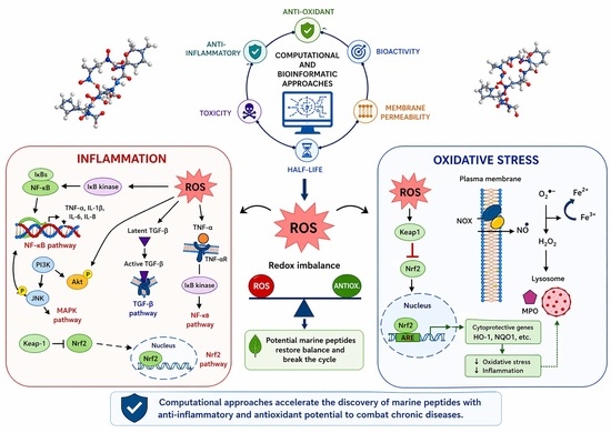
1. Introduction
2. Results
3. Discussion
4. Materials and Methods
4.1. Animals
4.2. Sample Preparation
4.3. Proteome
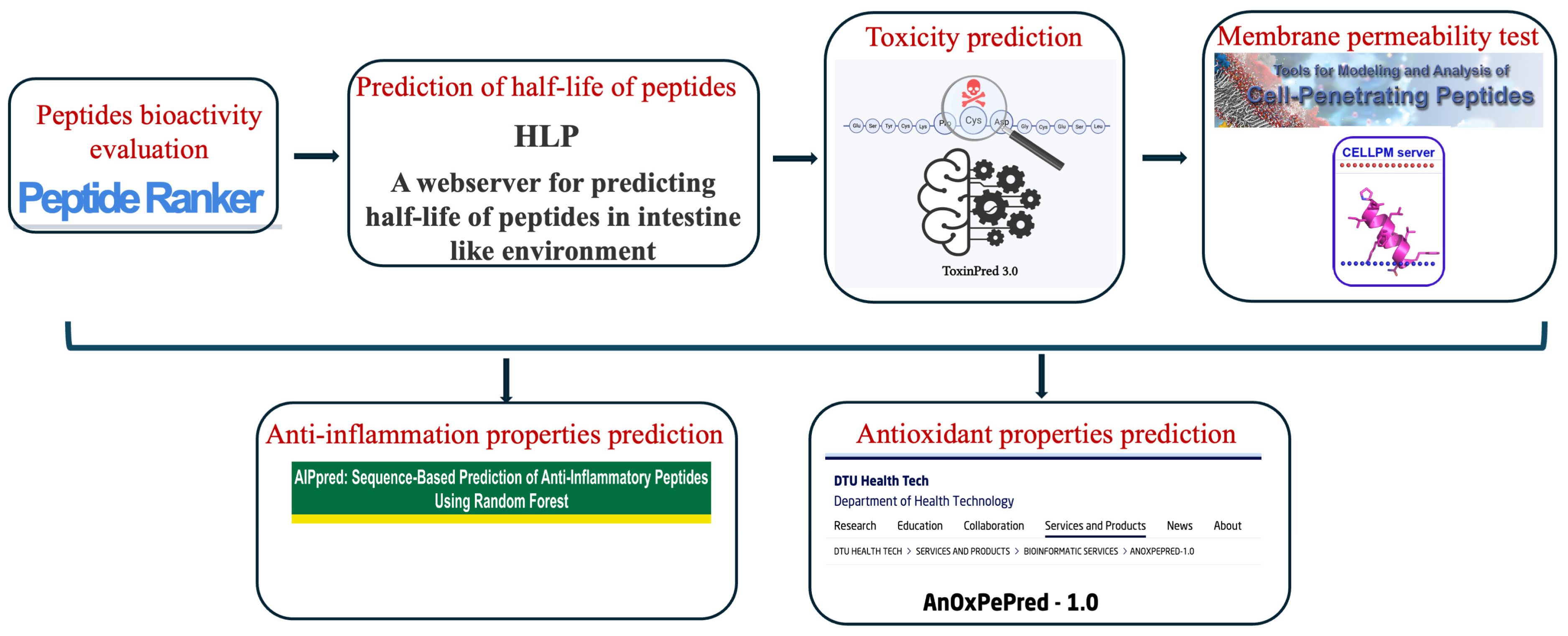
4.4. Peptide Bioinformatics Analysis Workflow
4.5. Bioinformatics Analysis
4.5.1. Peptides Bioactivity Evaluation
4.5.2. Prediction of Half-Life of Peptides
4.5.3. Peptides Toxicity Prediction
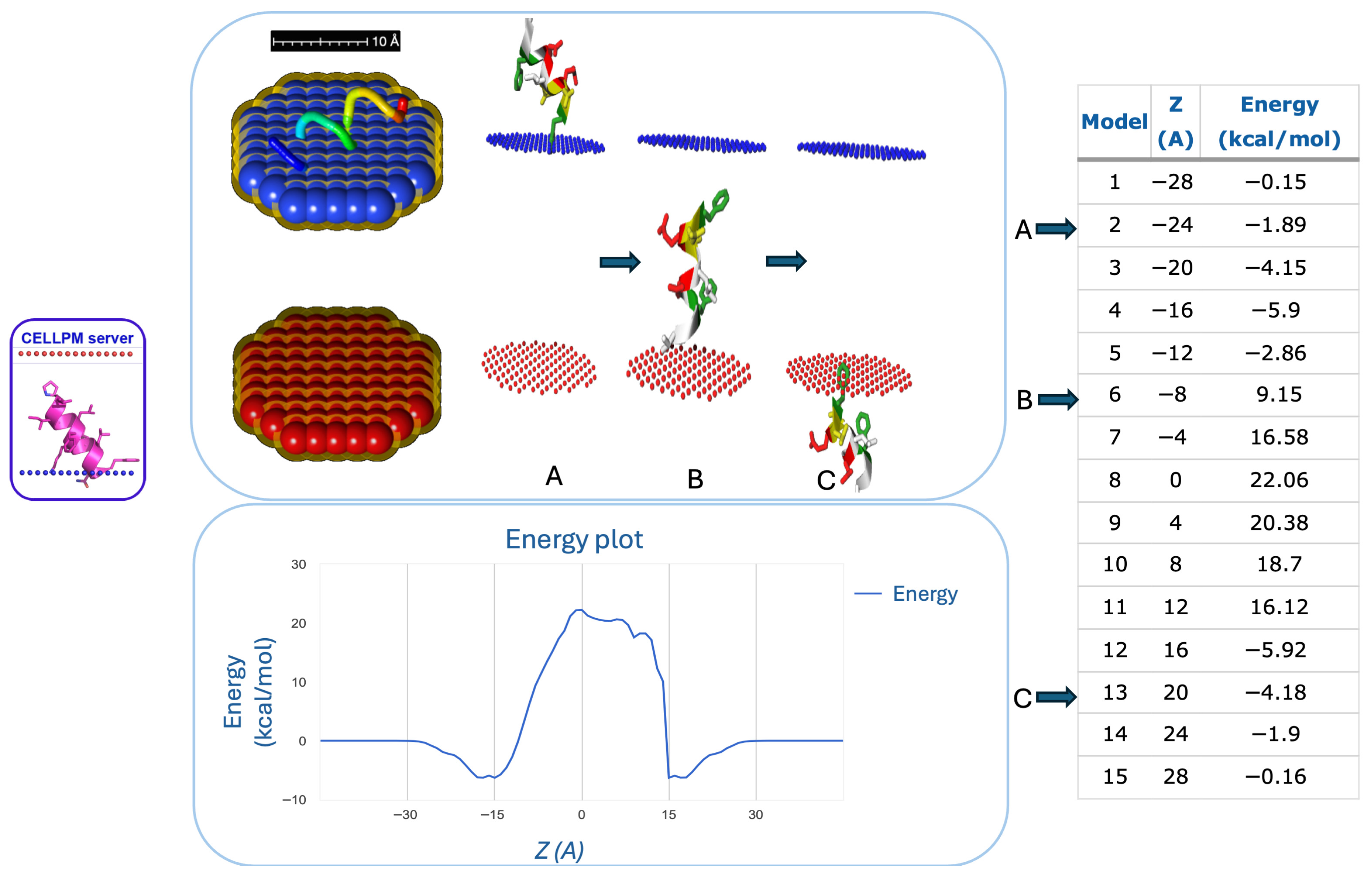
4.6. Membrane Permeability Test
4.7. Prediction of Anti-Inflammatory Properties of Peptides
4.8. Prediction of Antioxidative Properties of Peptides
4.9. Protein Annotation
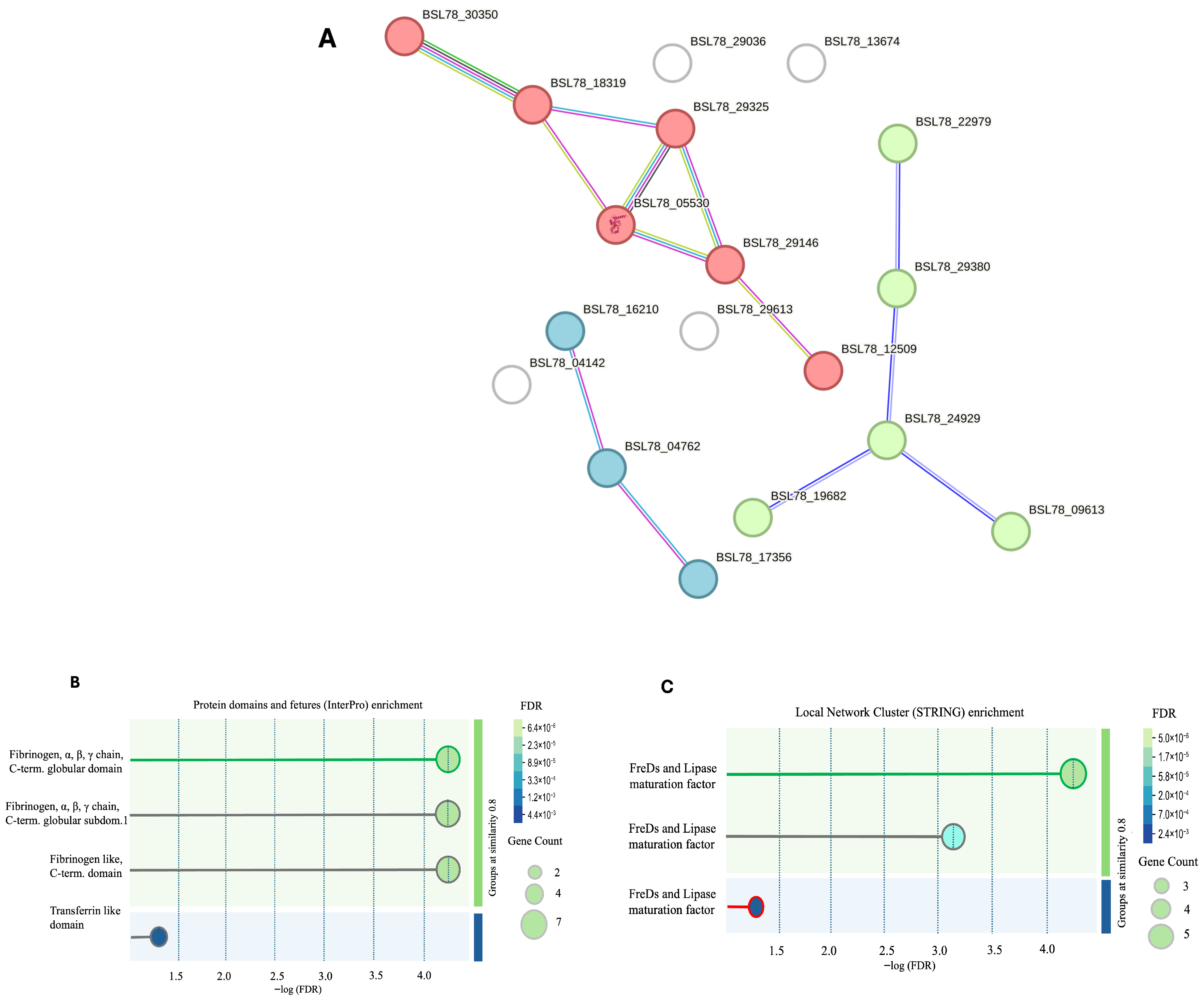
4.10. Protein–Protein Network Analysis
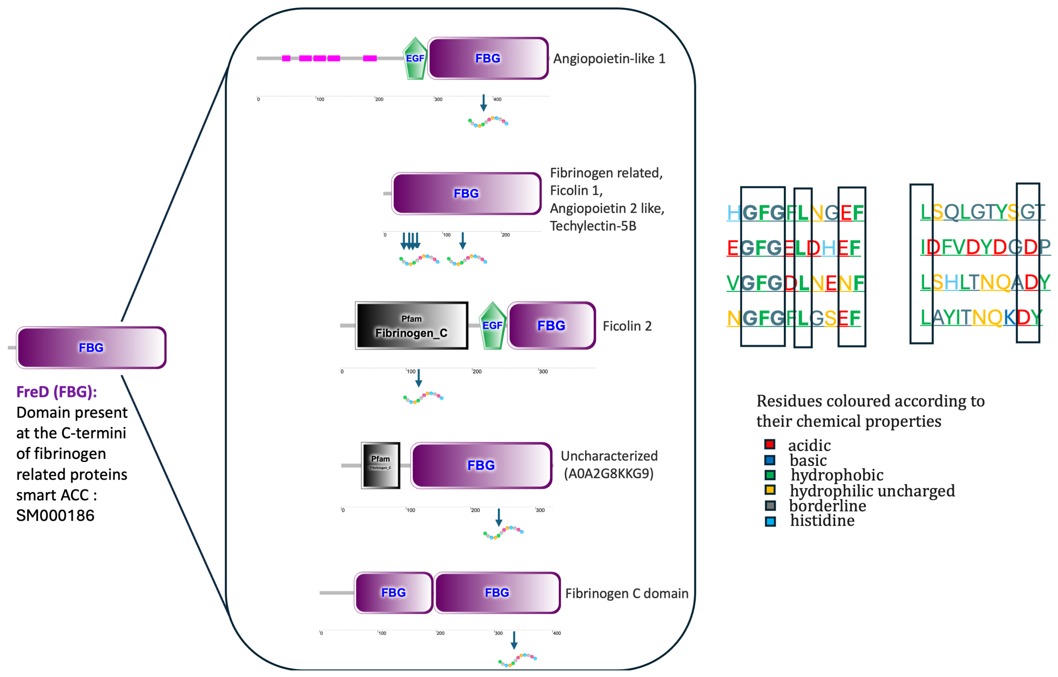
4.11. Protein Domains Analysis
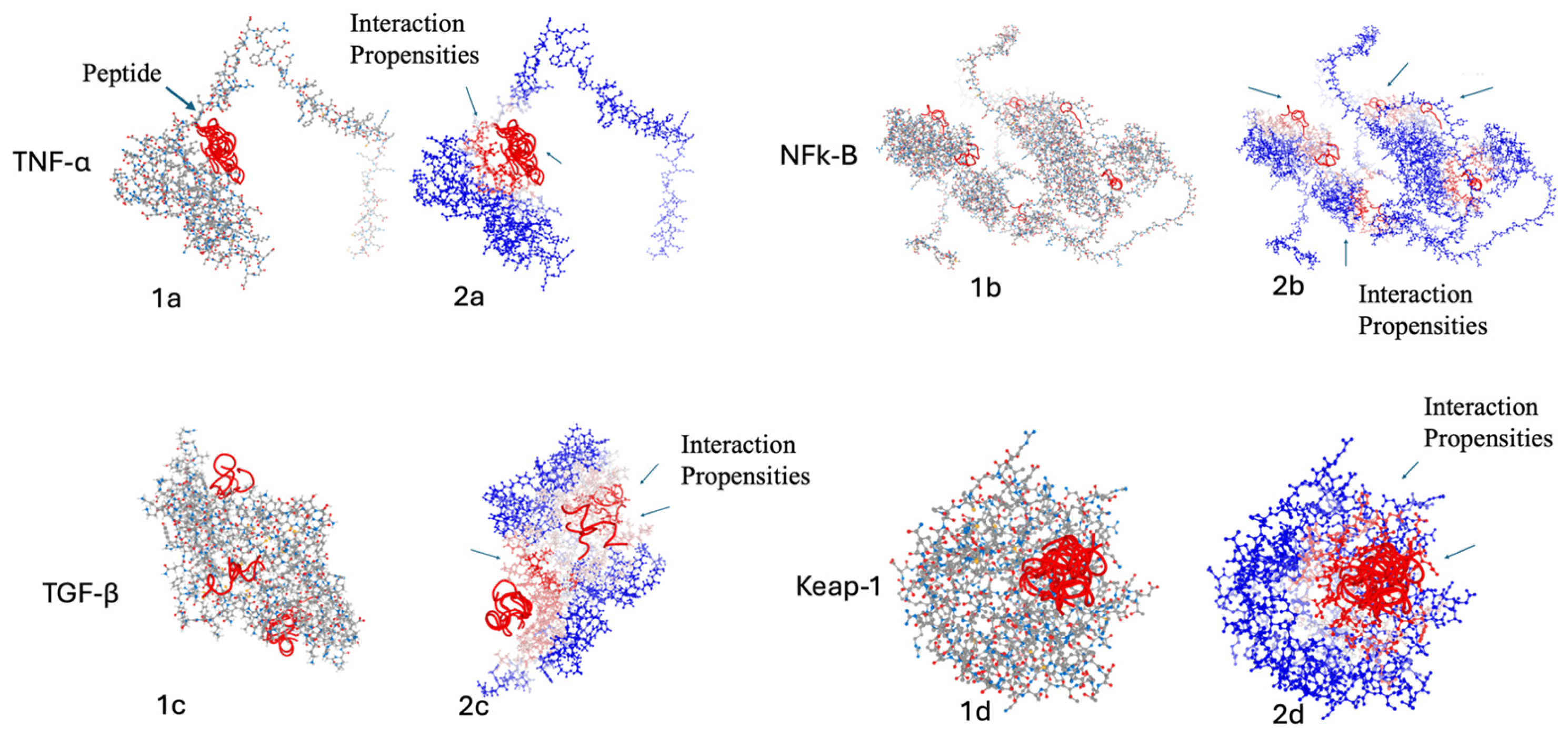
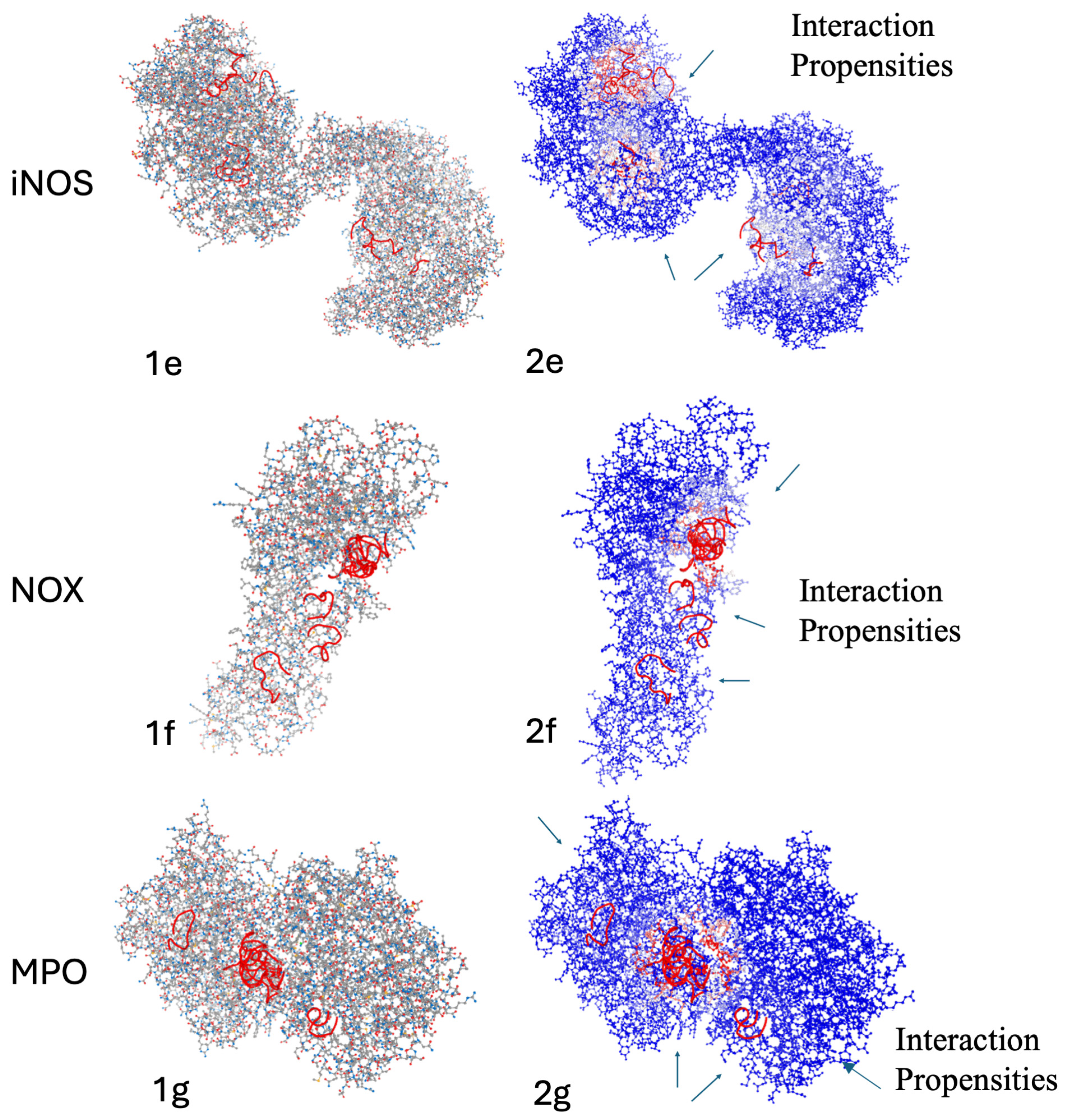
4.12. Peptide–Protein Interaction Prediction Through Dynamic Modeling
5. Conclusions and Further Directions
Supplementary Materials
Author Contributions
Funding
Institutional Review Board Statement
Data Availability Statement
Conflicts of Interest
References
- Liu, J.; Han, X.; Zhang, T.; Tian, K.; Li, Z.; Luo, F. Reactive oxygen species (ROS) scavenging biomaterials for anti-inflammatory diseases: From mechanism to therapy. J. Hematol. Oncol. 2023, 16, 116. [Google Scholar] [CrossRef] [Scilit]
- Bowie, A.G.; Unterholzner, L. Viral evasion and subversion of pattern-recognition receptor signalling. Nat. Rev. Immunol. 2008, 12, 911–922. [Google Scholar] [CrossRef] [Scilit] [PubMed] [PubMed Central]
- Takeuchi, O.; Akira, S. Pattern recognition receptors and inflammation. Cell 2010, 140, 805–820. [Google Scholar] [CrossRef] [Scilit] [PubMed]
- Zhao, H.; Wu, L.; Yan, G.; Chen, Y.; Zhou, M.; Wu, Y.; Li, Y. Inflammation and tumor progression: Signaling pathways and targeted intervention. Signal Transduct. Target. Ther. 2021, 6, 263. [Google Scholar] [CrossRef] [Scilit] [PubMed]
- Yeung, Y.T.; Aziz, F.; Guerrero-Castilla, A.; Arguelles, S. Signaling Pathways in Inflammation and Anti-inflammatory Therapies. Curr. Pharm. Des. 2018, 24, 1449–1484. [Google Scholar] [CrossRef] [Scilit]
- Bondia-Pons, I.; Ryan, L.; Martinez, J.A. Oxidative stress and inflammation interactions in human obesity. J. Physiol. Biochem. 2012, 68, 701–711. [Google Scholar] [CrossRef] [Scilit]
- Reuter, S.; Gupta, S.C.; Chaturvedi, M.M.; Aggarwal, B.B. Oxidative stress, inflammation, and cancer: How are they linked? Free Radic. Biol. Med. 2010, 49, 1603–1616. [Google Scholar] [CrossRef] [Scilit]
- Ďuračková, Z. Some current insights into oxidative stress. Physiol. Res. 2010, 59, 459–469. [Google Scholar] [CrossRef] [Scilit]
- Jones, D.P. Redefining oxidative stress. Antioxid. Redox Signal. 2006, 8, 1865–1879. [Google Scholar] [CrossRef] [Scilit] [PubMed]
- Klotz, L.O.; Steinbrenner, H. Cellular adaptation to xenobiotics: Interplay between xenosensors, reactive oxygen species and FOXO transcription factors. Redox Biol. 2017, 13, 646–654. [Google Scholar] [CrossRef] [Scilit]
- Schieber, M.; Chandel, N.S. ROS function in redox signaling and oxidative stress. Curr. Biol. 2014, 24, R453–R462. [Google Scholar] [CrossRef] [Scilit]
- D’Autréaux, B.; Toledano, M.B. ROS as signalling molecules: Mechanisms that generate specificity in ROS homeostasis. Nat. Rev. Mol. Cell Biol. 2007, 10, 813–824. [Google Scholar] [CrossRef] [Scilit]
- Okado-Matsumoto, A.; Fridovich, I. Subcellular Distribution of Superoxide Dismutases (SOD) in Rat Liver: Cu,Zn-SOD INMITOCHONDRIA. J. Biol. Chem. 2001, 276, 38388–38393. [Google Scholar] [CrossRef] [Scilit]
- Vives-Bauza, C.; Starkov, A.; Garcia-Arumi, E. Measurements of the antioxidant enzyme activities of superoxide dismutase, catalase, and glutathione peroxidase. Methods Cell Biol. 2007, 80, 379–393. [Google Scholar] [CrossRef] [Scilit]
- Cnubben, N.H.; Rietjens, I.M.; Wortelboer, H.; van Zanden, J.; van Bladeren, P.J. The interplay of glutathione-related processes in antioxidant defense. Environ. Toxicol. Pharmacol. 2001, 10, 141–152. [Google Scholar] [CrossRef] [Scilit]
- Malla, R.; Surepalli, N.; Farran, B.; Malhotra, S.V.; Nagaraju, G.P. Reactive oxygen species (ROS): Critical roles in breast tumor microenvironment. Crit. Rev. Oncol. Hematol. 2021, 160, 103285. [Google Scholar] [CrossRef] [Scilit] [PubMed]
- Bensimon, A.; Aebersold, R.; Shiloh, Y. Beyond ATM: The protein kinase landscape of the DNA damage response. FEBS Lett. 2011, 585, 1625–1639. [Google Scholar] [CrossRef] [Scilit] [PubMed]
- Hayden, M.S.; Ghosh, S. NF-κB in immunobiology. Cell Res. 2011, 21, 23–44. [Google Scholar] [CrossRef] [Scilit]
- Oeckinghaus, A.; Hayden, M.S.; Ghosh, S. Crosstalk in NF-κB signaling pathways. Nat. Immunol. 2011, 12, 695–708. [Google Scholar] [CrossRef] [Scilit]
- Kapoor, S.; Nailwal, N.; Kumar, M.; Barve, K. Recent Patents and Discovery of Anti-inflammatory Agents from Marine Source. Recent Pat. Inflamm. Allergy Drug Discov. 2019, 3, 105–114. [Google Scholar] [CrossRef] [Scilit]
- Tian, X.R.; Tang, H.F.; Tian, X.L.; Hu, J.J.; Huang, L.L.; Gustafson, K.R. Review of bioactive secondary metabolites from marine bryozoans in the progress of new drugs discovery. Future Med. Chem. 2018, 10, 1497–1514. [Google Scholar] [CrossRef] [Scilit]
- Cheung, R.C.F.; Ng, T.B.; Wong, J.H.; Chen, Y.; Chan, W.Y. Marine natural products with anti-inflammatory activity. Appl. Microbiol. Biotechnol. 2016, 100, 1645–1666. [Google Scholar] [CrossRef] [Scilit] [PubMed]
- Janakiram, N.B.; Mohammed, A.; Zhang, Y.; Choi, C.I.; Woodward, C.; Collin, P.; Steele, V.E.; Rao, C.V. Chemopreventive effects of Frondanol A5, a Cucumaria frondosa extract, against rat colon carcinogenesis and inhibition of human colon cancer cell growth. Cancer Prev. Res. 2010, 3, 82–91. [Google Scholar] [CrossRef] [Scilit] [PubMed]
- Pangestuti, R.; Arifin, Z. Medicinal and health benefit effects of functional sea cucumbers. J. Tradit. Complement. Med. 2017, 8, 341–351. [Google Scholar] [CrossRef] [Scilit]
- Bordbar, S.; Anwar, F.; Saari, N. High-Value Components and Bioactives from Sea Cucumbers for Functional Foods—A Review. Mar. Drugs 2011, 9, 1761–1805. [Google Scholar] [CrossRef] [Scilit] [PubMed]
- Jin, J.O.; Shastina, V.V.; Shin, S.W.; Xu, Q.; Park, J.I.; Rasskazov, V.A.; Avilov, S.A.; Fedorov, S.N.; Stonik, V.A.; Kwak, J.Y. Differential effects of triterpene glycosides, frondoside A and cucumarioside A2-2 isolated from sea cucumbers on caspase activation and apoptosis of human leukemia cells. FEBS Lett. 2009, 583, 697–702. [Google Scholar] [CrossRef] [Scilit]
- Ma, X.; Kundu, N.; Collin, P.D.; Goloubeva, O.; Fulton, A.M. Frondoside A inhibits breast cancer metastasis and antagonizes prostaglandin E receptors EP4 and EP2. Breast Cancer Res. Treat. 2012, 132, 1001–1008. [Google Scholar] [CrossRef] [Scilit]
- Al Marzouqi, N.; Iratni, R.; Nemmar, A.; Arafat, K.; Ahmed Al Sultan, M.; Yasin, J.; Collin, P.; Mester, J.; Adrian, T.E.; Attoub, S. Frondoside A inhibits human breast cancer cell survival, migration, invasion and the growth of breast tumor xenografts. Eur. J. Pharmacol. 2011, 668, 25–34. [Google Scholar] [CrossRef] [Scilit]
- Li, X.; Roginsky, A.B.; Ding, X.Z.; Woodward, C.; Collin, P.; Newman, R.A.; Bell, R.H., Jr.; Adrian, T.E. Review of the apoptosis pathways in pancreatic cancer and the anti-apoptotic effects of the novel sea cucumber compound. Frondoside A. Ann. N. Y. Acad. Sci. 2008, 1138, 81–98. [Google Scholar] [CrossRef] [Scilit]
- Dyshlovoy, S.A.; Menchinskaya, E.S.; Venz, S.; Rast, S.; Amann, K.; Hauschild, J.; Otte, K.; Kalinin, V.I.; Silchenko, A.S.; Avilov, S.A.; et al. The marine triterpene glycoside frondoside A exhibits activity in vitro and in vivo in prostate cancer. Int. J. Cancer 2016, 138, 2450–2465. [Google Scholar] [CrossRef] [Scilit]
- Janakiram, N.B.; Mohammed, A.; Rao, C.V. Sea Cucumbers Metabolites as Potent Anti-Cancer Agents. Mar. Drugs 2015, 13, 2909–2923. [Google Scholar] [CrossRef] [Scilit]
- Tahseen, Q. Coelomocytes: Biology and Possible Immune Functions in Invertebrates with Special Remarks on Nematodes. Int. J. Zool. 2009, 2009, 11–23. [Google Scholar] [CrossRef] [Scilit]
- Sun, Y.; Jin, L.; Wang, T.; Xue, J.; Liu, G.; Li, X.; You, J.; Li, S.; Xu, Y. Polysaccharides from Astragalus membranaceus promote phagocytosis and superoxide anion (O2−) production by coelomocytes from sea cucumber Apostichopus japonicus in vitro. Comp. Biochem. Physiol. C Toxicol. Pharmacol. 2008, 147, 293–298. [Google Scholar] [CrossRef] [Scilit] [PubMed]
- Pagliara, P.; Carnevali, C.; Burighel, P.; Ballarin, L. The spherule cells of Holothuria polii Delle Chiaie, 1823 (Aspidochirota, Holothuroidea) during brown body formation: An ultrastructural study. J. Submicrosc. Cytol. Pathol. 2003, 35, 295–301. [Google Scholar] [PubMed]
- Canicatti, C.; D’Ancona, G. Cellular aspects of Holothuria polii immune response. J. Invertebr. Pathol. 1989, 53, 152–158. [Google Scholar] [CrossRef] [Scilit]
- Canicatti, C. Hemolysins: Pore-forming proteins in invertebrates. Experientia 1990, 46, 239–244. [Google Scholar] [CrossRef] [Scilit]
- Smith, V.J.; Söderhäll, K. A comparison of phenoloxidase activity in the blood of marine invertebrates. Dev. Comp. Immunol. 1991, 15, 251–261. [Google Scholar] [CrossRef] [Scilit]
- Canicattì, C.; Seymour, J. Evidence for phenoloxidase activity in Holothuria tubulosa (Echinodermata) brown bodies and cells. Parasitol. Res. 1991, 77, 50–53. [Google Scholar] [CrossRef] [Scilit]
- Canicatti, C.; Roch, P. Studies on Holothuria polii (Echinodermata) antibacterial proteins. I. Evidence for and activity of a coelomocyte lysozyme. Experientia 1989, 45, 4. [Google Scholar] [CrossRef] [Scilit]
- Haug, T.; Kjuul, A.K.; Styrvold, O.B.; Sandsdalen, E.; Olsen, Ø.M.; Stensvåg, K. Antibacterial activity in Strongylocentrotus droebachiensis (Echinoidea), Cucumaria frondosa (Holothuroidea), and Asterias rubens (Asteroidea). J. Invertebr. Pathol. 2002, 81, 94–102. [Google Scholar] [CrossRef] [Scilit]
- Schillaci, D.; Cusimano, M.G.; Cunsolo, V.; Saletti, R.; Russo, D.; Vazzana, M.; Vitale, M.; Arizza, V. Immune mediators of sea-cucumber Holothuria tubulosa (Echinodermata) as source of novel antimicrobial and anti-staphylococcal biofilm agents. AMB Express 2013, 3, 35. [Google Scholar] [CrossRef] [Scilit]
- García-Arrarás, J.E.; Schenk, C.; Rodrígues-Ramírez, R.; Torres, I.I.; Valentín, G.; Candelaria, A.G. Spherulocytes in the echinoderm Holothuria glaberrima and their involvement in intestinal regeneration. Dev. Dyn. 2006, 235, 3259–3267. [Google Scholar] [CrossRef] [Scilit] [PubMed]
- San Miguel-Ruiz, J.E.; García-Arrarás, J.E. Common cellular events occur during wound healing and organ regeneration in the sea cucumber Holothuria glaberrima. BMC Dev. Biol. 2007, 7, 115. [Google Scholar] [CrossRef] [Scilit]
- La Paglia, L.; Vazzana, M.; Mauro, M.; Urso, A.; Arizza, V.; Vizzini, A. Bioactive Molecules from the Innate Immunity of Ascidians and Innovative Methods of Drug Discovery: A Computational Approach Based on Artificial Intelligence. Mar. Drugs 2023, 22, 6. [Google Scholar] [CrossRef] [Scilit]
- Ao, J.; Li, B. Amino acid composition and antioxidant activities of hydrolysates and peptide fractions from porcine collagen. Food Sci. Technol. Int. 2012, 8, 425–434. [Google Scholar] [CrossRef] [Scilit]
- Liu, J.; Zhang, D.; Zhu, Y.; Wang, Y.; He, S.; Zhang, T. Enhancing the in vitro Antioxidant Capacities via the interaction of amino acids. Emir. J. Food Agric. 2018, 30, 224–231. [Google Scholar] [CrossRef] [Scilit]
- Shi, C.; Liu, M.; Zhao, H.; Lv, Z.; Liang, L.; Zhang, B. A Novel Insight into Screening for Antioxidant Peptides from Hazelnut Protein: Based on the Properties of Amino Acid Residues. Antioxidants 2022, 11, 127. [Google Scholar] [CrossRef] [Scilit]
- Zhu, Y.; Wang, K.; Jia, X.; Fu, C.; Yu, H.; Wang, Y. Antioxidant peptides, the guardian of life from oxidative stress. Med. Res. Rev. 2024, 44, 275–364. [Google Scholar] [CrossRef] [Scilit]
- Kannan, T.J.; Balde, A.; Nazeer, R.A. A review on anti-inflammatory and antioxidant peptides derived from marine organisms: Mechanism of action and therapeutic applications. Food Biosci. 2025, 63, 105745. [Google Scholar] [CrossRef] [Scilit]
- Kumar, S.; Gupta, S. Thymosin Beta 4 Prevents Oxidative Stress by Targeting Antioxidant and Anti-Apoptotic Genes in Cardiac Fibroblasts. PLoS ONE 2011, 6, e26912. [Google Scholar] [CrossRef] [Scilit]
- Qiu, P.; Wheater, M.K.; Qiu, Y.; Sosne, G. Thymosin beta4 inhibits TNF-alpha-induced NF-kappaB activation, IL-8 expression, and the sensitizing effects by its partners PINCH-1 and ILK. FASEB J. 2011, 25, 1815–1826. [Google Scholar] [CrossRef] [Scilit]
- Chang, S.; Zhang, M.; Liu, C.; Li, M.; Lou, Y.; Tan, H. Redox mechanism of glycerophospholipids and relevant targeted therapy in ferroptosis. Cell Death Discov. 2025, 11, 358. [Google Scholar] [CrossRef] [Scilit] [PubMed]
- Del Valle-Pérez, B.; Martínez, V.G.; Lacasa-Salavert, C.; Figueras, A.; Shapiro, S.S.; Takafuta, T.; Casanovas, O.; Capellà, G.; Ventura, F.; Viñals, F. Filamin B Plays a Key Role in Vascular Endothelial Growth Factor-induced Endothelial Cell Motility through Its Interaction with Rac-1 and Vav-2. Cell Biol. 2010, 285, 10748–10760. [Google Scholar] [CrossRef] [Scilit]
- Ferro, E.; Goitre, L.; Retta, S.F.; Trabalzini, L. The Interplay between ROS and Ras GTPases: Physiological and Pathological Implications. J. Signal Transduct. 2012, 2012, 365769. [Google Scholar] [CrossRef] [Scilit] [PubMed]
- Chen, J.-X.; Zeng, H.; Lawrence, M.L.; Blackwell, T.S.; Meyrick, B. Angiopoietin-1-induced angiogenesis is modulated by endothelial NADPH oxidase. Am. J. Physiol.-Heart Circ. Physiol. 2005, 291, H1563–H1572. [Google Scholar] [CrossRef] [Scilit]
- Kim, S.; Lee, K.; Park, S.; Min, K.H.; Lee, K.Y.; Choe, Y.H.; Hong, S.H.; Koh, G.Y.; Lee, Y.C. Angiopoietin-1 variant, COMP-Ang1 attenuates hydrogen peroxide-induced acute lung injury. Exp. Mol. Med. 2008, 40, 320–331. [Google Scholar] [CrossRef] [Scilit]
- Ismail, N.S.; Pravda, E.A.; Li, D.; Shih, S.-C.; Dallabrida, S.M. Angiopoietin-1 Reduces H2O2-Induced Increases in Reactive Oxygen Species and Oxidative Damage to Skin Cells. J. Investig. Dermatol. 2009, 130, 1307–1317. [Google Scholar] [CrossRef] [Scilit] [PubMed]
- Wagner, S.; Ruff, H.M.; Weber, S.L.; Bellmann, S.; Sowa, T.; Schulte, T.; Anderson, M.E.; Grandi, E.; Bers, D.M.; Backs, J.; et al. Reactive Oxygen Species–Activated Ca/Calmodulin Kinase IIδ Is Required for Late INa Augmentation Leading to Cellular Na and Ca Overload. Circ. Res. 2011, 108, 555–565. [Google Scholar] [CrossRef] [Scilit]
- Luo, S.; Levine, R.L. Methionine in proteins defends against oxidative stress. FASEB J. 2009, 23, 464–472. [Google Scholar] [CrossRef] [Scilit]
- Chen, J.; Bai, W.; Cai, D.; Yu, Z.; Xu, B. Characterization and identification of novel anti-inflammatory peptides from Baijiao sea bass (Lateolabrax maculatus). LWT 2021, 147, 111521. [Google Scholar] [CrossRef] [Scilit]
- Biji, C.A.; Balde, A.; Nazeer, R.A. Anti-inflammatory peptide therapeutics and the role of sulphur-containing amino acids (cysteine and methionine) in inflammation suppression: A review. Inflamm. Res. 2024, 73, 1203–1221. [Google Scholar] [CrossRef] [Scilit] [PubMed]
- Huang, Z.; Aweya, J.J.; Zhu, C.; Tran, N.T.; Hong, Y.; Li, S.; Yao, D.; Zhang, Y. Modulation of crustacean innate immune response by amino acids and their metabolites: Inferences from other species. Front. Immunol. 2020, 11, 574721. [Google Scholar] [CrossRef] [Scilit] [PubMed]
- Guo, H.; Kouzuma, Y.; Yonekura, M. Structures and properties of antioxidative peptides derived from royal jelly protein. Food Chem. 2009, 13, 238–245. [Google Scholar] [CrossRef] [Scilit]
- Hsu, K.-C. Purification of antioxidative peptides prepared from enzymatic hydrolysates of tuna dark muscle by-product. Food Chem. 2010, 122, 42–48. [Google Scholar] [CrossRef] [Scilit]
- Saiga, A.; Tanabe, S.; Nishimura, T. Antioxidant activity of peptides obtained from porcine myofibrillar proteins by protease treatment. J. Agric. Food Chem. 2003, 51, 3661–3667. [Google Scholar] [CrossRef] [Scilit] [PubMed]
- Guidea, A.; Zăgrean-, C.; Tuza Moț, A.C.; Sârbu, C. Comprehensive evaluation of radical scavenging, reducing power and chelating capacity of free proteinogenic amino acids using spectroscopic assays and multivariate exploratory techniques. Spectrochim. Acta Part A Mol. Biomol. Spectrosc. 2020, 233, 118158. [Google Scholar] [CrossRef] [Scilit]
- Walters, M.; Esfandi, R.; Tsopmo, A. Potential of food hydrolyzed proteins and peptides to chelate iron or calcium and enhance their absorption. Foods 2018, 7, 172. [Google Scholar] [CrossRef] [Scilit]
- Durand, R.; Pellerin, G.; Thibodeau, J.; Fraboulet, E.; Marette, A.; Bazinet, L. Screening for metabolic syndrome application of a herring by-product hydrolysate after its separation by electrodialysis with ultrafiltration membrane and identification of novel anti-inflammatory peptides. Sep. Purif. Technol. 2020, 235, 116205. [Google Scholar] [CrossRef] [Scilit]
- Mayer, A.; Mayer, V.; Swanson-Mungerson, M.; Pierce, M.; Rodríguez, A.; Nakamura, F.; Taglialatela-Scafati, O. Marine pharmacology in 2019–2021: Marine compounds with antibacterial, antidiabetic, antifungal, anti-inflammatory, antiprotozoal, antituberculosis and antiviral activities; Affecting the immune and nervous systems, and other miscellaneous mechanisms of action. Mar. Drugs 2024, 22, 309. [Google Scholar] [CrossRef] [Scilit]
- Vogel, H.J.; Schibli, D.J.; Jing, W.; Lohmeier-Vogel, E.M.; Epand, R.F.; Epand, R.M. Towards a structure-function analysis of bovine lactoferricin and related tryptophan- and arginine-containing peptides. Biochem. Cell Biol. 2002, 80, 49–63. [Google Scholar] [CrossRef] [Scilit]
- Joshi, I.; Mohideen, H.S.; Nazeer, R.A. A Meretrix meretrix visceral mass derived peptide inhibits lipopolysaccharide-stimulated responses in RAW264.7 cells and adult zebrafish model. Int. Immunopharmacol. 2021, 90, 107140. [Google Scholar] [CrossRef] [Scilit]
- Akbarian, M.; Khani, A.; Eghbalpour, S.; Uversky, V.N. Bioactive peptides: Synthesis, sources, applications, and proposed mechanisms of action. Int. J. Mol. Sci. 2022, 23, 1445. [Google Scholar] [CrossRef] [Scilit] [PubMed]
- Roterman, I.; Stapor, K.; Fabian, P.; Konieczny, L. The Functional Significance of Hydrophobic Residue Distribution in Bacterial Beta-Barrel Transmembrane Proteins. Membranes 2021, 11, 580. [Google Scholar] [CrossRef] [Scilit]
- Mao, H.; Zhao, X.; Sun, S.C. NF-κB in inflammation and cancer. Cell Mol. Immunol. 2025, 22, 811–839. [Google Scholar] [CrossRef] [Scilit]
- Dash, S.; Sahu, A.K.; Srivastava, A.; Chowdhury, R.; Mukherjee, S. Exploring the extensive crosstalk between the antagonistic cytokines- TGF-β and TNF-α in regulating cancer pathogenesis. Cytokine 2020, 138, 155348. [Google Scholar] [CrossRef] [Scilit] [PubMed]
- Hogan, K.A.; Ravindran, A.; Podolsky, M.A.; Glick, A.B. The TGFβ1 pathway is required for NFκB dependent gene expression in mouse keratinocytes. Cytokine 2013, 64, 652–659. [Google Scholar] [CrossRef] [Scilit] [PubMed]
- Steel, R.; Cowan, J.; Payerne, E.; O’Connell, M.A.; Searcey, M. Anti-inflammatory Effect of a Cell-Penetrating Peptide Targeting the Nrf2/Keap1 Interaction. ACS Med. Chem. Lett. 2012, 3, 407–410. [Google Scholar] [CrossRef] [Scilit]
- Abed, D.A.; Goldstein, M.; Albanyan, H.; Jin, H.; Hu, L. Discovery of direct inhibitors of Keap1–Nrf2 protein–protein interaction as potential therapeutic and preventive agents. Acta Pharm. Sin. B 2015, 5, 285–299. [Google Scholar] [CrossRef] [Scilit]
- Robinson, J.M. Reactive oxygen species in phagocytic leukocytes. Histochem. Cell Biol. 2008, 130, 281–297. [Google Scholar] [CrossRef] [Scilit]
- Sedeek, M.; Nasrallah, R.; Touyz, R.M.; Hebert, R.L. NADPH oxidases, reactive oxygen species, and the kidney: Friend and foe. J. Am. Soc. Nephrol. 2013, 24, 1512–1518. [Google Scholar] [CrossRef] [Scilit]
- Granger, D.N.; Kvietys, P.R. Reperfusion injury and reactive oxygen species: The evolution of a concept. Redox Biol. 2015, 6, 524–551. [Google Scholar] [CrossRef] [Scilit]
- Vazquez-Medina, J.P. Chapter 3—Redox Signaling and the Onset of the Inflammatory Cascade. In Immunity and Inflammation in Health and Disease; Chatterjee, S., Jungraithmayr, W., Bagchi, D., Eds.; Academic Press: Cambridge, MA, USA, 2018. [Google Scholar] [CrossRef] [Scilit]
- La Paglia, L.; Mauro, M.; Arizza, V.; Urso, A.; Simon, S.; Drahos, L.; Di Stefano, V.; Luparello, C.; Vazzana, M.; Vizzini, A. Bioinformatics analyses of the proteome of Holothuria tubulosa coelomic fluid and the first evidence of primary cilium in coelomocyte cells. Front. Immunol. 2025, 16, 539751. [Google Scholar] [CrossRef] [Scilit] [PubMed]
- Mooney, C.; Haslam, N.J.; Pollastri, G.; Shields, D.C. Towards the Improved Discovery and Design of Functional Peptides: Common Features of Diverse Classes Permit Generalized Prediction of Bioactivity. PLoS ONE 2012, 7, e45012. [Google Scholar] [CrossRef] [Scilit]
- Sharma, A.; Singla, D.; Rashid, M.; Raghava, G.P.S. Designing of peptides with desired half-life in intestine-like environment. BMC Bioinform. 2014, 15, 282. [Google Scholar] [CrossRef] [Scilit]
- Gupta, S.; Kapoor, P.; Chaudhary, K.; Gautam, A.; Kumar, R.; Open Source Drug Discovery Consortium; Raghava, G.P. In silico approach for predicting toxicity of peptides and proteins. PLoS ONE 2013, 8, e73957. [Google Scholar] [CrossRef] [Scilit] [PubMed]
- Rathore, A.S.; Choudhury, S.; Arora, A.; Tijare, P.; Raghava, G.P.S. ToxinPred 3.0: An improved method for predicting the toxicity of peptides. Comput. Biol. Med. 2024, 179, 108926. [Google Scholar] [CrossRef] [Scilit]
- Lomize, A.L.; Hage, J.M.; Schnitzer, K.; Golobokov, K.; LaFaive, M.B.; Forsyth, A.C.; Pogozheva, I.D. PerMM: A Web Tool and Database for Analysis of Passive Membrane Permeability and Translocation Pathways of Bioactive Molecules. J. Chem. Inf. Model. 2019, 59, 3094–3099. [Google Scholar] [CrossRef] [Scilit] [PubMed]
- Manavalan, B.; Shin, T.H.; Kim, M.O.; Lee, G. AIPpred: Sequence-Based Prediction of Anti-inflammatory Peptides Using Random Forest. Front. Pharmacol. 2018, 9, 276. [Google Scholar] [CrossRef] [Scilit]
- Olsen, T.H.; Yesiltas, B.; Marin, F.I.; Pertseva, M.; García-Moreno, P.J.; Gregersen, S.; Overgaard, M.T.; Jacobsen, C.; Lund, O.; Hansen, E.B.; et al. AnOxPePred: Using deep learning for the prediction of antioxidative properties of peptides. Sci. Rep. 2020, 10, 21471. [Google Scholar] [CrossRef] [Scilit]
- UniProt Consortium. UniProt: The Universal Protein Knowledgebase in 2025. Nucleic Acids Res. 2025, 53, D609–D617. [Google Scholar] [CrossRef] [Scilit]
- Szklarczyk, D.; Kirsch, R.; Koutrouli, M.; Nastou, K.; Mehryary, F.; Hachilif, R.; Annika, G.L.; Fang, T.; Doncheva, N.T.; Pyysalo, S.; et al. The STRING database in 2023: Protein–protein association networks and functional enrichment analyses for any sequenced genome of interest. Nucleic Acids Res. 2023, 51, D638–D646. [Google Scholar] [CrossRef] [Scilit]
- Letunic, I.; Bork, P. SMART v10: Three decades of the protein domain annotation resource. Nucleic Acids Res. 2025, 54, D499–D503. [Google Scholar] [CrossRef] [Scilit]
- Saladin, A.; Rey, J.; Thévenet, P.; Zacharias, M.; Moroy, G.; Tufféry, P. PEP-SiteFinder: A tool for the blind identification of peptide binding sites on protein surfaces. Nucleic Acids Res. 2014, 42, W221–W226. [Google Scholar] [CrossRef] [Scilit]
- Maupetit, J.; Derreumaux, P.; Tuffery, P. PEP-FOLD: An online resource for de novo peptide structure prediction. Nucleic Acids Res. 2009, 37, W498–W503. [Google Scholar] [CrossRef] [Scilit]
- Zacharias, M. Protein-protein docking with a reduced protein model accounting for side-chain flexibility. Protein Sci. 2003, 6, 1271–1282. [Google Scholar] [CrossRef] [Scilit]



| Sequence | Cell Pellet/Uniprot ID Cell Free | Name | Bioactivity | Toxicity | Anti-Inflammatory | FSR AnOx | Chel AnOx | Half Life | |||
|---|---|---|---|---|---|---|---|---|---|---|---|
| Prediction | AIP or Non-AIP | Prob | Half-Life [s] | Stability | |||||||
| VGFGDLNENF | cell free | A0A2G8L651_STIJA | Ficolin-1 | 0.83 | Non-Toxin | AIP | 0.43 | 0.47 | 6.29 | High | |
| NGFGFLNTEF | cell free | A0A2G8K011_STIJA | Fibrinogen-like | 0.73 | Non-Toxin | AIP | 0.41 | 0.46 | 5.58 | High | |
| HGFGFLNGEF | cell free | A0A2G8K2I6_STIJA | Fibrinogen C-terminal domain-containing protein | 0.73 | Non-Toxin | AIP | 0.39 | 0.45 | 5.22 | High | |
| NGFGFLGSEF | cell free | A0A2G8JM67_STIJA | Putative ficolin-2-like | 0.69 | Non-Toxin | AIP | 0.50 | 0.45 | 4.72 | High | |
| FGTIGFDDGI | cell pellet | A0A2G8LDJ7_STIJA | Alkaline phosphatase | 0.56 | Non-Toxin | AIP | 0.42 | 0.40 | 3.92 | High | |
| DSYVGDEAQS | cell free | Q26378_TRIGR | CyI actin | 0.56 | Non-Toxin | AIP | 0.38 | 0.33 | 0.23 | 3.90 | High |
| LFDSAGYTDS | cell free | A0A0K1Z5K0_HOLGL | Melanotransferrin 2 | 0.53 | Non-Toxin | AIP | 0.39 | 0.40 | 3.69 | High | |
| DSNFGINDLE | cell pellet | A0A0K1Z5K0_HOLGL | Melanotransferrin 2 | 0.41 | Non-Toxin | AIP | 0.38 | 0.24 | 3.37 | High | |
| ESINVEQAFQ | cell pellet | A0A2G8LEX4_STIJA | Putative ras-related protein | 0.37 | Non-Toxin | AIP | 0.52 | 0.32 | 0.25 | 3.34 | High |
| EGFGELDHEF | cell free | A0A2G8KAK2_STIJA | Angiopoietin-like 1 | 0.37 | Non-Toxin | AIP | 0.42 | 0.52 | 3.33 | High | |
| NGFGELDHEF | cell free | A0A2G8L6S5_STIJA | Putative ficolin-2-like | 0.34 | Non-Toxin | AIP | 0.38 | 0.49 | 3.24 | High | |
| NTLPTKDTID | cell free | A7LGB0_HOLGL | Thymosin beta | 0.31 | Non-Toxin | AIP | 0.41 | 0.22 | 2.79 | High | |
| ADLITLDGGD | cell free | A0A0K1Z5K0_HOLGL | Melanotransferrin 2 | 0.31 | Non-Toxin | AIP | 0.45 | 0.41 | 0.23 | 2.76 | High |
| LFDSEDYEGS | cell free | A0A0K1Z4Z0_HOLGL | Melanotransferrin 4 | 0.30 | Non-Toxin | AIP | 0.40 | 0.46 | 2.75 | High | |
| IDFVDYDGDP | cell free | A0A2G8LI29_STIJA | Fibrinogen C-terminal domain-containing protein | 0.29 | Non-Toxin | AIP | 0.38 | 0.57 | 2.71 | High | |
| YPIEHGIITN | cell free | W4ZD41_STRPU | Uncharacterized protein | 0.23 | Non-Toxin | AIP | 0.45 | 0.24 | 2.58 | High | |
| DGNGFISAAE | cell free | A0A2G8LBK1_STIJA | Putative calmodulin-like | 0.23 | Non-Toxin | AIP | 0.47 | 0.250 | 2.57 | High | |
| ETDSEEEIRE | cell pellet | A0A2G8LBK1_STIJA | Putative calmodulin-like | 0.21 | Non-Toxin | AIP | 0.43 | 0.23 | 2.55 | High | |
| ASLPVEFTVD | cell pellet | A0A2G8KHF6_STIJA | Putative filamin-B | 0.18 | Non-Toxin | AIP | 0.39 | 0.37 | 0.20 | 2.59 | High |
| KEEIEGELSN | cell pellet | A0A2G8KVL2_STIJA | Uncharacterized protein | 0.18 | Non-Toxin | AIP | 0.44 | 0.34 | 0.22 | 2.43 | High |
| AMSIMNSFVN | cell free | H2B_ASTRU | Histone H2B, gonadal | 0.17 | Non-Toxin | AIP | 0.37 | 0.35 | 0.20 | 2.43 | High |
| LAYITNQKDY | cell pellet | A0A2G8LHI5_STIJA | Techylectin-5B | 0.16 | Non-Toxin | AIP | 0.46 | 0.39 | 2.42 | High | |
| PSSESSIQDD | cell free | A0A2G8KCF3_STIJA | Putative solute carrier family 15member 4-like | 0.16 | Non-Toxin | AIP | 0.41 | 0.27 | 0.26 | 2.21 | High |
| YPIEHGIVTN | cell free | Q26378_TRIGR | CyI actin | 0.15 | Non-Toxin | AIP | 0.38 | 0.46 | 0.22 | 2.17 | High |
| TSGWIVPIGL | cell free | A0A0K1Z5K0_HOLGL | Melanotransferrin 2 | 0.15 | Non-Toxin | AIP | 0.48 | 0.44 | 0.22 | 2.07 | High |
| LSISTLEDTI | cell free | W4XW27_STRPU | Uncharacterized protein | 0.13 | Non-Toxin | AIP | 0.47 | 0.28 | 0.23 | 2.07 | High |
| VFDKDGNGFI | cell free | A0A2G8LBK1_STIJA | Putative calmodulin-like | 0.13 | Non-Toxin | AIP | 0.40 | 0.38 | 0.21 | 2.07 | High |
| GIHVPGSPFQ | cell pellet | A0A2G8JE62_STIJA | Putative filamin-B | 0.12 | Non-Toxin | AIP | 0.38 | 0.40 | 2.06 | High | |
| ATAEEGAEFL | cell free | A0A2G8KN50_STIJA | Putative nucleolin | 0.11 | Non-Toxin | AIP | 0.43 | 0.42 | 2.04 | High | |
| SLGQNPTEAE | cell pellet | A0A2G8LBK1_STIJA | Putative calmodulin-like | 0.11 | Non-Toxin | AIP | 0.47 | 0.44 | 0.27 | 2.00 | High |
| FEVDVQSDGR | cell pellet | A0A2G8KRL6_STIJA | Protein singed | 0.11 | Non-Toxin | AIP | 0.40 | 0.31 | 0.20 | 1.99 | High |
| VISVGTTENK | cell free | C4TQH8_STIJA | Major yolk protein 2 | 0.11 | Non-Toxin | AIP | 0.43 | 0.35 | 0.21 | 1.96 | High |
| EAADVIAESP | cell pellet | A0A2D3HXS4_STIJA | Stomatin | 0.10 | Non-Toxin | AIP | 0.44 | 0.23 | 1.92 | High | |
| LSHLTNQADY | cell free | A0A2G8KZU0_STIJA | Putative angiopoietin-2-like | 0.10 | Non-Toxin | AIP | 0.40 | 0.23 | 1.89 | High | |
| EAFTDADNFG | cell pellet | W4XKI6_STRPU | Phospholipid scramblase | 0.08 | Non-Toxin | AIP | 0.42 | 0.46 | 1.85 | High | |
| EGGDEAIIAQ | cell pellet | W4ZL48_STRPU | Helicase C-terminal domain-containing protein | 0.08 | Non-Toxin | AIP | 0.47 | 0.27 | 1.79 | High | |
| TAGWNIPVGY | cell free | A0A0K1Z5K0_HOLGL | Melanotransferrin | 0.06 | Non-Toxin | AIP | 0.47 | 0.54 | 1.77 | High | |
| LSQLGTYSGT | cell free | A0A2G8KKG9_STIJA | Uncharacterized protein | 0.05 | Non-Toxin | AIP | 0.37 | 0.44 | 1.7 | High | |
| EGQADLITLD | cell free | C6KJ79_HOLGL | Melanotransferrin 1 | 0.05 | Non-Toxin | AIP | 0.46 | 0.28 | 0.21 | 1.707 | High |
| IGPQHSPNNP | cell pellet | A0A2G8K9B6_STIJA | Putative ras-related protein | 0.05 | Non-Toxin | AIP | 0.44 | 0.39 | 0.24 | 1.696 | High |
| Peptide Seq. | CF or CP | Uniprot ID | Protein Name | Species |
|---|---|---|---|---|
| VGFGDLNENF | cell free | A0A2G8L651 | Ficolin-1 | Stichopus japonicus (S. japonicus) |
| NGFGFLNTEF | cell free | A0A2G8K011 | Fibrinogen-like | S. japonicus |
| HGFGFLNGEF | cell free | A0A2G8K2I6 | Fibrinogen C-terminal domain-containing protein | S. japonicus |
| NGFGFLGSEF | cell free | A0A2G8JM67 | Putative ficolin-2-like | S. japonicus |
| FGTIGFDDGI | cell pellet | A0A2G8LDJ7 | Alkaline phosphatase | S. japonicus |
| DSYVGDEAQS | cell free | Q26378 | CyI actin | Tripneustes gratilla (T. gratilla) |
| LFDSAGYTDS | cell free | A0A0K1Z5K0 | Melanotransferrin 2 | Holothuria glaberrima (H. glaberrima) |
| DSNFGINDLE | cell pellet | A0A0K1Z5K0 | Melanotransferrin 2 | Holothuria glaberrima (H. glaberrima) |
| ESINVEQAFQ | cell pellet | A0A2G8LEX4 | Putative ras-related protein | S. japonicus |
| EGFGELDHEF | cell free | A0A2G8KAK2 | Angiopoietin-like 1 | S. japonicus |
| NGFGELDHEF | cell free | A0A2G8L6S5 | Putative ficolin-2-like | S. japonicus |
| NTLPTKDTID | cell free | A7LGB0 | Thymosin beta | Holothuria glaberrima (H. glaberrima) |
| ADLITLDGGD | cell free | A0A0K1Z5K0 | Melanotransferrin 2 | Holothuria glaberrima (H. glaberrima) |
| LFDSEDYEGS | cell free | A0A0K1Z4Z0 | Melanotransferrin 4 | Holothuria glaberrima (H. glaberrima) |
| IDFVDYDGDP | cell free | A0A2G8LI29 | Fibrinogen C-terminal domain-containing protein | S. japonicus |
| YPIEHGIITN | cell free | W4ZD41 | Uncharacterized protein | Strongylocentrotus purpuratus (S. purpuratus) |
| DGNGFISAAE | cell free | A0A2G8LBK1 | Putative calmodulin-like | S. japonicus |
| ETDSEEEIRE | cell pellet | A0A2G8LBK1 | Putative calmodulin-like | S. japonicus |
| ASLPVEFTVD | cell pellet | A0A2G8KHF6 | Putative filamin-B | S. japonicus |
| KEEIEGELSN | cell pellet | A0A2G8KVL2 | Uncharacterized protein | S. japonicus |
| AMSIMNSFVN | cell free | P02286 | Histone H2B, gonadal | Asterias rubens (A. rubens) |
| LAYITNQKDY | cell pellet | A0A2G8LHI5 | Techylectin-5B | S. japonicus |
| PSSESSIQDD | cell free | A0A2G8KCF3 | Putative solute carrier family 15member 4-like | S. japonicus |
| YPIEHGIVTN | cell free | Q26378 | CyI actin | Tripneustes gratilla (T. gratilla) |
| TSGWIVPIGL | cell free | A0A0K1Z5K0 | Melanotransferrin 2 | Holothuria glaberrima (H. glaberrima) |
| LSISTLEDTI | cell free | W4XW27 | Uncharacterized protein | Strongylocentrotus purpuratus (S. purpuratus) |
| VFDKDGNGFI | cell free | A0A2G8LBK1 | Putative calmodulin-like | S. japonicus |
| GIHVPGSPFQ | cell pellet | A0A2G8JE62 | Putative filamin-B | S. japonicus |
| ATAEEGAEFL | cell free | A0A2G8KN50 | Putative nucleolin | S. japonicus |
| SLGQNPTEAE | cell pellet | A0A2G8LBK1 | Putative calmodulin-like | S. japonicus |
| FEVDVQSDGR | cell pellet | A0A2G8KRL6 | Protein singed | S. japonicus |
| VISVGTTENK | cell free | C4TQH8 | Major yolk protein 2 | S. japonicus |
| EAADVIAESP | cell pellet | A0A2D3HXS4 | Stomatin | S. japonicus |
| LSHLTNQADY | cell free | A0A2G8KZU0 | Putative angiopoietin-2-like | S. japonicus |
| EAFTDADNFG | cell pellet | W4XKI6 | Phospholipid scramblase | Strongylocentrotus purpuratus (S. purpuratus) |
| EGGDEAIIAQ | cell pellet | W4ZL48 | Helicase C-terminal domain-containing protein | Strongylocentrotus purpuratus (S. purpuratus) |
| TAGWNIPVGY | cell free | A0A0K1Z5K0 | Melanotransferrin | S. japonicus |
| LSQLGTYSGT | cell free | A0A2G8KKG9 | Uncharacterized protein | Holothuria glaberrima (H. glaberrima) |
| EGQADLITLD | cell free | C6KJ79 | Melanotransferrin 1 | Holothuria glaberrima (H. glaberrima) |
| IGPQHSPNNP | cell pellet | A0A2G8K9B6 | Putative ras-related protein | S. japonicus |
Disclaimer/Publisher’s Note: The statements, opinions and data contained in all publications are solely those of the individual author(s) and contributor(s) and not of MDPI and/or the editor(s). MDPI and/or the editor(s) disclaim responsibility for any injury to people or property resulting from any ideas, methods, instructions or products referred to in the content. |
© 2026 by the authors. Licensee MDPI, Basel, Switzerland. This article is an open access article distributed under the terms and conditions of the Creative Commons Attribution (CC BY) license.
Share and Cite
La Paglia, L.; Vazzana, M.; Mauro, M.; Dumas, F.; Urso, A.; Simon, S.; Drahos, L.; Vizzini, A. Identification of Antioxidant and Anti-Inflammatory Activity of Sea Cucumber (Holothuria tubulosa) Active Peptides by a Combined Approach of Omics Data and Bioinformatics Analysis. Mar. Drugs 2026, 24, 158. https://doi.org/10.3390/md24050158
La Paglia L, Vazzana M, Mauro M, Dumas F, Urso A, Simon S, Drahos L, Vizzini A. Identification of Antioxidant and Anti-Inflammatory Activity of Sea Cucumber (Holothuria tubulosa) Active Peptides by a Combined Approach of Omics Data and Bioinformatics Analysis. Marine Drugs. 2026; 24(5):158. https://doi.org/10.3390/md24050158
Chicago/Turabian StyleLa Paglia, Laura, Mirella Vazzana, Manuela Mauro, Francesca Dumas, Alfonso Urso, Sugár Simon, Laszlo Drahos, and Aiti Vizzini. 2026. "Identification of Antioxidant and Anti-Inflammatory Activity of Sea Cucumber (Holothuria tubulosa) Active Peptides by a Combined Approach of Omics Data and Bioinformatics Analysis" Marine Drugs 24, no. 5: 158. https://doi.org/10.3390/md24050158
APA StyleLa Paglia, L., Vazzana, M., Mauro, M., Dumas, F., Urso, A., Simon, S., Drahos, L., & Vizzini, A. (2026). Identification of Antioxidant and Anti-Inflammatory Activity of Sea Cucumber (Holothuria tubulosa) Active Peptides by a Combined Approach of Omics Data and Bioinformatics Analysis. Marine Drugs, 24(5), 158. https://doi.org/10.3390/md24050158