3. Discussion
Highly metastatic melanoma cells express large amounts of TF, an initiator of the blood coagulation cascade that significantly enhances the coagulation and inflammatory responses of the host [
8]. Melanoma cell metastasis is promoted in a coagulation-dependent manner, and activated coagulation factors, thrombin and factor Xa, play important roles in tumor growth and metastasis [
8,
21]. The thrombin receptor PAR1 and factor Xa receptor PAR2 are involved in tumor cell growth and metastasis and are widely expressed on both tumor cells and host tissue cells [
10,
22]. Thrombin increases the expression of PAR1 [
13], whereas factor Xa increases the expression of PAR2. These PAR1- and PAR2-mediated signaling pathways activate different transcription factors and genes, thereby promoting tumor proliferation, increasing the expression of adhesion molecules [
11], promoting tumor cell adhesion and tissue invasion [
12], inducing EMT in surrounding tissues and tumor angiogenesis, and ultimately promoting metastasis [
14,
23]. Furthermore, the fibrinolytic system, involving plasmin and its regulatory factors, and the complement system, which induces host inflammation and promotes cancer cell proliferation and metastasis, play important roles in tumor angiogenesis, cancer progression, and tumor cell destruction associated with cancer therapy [
24].
We previously found that edoxaban, a factor Xa inhibitor among direct oral anticoagulants (such as dabigatran etexilate, rivaroxaban, and edoxaban), reduced PAR2 expression and suppressed coagulation and inflammatory responses via the PAR2 signaling pathway, thereby inhibiting tumor growth in mice implanted with non-metastatic colon cancer cells (Colon26 cells) [
25]. This study showed that edoxaban induces apoptosis by enhancing the PAR1 pathway, which antagonizes the PAR2 pathway. Furthermore, we recently reported that edoxaban inhibits the metastasis of highly metastatic B16 melanoma cells by suppressing the PAR2 pathway and its downstream TGFβ pathway and inhibits EMT and angiogenesis in cells surrounding B16 cells [
26]. This study also showed that the thrombin inhibitor dabigatran etexilate significantly suppressed B16 cell metastasis by inhibiting the PAR1 pathway, although the inhibitory effect was less than that of edoxaban.
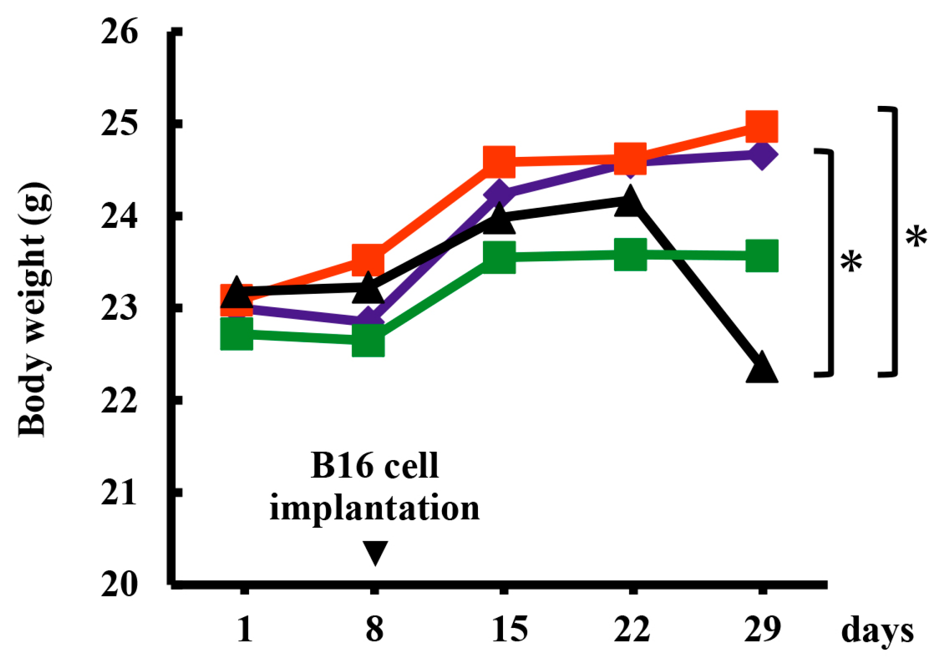
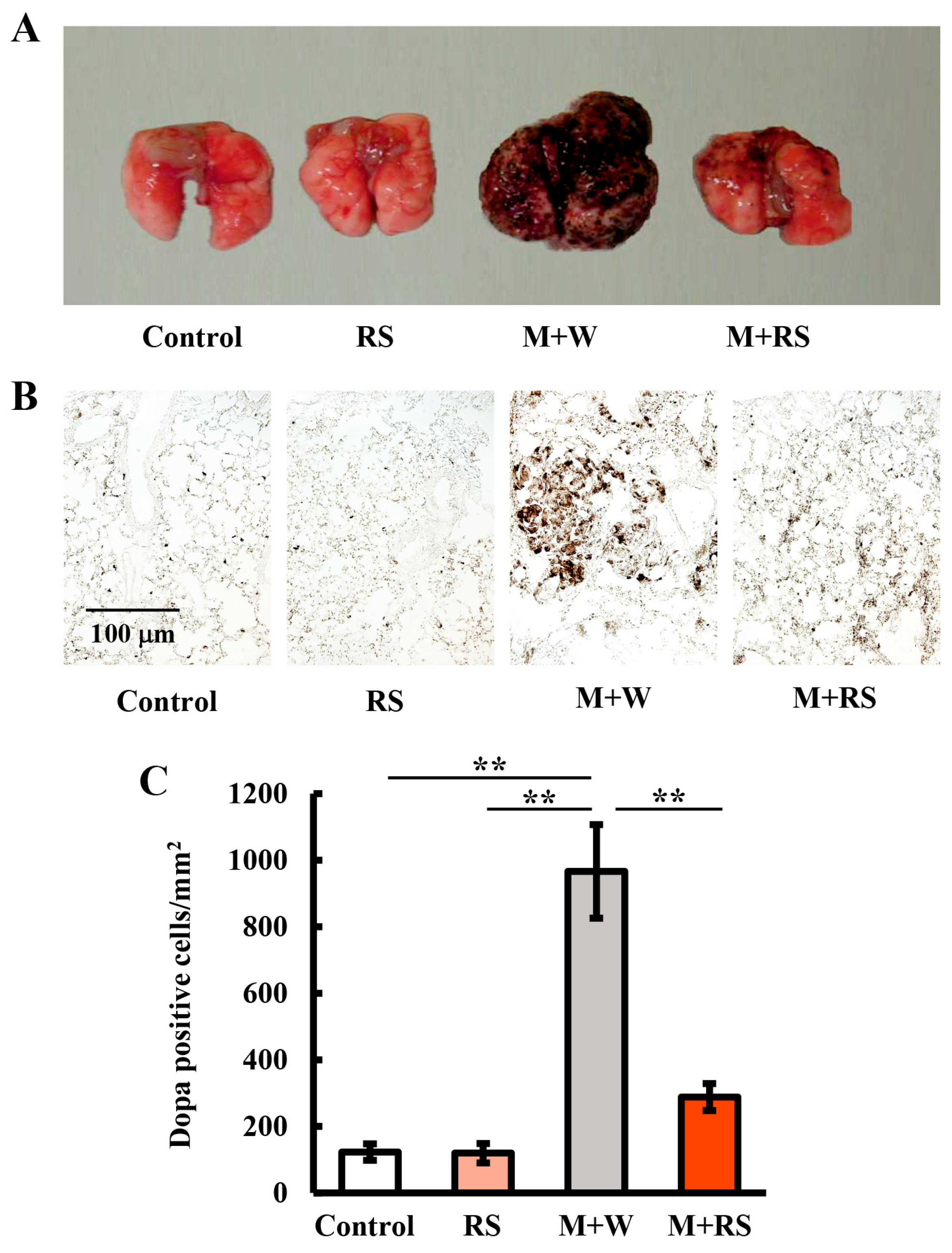
In the present study, we investigated the effects of orally administered RS on B16 cell metastasis in mice implanted with B16 cells. The mean body weight of mice in the M + W group on day 29 was significantly lower than that of mice in the control and RS groups, whereas the M + RS group showed a tendency toward improvement, although the difference was not significant. Furthermore, the M + RS group significantly suppressed the increase in B16 cell (DOPA-positive cell) clusters in the lung tissue.
Factors related to inflammation, tumor invasion, and metastasis in plasma (IL-6, uPA, MMP-2, and MMP-9) and lung tissue (PAR1, PAR2, and TGFβ1) were all significantly increased in the M + W group. However, in the M + RS group, all plasma factors and PAR1 levels were significantly decreased, whereas PAR2 and TGFβ1 were not. PAR2 and TGFβ1 are known to be interdependently involved in tumor angiogenesis and metastasis [
26,
27,
28], suggesting that RS ingestion does not affect the factor Xa-PAR2-dependent pathway or the PAR2-TGFβ1-dependet pathway. In contrast, RS ingestion may suppress the activation and expression of PAR1 via thrombin produced in the host, thereby suppressing B16 cell invasion and metastasis.
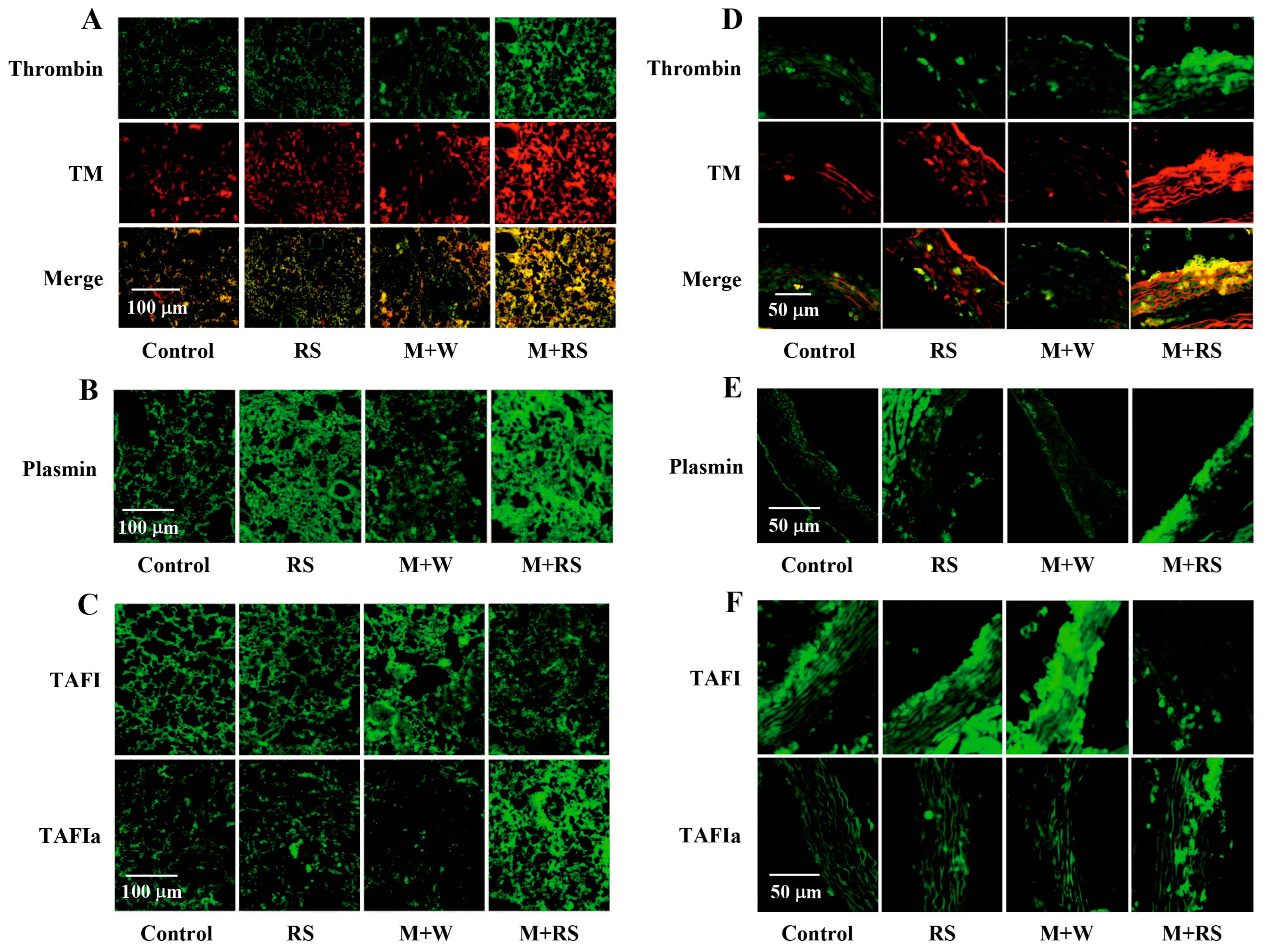
Regarding factors related to inflammation, coagulation, and fibrinolysis in both lung and aortic tissues, TNFα, TF, and the inactive form of TAFI were significantly increased in the M + W group and significantly decreased in the M+RS group compared to the M + W group. The expression levels of thrombin, TM, and plasmin in the M + W group remained unchanged, whereas these factors, along with TAFIa, were significantly increased in the M + RS group. Merged image analysis of thrombin and TM suggests that they may form a complex in the same tissue, contributing to TAFI activation, as the thrombin-TM complex is known to activate TAFI [
29]. These results suggest that RS ingestion enhances both fibrinolysis activation (increased plasmin) and fibrinolysis regulation (increased TAFIa) in melanoma-bearing mice, although the underlying mechanisms remain unclear.
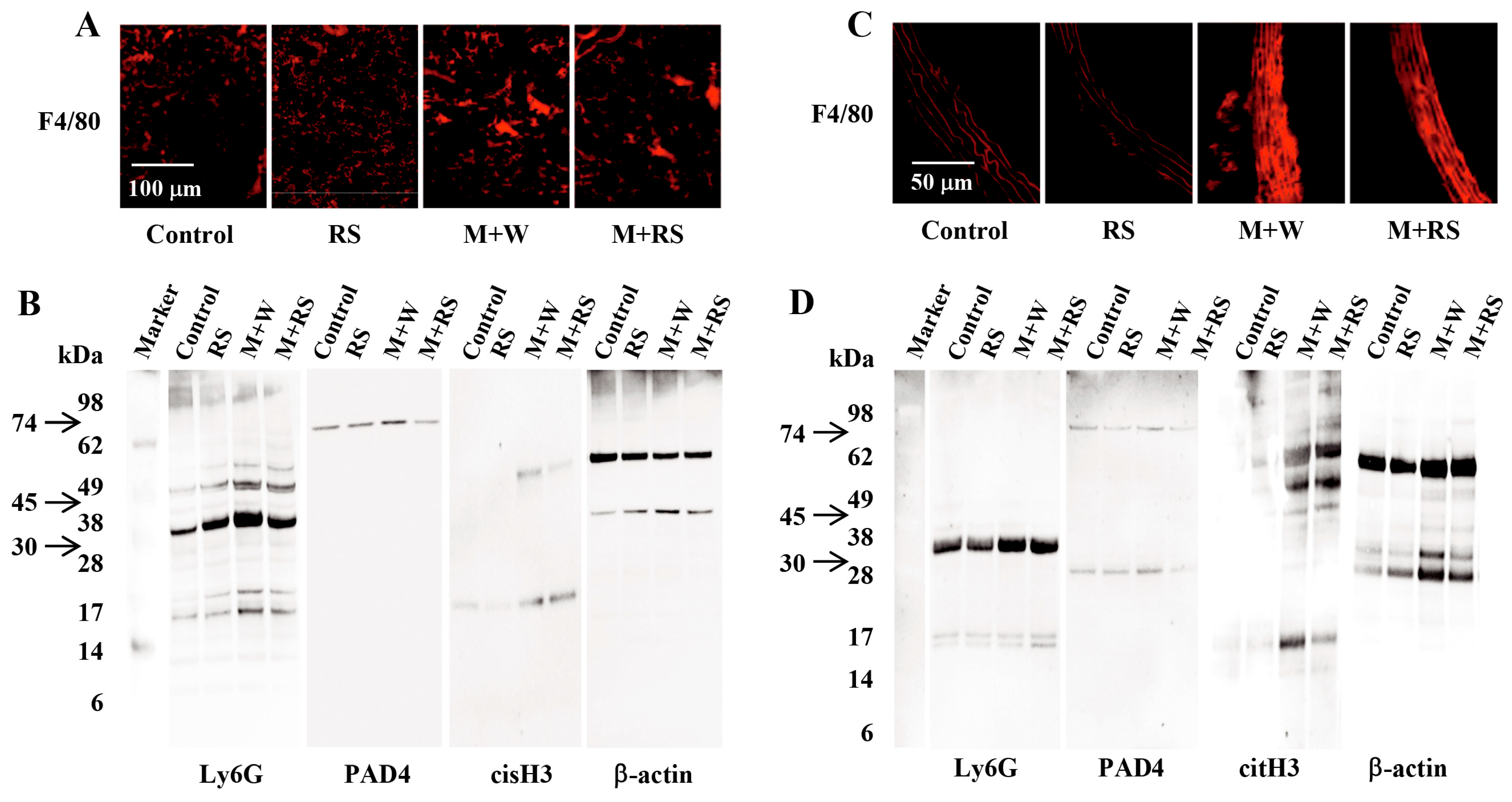
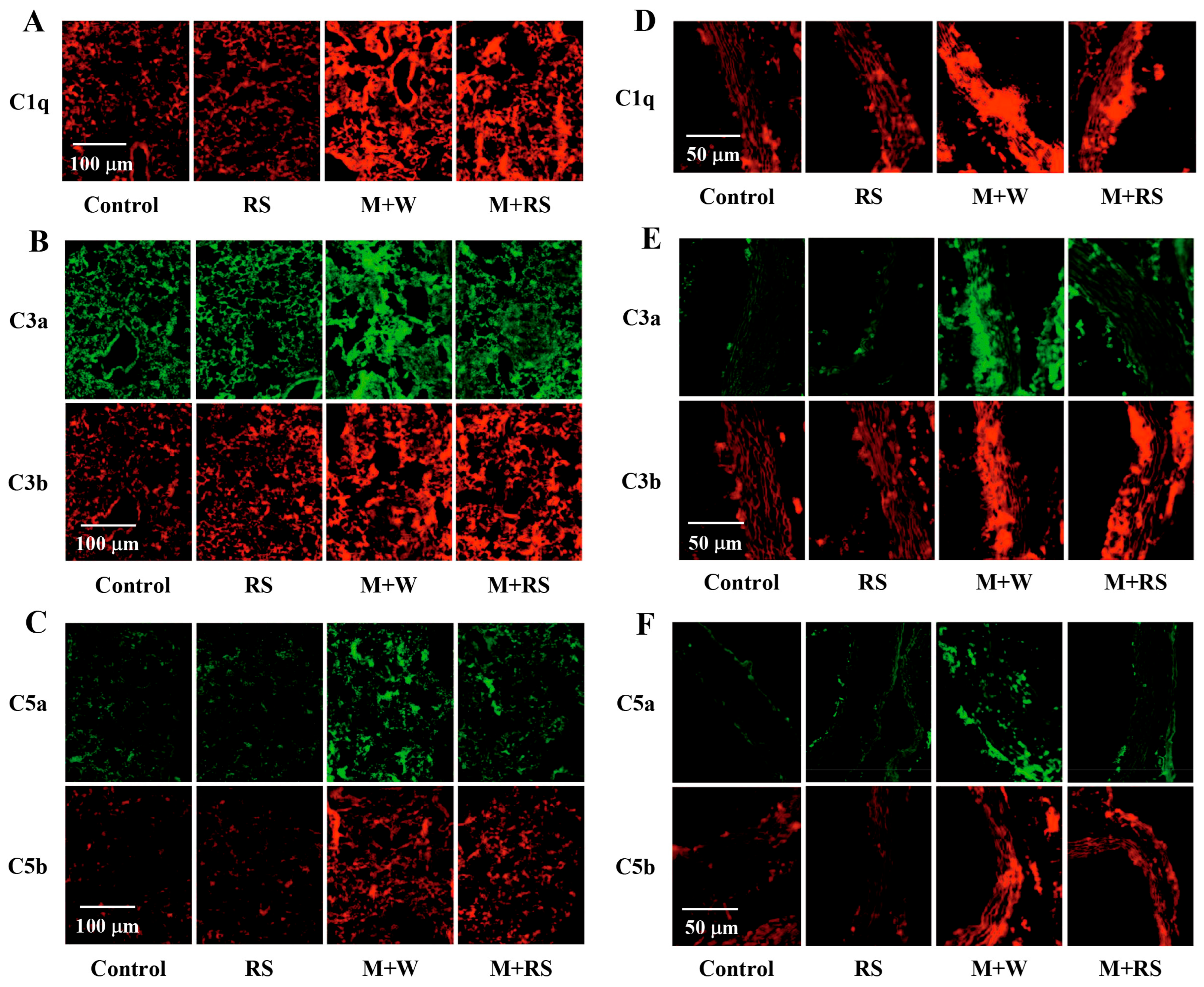
Tumor-associated leukocyte (macrophages and activated neutrophils)-related factors were significantly increased in the lung and aortic tissues of the M + W group, whereas most of these factors were significantly decreased in the M + RS group. Tumor-associated macrophages are known to release C1q, which activates the complement system. The complement system is closely linked to the coagulation and fibrinolysis systems, and C3b forms complexes with factor H and regulates the complement system via TM [
30]. Furthermore, TAFIa, activated by the thrombin–TM complex or plasmin, proteolytically inactivates C3a, C5a, and bradykinin, thereby suppressing vascular inflammation [
31].
All complement factors (C1q, C3a, C3b, C5a, and C5b) measured in this study were significantly increased in the lung and aorta tissues of the M + W group. In the M + RS group, C3b and C5b did not show significant changes compared to the M + W group, whereas C1q, C3a, and C5a were significantly decreased, with particularly marked reductions in C3a and C5a. The increased levels of plasmin, thrombin–TM complex, and TAFIa suggest that degradation of C3a and C5a by TAFIa may be involved. These results suggest that RS ingestion may inhibit melanoma cell proliferation and metastasis by promoting the degradation of C3a and C5a while maintaining C3b and C5b levels.
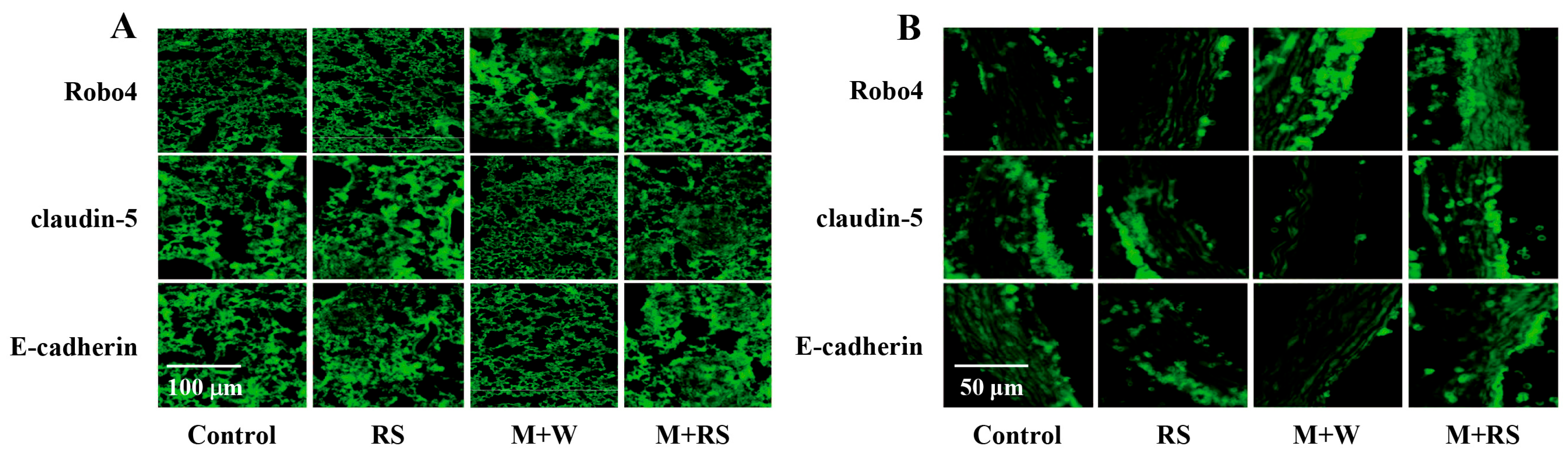
Next, we analyzed the effects of RS ingestion on angiogenesis-related factors derived from tumor cells and surrounding tissue cells and their involvement in B16 cell invasion and metastasis. The levels of Ang-2, bFGF, and Robo4, as well as TGFβ1, MMP-2, and MMP-9, which are involved in tumor-associated angiogenesis, vascular permeability, and tumor invasion, were significantly increased in the M + W group compared to the control group. In contrast, claudin 5 and E-cadherin, which are involved in maintaining tight junctions between epithelial cells, were significantly decreased in the M + W group but significantly increased in the M + RS group. These findings suggest that RS ingestion suppresses B16 cell invasion into surrounding tissues and tumor-associated angiogenesis by increasing the expression of intercellular tight junction molecules such as claudin 5 and E-cadherin.
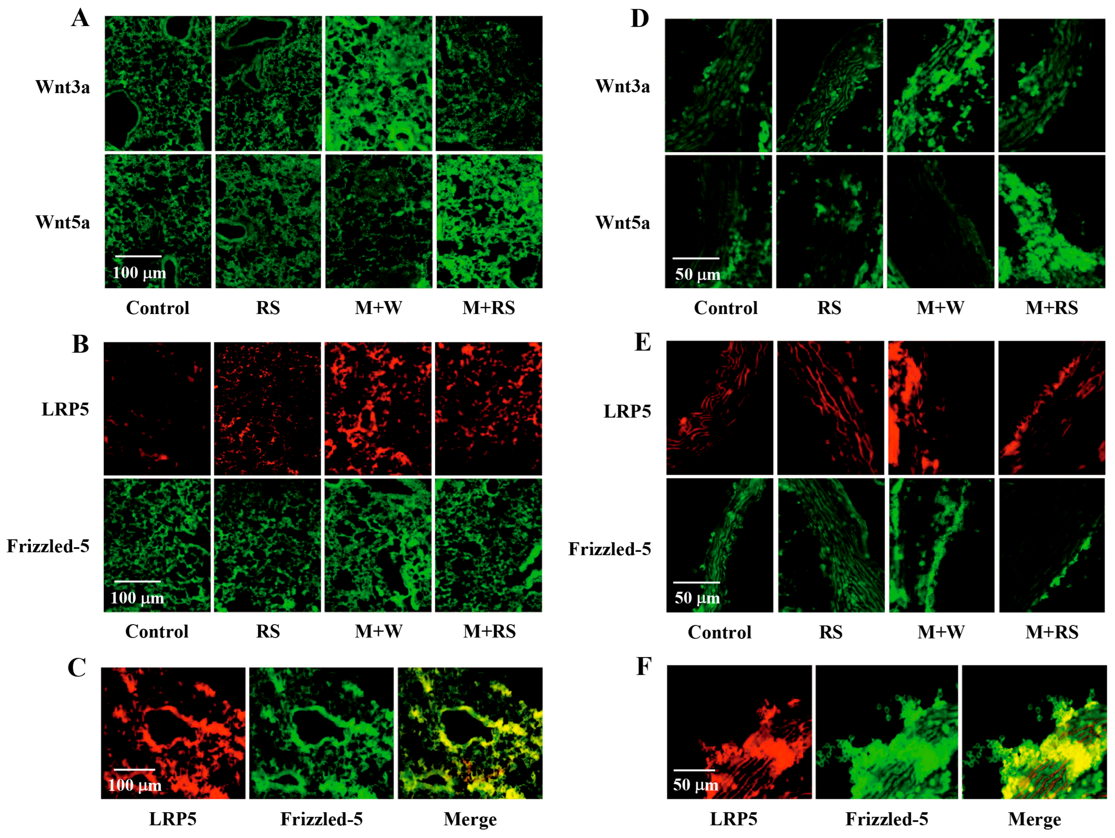
Finally, we analyzed the effects of RS ingestion on the expression of EMT-related factors, which are closely associated with tumor invasion and metastasis, in the tissues of B16 cell-implanted mice. The results showed that the expression of β-catenin, vimentin, Snail-1, and Wnt3a, which are involved in promoting EMT, was significantly increased in the lung and aortic tissue of the M + W group. This increase in expression was consistent with the increase in the expression of uPA, which promotes tumor cell migration, invasion, and proliferation by converting plasminogen to plasmin, activating MMPs, and degrading the extracellular matrix. In contrast, Wnt5a, which is involved in suppressing EMT, was significantly decreased. In the M + RS group, all factors except Wnt5a were significantly decreased, indicating that RS ingestion affects the expression of EMT-related factors associated with melanoma cell metastasis. Furthermore, the fact that RS ingestion significantly decreased Wnt3a expression and significantly increased Wnt5a expression suggests that the thrombin-PAR1 signaling pathway may be involved in the regulation of the Wnt signaling pathway. In addition, the expression levels of LRP5 and Frizzled-5, which are involved in the Wnt signaling pathway, were increased in the M + W group and decreased in the M + RS group. Image analysis integrating the expression of these two factors suggested that both factors are co-expressed within the same cell. These results suggest that RS ingestion reduces the binding of extracellular Wnt ligands to LRP5 and Frizzled-5 coreceptors in vascular endothelial cells of tumor-carrying mice, thereby suppressing the Wnt/β-catenin signaling pathway that promotes EMT.
Based on these considerations,
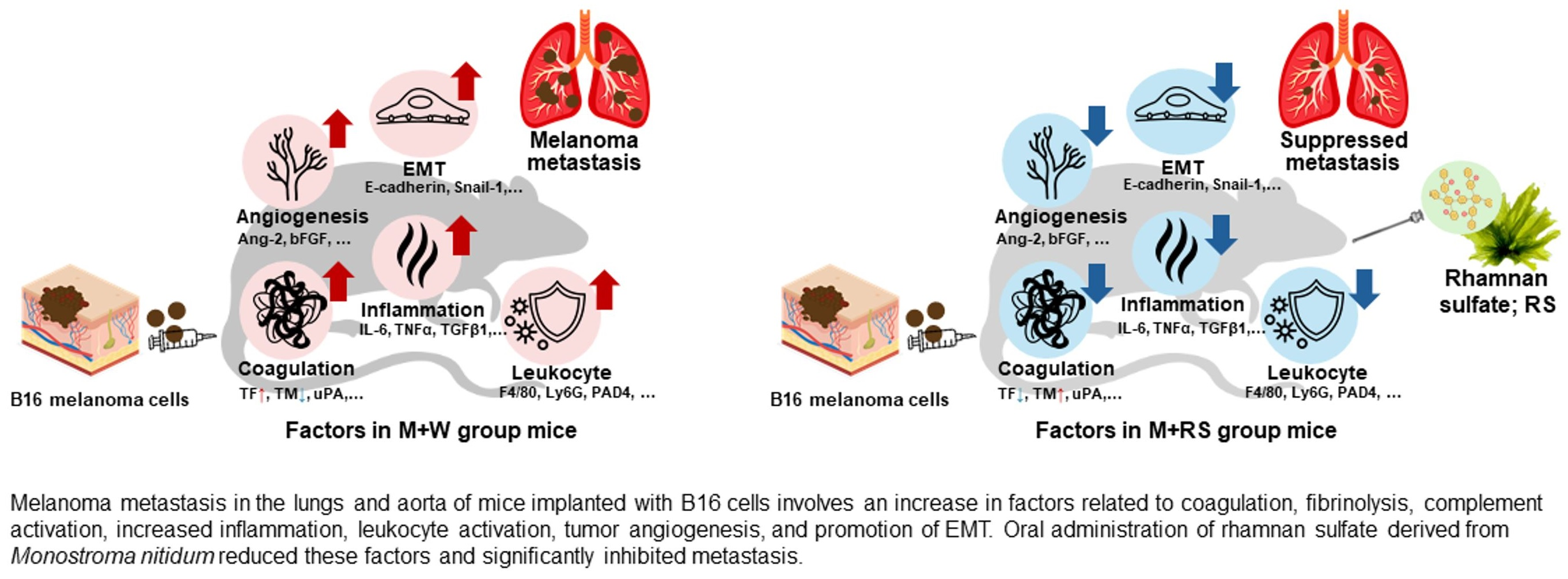
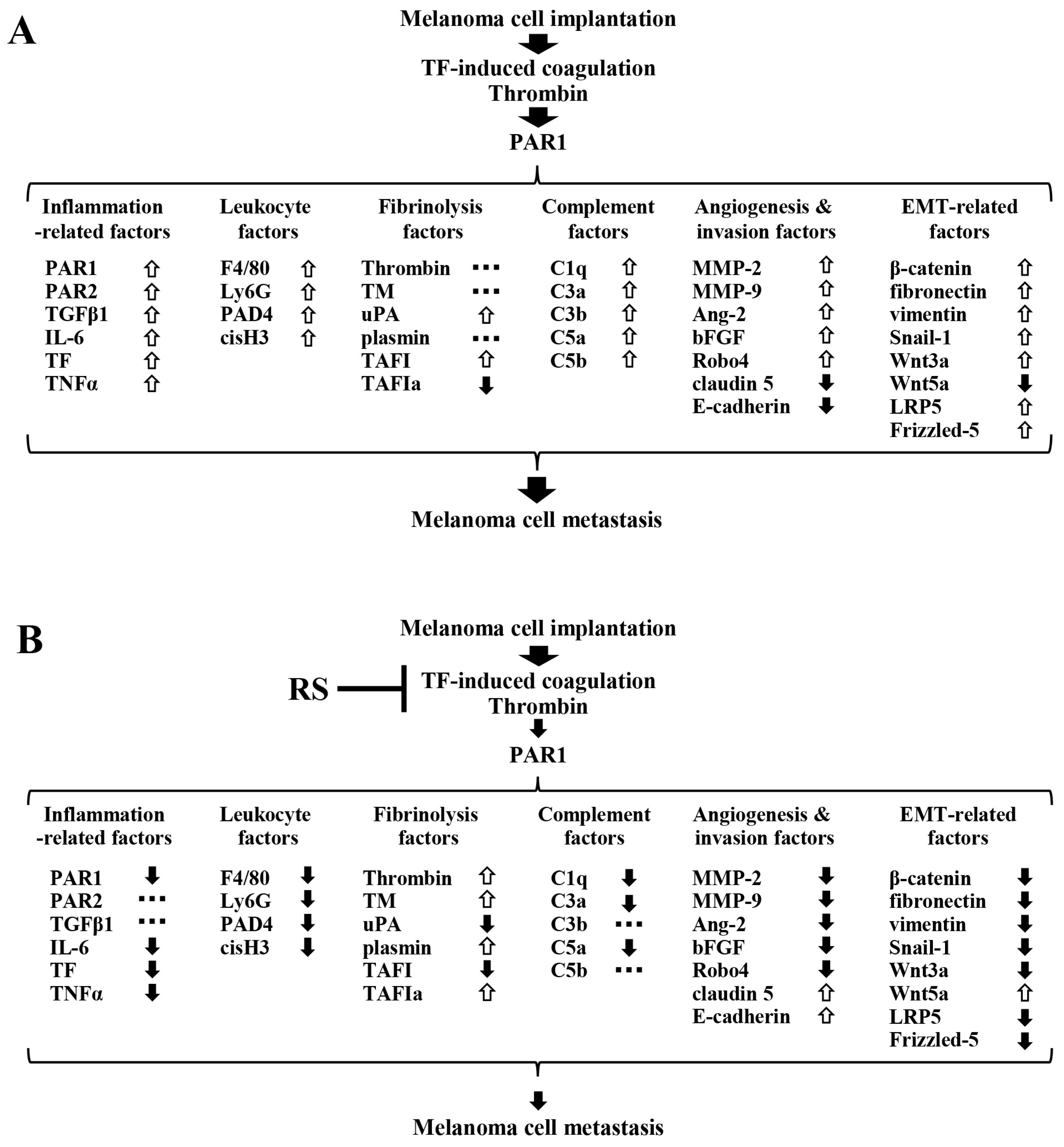
Figure 8A shows that, in mice in the M + W group, TF derived from implanted B16 cells induced blood coagulation, and the generated thrombin activated PAR1 in vasculature cells, causing changes in the expression of factors related to inflammation, leukocyte activation, fibrinolysis, complement, tumor angiogenesis and tissue invasion, and EMT. These changes collectively promote B16 cell metastasis. Upward arrows indicate increased expression, downward arrows indicate decreased expression, and horizontal arrows indicate no change in expression compared to the control group. Among these factors, only TAFIa, claudin 5, E-cadherin, and Wnt5a were significantly decreased compared to the control group, whereas all other factors, except thrombin, TM, and plasmin, were significantly increased compared to the control group.
Figure 8B shows factors whose expression levels were significantly altered in the M + RS group compared to the M + W group. These changes in factors are thought to be involved in the suppression of metastasis of implanted B16 cells by RS ingestion. Upward arrows indicate increased expression, downward arrows indicate decreased expression, and horizontal arrows indicate no change in expression. All inflammatory and leukocyte activation-related factors, except for PAR2 and TGFβ1, were significantly decreased. Among fibrinolysis- and complement-related factors, uPA, TAFI, C1q, C3a, and C5a were significantly decreased, whereas C3b and C5b remained unchanged. Thrombin, TM, plasmin, and TAFIa were significantly increased.
Regarding tumor angiogenesis- and tissue invasion-related factors, the tight junction molecules claudin 5 and E-cadherin were increased, whereas all other pro-angiogenic factors were significantly decreased. Furthermore, among EMT-related factors, Wnt5a, which inhibits EMT, was increased, whereas all other EMT-promoting factors were decreased. Changes in the expression of these factors induced by RS ingestion collectively suppressed the metastasis of implanted B16 cells.
The effects of RS are dose-dependent. Orally administered RS (0.25–2.5 mg RS /mouse) dose-dependently suppressed the increased vascular permeability observed in LPS-treated mice, restoring it to control levels. It also reduced various inflammatory markers, such as IL-6 and TF, demonstrating a dose-dependent anti-inflammatory effect of RS [
5]. Furthermore, orally administered RS (0.25–7.5 mg RS/mouse) dose-dependently suppressed the increase in blood TNFα levels in hyperglycemic diabetic mice, whereas excessive RS administration attenuated this effect [
7]. These results suggest that the bio-protective effect of RS depends on the appropriate amount of RS ingested.
The mechanisms by which orally administered RS suppresses tumor cell-derived TF-induced blood coagulation and tissue inflammation in mice implanted with B16 cells are unclear, but previous studies by our group and others have suggested several possibilities.
First, RS inhibits thrombin through an antithrombin-dependent mechanism [
4]. In vitro experiments using purified coagulation factors have shown that RS potently inhibits thrombin in an antithrombin-dependent manner, like unfractionated heparin, while inhibiting factor Xa very weakly. Furthermore, experiments using cultured human umbilical vein endothelial cells have shown that RS dose-dependently inhibits the increased expression of TF and the platelet aggregation factor von Willebrand factor in unstimulated endothelial cells and in endothelial cells stimulated with thrombin, TNFα, or LPS.
Second, orally administered RS in mice potently suppresses endothelial cell inflammation [
5]. RS suppressed intraperitoneally administered LPS-induced vascular hyperpermeability and neutrophil infiltration into organs, including the lungs and liver, and significantly reduced plasma levels of the inflammatory molecular markers, IL-6 and TF. Furthermore, RS maintained the expression level of syndecan-4 in endothelial cells and significantly inhibited the loss of the glycocalyx layer, thereby suppressing inflammatory damage to endothelial cells.
Third, RS inhibits hyaluronidase in a concentration-dependent manner [
32]. Hyaluronidase degrades and strips hyaluronic acid, the main component of the glycocalyx layer of vascular endothelial cells, increasing vascular permeability and inducing endothelial dysfunction. Therefore, RS may play a role in protecting the vascular endothelium by inhibiting hyaluronidase.
Another possibility is that RS ingestion in humans modulates the composition of the gut microbiota, improves intestinal function, and suppresses systemic inflammation [
33]. Indeed, administration of RS to mice has been shown to induce a healthy gut microbiota, which may be beneficial for health [
34].
A limitation of this study was the inability to directly measure the blood concentrations of RS and its metabolites after oral administration. Therefore, it was not possible to determine the systemic distribution of RS or the amount of active RS present in the lungs and aorta. However, previous studies have confirmed that orally administered fluorescein isothiocyanate-labeled RS co-localizes with M cells and is taken up by Peyer’s patches [
35], suggesting the possibility of intestinal absorption of RS. This possibility is supported by studies on fucoidan, another high molecular weight sulfated polysaccharide. Orally administered fucoidan has been detected in serum and urine and has been reported to be taken up by the intestines and liver tissue despite its low intestinal permeability [
36,
37]. Therefore, although direct pharmacokinetic data of RS are lacking, it is quite possible that some orally administered RS is absorbed into the body, transported to immune-related tissues, and thereby contributes to the systemic suppression of melanoma cell metastasis.
In conclusion, the results of this study suggest that orally administered RS inhibits thrombin generated during blood coagulation induced by implanted B16 cells via antithrombin, thereby suppressing the activation and expression of PAR1 in tissue cells and inhibiting vascular endothelial inflammation and complement system activation. RS also increases TAFIa production via plasmin and the thrombin–TM, thereby regulating tissue fibrinolysis. These effects of RS may collectively inhibit EMT and tumor angiogenesis, thereby suppressing melanoma cell invasion and metastasis.
Although this study was conducted using mice, the results suggest that oral administration of RS may suppress tumor growth and metastasis in patients with cancers such as melanoma. Further basic research and subsequent clinical studies are needed to understand the inhibitory effects of RS and other seaweed components on cancer cell growth and metastasis, their mechanisms of action, and their impact on human patients.
4. Materials and Methods
4.1. Animals and Melanoma Cells
Allogeneic transplantation studies were performed using specific pathogen-free (SPF) male 8-week-old C57BL/6J mice (SLC, Hamamatsu, Japan). Mice were housed in individual cages in an air-conditioned, SPF-controlled room at 23 ± 1 °C with a 12 h light/dark cycle (lights on at 08:00). Animals had ad libitum access to food and water.
A metastatic mouse melanoma cell line, B16 cells, established from a C57BL/6 mouse tumor [
15] and obtained from the Japanese Collection of Research Bioresources Cell Bank (Osaka, Japan), was used at passages 5 to 15. The cells were cultured in Eagle’s minimum essential medium (Sigma-Aldrich, Darmstadt, Germany) supplemented with 10% serum and l-glutamine. The cells were tested periodically to ensure that they were free of Mycoplasma, mouse viruses, or tumorigenic contamination. Subconfluent monolayers were harvested after treatment with 1 mM 0.25% trypsin and 0.02% ethylenediaminetetraacetic acid (Sigma-Aldrich). The trypsinized cells were washed and resuspended in phosphate-buffered saline (PBS; Ca
2+, Mg
2+-free; Sigma-Aldrich Chemical, St. Louis, MO, USA).
4.2. RS Sample
RS is composed of α-1,3-linked L-rhamnose residues, some of which are sulfated mainly at the O-2 position, with trace amounts of 1,2-rhamnose and branched rhamnose residues [
3]. In this study, we used an RS sample (purity 94%, molecular weight ranging from tens of thousands to hundreds of thousands, with an average molecular weight of approximately 150 kDa, and approximately 32% sulfate groups) purified from hot-water extracts of
M. nitidum by Konan Chemical Manufacturing Co., Ltd. (Yokkaichi, Mie, Japan) using a previously published method [
6].
4.3. Experiment on the Effect of Orally Administered RS on B16 Cell Metastasis in Mice and Sample Collection
Regarding the oral dosage of RS in mice, previous studies have shown that a dose of approximately 230 mg/kg body weight per day (6 mg per mouse) sufficiently improves inflammatory disorders without causing side effects such as bleeding [
5,
6,
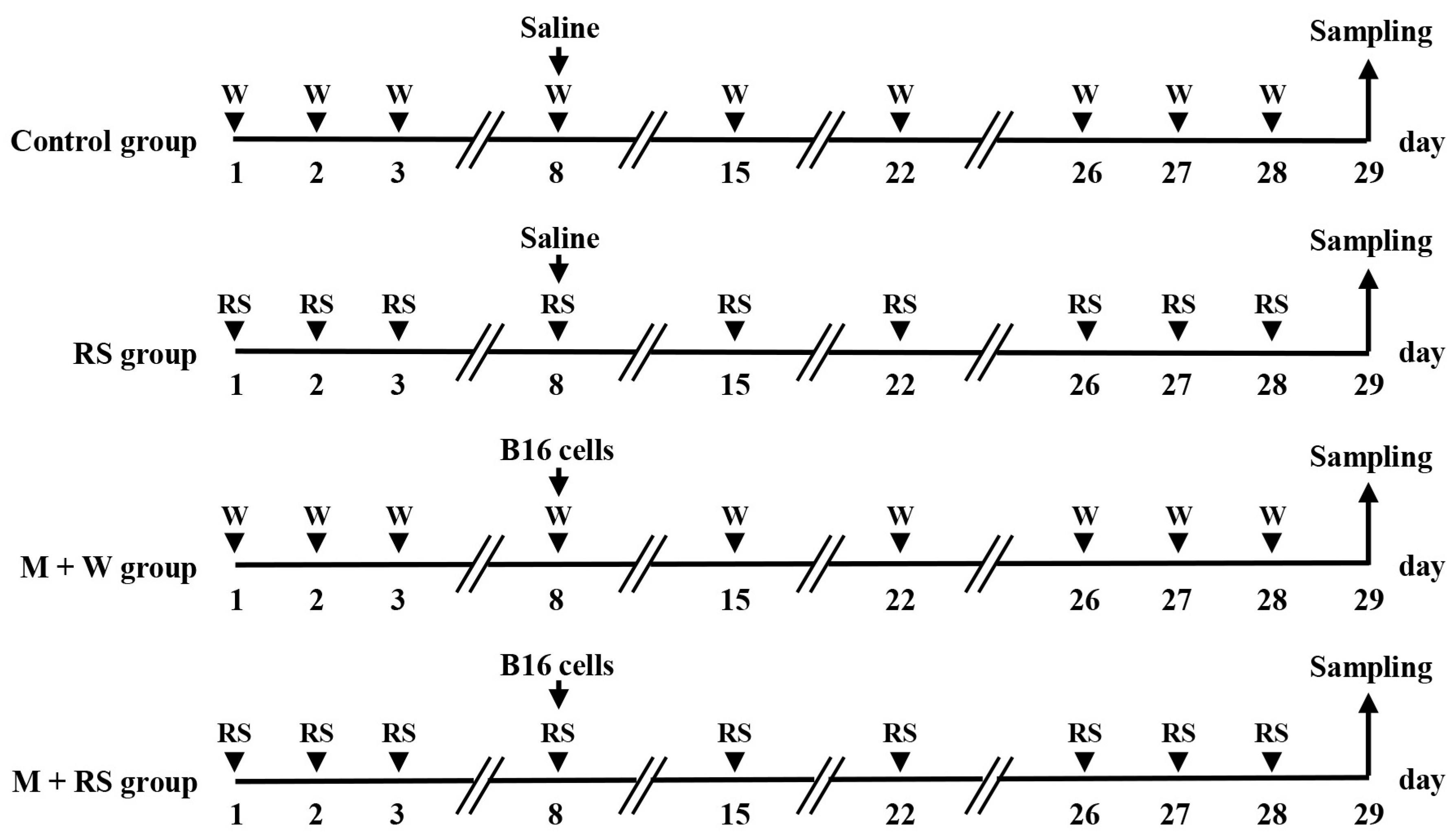
7], and this dosage was used in this study. An outline of the experimental method is shown in
Figure 9.
Eight-week-old male C57BL/6J mice were orally administered water or RS (6 mg/100 μL/day) daily. On day 8, saline (100 μL) or B16 melanoma cells (M) (1 × 10⁶ cells/100 μL) were injected into the tail vein, and water or RS was then administered daily by feeding needle for 21 days to create four groups; water-only (W) group (control), RS-only group, M + W group, and M + RS group, n = 5/group).
Body weight was measured on days 1, 8, 15, 22, and 29. On day 29, the mice were anesthetized, and blood and organs were collected. Blood collected from the aorta via abdominal incision was mixed with 1/10 volume of 100 IU/mL heparin sodium and centrifuged at 3000× g at 4 °C for 10 min using a refrigerated centrifuge. The heparinized plasma was stored at −80 °C for later use. After blood collection, the lungs and aorta were harvested, photographed, and frozen at −80 °C for later use. Organ tissue samples were fixed in PBS containing 4% paraformaldehyde (Fujifilm Wako Pure Chemicals, Osaka, Japan).
4.4. Identification of Melanoma Cells in Organ Tissues and Immunohistochemical Analysis of Tumor-Associated Factors
Fixed tissue specimens were embedded in frozen Tissue–Tek OCT Compound (Sakura Finetek, Tokyo, Japan) and sliced into 5 µm thick sections. To identify melanoma cells in the tissue, DOPA-positive cells, which indicate melanocyte tyrosinase activity, were examined. DOPA-positive melanocytes were stained as previously described [
26]. The tissue was washed with PBS and incubated in PBS containing 0.1% L-DOPA at 37 °C (Sigma-Aldrich Chemical, St. Louis, MO, USA). The specimens were washed with PBS for microscopic examination.
To visualize tumor-associated factors using immunohistochemical analysis, tissue specimens were incubated with factor-specific antibodies: rabbit polyclonal anti-thrombin (1:100; GTX610270, GeneTex, Irvine, CA, USA), rabbit polyclonal anti-plasmin (1:100; ABV11485, ABGENT, San Diego, CA, USA), rabbit polyclonal ant-TAFI (1:100; 55201-1-AP, Proteintech, Rosemont, IL, USA), rabbit polyclonal anti-TAFIa (1:100; 10672-1-AP, Proteintech), mouse monoclonal anti-C1q (1:100; ab71940, Abcam, Cambridge, UK), rat monoclonal anti-C3a (1:100; HM1072, Hycult Biotech, Wayne, PA, USA), rabbit polyclonal anti-C3b (1:100; GTX101316, GeneTex), rabbit polyclonal anti-C5a (1:100; bs-0324R, Bioss Antibodies, Woburn, MA, USA), mouse monoclonal anti-C5b (1:100; ab66768, Abcam), rabbit monoclonal anti-Wnt3a (1:100; #2721, Cell Signaling Technology, Danvers, MA, USA), rabbit polyclonal anti-Wnt5a (1:100; ab235966, Abcam), rabbit polyclonal anti-LRP5 (1:100; 24899-1-AP, Proteintech), rabbit polyclonal anti-Frizzled-5 (1:100; 21519-1-AP, Proteintech), goat polyclonal anti-TM (1:100; AF3894, R&D systems, Minneapolis, MN, USA), mouse monoclonal anti-Robo4 (1:50; sc-166872, Santa Cruz Biotechnology, Santa Cruz, CA, USA), rabbit monoclonal anti-claudin 5 (1:100; ab131259, Abcam), and rabbit monoclonal anti-E-cadherin (1:100; #3195, Cell Signaling Technology).
Sections were then incubated with the appropriate secondary antibodies (1:30 dilution; fluorescein isothiocyanate-conjugated anti-rabbit, anti-mouse, anti-rat, or anti-goat secondary antibody; Dako Cytomation, Glostrup, Denmark) for 2 h in the dark. Fluorescence intensity was quantified from five random fields of constant area using ImageJ software (version 2.1.53; NIH, Bethesda, MD, USA). The original files were converted to monochrome 8-bit images, and an arbitrary fluorescence intensity threshold was set. Areas exceeding the threshold (referred to as “intensity”) were measured for each sample.
4.5. Measurement of the Levels of Tumor-Associated Factors Using ELISA
Blood, liver, and aortic samples were collected at the end of the experiment. Plasma was separated from blood samples by centrifugation at 3000× g at 4 °C for 10 min., and the supernatant was used for further analysis. Plasma levels of uPA, IL-6, MMP-2, and MMP-9 were determined using commercially available ELISA kits according to the manufacturer’s instructions: uPA (IMSUPAKTT, Innovative Research Inc., Novi, MI, USA), IL-6 (M6000B; R&D Systems), MMP-2 (ab254516; Abcam), and MMP-9 (ab253227; Abcam).
Liver and aortic samples were homogenized at 15,000× g for 15 min at 4 °C (Tomy MX-201; Tomy Digital Biology, Tokyo, Japan), and the supernatant was collected for analysis. The levels of PAR1, PAR2, TF, TNF-α, TGF-β1, Ang-2, bFGF, β-catenin, vimentin, fibronectin, and Snail-1 in tissues were determined using commercially available ELISA kits: PAR1 (MBS753326, MyBioSource, San Diego, CA, USA), PAR2 (MBS4501658; MyBioSource), TF (ab214091; Abcam), TNF-α (KE10002, Proteintech), TGFβ1 (E-EL-M0051, Elabscience, Houston, TX, USA), Ang-2 (MANG20, R&D Systems), bFGF (bs-0217R, Bioss Antibodies), β-catenin (ADI-900-135; Enzo Life Sciences, Executive Blvd Farmingdale, NY, USA), fibronectin (OKCD05702, Aviva Systems Biology, San Diego, CA, USA), vimentin (ELK3731, ELK Biotechnology, Denver, CO, USA), and Snail-1 (LS-F2317-1, LS Bio, Shirley, MA, USA). The optical density was measured using a microplate reader (Molecular Devices, Sunnyvale, CA, USA).
4.6. Western Blotting Analysis of the Lungs and Aorta
Lung and aortic samples were homogenized in lysis buffer (Kurabo Industries, Osaka, Japan) and centrifuged to obtain the supernatants. Western blotting was performed as previously described [
38]. After electrophoresis, membranes were incubated with primary antibodies against Ly6G (neutrophil activation marker, 1:1000; 551459, BD Biosciences, Franklin Lakes, NJ, USA), citH3 (neutrophil activation marker, 1:1000; ab281584, Abcam), PAD4 (neutrophil activation marker, 1:1000; ab214810, Abcam), and β-actin (1:5000; #58169, Cell Signaling) for 1 h at room temperature. β-actin was used as a loading control.
Membranes were washed and incubated with horseradish peroxidase-conjugated secondary antibodies (Novex, Frederick, MD, USA). Immune complexes were detected using ImmunoStar Zeta reagent (Wako Pure Chemical Industries, Osaka, Japan), and images were acquired using Multi Gauge software v3.0 (Fujifilm, Greenwood, SC, USA).
4.7. Statistical Analysis
All data are presented as mean ± standard deviation (SD). Microsoft Excel 2010 (Microsoft Corp., Redmond, WA, USA) and SPSS version 20 (SPSS Inc., Chicago, IL, USA) were used for statistical analysis. One-way analysis of variance followed by Tukey’s post hoc test was performed. Results were considered statistically significant at α = 0.05 (p < 0.05) or α = 0.01 (p < 0.01).
4.8. Ethical Statement for Animal Studies
All animal experiments were conducted in strict accordance with the recommendations of the Suzuka University of Medical Science Animal Experiment Ethics Committee (Approval no. 84) and prepared according to the guidelines of the Ministry of Education, Culture, Sports, Science, and Technology of Japan. Surgery was performed under pentobarbital anesthesia, and every effort was made to minimize pain.






 ), RS group (
 ), M + W group (
 ), and M + RS group (
 ). * p < 0.05.