Article

New Strategy for the Degradation of High-Concentration Sodium Alginate with Recombinant Enzyme 102C300C-Vgb and the Beneficial Effects of Its Degradation Products on the Gut Health of Stichopus japonicus

by
Ziqiang Gu
,
Feiyu Niu
,
Peng Yang
,
Wenling Gong
,
Hina Mukhtar
,
Siyu Li
,
Yanwen Zheng
,
Yiling Zhong
,
Hanyi Cui
,
Jichao Li
,
Haijin Mou
* and
Dongyu Li
*
College of Food Science and Engineering, Ocean University of China, No.1299 Sansha Road, Qingdao 266003, China
*
Authors to whom correspondence should be addressed.
Mar. Drugs 2025, 23(9), 339; https://doi.org/10.3390/md23090339
Submission received: 25 July 2025 / Revised: 22 August 2025 / Accepted: 22 August 2025 / Published: 25 August 2025
(This article belongs to the Section Biomaterials of Marine Origin)

Abstract

High viscosity of alginate means a relatively low substrate concentration, which limits the efficiency of hydrolysis, resulting in one of the main challenges for the large-scale production of alginate oligosaccharides (AOS). In this study, a pilot-scale degradation product (PSDP) of the recombinant enzyme 102C300C-Vgb was produced for the first time at a substrate concentration of up to 20% sodium alginate. The optimal conditions for SA digestion were: enzyme dosage of 25 U/g, enzymatic temperature of 45 °C, enzymatic pH of 7.0, and enzymatic time of 24 h. Under these conditions, the yield of enzymatic hydrolysis was consistently in the range of 69% to 70%. The average molecular weight (Mw) of PSDP was 1496.36 Da, mainly containing oligosaccharides with degrees of polymerization ranging from 2 to 4. The low-Mw PSDP was subsequently applied in the diet of sea cucumber Stichopus japonicus. The results showed that the body wall weight of S. japonicus increased significantly after 40 days of feeding with a 0.09% PSDP-supplemented diet. Furthermore, PSDP-supplemented diet significantly increased the thickness of the serosal and submucosal layers and the width folds of mucosa of the sea cucumber gut. The abundance of pathogenic bacteria was reduced effectively, and that of beneficial bacteria increased significantly after being fed with PSDP. The results demonstrated that PSDP can serve as a digestive health enhancer for sea cucumbers, promoting their healthy growth.

Graphical Abstract

1. Introduction

Sea cucumbers Stichopus japonicus (also called Apostichopus japonicus) are a prevalent group of marine invertebrates [1,2]. This species is also extensively cultivated in China, with production volumes reaching 248,000 tons in 2022, underscoring its significant economic value [3]. Sea cucumbers are rich in bioactive compounds, including polysaccharides and collagen, demonstrating a range of biological activities such as anticancer, antihypertensive, antibacterial, and antioxidant effects [4]. In particular, sea cucumber collagen is free from health-related risks and religious restrictions, making it a promising alternative to mammalian collagen [5].
However, intensification of aquaculture systems, unfavorable environmental conditions, and poor management practices have significantly exacerbated disease outbreaks in aquaculture farms, thus constraining the development of the sea cucumber farming industry [6]. In routine management, antibiotics are commonly used to control bacterial diseases, but their overuse can lead to the emergence of antibiotic-resistant pathogenic strains, posing a threat to the sustainability of sea cucumber farming [7,8]. Consequently, there is an urgent need to identify efficient, safe, and sustainable alternatives to antibiotics to promote the growth and yield of S. japonicus. Research indicates that strategic addition of certain prebiotics or bioactive compounds can enhance feed quality, optimize the gut microecological environment, and promote organism growth and health [9,10]. Consequently, external interventions, such as the incorporation of prebiotics to maintain the balance of the microbial flora in the gut, play a crucial role in the cultivation of S. japonicus [11].
The prebiotics extracted from brown algae have demonstrated promising activity in the aquaculture industry [12]. Alginate oligosaccharides (AOS), the low molecular weight degradation products of alginate from brown algae and bacteria, are characterized by their improved solubility, high safety profile, and remarkable stability [13,14]. Recent studies have demonstrated that dietary supplementation with AOS also reduces Salmonella colonization, improving gut barrier function and poultry production performance [15,16]. A moderate amount of AOS can improve lipid metabolism and immune responses in aquatic animals, alter gut bacterial communities, and aid in combating bacterial infections [17]. Furthermore, AOS, as a dietary supplement, can enhance growth performance, non-specific immune response, and disease resistance of aquatic animals [18]. Collectively, these findings underscore the potential of AOS as a feed additive with substantial promise in improving the health and disease resistance of farmed animals. Currently, enzyme hydrolysis, a key technique in the biological method, is considered an effective strategy for the depolymerization of polysaccharides to produce specific oligosaccharides with defined degrees of polymerization [19], offering advantages such as mild reaction conditions, avoidance of radiation, low energy consumption, and high yield compared to chemical and physical methods [20,21,22]. However, the high viscosity of alginate often limits substrate concentration during enzymatic hydrolysis, resulting in reduced AOS yields [23].
In this study, the degradation process of sodium alginate was optimized using a highly active alginate lyase 102C300C-Vgb and scaled up for the pilot-scale experiment to improve substrate concentration during enzyme hydrolysis and result in high product yield. Furthermore, the pilot-scale degradation product (PSDP) was conducted with a detailed analysis of the structure and composition, and its effectiveness was evaluated in promoting the healthy aquaculture of S. japonicus.

2. Results and Discussion

2.1. Optimal Enzymatic Hydrolysis Conditions

The enzymatic preparation of AOS is considered an efficient, energy-saving, and environmentally friendly method. Furthermore, alginate lyase activity is critical to the efficiency of AOS production [24]. Additionally, increasing substrate concentration can enhance the AOS yield within a single batch of enzymatic hydrolysis, significantly reducing industrial production costs [25]. However, the high substrate concentration severely increases the viscosity of the SA solution, limiting the mobility of alginate lyase and affecting its ability to interact with the substrate [23]. Furthermore, current alginate lyases suffer from poor substrate specificity, low activity, and limited stability, failing to meet the demands of industrial production [26].
In this study, the substrate concentration of 20% SA was hydrolyzed by high-activity enzyme 102C300C-Vgb with five different dosages. As shown in Figure 1A, the yield of enzymatic hydrolysis increased with the addition of enzyme dosage. When the enzyme dosage reached 25 U/g, the yield was 59.98 ± 0.03%. Further, an increase in enzyme addition did not result in a significant change in yield. Similarly, the results for the proportion of RSC (Reducing sugar content) indicated that when the enzyme dosage exceeded 25 U/g, the RSC obtained from enzymatic hydrolysis decreased (Figure 1A), demonstrating that an excessive dosage of enzyme failed to further enhance enzymatic hydrolysis efficiency. Therefore, 25 U/g was chosen as the optimal enzyme dosage.
The selection of temperature in the enzymatic hydrolysis process of alginate lyase was extremely important. To determine the optimal hydrolysis temperature, this study set up five temperature gradients. As shown in Figure 1B, the yield of enzymatic hydrolysis increased with temperature. When the temperature reached 45 °C, the yield was 65.72 ± 1.62%. Further increasing the temperature resulted in a significant decrease in yield (p < 0.05). In the same way, the results for the RSC indicated that when the hydrolysis temperature exceeded 45 °C, the RSC obtained from enzymatic hydrolysis decreased (Figure 1B), demonstrating that the enzyme became denatured and lost activity at excessive temperature.
Most known alginate lyases exhibited optimal catalytic activity at pH between 6.0 and 8.0 [27]. To determine the optimal hydrolysis pH, this study set up five pH gradients. The highest yield of enzymatic hydrolysis (70.01 ± 0.33%) was observed at pH 7.0 (Figure 1C). The results also indicated that the RSC obtained from enzymatic hydrolysis at pH 7.0 was significantly higher than at other pH conditions (p < 0.05) (Figure 1C), demonstrating that this alginate lyase preferred a neutral environment, and that both excessively high and low pH negatively affect its enzymatic efficiency.
To determine the optimal hydrolysis time, this study set up five hydrolysis time gradients. As shown in Figure 1D, the yield of enzymatic hydrolysis increased with time. When the hydrolysis time reached 24 h, the yield was highest, at 69.07 ± 0.06%, which was consistent with the results for the RSC (Figure 1D). Most known alginate lyases had poor thermal stability, exhibiting optimal catalytic activity only between 30 °C and 40 °C. Currently, there are few thermophilic alginate lyases, with recombinant alginate lyase produced by thermophilic degrading bacteria showing an optimal catalytic temperature of 50 °C. However, after reacting at 50 °C for 30 min, it lost 58% of its initial activity [28]. The alginate lyase 102C300C-Vgb used in this study can maintain its activity for 24 h at 45 °C, indicating its thermal stability [27].
In summary, optimal conditions for SA digestion using the recombinant enzyme 102C300C-Vgb with a substrate concentration of 20% SA were: enzyme dosage of 25 U/g, enzymatic temperature of 45 °C, enzymatic pH of 7.0, and enzymatic time of 24 h. Under these conditions, the yield of enzymatic hydrolysis was consistently in the range of 69% to 70%.

2.2. Structural Analysis of Degradation Product

2.2.1. FTIR Analysis

The degradation product was compared with SA using FTIR. In the IR spectrum (Figure 2), the peak at 3369.53 cm−1 corresponded to O-H stretching vibrations, while the peak at 2924.23 cm−1 was attributed to C-H stretching vibrations. The peaks at 1608.59 cm−1 and 1414.86 cm−1 represented asymmetrical and symmetrical O-C-O stretching vibrations, respectively. Additionally, the peaks at 868.95 cm−1 and 796.66 cm−1 were indicative of the presence of mannuronic acid and guluronic acid, respectively, according to previous findings [29]. The results indicate that the product retained the characteristic features of alginate, with no significant alterations observed in the spectra of the side groups [27].

2.2.2. HPLC Analysis of Degradation Product

The Mw distribution of the degradation product was shown in Figure 3(A-1). Based on the elution time and the standard curve, the Mw range of the product was 1934–5418 Da, with an average Mw of 2575 Da. Among these, the fraction with Mw between 1000–3000 Da represented the largest proportion at 70.40%, followed by the fraction with Mw between 3000–5000 Da, which accounted for 29%.

2.2.3. ESI-MS Analysis of Degradation Product

The ESI-MS spectrum for the degradation product is shown in Figure 3(A-2), indicating that the product was mainly composed of oligosaccharides with degrees of polymerization ranging from 2 to 4. AOS with this degree of polymerization was considered to possess significant biological activity [30]. The ions at 351.1 m/z, 571.1 m/z, 769.1 m/z, 967.1 m/z, and 1165.1 m/z represented unsaturated disaccharide (374 Da), unsaturated trisaccharide (572 Da), unsaturated tetrasaccharide (770 Da), unsaturated pentasaccharide (968 Da), and hexasaccharide (1166 Da), respectively. It verified that 102C300C-Vgb cleaved the glycosidic bonds in SA by β-elimination reaction.

2.3. Preparation and Compositional Analysis of PSDP

Currently, the high viscosity of the SA substrate imposes a significant limitation on the substrate concentration during the enzymatic production of AOS (typically below 5% w/v SA). Conventional approaches, such as increasing temperature, have proven inadequate in addressing this issue [31,32]. In this study, the PSDP was demonstrated at a substrate concentration of 20% w/v SA with a yield of 70%. Despite certain losses during production (e.g., residuals in pipelines and losses in the spray tower), this yield was generally consistent with the results obtained from high-concentration enzymatic hydrolysis of SA in laboratory optimization experiments, presenting substantial commercial potential.
Sensory, physicochemical, and contaminant analyses were performed on the PSDP, with results presented in Table 1. The color of the product changed from pale yellow-white to pale yellow-brown, attributed to the formation of unsaturated double bonds during the enzymatic hydrolysis process, which darkened the product [23]. The AOS content and water content in the product were 66.7 ± 0.1% and 5.10 ± 0.85%, which was influenced by factors such as the solid content of the solution before spray drying, the inlet and outlet temperatures of the spray tower. These parameters could affect the storage stability of the product [33]. The ash content of PSDP was 20.95 ± 3.32%, with a total arsenic content of 0.29 ± 0.01 mg/kg. Furthermore, no lead or Salmonella was detected, all of which met the standards for the use of feed additives.

2.4. Structural Analysis of PSDP

2.4.1. HPLC Analysis of PSDP

Based on the elution time and the standard curve, the Mw range of PSDP was 830.51–4076.73 Da, with an average Mw of 1496.36 Da (Figure 3(B-1)). Among these, molecules with an Mw below 1000 Da constituted the largest proportion, accounting for 53.70%, followed by those in the 1000–3000 Da range, comprising 44.70%. The average Mw of PSDP was lower than that of the degradation product prepared in the laboratory, with a higher proportion of low-Mw fragments.

2.4.2. ESI-MS Analysis of PSDP

The structural diagram of the PSDP was shown in Figure 3(B-2). Combined with the analysis result (Figure 3(A-1,A-2)), this further confirmed that PSDP underwent more extensive enzymatic hydrolysis compared to the degradation product prepared under laboratory conditions. The results might be attributed to more clearly defined reaction conditions in pilot-scale equipment under the same process parameters, such as the stirring speed in the reaction vessel and heating efficiency in the enzymatic hydrolysis tank.

2.5. Evaluation of the Feeding Effect of PSDP

2.5.1. Growth Performance

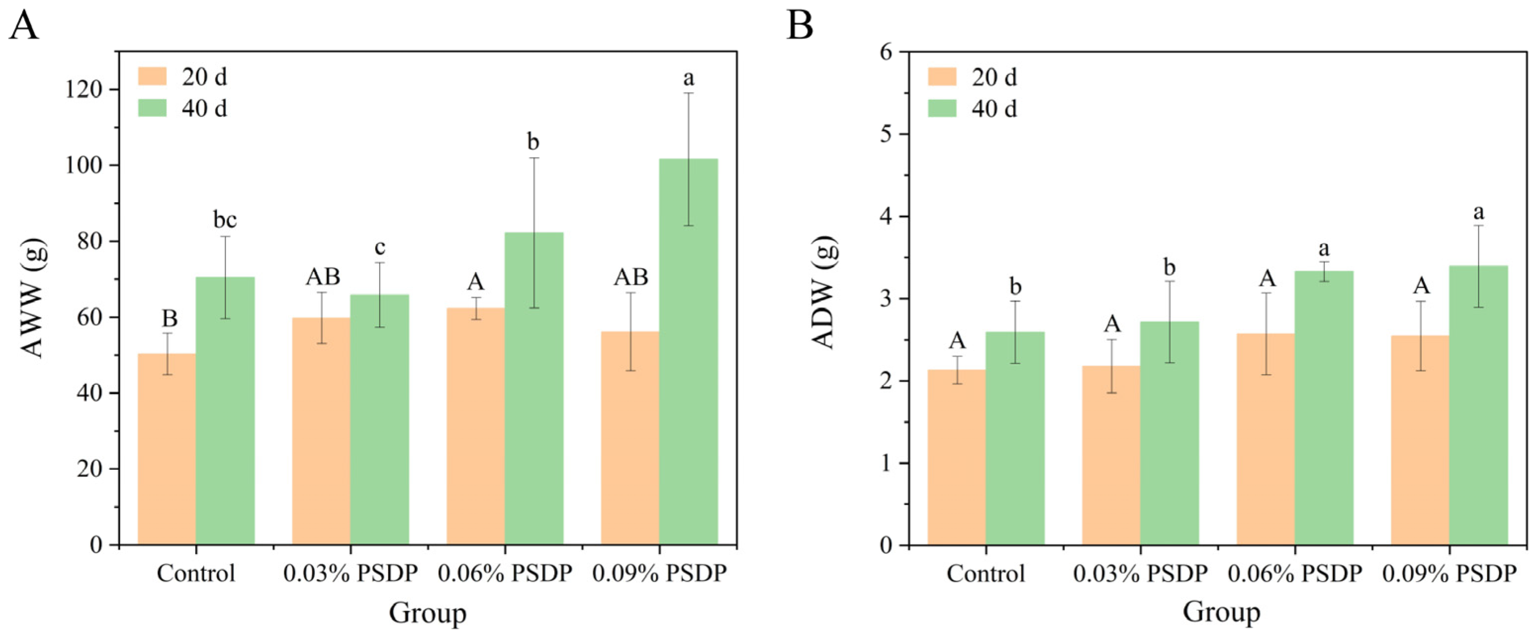
AOS exhibits excellent solubility, strong stability, ease of absorption, safety, and nontoxicity, along with a range of biological activities. These properties have led to their widespread application in fields such as biotechnology, medicine, food, and agriculture [19,34,35,36]. In the field of animal husbandry, research on AOS has primarily focused on their biological functions in poultry, piglets, and aquatic animals [15,37,38]. In this study, three proportions of PSDP were added to commercial feed to assess its growth-promoting effects on the sea cucumbers S. japonicus. Changes in the AWW of sea cucumbers after 20 days and 40 days of cultivation are shown in Figure 4A. After 20 days of cultivation, the wet weight of sea cucumbers fed with different concentrations of PSDP increased compared to the control group (50.29 ± 5.48 g), with the 0.06% PSDP group showing the highest weight of 62.26 ± 2.89 g (p < 0.05). After 40 days, the AWW of the individuals in the 0.09% PSDP group reached 101.57 ± 17.46 g, significantly higher than that of the other groups (p < 0.05) and 44.30% higher than that of the control group. The 0.06% PSDP group followed, with a wet weight of 82.19 ± 19.75 g. As the primary defense mechanism against pathogens, the body wall of the sea cucumber exhibits antibacterial properties and demonstrates a significant inhibitory effect on both bacteria and fungi [39]. The ADW results of the body walls for each group were presented in Figure 4B. After 20 days of cultivation, the dry weight of body walls in the groups fed by PSDP showed varying degrees of increase compared to the control group. The highest was observed in the 0.06% PSDP group (2.57 ± 0.50 g), followed by the 0.09% PSDP group (2.54 ± 0.42 g). After 40 days of cultivation, the 0.06% PSDP group exhibited a dry weight of body wall of 3.33 ± 0.12 g, an increase of 28.47% compared to the control group (2.59 ± 0.38 g) (p < 0.05), while the 0.09% PSDP group showed a dry weight of 3.39 ± 0.50 g, marking a 30.91% increase (p < 0.05). Combined with the AWW results (Figure 4A), these findings demonstrated that PSDP, as a feed additive, effectively promoted the growth of sea cucumbers S. japonicus. Gao [40] found that the addition of 4 g/kg of AOS enriched with polyM fragments improved the growth performance of A. japonicus, while higher doses had a negative impact on growth. In our experiment, the highest PSDP concentration used was 0.09%, which did not reach the levels reported to be excessive.

2.5.2. Body Wall Composition

Protein Aspect
The chemical composition of sea cucumbers can vary significantly across species, dietary sources, aquaculture practices, and geographic regions [41,42,43]. Changes in the protein content of the body walls of sea cucumbers during cultivation were shown in Figure 5(A-1). After 20 days of cultivation, all experimental groups fed with PSDP exhibited varying degrees of increase in total protein content of body walls. In particular, the total protein content in the body walls of sea cucumbers fed with 0.09% PSDP reached 1.66 ± 0.23 g, significantly higher than that of the control group at 1.20 ± 0.12 g (p < 0.05). After 40 days of cultivation, the protein content in the body walls of sea cucumbers fed with the standard diet increased to 1.84 ± 0.39 g, while the 0.06% and 0.09% PSDP groups saw increases to 2.38 ± 0.09 g and 2.41 ± 0.35 g, respectively, representing 1.29 and 1.31 times that of the standard diet group. Taking into account the ADW results, the proportion of proteins in the body wall of the individual sea cucumber was calculated (Figure 5(A-2)). Throughout the cultivation period, the protein proportion remained between 60% and 70%, with no significant differences among groups. This suggested that PSDP feeding did not affect the proportion of protein in the body walls of sea cucumbers. Instead, protein content increased with the growth in body weight, indirectly confirming the growth-promoting effect of PSDP on sea cucumbers. Furthermore, previous research has demonstrated that the protein content in the body wall of commercial sea cucumber species typically exceeds 40%, positioning them as a superior source of crude protein relative to most of the seafoods currently utilized [44]. In this study, cultured S. japonicus fed with PSDP exhibited protein levels greater than 60% of dry weight, exceeding other species of sea cucumbers such as Thelenota anax, Holothuria spinifera, Holothuria scabra, and Bohadschia sp. 1 [43,45,46].
Fat Aspect
Changes in the fat content of the body walls of sea cucumbers during cultivation were shown in Figure 5(B-1). After 20 days of cultivation, the group fed with 0.09% PSDP exhibited the highest average fat content in the body walls at 0.28 ± 0.033 g, which was significantly higher than that of the control group (0.22 ± 0.015 g) (p < 0.05). After 40 days of cultivation, the fat content in sea cucumbers fed with 0.06% and 0.09% PSDP increased to 0.37 ± 0.01 g and 0.34 ± 0.06 g, respectively, both significantly higher than that of the control group (p < 0.05). Based on the ADW results, the proportion of fat in the body wall of individual sea cucumbers was calculated (Figure 5(B-2)). The fat content of sea cucumbers typically ranges between 0.3% and 1.9% [43]. Throughout the cultivation period, the fat proportion remained around 10%, with no significant differences among groups. This is directly related to the species of sea cucumbers [47].
Amino-Acid Composition
Table 2 illustrates the changes in the amino acid composition of the body walls of sea cucumbers as a percentage of the total weight during cultivation. Glutamic acid and glycine were the most abundant amino acids, followed by aspartic acid, alanine, and arginine, while cysteine and histidine were the least abundant, at levels similar to Li’s report [47]. However, analyses of A. japonicus from Korea revealed that the predominant amino acids in its body wall were glutamic acid, arginine, aspartic acid, and glycine. These variations may be attributed to differences in habitat and feeding conditions [48]. The most abundant amino acids (glutamic acid and glycine) in the samples play a significant role in immune regulation [44], making all cultured sea cucumbers have a greater market value. Furthermore, the low Lys/Arg ratio of a protein in the samples contributes to the hypocholesterolemic effect [49], making it an ideal dietary option for individuals with high cholesterol levels. Comparing the results across the four groups on 20 days and 40 days, no significant increases or decreases in amino acid composition percentages were observed, suggesting that the amino acid composition of the body walls of sea cucumbers was not affected by PSDP supplementation. This was consistent with the results for the proportions of protein and fat.

2.5.3. Gut Health

Intestinal Morphology Analysis
Intestinal morphology is a crucial indicator of intestinal health [50], closely related to the ability to digest and absorb nutrients [51]. Incorporation of AOS into feed has been consistently shown to promote intestinal development in farmed animals [52,53]. Compared to the control group, the sea cucumbers fed with PSDP exhibited a more regular and intact intestinal structure (Figure 6). As shown in Table 3, the thickness of the serosa layer and the width of the mucosal fold in the 0.09% PSDP group were significantly greater than those in the other groups (p < 0.05). Additionally, the thickness of the submucosa in all PSDP-supplemented diet groups was markedly higher than that in the control group (p < 0.05). However, there was no significant difference in the thickness of the muscularis propria among the groups. In this study, PSDP supplementation increased the thickness of the intestinal wall and the width of the mucosal folds in sea cucumbers, indicating enhanced intestinal activity and increased feed intake. The increase in intestinal folds of sea cucumbers is likely to enhance carbohydrate absorption and utilization, enabling the conversion of a portion of the increased feed intake into glycogen, thus maintaining carbohydrate balance [54].
Gut Microbial Diversity Analysis
Bacterial communities constitute a significant portion of the gut microbiota in sea cucumbers [55], playing a crucial role in digestion and nutrient absorption, as well as providing more than 70% of the energy requirement of sea cucumbers [56]. Additionally, the gut microbiota is closely related to the overall health and growth of sea cucumbers [57]. Based on the previous findings, the control group (CT), the 0.06% PSDP group (PSDP6), and the 0.09% PSDP group (PSDP9) were selected to assess the impact of PSDP on the intestinal microbiota of sea cucumbers.
The number of ASVs, as well as the Shannon, ACE, Chao1, and Simpson indices obtained for each sample from each group, is presented in Table S1. Studies have shown that the addition of AOS enriched with polyM fragments to sea cucumber diets could balance the abundance and diversity of the gut microbiota [40]. Compared to the CT group, there were no significant differences in various diversity indices in the PSDP-treated groups, indicating that the PSDP6 and PSDP9 did not affect the diversity of the gut microbiota in sea cucumbers.
A total of 32 bacterial phyla, 106 classes, 263 orders, 428 families, 901 genera, and 1385 species were identified in all samples. The Venn diagram showed that 360 ASVs were shared between the CT and the PSDP-treated groups (Figure S2).
At the phylum level, the predominant bacterial phyla detected in the gut of sea cucumbers were Firmicutes (45.5 ± 4.27%), followed by Bacteroidetes (22.98 ± 7.95%), Proteobacteria (12.49 ± 3.07%), and Actinobacteria (11.08 ± 0.74%) (Table S2). Upon administration of PSDP6 and PSDP9, the relative abundance of Bacteroidetes in the gut exhibited an upward trend in all experimental groups. Previous studies have demonstrated that Bacteroidetes are capable of degrading complex organic materials, such as polysaccharides and hydrocarbons [58]. Additionally, AOS has been found to upregulate Bacteroidetes, which can play a crucial role in the probiotic activity of AOS in vivo [59]. Consistent with these findings, this study revealed that the relative abundance of Bacteroidetes increased to varying extents in the gut of sea cucumbers fed with PSDP, indicating that PSDP could positively contribute to gut health in sea cucumbers. In particular, the relative abundance of Firmicutes in the PSDP9 group increased compared to the CT group. In contrast, the relative abundances in Proteobacteria, Actinobacteria, and Fusobacteria were reduced (Figure S3). Firmicutes are involved in the breakdown of carbohydrates and the degradation of the components of the cell wall, leading to the production of short-chain fatty acids that serve as vital nutrients for the host [60]. Furthermore, certain Firmicutes have been shown to enhance host immunity by upregulating the expression of immune-related genes [61].
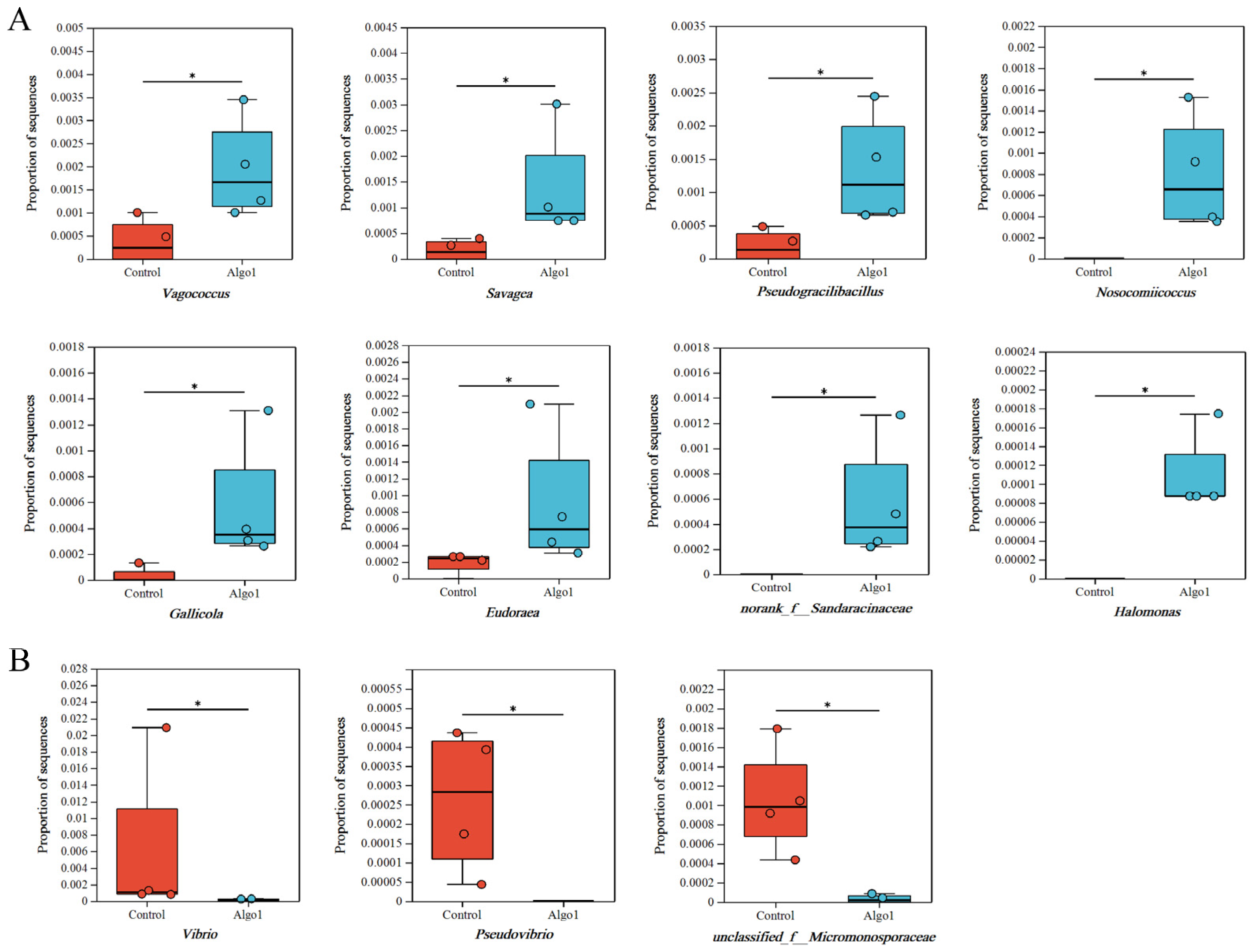
At the genus level, significant differences were observed in the relative abundance of microbial communities in the gut of sea cucumbers fed with PSDP compared to the CT group. In the PSDP6 group, the relative abundances of the genera Vagococcus, Savagea, Pseudogracilibacillus, Nosocomiicoccus, and Gallicola in Firmicutes, as well as the genus Eudoraea in Bacteroidetes, and the genera norank f Sandaracinaceae and Halomonas in Proteobacteria, increased significantly (p < 0.05). Conversely, the relative abundances of the genera Vibrio and Pseudovibrio in Proteobacteria, and unclassified f Micromonosporaceae in Actinobacteria, decreased significantly (p < 0.05) (Figure 7). In the PSDP9, the relative abundances of genera norank f Limnochordaceae and Nosocomiicoccus in Firmicutes, as well as the genus norank f Cellvibrionaceae in Proteobacteria, elevated significantly (p < 0.05). In contrast, the relative abundances of the genus Weissella in Firmicutes and genera unclassified f Geodermatophilaceae, unclassified f Micromonosporaceae, Pseudovibrio, and Vibrio in Proteobacteria reduced significantly (p < 0.05) (Figure 8). Gut microbiota plays a crucial role in maintaining gut health by facilitating digestion and regulating the homeostasis of the organism’s micro-ecological system [62,63]. They can reshape the beneficial effects of AOS on aquatic animals [64]. As shown in Figure 7 and Figure 8, feeding sea cucumbers with PSDP6 and PSDP9 consistently promoted the growth of the genus Nosocomiicoccus, while simultaneously inhibiting the relative abundance of the genera Vibrio, Pseudovibrio, and unclassified f Micromonosporaceae. Vibrio is generally regarded as a primary opportunistic pathogen that contributes to disease and mortality in aquatic animals [65]. Moreover, acting as the primary pathogenic bacteria for sea cucumbers, the significant reduction in the abundance of Vibrio in the samples mitigates the risk of disease in the cultivation of S. japonicus [66]. Pseudovibrio strains can synthesize secondary metabolites and contribute essential cofactors to their hosts through nonribosomal peptide synthetase and polyketide synthase pathways [67]. The observed reduction in the abundance of Pseudovibrio in the samples can be attributed to factors such as the farmed species and environmental conditions [68]. There were numerous reports on the interaction between AOS and gut microbiota, as well as its regulatory effects on various diseases. AOS exhibited functions in microbiota modulation similar to those of alginate. For instance, low Mw alginate extracted from kelp not only reduced obesity symptoms in HFD-fed BALB/c mice but also modulated the gut microbiota by increasing the abundance of Bacteroidales and decreasing Clostridia, thus improving the physiological state of the mice [64]. The regulation of the gut microbiota also contributed to the anti-obesity effects of AOS, significantly promoting the growth of Akkermansia muciniphila, Lactobacillus reuteri, and Lactobacillus gasseri. Furthermore, the AOS intervention significantly increased the concentration of short-chain fatty acids (SCFAs), such as acetate, propionate, and butyrate, while reducing endotoxin levels [69]. In colitis mouse models, treatment with unsaturated AOS inhibited immune damage and maintained mucosal barrier function by modulating gut microbiota, increasing the abundance of Firmicutes and Actinobacteria, while decreasing the abundance of Bacteroidetes [18]. Additionally, AOS supported the growth of beneficial gut bacteria [70,71]. Collectively, the results suggested that AOS promoted the growth of beneficial bacteria and enhanced gut health in the gut of S. japonicus. This could potentially bolster growth and augment non-specific immunity in S. japonicus.

3. Materials and Methods

3.1. Experimental Materials

The alginate lyase used in this study was 102C300C-Vgb (203 U/mL), developed by the Laboratory of Marine Microbial Engineering at Ocean University of China [72]. Sea cucumbers S. japonicus and experimental diets were provided by Yantai Huakang Biomedical Technology Co., Ltd., Yantai, Shandong (China).

3.2. Optimization of Enzymatic Conditions

3.2.1. Enzyme Dosage

Different enzyme dosages (each g of raw material: 2.5 U, 12.5 U, 25 U, 37.5 U, and 50 U) of 102C300C-Vgb were mixed with 20% sodium alginate (SA) and incubated at 45 °C, pH 7.0, for 24 h in a 250 mL shaker to obtain the hydrolysis sample. Subsequently, the sample yield and reducing sugar content were determined to investigate the effect of the amount of addition.

3.2.2. Enzymatic Temperature

The enzymatic temperature (35 °C, 40 °C, 45 °C, 50 °C, and 55 °C) was optimized based on the optimal amount of enzyme addition (25 U/g). Other operations were the same as those in the above section.

3.2.3. Enzymatic pH

Based on the above optimum enzymatic conditions (enzyme dosage of 25 U/g and enzymatic temperature of 45 °C), the pH of the enzymatic hydrolysis system was adjusted to 5.5, 6.0, 6.5, 7.0, and 7.5, respectively, by varying the amount of phosphate added to the culture medium.

3.2.4. Enzymatic Time

The enzymatic time (6 h, 12 h, 18 h, 24 h, and 30 h) was optimized according to the optimum enzymatic conditions mentioned above (enzyme dosage of 25 U/g, enzymatic temperature of 45 °C, and enzymatic pH of 7.0). The sample was subsequently vacuum freeze-dried for further analysis.

3.3. 250-L Enlargement Experiment

The PSDP preparation process optimized at the laboratory level was scaled up for pilot production using the equipment shown in Figure 9. In this process, 50 kg of SA was mixed with 200 L of water at a material-to-water ratio of 1:4, with simultaneous addition and uniform stirring. The mixture was then subjected to enzymatic hydrolysis at 45 °C for 24 h in an enzymatic hydrolysis tank. After the hydrolysis, the temperature was raised to 85 °C for 10 min to deactivate the enzymes. The supernatant, obtained after centrifugation to remove impurities, was concentrated to one-third of its original volume, followed by UHT sterilization, and then subjected to spray drying.

3.4. S. japonicus Feeding Experiment

The sea cucumbers were cultured for 40 days in the sea area of Longkou Economic Development Zone (120°28′ E, 37°58′ N). Four basal diets were supplemented with a diet of 0, 300, 600, and 900 mg PSDP/kg (hereinafter referred to as the control, 0.03% PSDP, 0.06% PSDP, and 0.09% PSDP groups). Before the start of the experiment, sea cucumbers were fed the control diet for 1 week to acclimatize to the experimental conditions. Each group has two replicates, and each of similar-sized 2000 S. japonicus (40.0 ± 1.0 g of wet body weight) was selected out and randomly distributed into 8 aquaculture cages (length × width × height: 4.00 × 4.00 × 2.80 m), under a natural photoperiod (12 dark:12 light). The sea cucumbers were fed on experimental diets once a day at 10:00 am to ad libitum for a consecutive 40 days. The daily feeding rate was 3% of the total body weight.

3.5. Data Measurement

3.5.1. Yield Analysis of Enzymatic Hydrolysis

The enzymatic supernatant, which has been centrifuged to remove the precipitate, was lyophilized or spray-dried. Subsequently, it was dried to constant weight using a weighing bottle. The yield of enzymatic hydrolysis was calculated according to the following formulas:
Y i e l d % = P 100 % / M
P: degradation product weight; M: SA weight.

3.5.2. Reducing Sugar Content Analysis

Reducing sugar was determined by the 3,5-dinitrosalicylic acid (DNS) method [73]. 2 mL of DNS solution was added to the appropriately diluted enzymatic hydrolysate. The mixture was incubated in a boiling water bath for 2 min and ddH2O was added to a total volume of 15 mL. Absorbance was measured at 540 nm. Reducing sugar content (RSC) was calculated according to the following formulas:
R S C ( % ) = R S 100 % / M
RS: reducing sugar weight; M: SA weight.

3.5.3. Fourier Transform Infrared Analysis

Fourier transform infrared (FTIR) spectra were obtained using a Magna-IR560 spectrometer (Nicolet, Madison, WI, USA). For the analysis, 2 mg of the sample was homogenized with 200 mg of KBr and subsequently compressed into a disk. Spectral data were acquired across the 4000–400 cm−1 range, employing a resolution of 4 cm−1.

3.5.4. High-Performance Liquid Chromatography Analysis

The molecular weight (Mw) of the degradation product was determined using high-performance liquid chromatography on an Agilent 1260 Infinity system equipped with a PL Aquagel-OH 30 column (Agilent, Santa Clara, CA, USA). A mobile phase consisting of 200 mM NaNO3 and 10 mM NaH2PO4 was employed at a flow rate of 0.5 mL/min, with the column maintained at 25 °C. Detection was carried out using a refractive index detector (RID). Dextran standards with molecular weights of 1 kDa, 3.65 kDa, 5 kDa, and 12 kDa were utilized for calibration. Prior to injection, the precipitates were dissolved in the mobile phase and filtered through a 0.22 µm membrane.

3.5.5. Electrospray Ionization Mass Spectrometry Analysis

The dried samples were introduced by direct infusion into the electrospray ionization source (ESI), and mass spectra (MS) were collected for elucidating the structure of the ESI-MS peaks (Agilent 1290 Infinity II-6460, Frag = 175.0 V, m/z 100–2000 amu) [27].

3.5.6. Compositional Analysis of Pilot-Scale Products

Proximate composition was determined on the PSDP. The appropriate amount of the sample was placed in a clean and dry white porcelain dish for sensory evaluation. The AOS content was measured using the m-hydroxybiphenyl method [74]. Weight difference methods were used to determine moisture and ash content [75]. The particle size was calculated using an 80 mesh standard sieve pass rate. The total arsenic and lead were determined by Hydride Generation Atomic Fluorescence Spectrometry (HG-AFS) [76] and Atomic Absorption Spectroscopy (AAS) [77]. Additionally, samples were tested for the presence of Salmonella contamination.

3.5.7. Growth Performance

Ten sea cucumbers were randomly selected from 2 cages in each group and weighed to measure the growth parameters every 20 days. The average value of each 40 individual animals was considered as a valid value for each replicate. Average wet weight (AWW) was calculated according to the following formulas:
A W W ( g ) = T W W / N
TWW: total wet weight; N: the number of sea cucumbers
The individuals after weighed were dissected on ice, and the body walls obtained were boiled for 5 min to prevent autolysis. The body walls were subsequently lyophilized and pulverized. The average dry weight (ADW) was calculated after taking a portion of it for constant weight:
A D W g = T D W / N
ADW: total dry weight; N: sea cucumber number.

3.5.8. Analyses of Body Wall Compositions

The freeze-dried body walls were analyzed for protein, fat, and amino acid composition. Protein content was measured using a Kjeldahl nitrogen analyzer (SKD-100, Shanghai Peiou Analytical Instrument Co., Ltd., Shanghai, China) [78,79]. The crude fat was determined using Soxhlet extraction with petroleum ether as a solvent [80]. The amino acid composition of the samples was analyzed using a fully automated amino acid analyzer (L-8900, Hitachi High-Tech Science Corporation, Tokyo, Japan).

3.5.9. Intestinal Morphometry

The mid-intestinal section (3 cm) was fixed in Bouin’s fixative solution (Phygene, Wuhan, China), embedded in paraffin blocks, sliced to 4 μm thickness sections for hematoxylin and eosin staining (H and E; Wuhan Servicebio Biotechnology Co., Ltd., Wuhan, China), and examined under a light microscope (BX41, Olympus, Tokyo, Japan). Subsequently, the images were analyzed with AJ-VERT software, version 6.0. The thickness of the intestinal serosa, the muscularis propria and submucosa, and the width of the folds were measured (six measurements of each type of structure, five sea cucumbers per tank).

3.5.10. Intestinal DNA Extraction and Illumina Sequencing of 16S rRNA Genes

Total DNA was extracted from the gut using the E.Z.N.A TM bacterial Mag-Bind DNA Kit (OMEGA, Beijing, China) following the manufacturer’s instructions. DNA concentration was quantified using Qubit 3.0 (Invitrogen, Q32866, Waltham, MA, USA), while library concentration was quantified by a fluorescence instrument. Amplification and sequencing of the V3-V4 region of the 16S rRNA gene were performed using fusion primers, including 338F (5′-ACTCCTACGGGAGGCAGCAG-3′) and 806R (5′-GGACTACHVGGGTWTCTAAT-3′). PCR, PCR product purification, quantification, and high-throughput sequencing were performed using the Illumina MiSeq platform at Shanghai Majorbio Bio-Pharm Technology Co., Ltd., Shanghai, China. Bioinformatics analysis of the gut microbiota was performed using the Majorbio Cloud platform (URL accessed on 15 January 2024) (https://cloud.majorbio.com).

3.6. Statistical Analysis

Statistical analysis was performed using one-way analysis of variance (ANOVA) with post hoc multiple comparison LSD tests to detect significant differences.

4. Conclusions

In this study, recombinant alginate lyase 102C300C-Vgb was employed to achieve targeted enzymatic hydrolysis of oligosaccharides under 20% SA concentration, which is the highest substrate concentration reported to date, significantly improving the production efficiency of PSDP. In particular, when 0.06% PSDP was added to the diet of sea cucumber S. japonicus, the growth and gut health were all improved significantly. This study presents a novel method for the degradation of high concentrations of SA, which has good economic and application potential in industrial production and sustainable marine aquaculture.

Supplementary Materials

The following supporting information can be downloaded at: https://www.mdpi.com/article/10.3390/md23090339/s1, Figure S1: The principal coordinates analysis (PCoA) showing the separate distribution of gut microbiota composition on the plot among various PSDP treatment groups and the CT. Figure S2: Venn diagram showing the number of unique and shared ASVs of gut bacteria between the CT and the PSDP treatment groups. Figure S3: Relative abundance of bacteriuml communities in different samples at the top 10 phylum level. Table S1: An Illumina high-throughput sampling depth, richness, and diversity index of bacteriuml community in the gut of S. japonicus under different experimental groups. Table S2: Relative abundances of thirty-two different bacteria phyla under different experimental groups.

Author Contributions

Conceptualization, Z.G., H.M. (Haijin Mou) and D.L.; Data curation, Z.G.; Validation, Z.G., F.N., P.Y., W.G., Y.Z. (Yiling Zhong), H.C. and J.L.; Writing-original draft, Z.G.; Supervision, F.N.; Visualization, F.N., P.Y., W.G., Y.Z. (Yiling Zhong), H.C. and J.L.; Writing-review and editing, H.M. (Hina Mukhtar), S.L., Y.Z. (Yanwen Zheng), H.M. (Haijin Mou) and D.L.; Funding acquisition: H.M. (Haijin Mou). All authors have read and agreed to the published version of the manuscript.

Funding

This work was supported by Youth Innovation Team Program of Universities in Shandong Province (2023KJ040), Shandong Province Key R&D Program (2022TZXD005), Shandong Province Marine Aquaculture Innovation and Entrepreneurship Community Project (YZ2024002, YZ2024004), National Natural Science Foundation of China (42106110, 32202064), Natural Science Foundation of Jiangxi Province (20224BAB215044), and China Postdoctoral Science Foundation (2021M701547).

Institutional Review Board Statement

Not applicable.

Data Availability Statement

The original contributions presented in this study are included in the article. Further inquiries can be directed to the corresponding authors.

Conflicts of Interest

The authors declare no conflicts of interest.

Abbreviations

The following abbreviations are used in this manuscript:
S. japonicusStichopus japonicus
A. japonicusApostichopus japonicus
SASodium alginate
AOSAlginate oligosaccharides
PSDPPilot-scale degradation product
RSCReducing sugar content
FTIRFourier transform infrared
MwMolecular weight
AWWAverage wet weight:
ADWAverage dry weight

References

  1. Chen, L.; Wang, X.Y.; Liu, R.Z.; Wang, G.Y. Culturable microorganisms associated with sea cucumbers and microbial natural products. Mar. Drugs 2021, 19, 8. [Google Scholar] [CrossRef]
  2. Purcell, S.W.; Hair, C.A.; Mills, D.J. Sea cucumber culture, farming and sea ranching in the tropics: Progress, problems and opportunities. Aquaculture 2012, 368–369, 68–81. [Google Scholar] [CrossRef]
  3. Yin, H.W.; Yue, H.; Wang, M.; Zhang, T.Q.; Zhao, Y.T.; Liu, H.Y.; Wang, J.F.; Zheng, H.W.; Xue, C.H. Preparation of novel sea cucumber intestinal peptides to promote tibial fracture healing in mice by inducing differentiation of hypertrophic chondrocytes to the osteoblast lineage. Mol. Nutr. Food Res. 2024, 68, 2300344. [Google Scholar] [CrossRef] [PubMed]
  4. Hossain, A.; Dave, D.; Shahidi, F. Northern sea cucumber (Cucumaria frondosa): A potential candidate for functional food, nutraceutical, and pharmaceutical sector. Mar. Drugs 2020, 18, 274. [Google Scholar] [CrossRef] [PubMed]
  5. Senadheera, T.R.L.; Dave, D.; Shahidi, F. Sea cucumber derived type I collagen: A comprehensive review. Mar. Drugs 2020, 18, 471. [Google Scholar] [CrossRef]
  6. Park, M.; Shin, S.K.; Do, Y.H.; Yarish, C.; Kim, J.K. Application of open water integrated multitrophic aquaculture to intensive monoculture: A review of the current status and challenges in Korea. Aquaculture 2018, 497, 174–183. [Google Scholar] [CrossRef]
  7. Zhu, M.H.; Zhao, H.X.; Chen, J.W.; Xie, H.J.; Du, J. Investigation of antibiotics in sea cucumbers: Occurrence, pollution characteristics, and human risk assessment. Int. J. Environ. Sci. Technol. 2018, 25, 32081–32087. [Google Scholar] [CrossRef]
  8. Zhu, M.H.; Wang, Z.Y.; Chen, J.W.; Xie, H.J.; Zhao, H.X.; Yuan, X.T. Bioaccumulation, biotransformation, and multicompartmental toxicokinetic model of antibiotics in sea cucumber (Apostichopus japonicus). Environ. Sci. Technol. 2020, 54, 13175–13185. [Google Scholar] [CrossRef]
  9. Holscher, H.D. Dietary fiber and prebiotics and the gastrointestinal microbiota. Gut Microbes 2017, 8, 172–184. [Google Scholar] [CrossRef]
  10. Li, W.; Liu, B.D.; Liu, A.P.; Han, M.L.; Yin, Y.L.; Xu, G.H.; Cheng, W.X.; Xie, L.W. Dietary supplementation of N-carbamylglutamate promotes growth performance by modulating the homeostasis of gut microbiota in tilapia (Oreochromis niloticus). Aquacult. Rep. 2021, 20, 100750. [Google Scholar] [CrossRef]
  11. Huang, X.L.; Xie, R.B.; Zou, A.G.; Zhang, S.Q.; Xu, X.H.; Sun, G.H.; Yang, J.M. Effects of polypeptidin feeding on growth and intestinal flora of Apostichopus japonicas. Aquacult. Rep. 2024, 38, 102284. [Google Scholar]
  12. Amillano-Cisneros, J.M.; Fuentes-Valencia, M.A.; Leyva-Morales, J.B.; Davizón, Y.A.; Marquéz-Pacheco, H.; Valencia-Castañeda, G.; Maldonado-Coyac, J.A.; Ontiveros-García, L.A.; Badilla-Medina, C.N. Prebiotics in Global and Mexican Fish Aquaculture: A Review. Animals 2023, 13, 3607. [Google Scholar] [CrossRef]
  13. Huang, H.; Li, S.; Bao, S.; Mo, K.; Sun, D.; Hu, Y. Expression and characterization of a cold-adapted alginate Lyase with Exo/Endo-type activity from a novel marine bacterium Alteromonas portus HB161718T. Mar. Drugs 2021, 19, 155. [Google Scholar] [CrossRef] [PubMed]
  14. Cao, S.S.; Li, Q.; Xu, Y.X.; Tang, T.C.; Ning, L.M.; Zhu, B.W. Evolving strategies for marine enzyme engineering: Recent advances on the molecular modifcation of alginate lyase. Mar. Life Sci. Technol. 2022, 4, 106–116. [Google Scholar] [CrossRef] [PubMed]
  15. Yan, G.L.; Guo, Y.M.; Yuan, J.M.; Liu, D.; Zhang, B.K. Sodium alginate oligosaccharides from brown algae inhibit Salmonella Enteritidis colonization in broiler chickens. Poultry Sci. 2011, 90, 1441–1448. [Google Scholar] [CrossRef] [PubMed]
  16. Cheng, J.Y.; Xiao, M.S.; Ren, X.M.; Secundo, F.; Yu, Y.; Nan, S.H.; Chen, W.M.; Zhu, C.L.; Kong, Q.; Huang, Y.T.; et al. Response of Salmonella enterica serovar Typhimurium to alginate oligosaccharides fermented with fecal inoculum: Integrated transcriptomic and metabolomic analyses. Mar. Life Sci. Technol. 2023, 5, 242–256. [Google Scholar]
  17. Li, F.L.; Tang, Y.; Wei, L.X.; Yang, M.X.; Lu, Z.J.; Shi, F.; Zhan, F.B.; Li, Y.A.; Liao, W.C.; Lin, L.; et al. Alginate oligosaccharide modulates immune response, fat metabolism, and the gut bacterial community in grass carp (Ctenopharyngodon idellus). Fish Shellfish Immun. 2022, 130, 103–113. [Google Scholar] [CrossRef]
  18. He, N.; Yang, Y.; Wang, H.; Liu, N.; Yang, Z.; Li, S. Unsaturated alginate oligosaccharides (UAOS) protects against dextran sulfate sodium-induced colitis associated with regulation of gut microbiota. J. Funct. Foods 2021, 83, 104536. [Google Scholar] [CrossRef]
  19. Yuan, X.; Zheng, J.; Jiao, S. A review on the preparation of chitosan oligosaccharides and application to human health, animal husbandry and agricultural production. Carbohyd. Polym. 2019, 220, 60–70. [Google Scholar] [CrossRef]
  20. Feng, L.P.; Cao, Y.P.; Xu, D.X.; Wang, S.J.; Zhang, J. Molecular weight distribution, rheological property and structural changes of sodium alginate induced by ultrasound. Ultrason. Sonochem. 2017, 34, 609–615. [Google Scholar] [CrossRef]
  21. Vasudevan, U.M.; Lee, O.K.; Lee, E.Y. Alginate derived functional oligosaccharides: Recent developments, barriers, and future outlooks. Carbohyd. Polym. 2021, 267, 118158. [Google Scholar] [CrossRef] [PubMed]
  22. Zhu, B.W.; Ni, F.; Xiong, Q.; Yao, Z. Marine oligosaccharides originated from seaweeds: Source, preparation, structure, physiological activity and applications. Crit. Rev. Food Sci. 2021, 61, 60–74. [Google Scholar] [CrossRef] [PubMed]
  23. Liu, J.; Yang, S.; Li, X.; Yan, Q.; Reaney, M.J.; Jiang, Z. Alginate oligosaccharides: Production, biological activities, and potential applications. Compr. Rev. Food Sci. Food Saf. 2019, 18, 1859–1881. [Google Scholar] [CrossRef] [PubMed]
  24. Jiang, Z.; Zhang, X.; Wu, L.; Li, H.; Chen, Y.; Li, L.; Ni, H.; Li, Q.; Zhu, Y. Exolytic products of alginate by the immobilized alginate lyase confer antioxidant and antiapoptotic bioactivities in human umbilical vein endothelial cells. Carbohyd. Polym. 2021, 251, 116976. [Google Scholar] [CrossRef]
  25. Liu, Z.M.; Ning, C.; Yuan, M.X.; Yang, S.X.; Wei, X.Y.; Xiao, M.S.; Fu, X.D.; Zhu, C.L.; Mou, H.J. High-level expression of a thermophilic and acidophilic β-mannanase from Aspergillus kawachii IFO 4308 with significant potential in mannooligosaccharide preparation. Bioresour. Technol. 2020, 295, 122257. [Google Scholar] [CrossRef]
  26. Wang, M.; Chen, L.; Zhang, Z. Potential applications of alginate oligosaccharides for biomedicine-a mini review. Carbohyd. Polym. 2021, 271, 118408. [Google Scholar] [CrossRef]
  27. Yang, S.X.; Liu, Z.M.; Fu, X.D.; Zhu, C.L.; Kong, Q.; Yang, M.; Mou, H.J. Expression and characterization of an alginate lyase and its thermostable mutant in Pichia pastoris. Mar. Drugs 2020, 18, 305. [Google Scholar] [CrossRef]
  28. Han, W.J.; Gu, J.Y.; Chen, Y.Y.; Liu, H.H.; Li, Y.Z. Novel alginate lyase (Aly5) from a polysaccharide-degrading marine bacterium, Flammeovirga sp. strain MY04: Effects of module truncation on biochemical characteristics, alginate degradation patterns, and oligosaccharide-yielding properties. Appl. Environ. Microb. 2016, 82, 364–374. [Google Scholar] [CrossRef]
  29. Mackie, W. Semi-quantitative estimation of the composition of alginates by infra-red spectroscopy. Carbohyd. Res. 1971, 20, 413–415. [Google Scholar] [CrossRef]
  30. Hifney, A.F.; Fawzy, M.A.; Abdel-Gawad, K.M. Upgrading the antioxidant properties of fucoidan and alginate from Cystoseira trinodis by fungal fermentation or enzymatic pretreatment of the seaweed biomass. Food Chem. 2018, 269, 387–395. [Google Scholar] [CrossRef]
  31. Liang, Q.P.; Huang, Y.T.; Liu, Z.M.; Xiao, M.S.; Ren, X.M.; Liu, T.H.; Li, H.Y.; Yu, D.X.; Wang, Y.; Zhu, C.L. A recombinant alginate lyase Algt1 with potential in preparing alginate oligosaccharides at high concentration substrate. Foods 2023, 12, 4039. [Google Scholar] [CrossRef]
  32. Kaur, S.; Abraham, R.E.; Franco, C.M.M.; Puri, M. Production of alginate oligosaccharides (AOSs) using enhanced physicochemical properties of immobilized alginate lyase for industrial application. Mar. Drugs 2024, 22, 120. [Google Scholar] [CrossRef]
  33. Qiao, X.W.; Li, X.X.; Liu, S.Q.; Li, Y.; Zheng, M.L.; Fu, W.Y.; Long, H.M.; Luo, F.X. Study on storage stability of spray dried hemoglobin powder. Feed Res. 2023, 46, 107–111. (In Chinese) [Google Scholar]
  34. Falkeborg, M.; Cheong, L.Z.; Gianfico, C.; Sztukiel, K.M.; Kristensen, K.; Glasius, M.; Xu, X.B.; Guo, Z. Alginate oligosaccharides: Enzymatic preparation and antioxidant property evaluation. Food Chem. 2014, 164, 185–194. [Google Scholar] [CrossRef] [PubMed]
  35. Liu, N.; Wang, H.Y.; Yang, Z.Z.; Zhao, K.Y.; Li, S.Y.; He, N.N. The role of functional oligosaccharides as prebiotics in ulcerative colitis. Food Funct. 2022, 13, 6875–6893. [Google Scholar] [CrossRef] [PubMed]
  36. Riseh, R.S.; Vazvani, M.G.; Ebrahimi-Zarandi, M.; Skorik, Y.A. Alginate-induced disease resistance in plants. Polymers 2022, 14, 661. [Google Scholar] [CrossRef] [PubMed]
  37. Wan, J.; Zhang, J.; Xu, Q.; Yin, H.; Chen, D.; Yu, B.; He, J. Alginate oligosaccharide protects against enterotoxigenic Escherichia coli-induced porcine intestinal barrier injury. Carbohyd. Polym. 2021, 270, 118316. [Google Scholar] [CrossRef]
  38. Bai, N.; Deng, W.; Qi, Z.; Pan, S.; Li, Q.; Gu, M. The effect of alginate oligosaccharides on intestine barrier function and Vibrio parahaemolyticus infections in the white shrimp Litopenaeus vannamei. Fish Shellfish Immun. 2023, 141, 109011. [Google Scholar] [CrossRef]
  39. Shi, L.D.; Zhai, H.J.; Wei, L.B.; Zeng, F.S.; Ren, T.J.; Han, Y.Z. Effects of dietary folic acid on growth performance, body wall folate content, immune and antioxidant enzyme activity of juvenile sea cucumber, Apostichopus japonicas. Aquac. Res. 2022, 53, 6219–6226. [Google Scholar] [CrossRef]
  40. Gao, X.B.; Zhai, H.; Wei, L.; Shi, L.; Yan, L.; Peng, Z.; Wang, W.; Ren, T.; Han, Y. Effects of dietary mannan oligosaccharides on growth, non-specific immunity, and intestinal health in juveniles of the Japanese sea cucumber (Apostichopus japonicus). Aquacult. Int. 2023, 31, 1705–1727. [Google Scholar] [CrossRef]
  41. Feng, J.H.; Zhang, L.; Xia, X.B.; Hu, W.; Zhou, P. Effect of geographic variation on the proteome of sea cucumber (Stichopus japonicus). Food Res. Int. 2020, 136, 109498. [Google Scholar] [CrossRef]
  42. Li, M.; Qi, Y.; Mu, L.; Li, Z.; Zhao, Q.; Sun, J.; Jiang, Q. Effects of processing method on chemical compositions and nutritional quality of ready-to-eat sea cucumber (Apostichopus japonicus). Food Sci. Nutr. 2019, 7, 755–763. [Google Scholar] [CrossRef] [PubMed]
  43. Wen, J.; Hu, C.; Fan, S. Chemical composition and nutritional quality of sea cucumbers. J. Sci. Food Agr. 2010, 90, 2469–2474. [Google Scholar] [CrossRef] [PubMed]
  44. Bordbar, S.; Anwar, F.; Saari, N. High-value components and bioactives from sea cucumbers for functional foods-a review. Mar. Drugs 2011, 9, 1761–1805. [Google Scholar] [CrossRef] [PubMed]
  45. Gao, Y.; Li, Z.; Qi, Y.; Guo, Z.; Lin, Y.; Li, W.; Hu, Y.; Zhao, Q. Proximate composition and nutritional quality of deep sea growth sea cucumbers (Stichopus japonicus) from different origins. J. Sci. Food Agr. 2016, 96, 2378–2383. [Google Scholar] [CrossRef]
  46. Nishanthan, G.; Kumara, P.A.D.A.; de Croos, M.D.S.T.; Prasada, D.V.P.; Dissanayake, D.C.T. Effects of processing on proximate and fatty acid compositions of six commercial sea cucumber species of Sri Lanka. J. Food Sci. Technol. 2018, 55, 1933–1941. [Google Scholar] [CrossRef]
  47. Li, M.; Gao, Y.; Qi, Y.X.; Song, Z.Y.; Li, Z.B.; Lin, Y.T.; Zhao, Q.C. Assessment of the nutritional value of cultured sea cucumber Apostichopus japonicas. J. Aquat. Food Prod. T. 2021, 30, 868–879. [Google Scholar] [CrossRef]
  48. Lee, M.H.; Kim, Y.K.; Moon, H.S.; Kim, K.D.; Kim, G.G.; Cho, H.A.; Yoon, N.Y.; Sim, K.B.; Park, H.Y.; Lee, D.S. Comparison on proximate composition and nutritional profile of red and black sea cucumbers (Apostichopus Japonicus) from Ulleungdo (Island) and Dokdo (Island), Korea. Food Sci. Biotechnol. 2012, 21, 1285–1291. [Google Scholar] [CrossRef]
  49. Sugano, M.; Ishiwaki, N.; Nakashima, K. Dietary Protein-dependent modification of serum cholesterol level in rats. Significance of the arginine/lysine ratio. Ann. Nutr. Metab. 1984, 28, 192–199. [Google Scholar] [CrossRef]
  50. Heidarieh, M.; Mirvaghef, A.R.; Akbari, M.; Sheikhzadeh, N.; Kamyabi-Moghaddam, Z.; Askari, H.; Shahbazfar, A.A. Evaluations of Hilyses™, fermented Saccharomyces cerevisiae, on rainbow trout (Oncorhynchus mykiss) growth performance, enzymatic activities and gastrointestinal structure. Aquacult. Nutr. 2012, 19, 343–348. [Google Scholar] [CrossRef]
  51. Torrecillas, S.; Makol, A.; Caballero, M.J.; Montero, D.; Robaina, L.; Real, F.; Sweetman, J.; Tort, L.; Izquierdo, M.S. Immune stimulation and improved infection resistance in European sea bass (Dicentrarchus labrax) fed mannan oligosaccharides. Fish Shellfish Immun. 2007, 23, 969–981. [Google Scholar] [CrossRef] [PubMed]
  52. Wan, J.; Zhang, J.; Chen, D.; Yu, B.; He, J. Effects of alginate oligosaccharide on the growth performance, antioxidant capacity and intestinal digestion-absorption function in weaned pigs. Anim. Feed Sci. Tech. 2017, 234, 118–127. [Google Scholar] [CrossRef]
  53. Wan, J.; Zhang, J.; Chen, D.; Yu, B.; Huang, Z.; Mao, X.; Zheng, P.; Yu, J.; He, J. Alginate oligosaccharide enhances intestinal integrity of weaned pigs through altering intestinal inflammatory responses and antioxidant status. RSC Adv. 2018, 8, 13482–13492. [Google Scholar] [CrossRef] [PubMed]
  54. Huang, X.Y.; Zhao, Z.H.; Yang, Y.J.; Song, Q.Z.; Ding, J.; Han, B.; Zhao, C. Dietary allicin improves behavior, physiology, growth, and disease resistance in the sea cucumber Apostichopus japonicas. Aquaculture 2024, 593, 741321. [Google Scholar] [CrossRef]
  55. Chang, Y.Q.; Ding, J.; Song, J. Biological Research and Culture of Sea Cucumbers and Sea Urchins; China Ocean Press: Beijing, China, 2004. [Google Scholar]
  56. Zhou, M.; Hernandez-Sanabria, E.; Guan, L.L. Assessment of the microbial ecology of ruminal methanogens in cattle with different feed efficiencies. Appl. Environ. Microb. 2009, 75, 6524–6533. [Google Scholar] [CrossRef]
  57. Zhang, Z.; Xing, R.L.; Lv, Z.M.; Shao, Y.N.; Zhang, W.W.; Zhao, X.L.; Li, C.H. Analysis of gut microbiota revealed Lactococcus garvieae could be an indicative of skin ulceration syndrome in farmed sea cucumber Apostichopus japonicus. Fish Shellfish Immun. 2018, 80, 148–154. [Google Scholar] [CrossRef]
  58. Tsagaraki, T.M.; Pree, B.; Leiknes, Ø.; Larsen, A.; Bratbak, G.; Øvreås, L.; Egge, J.K.; Spanek, R.; Paulsen, M.L.; Olsen, Y.; et al. Bacterial community composition responds to changes in copepod abundance and alters ecosystem function in an arctic mesocosm study. ISME J. 2018, 12, 2694–2705. [Google Scholar] [CrossRef]
  59. Bi, D.C.; Yang, X.; Lu, J. Preparation and potential applications of alginate oligosaccharides. Crit. Rev. Food Sci. 2023, 63, 10130–10147. [Google Scholar] [CrossRef]
  60. Tao, L.; Chai, J.; Liu, H.Y.; Huang, W.H.; Zou, Y.; Wu, M.L.; Peng, B.Q.; Wang, Q.; Tang, K.Y. Characterization and dynamics of the gut microbiota in rice fishes at different developmental stages in rice-fish coculture systems. Microorganisms 2022, 10, 2373. [Google Scholar] [CrossRef]
  61. Foysal, M.J.; Alam, M.; Robiul Kawser, A.Q.M.; Hasan, F.; Rahman, M.M.; Tay, C.Y.; Prodhan, M.S.H.; Gupta, S.K. Meta-omics technologies reveals beneficiary effects of Lactobacillus plantarum as dietary supplements on gut microbiota, immune response and disease resistance of Nile tilapia (Oreochromis niloticus). Aquaculture 2020, 520, 734974. [Google Scholar] [CrossRef]
  62. Adak, A.; Khan, M.R. An insight into gut microbiota and its functionalities. Cell. Mol. Life Sci. 2019, 76, 473–493. [Google Scholar] [CrossRef] [PubMed]
  63. Yan, P.G.; Li, J.N. Advances in the understanding of the intestinal microenvironment and inflammatory bowel disease. Chin. Med. J. 2020, 133, 834–841. [Google Scholar] [CrossRef] [PubMed]
  64. Zhang, R.; Wang, X.W.; Zhu, J.Y.; Liu, L.L.; Liu, Y.C.; Zhu, H. Dietary sanguinarine affected immune response, digestive enzyme activity and intestinal microbiota of Koi carp (Cyprinus carpiod). Aquaculture 2018, 502, 72–79. [Google Scholar] [CrossRef]
  65. Thompson, F.L.; Iida, T.; Swings, J. Biodiversity of vibrios. Microbiol. Mol. Biol. R. 2004, 68, 403–431. [Google Scholar] [CrossRef]
  66. Li, Z.; Ren, H.Y.; Li, Q.; Murtaza, B.; Li, X.Y.; Zhang, J.C.; Xu, Y.P. Exploring the effects of phage cocktails in preventing Vibrio infections in juvenile sea cucumber (Apostichopus japonicus) farming. Aquaculture 2020, 515, 734599. [Google Scholar] [CrossRef]
  67. Bondarev, V.; Richter, M.; Romano, S.; Piel, J.; Schwedt, A.; Schulz-Vogt, H.N. The genus Pseudovibrio contains metabolically versatile bacteria adapted for symbiosis. Environ. Microbiol. 2013, 15, 2095–2113. [Google Scholar] [CrossRef]
  68. Domínguez-Borbor, C.; Ardiles, V.; Bermeo, M.; Bolívar-Alvarado, C.; Tomalá, C.; Sonnenholzner, S.; Rodríguez, J.A. The marine symbiont Pseudovibrio denitrificans, is effective to control pathogenic Vibrio spp. in shrimp aquaculture. Aquaculture 2019, 508, 127–136. [Google Scholar] [CrossRef]
  69. Wang, Y.; Li, L.; Ye, C.; Yuan, J.; Qin, S. Alginate oligosaccharide improves lipid metabolism and inflammation by modulating gut microbiota in high-fat diet fed mice. Appl. Microbiol. Biot. 2020, 104, 3541–3554. [Google Scholar] [CrossRef]
  70. Zhang, P.; Liu, J.; Xiong, B.; Zhang, C.; Kang, B.; Gao, Y.; Li, Z.; Ge, W.; Cheng, S.; Hao, Y. Microbiota from alginate oligosaccharide-dosed mice successfully mitigated small intestinal mucositis. Microbiome 2020, 8, 112. [Google Scholar] [CrossRef]
  71. Zhang, C.; Xiong, B.; Chen, L.; Ge, W.; Yin, S.; Feng, Y.; Sun, Z.; Sun, Q.; Zhao, Y.; Shen, W. Rescue of male fertility following faecal microbiota transplantation from alginate oligosaccharide-dosed mice. Gut 2021, 70, 2213–2215. [Google Scholar] [CrossRef]
  72. Gu, Z.Q.; Niu, F.Y.; Yu, Z.H.; Bao, Z.; Mukhtar, H.; Yang, P.; Wang, S.T.; Mou, H.J.; Yang, M. High-efficiency expression of alginate lyase in Pichia pastoris facilitated by Vitreoscilla hemoglobin. Int. J. Biol. Macromol. 2024, 282, 137027. [Google Scholar] [CrossRef]
  73. Miller, G.L. Use of dinitrosalicylic acid reagent for determination of reducing sugar. Anal. Chem. 1959, 31, 426–428. [Google Scholar] [CrossRef]
  74. Zhao, M.J.; Kuang, F.Y.; Zhang, Y.Y.; Lv, G.P. Effects of hydrolysis condition and detection method on the monosaccharide composition analysis of polysaccharides from natural sources. Separations 2024, 11, 2. [Google Scholar] [CrossRef]
  75. Acheampong, R.; Tutu, C.O.; Amissah, J.G.N.; Danquah, A.O.; Saalia, F.K. Physicochemical and sensory characteristics of a breakfast cereal made from sprouted finger millet-maize composite flour. Cogent Food Agricult. 2024, 10, 2363003. [Google Scholar] [CrossRef]
  76. Liu, W.; Pan, Q.S.; Zhang, P.; Huang, D.S.; Fan, A.P.; Yi, P. Determination of total arsenic in Chinese traditional herbs by high pressure digestion-hydride generation atomic fluorescence spectrometry. Adv. Mat. Res. 2012, 554–556, 1967–1970. [Google Scholar] [CrossRef]
  77. Hodges, D.J.; Skelding, D. Determination of lead in blood by atomic-absorption spectroscopy with electrothermal atomisation. Analyst 1983, 108, 813–820. [Google Scholar] [CrossRef]
  78. Marzo, L.D.; Pranata, J.; Barbano, D.M. Measurement of casein in milk by Kjeldahl and sodium dodecyl sulfate-polyacrylamide gel electrophoresis. J. Dairy Sci. 2021, 104, 7448–7456. [Google Scholar] [CrossRef]
  79. Marco, A.; Rubio, R.; Compañó, R.; Casals, I. Comparison of the Kjeldahl method and a combustion method for total nitrogen determination in animal feed. Talanta 2002, 57, 1019–1026. [Google Scholar] [CrossRef]
  80. Shin, J.M.; Hwang, Y.O.; Tu, O.J.; Jo, H.B.; Kim, J.H.; Chae, Y.Z.; Rhu, K.H.; Park, S.K. Comparison of different methods to quantify fat classes in bakery products. Food Chem. 2013, 136, 703–709. [Google Scholar] [CrossRef]
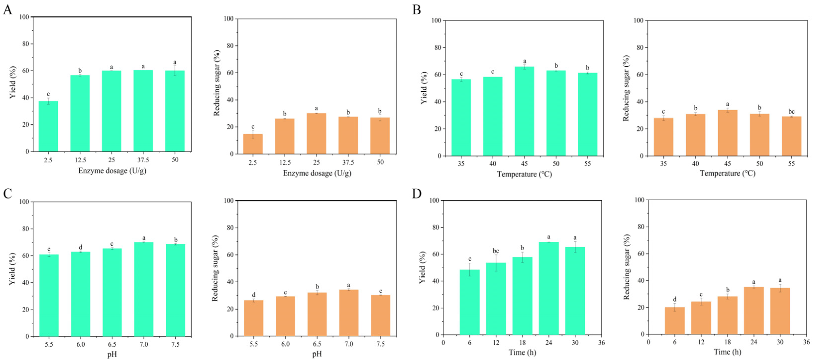
Figure 1. The yield and proportion of reducing sugar of digested products under different conditions. (A) Enzyme dosage; (B) Temperature; (C) pH; (D) Time. Different letters displayed above the bars on the column chart denote significant differences among treatments (n = 3) (mean ± SE).
Marinedrugs 23 00339 g001
Figure 2. FTIR analyses of SA and its degradation product.
Marinedrugs 23 00339 g002
Figure 3. The Mw distribution (1) and ESI-MS (2) of the degradation product (A) and PSDP (B).
Marinedrugs 23 00339 g003
Figure 4. The changes of average wet weight (A) and average dry weight in body wall (B) of S. japonicus in different feeding groups. Different letters (uppercase letters: 20 days of cultivation; lowercase letters: 40 days of cultivation) displayed above the bars on the column chart denote significant differences among treatments (n = 10) (mean ± SE).
Marinedrugs 23 00339 g004
Figure 5. The changes of the protein (A)/fat (B) content (1) and proportion (2) in body wall of S. japonicus in different feeding groups. The protein/fat proportion of the body wall was calculated by dividing the protein/fat content of the individual by the dry weight of the body wall. Different letters (uppercase letters: 20 days of cultivation; lowercase letters: 40 days of cultivation) displayed above the bars on the column chart denote significant differences among treatments (n = 10) (mean ± SE).
Marinedrugs 23 00339 g005
Figure 6. Intestinal tissue sections of S. japonicus fed diets with different PSDP levels. SE: Serosal layer; MU: Muscularis propria; SU: Submucosa; FO: Fold width.
Marinedrugs 23 00339 g006
Figure 7. Analysis of differences in abundance between PSDP6 and the CT at the genus level: (A). compared to CT, the bacteriuml communities of increased abundance in PSDP6; (B). compared to CT, the bacteriuml communities of decreased abundance in PSDP6. Values with * were p < 0.05 between two treatments (n = 4) (mean ± SE).
Marinedrugs 23 00339 g007
Figure 8. Analysis of differences in abundance between PSDP9 and the CT at the genus level: (A). compared to CT, the bacteriuml communities of increased abundance in PSDP9; (B). compared to CT, the bacteriuml communities of decreased abundance in PSDP9. Values with * were p < 0.05 between two treatments (n = 4) (mean ± SE).
Marinedrugs 23 00339 g008
Figure 9. Flow chart of pilot production equipment.
Marinedrugs 23 00339 g009
Table 1. Detection of related index of PSDP.
IndexDetection Result
Sensory requirements
SensationPowder, light yellowish-brown in color, free from mold, spoilage, and off odors or smells
Physicochemical parameters
AOS content (%)66.7 ± 0.1
Water content (%)5.10 ± 0.85
Crude ash content (%)20.95 ± 3.32
Particle size (80 mesh pass rate, %)99.50 ± 0.42
Sanitary indicators
Total arsenic (mg/kg)0.29 ± 0.01
Lead (mg/kg)ND
Salmonella (/25g)ND
Values expressed in mean ± SD. ND means no data.
Table 2. Amino-acid composition of body wall of S. japonicus.
20 DayControl (%)0.03% PSDP
(%)
0.06% PSDP
(%)
0.09% PSDP
(%)
40 DayControl
(%)
0.03% PSDP
(%)
0.06% PSDP
(%)
0.09% PSDP
(%)
Asp4.6044.804.70Asp4.804.704.304.50
Thr2.402.102.502.60Thr2.502.502.202.30
Ser2.3022.402.50Ser2.302.302.12.30
Glu7.206.207.407.06Glu7.407.306.607
Gly6.505.606.607.10Gly6.106.505.906.10
Ala32.6033.70Ala2.9032.702.70
Cys0.600.400.500.80Cys0.400.300.300.30
Val1.901.601.901.90Val21.801.601
Met0.800.700.701.20Met0.900.800.700.40
Ile1.401.501.501.90Ile1.501.301.200.70
Leu222.202.70Leu2.402.2022.10
Tyr1.501.201.401.90Tyr1.401.301.201.90
Phe1.501.201.501.50Phe1.501.301.301.80
Lys1.901.501.901.80Lys21.801.601.50
His0.500.500.600.70His0.600.700.500.30
Arg3.402.903.503.50Arg3.402.403.102.90
Hypro2.4022.302.90Hypro22.402.102.20
Pro3.202.803.303.20Pro3.103.4032.80
The color in the table represents the change in the content of each amino acid. The redder the color, the higher the content; the whiter the color, the lower the content.
Table 3. Effects of dietary PSDP on intestinal morphology structure of S. japonicus.
GroupSerosa Layer Thickness (μm)Muscularis Propria Thickness (μm)Submucosa Thickness (μm)Folds Width
(μm)
Control22.25 ± 10.37 b9.64 ± 1.7 b23.61 ± 3.42 b211.85 ± 25.8 c
0.03% PSDP25.45 ± 2.58 b9.41 ± 1.55 b37.78 ± 9.06 a184.23 ± 13.5 c
0.06% PSDP29.49 ± 9.99 b12.58 ± 2.13 a43.83 ± 7.82 a246.62 ± 23.4 b
0.09% PSDP39.72 ± 3.55 a11.92 ± 2.56 ab41.26 ± 3.89 a311.01 ± 3.99 a
Different letters within a line denote significant differences (p < 0.05). Values expressed in mean ± SD.
Disclaimer/Publisher’s Note: The statements, opinions and data contained in all publications are solely those of the individual author(s) and contributor(s) and not of MDPI and/or the editor(s). MDPI and/or the editor(s) disclaim responsibility for any injury to people or property resulting from any ideas, methods, instructions or products referred to in the content.