Review

Jellyfish Venom-Induced Cardiotoxicity and Immune Responses: Mechanisms and Potential Therapeutic Strategies

1
Naval Medical Center of PLA, Naval Medical University, Shanghai 200052, China
2
Department of Nanomedicine & Shanghai Key Laboratory of Nautical Medicine and Translation of Drugs and Medical Devices, Naval Medical University, Shanghai 200433, China
3
School of Health Science and Engineering, University of Shanghai for Science and Technology, Shanghai 200093, China
*
Authors to whom correspondence should be addressed.
These authors contributed equally to this work.
Mar. Drugs 2025, 23(10), 369; https://doi.org/10.3390/md23100369
Submission received: 25 August 2025 / Revised: 15 September 2025 / Accepted: 18 September 2025 / Published: 23 September 2025
(This article belongs to the Special Issue Jellyfish-Derived Compounds)

Abstract

Jellyfish stings represent a significant global marine hazard, causing injuries from localized skin damage to fatal systemic complications. While skin reactions are the most common symptom, heart toxicity (cardiotoxicity) is the primary cause of death. A growing body of evidence shows that the immune system’s response worsens this venom-induced heart damage. However, current research remains disproportionately focused on cutaneous inflammatory responses, leaving systemic immunopathological processes—especially those potentiating cardiotoxicity—poorly understood. Moreover, few jellyfish toxins (like those from the Chironex fleckeri) have been thoroughly studied, and the molecular mechanisms of heart injury remain largely unknown. This review introduces a novel pathophysiological classification of jellyfish envenomation into three distinct categories—immunotoxicity-dominant, cardiotoxicity-dominant, and dual-mechanism synergistic—based on clinical and mechanistic profiles. By synthesizing current knowledge on venom components and their multi-system interaction, we aim to identify actionable therapeutic targets and propose mechanism-driven treatment strategies. This refined classification offers a foundation for future clinical decision-making and the development of targeted therapies, potentially improving patient outcomes through more personalized envenomation management.

1. Introduction

Jellyfish, as ancient members of the phylum Cnidaria, have evolved complex venoms consisting of multiple bioactive toxin components, which mediate diverse pathological effects ranging from localized tissue necrosis to life-threatening systemic envenomation [1,2]. In recent years, jellyfish blooms have become more frequent worldwide due to climate change, eutrophication, and human activities [3], posing significant threats to human health. Against the broad global context of increasing jellyfish blooms, coastal areas—particularly public beaches, seaside resorts, and tourist vacation zones—are increasingly becoming high-risk regions for jellyfish–human encounters and related envenomation incidents [4,5]. According to Boulware, an estimated 150 million jellyfish stings occur worldwide each year [6]. Regionally, in China’s coastal waters alone, Cyanea nozakii is responsible for hundreds of thousands of envenomation cases annually [7]. When the skin comes into contact with a jellyfish, individuals typically experience immediate local pain, followed by the development of wheals [8]. Although rare, in some cases, exposure to jellyfish venom can trigger anaphylactic shock [9]. Envenomation can lead not only to irritation [10] but also to severe pain, hemolysis, neurotoxicity, myotoxicity, and cardiac injury (Figure 1). While dermatotoxicity is the most common early symptom [11], cardiac injury is the most serious complication and the primary cause of jellyfish sting-related mortality [12]. Evidence shows that various jellyfish toxins, especially those from cubozoan species, cause both direct and indirect damage to cardiac tissue, leading to myocardial dysfunction and injury [13,14].
Critically, jellyfish venoms exhibit pronounced interspecies variability and possess complex, multi-component compositions. For instance, the venom of Chironex fleckeri is dominated by pore-forming toxins such as CfTX-1/2, which cause life-threatening cardiac toxicity in humans [14], whereas the venom of Nemopilema nomurai is rich in phospholipases A2 and metalloproteinases that contribute to severe inflammatory reactions and tissue damage [15]. Current research on jellyfish envenomation remains predominantly focused on localized cutaneous inflammatory responses, with most studies exclusively associating venom-induced immune reactions with dermatological manifestations [16,17]. However, the mechanisms underlying jellyfish venom-mediated cardiotoxicity remain poorly understood. Jellyfish envenomation-induced cardiotoxicity may be aggravated by concurrent inflammatory responses and oxidative stress [15,18]. This review systematically evaluates representative jellyfish species, with particular emphasis on venom-triggered immune responses and cardiotoxic effects. Through systematic analysis of toxic venom constituents, we delineate and contrast their distinct immunotoxic and cardiotoxic mechanisms. The synthesized evidence provides valuable therapeutic targets for developing drugs against both venom-induced immune dysregulation and cardiac damage, thereby advancing treatment strategies for jellyfish envenomation.

2. Jellyfish Morphology and Nematocyst Ultrastructure

Jellyfish, members of the subphylum Medusozoa within the phylum Cnidaria, exhibit a simple anatomical organization characterized by a mesogleal layer sandwiched between two epithelial tissues (the epidermis and gastrodermis). Their bodies are translucent and flexible, consisting of four main parts: an umbrella-shaped bell, oral arms, tentacles, and accessory organs [19]. Bell diameter varies widely across species, ranging from 2 cm to 2 m. Situated under the bell, the oral arms facilitate both feeding and waste expulsion [20]. Tentacle number and shape also differ significantly among species and are entirely absent in rhizostomeae [21]. As an ancient member of this phylum, jellyfish inherently possess cnidocysts. Their tentacles and perioral tissues are densely populated with specialized nematocytes—stinging cells with unique structural and functional characteristics [22]. These cells play pivotal roles in various biological processes, including prey capture, defense mechanisms, locomotion [23], and mating behaviors [24]. Each nematocyte contains a highly specialized organelle called the nematocyst, which serves as both the venom reservoir and delivery apparatus.
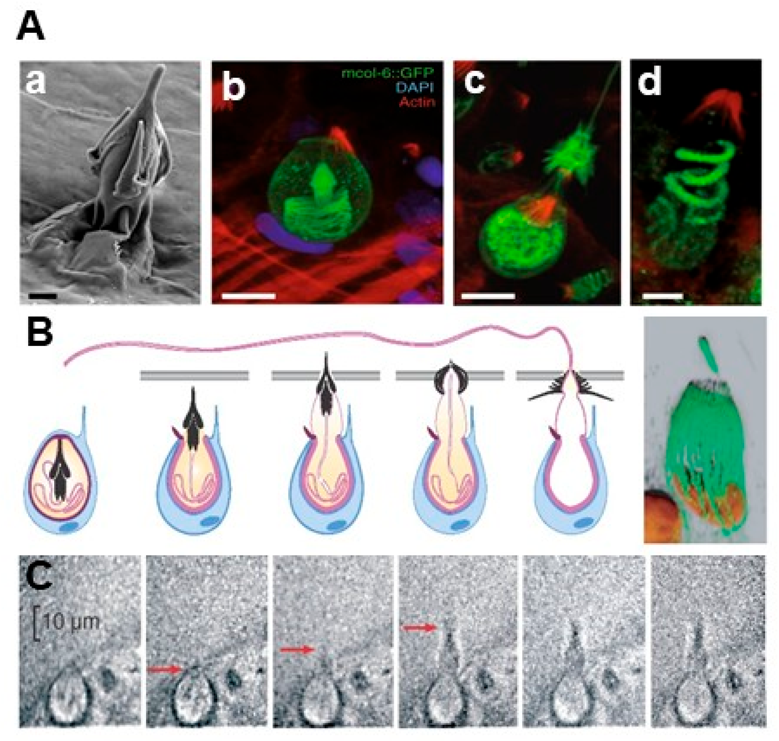
The nematocyst is a complex secretory organelle derived from Golgi vesicles [25,26]. It consists of a collagenous capsule enclosing a tightly coiled, rapidly evertible tubule—known as the nematocyst thread—which may be either permeable or impermeable. These stinging organelles are distributed differentially across cnidarians: typically concentrated on tentacles [27], they are found mainly on the oral arms in scyphozoans such as Pelagia noctiluca, though they may also occur on the bell and body [28]. Structurally, the nematocyst comprises three key components: the operculum, capsule, and thread. Prior to discharge, the thread remains coiled within the capsule. Upon stimulation, it everts at extreme speeds—completing deployment in approximately 700 nanoseconds, with velocities reaching 18.6 m/s [29] (Figure 2). This phenomenon, termed discharge, represents one of the fastest biological events known [30]. Nematocysts function as dual chemosensory and mechanosensory cells [7,16], responsive to both physical and chemical cues. During discharge, the thread accelerates instantaneously at up to 5.4 × 10⁶ g, producing impact pressures of 7.7 GPa [31]. This allows it to penetrate diverse biological barriers, from human skin to crustacean exoskeletons. Once embedded, the barbed tubule delivers venom efficiently into the target, initiating rapid envenomation for both predation and defense.

3. Jellyfish Venom Composition

Jellyfish venom is the primary cause of toxic reactions and lethal outcomes following stings. Recent ecological changes, including global increases in jellyfish blooms and associated sting incidents, have driven significant research efforts to characterize the venom’s bioactive components. Nevertheless, the full composition remains poorly characterized, with only a limited number of constituents clearly identified (Table 1). Biochemically, the venom is a complex mixture of structurally novel and highly potent molecules. It is composed mainly of peptides and proteins, but also contains thermolabile enzymes, vasoactive amines (e.g., histamine and 5-hydroxytryptamine), kinin-like compounds, bioactive lipids, and other low-molecular-weight effectors [34]. Both proteinaceous and non-proteinaceous fractions contribute to the venom’s diverse toxic effects—including cardiac injury.

3.1. Matrix Metalloproteinases

Metalloproteases are a class of proteolytic enzymes dependent on metal ions (particularly Zn2+ and Ca2+), which are widely distributed in the venoms of various animals such as centipedes [35], snakes [36], and spiders [37]. Among these enzymes, matrix metalloproteinases (MMPs, also known as matrixins)—a specialized Zn2+-dependent subgroup [38]—stand out as particularly important toxic components in jellyfish venom [39]. Proteomic analyses have demonstrated that matrix metalloproteinase-14 and astacin-like metalloprotease toxin 3 precursor form the predominant protein components in Nemopilema nomurai venom [39]. Similarly, matrix metalloproteinases comprise approximately 15% of the total protein content in Stomolophus meleagris venom [40]. Comparative studies have identified seven and nine distinct MMPs in Rhopilema esculentum and Sanderia malayensis, respectively [41]. Furthermore, metalloprotease activity has been confirmed in the key lethal toxins of Cyanea nozakii [42].
MMPs represent the primary pathogenic mediators in jellyfish envenomation, capable of inducing diverse pathophysiological processes, including severe cardiotoxicity. Notably, Prakash et al. [43] isolated a cardiotoxic component (NnTP) from N. nomurai venom (NnV) using chromatographic techniques, subsequently identifying its toxic protease activity. Although direct evidence in jellyfish envenomation remains limited, metalloproteinases are known to exert significant effects on the extracellular matrix, thereby potentially impairing tissue repair processes such as muscle regeneration. For instance, studies on snake venom metalloproteinases—such as those isolated from Bothrops asper—have demonstrated that intramuscular injection leads to dysregulated regeneration accompanied by fibrosis and atrophic fibers, suggesting a conserved pathomechanism that may also contribute to the sustained tissue damage observed in severe jellyfish stings [44]. Beyond that, MMPs have been strongly linked to multiple pathological manifestations such as cutaneous damage, inflammatory responses, and secondary infections. Yu et al. further demonstrated that venom metalloproteases critically regulate both pro-inflammatory responses and anti-inflammatory modulation in jellyfish sting-induced dermatitis [45], with pro-inflammatory factor expression primarily controlled through the PI3K-Akt pathway, while anti-inflammatory feedback appears to be mediated via the NF-κB p65 signaling pathway.

3.2. Phospholipase A2, PLA2

Phospholipase A2 (PLA2) represents another crucial enzymatic component in jellyfish venom, with widespread distribution across various jellyfish species. Research on jellyfish venom enzymes has predominantly investigated PLA2 activities, with demonstrated presence in nematocysts from both fishing and mesenteric tentacles of Cyanea capillata and Cyanea lamarckii [46]. Similar PLA2 activity has been confirmed in C. nozakii nematocyst venom [47], consistent with the identification of phospholipase motifs in its key lethal toxin (Letoxcn) alongside metalloproteases [42]. Compositional analyses reveal PLA2 represents over 20% of the venom components in S. meleagris [40]. Further characterization by Heo et al. [48] identified two PLA2 subtypes in N. nomurai venom: acidic PLA2 PA4 and PLA2 PA3A/PA3B/PA5 variants.
Phospholipase A2 (PLA2) mediates the hydrolysis of glycerophospholipids at the sn-2 position, releasing lysophospholipids and free fatty acids, including arachidonic acid [49]. This enzyme exhibits particularly high activity in cnidarian tentacles and appendages, where it facilitates critical biological functions such as prey capture, food digestion, and defensive mechanisms. Following cnidarian envenomation, PLA2 has been directly linked to diverse pathological effects encompassing pain induction, inflammatory responses, myotoxicity, neurotoxicity, and cytolytic damage [50]. Research demonstrates that PLA2 induces hemolysis through two distinct pathways [51,52]—direct enzymatic degradation of erythrocyte membranes and indirect membrane disruption mediated by lytic lipid byproducts generated from phospholipid hydrolysis. The released arachidonic acid and related hydrolysis products further promote the biosynthesis of inflammatory lipid mediators like prostaglandins and leukotrienes, which subsequently propagate inflammatory cascades through their enzymatic activities. These toxic effects may arise either from PLA2’s direct action or through synergistic interactions with other venom protein components [53].

3.3. Serine Protease

Serine proteases, well-characterized across diverse venomous species including snakes, scorpions, and spiders, primarily manifest hematotoxic effects through coagulation cascade disruption [54]. Recent integrative transcriptomic and proteomic studies have identified these enzymes as additional toxic components in jellyfish venom [40]. Supporting this finding, enzymatic analyses of N. nomurai nematocyst extracts detected chymotrypsin-like amidolytic activity that was markedly suppressed by serine protease inhibitors, confirming the presence of chymotrypsin-like serine proteases in jellyfish venom. cDNA sequence analysis revealed that this enzyme shares 41% amino acid sequence identity with human chymotrypsin-like proteins (CTRL and CTRL-1) [55]. Further investigations by Seong Kyeong Bae et al. demonstrated the fibrinolytic capacity of this chymotrypsin-like serine protease, highlighting its potential therapeutic value for thrombotic conditions [56]. Although this protease may participate in jellyfish envenomation-induced inflammatory responses, this potential association awaits further experimental confirmation.

3.4. Pore-Forming Toxins, PFTs

Jellyfish pore-forming toxins (PFTs) represent a class of cytolytic proteins capable of directly perforating cellular membranes, exhibiting significant toxicity against target cells and organisms [57]. Initially characterized from lethal box jellyfish nematocysts, including Carybdea rastonii [58], Carybdea alata [59], Chiropsalmus quadrigatus [60], and C. fleckeri [61], these toxins demonstrate potent cytotoxicity and lethality across phylogenetically diverse species such as mice, lobsters, and humans [62]. While originally believed to be unique to cubozoans, subsequent research has identified PFTs in other cnidarian classes, including scyphozoans [61] (e.g., Aurelia spp. [63], Cassiopea xamachana [64]), and hydrozoans (e.g., Hydra vulgaris [65]).
Mechanistically, jellyfish PFTs display marked hemolytic and cardiotoxic activities through specific interactions with cellular membranes. These interactions fundamentally compromise membrane integrity and permeability via the formation of transmembrane pores, leading to disruption of ionic gradients. The consequent osmotic imbalance induces cellular swelling and ultimately results in lytic cell death [62].

3.5. Others

In addition to the aforementioned proteinaceous and enzymatic components, jellyfish venom contains a diverse array of bioactive toxins with significant pharmacological properties. Notably, potent neurotoxins have been identified that disrupt ion transport dynamics (particularly sodium and calcium fluxes) by inducing channel or pore formation in neuronal and muscle cell membranes [66]. These neurotoxic components can elicit a spectrum of toxicological manifestations, including neurological dysfunction, skeletal myotoxicity, and cardiotoxic effects [67].
Table 1. Identified active components of jellyfish venom.
ClassSpeciesSourceProtein ComponentMolecular Weight (kDa)Bioactivity/Toxicological manifestaionMajor Potential Toxin FamiliesRef.
Cardiotoxicity-dominantCarukia barnesiDouble Island (northern Australia)Crude venom25–250pulmonary hypertension; tachycardiaNeural sodium channel activator[68]
Chrysaora quinquecirrhaBay of Bengal (India)Frc-1105antioxidant potentialU[69,70]
Frc-265
Frc-39
Meredith Creek (Maryland)SNLF100cardiotoxic; neurotoxic
190
Physalia physalisMiami (Florida)Physalitoxin240haemolytic activityU[71]
Immunotoxicity-dominantStomolophus meleagrisQingdao (China)SmTX ~45haemolytic activityU[72]
52
Pelagia noctilucaSicilian coasts (Strait of Messina)Crude venom20–100haemolytic activityU[73]
Dual-mechanism synergistic toxicityChironex fleckeriCairns, Townsville, and Weipa (Queensland, Australia)CfTX-143pore-forming toxins; haemolytic activityU[61,74]
CfTX-245
Weipa (Queensland, Australia)CfTX-A~40
CfTX-B~42
Nemopilema nomuraiYellow Sea (South Korea)Crude venom15–250 cardiotoxic, hepatotoxic, hemolytic and cytotoxic biological activitiesMetalloproteinase, PLA2 activity[75]
ROK CoastJVMP17-1~25dermotoxicity, cytotoxicity, and lethalityMetalloproteinase
JVMP17-250–70
Cyanea capillataIsle of Lewis (Western Isles, Scotland)CcTX-131.17haemolytic activityHaemolytic proteins[76,77]
Isle of Lewis, Rousay (Scotland)CcNT8.22neurotoxinNeurotoxic peptide
UnkownCyanea nozakiiQingdao (China)Letoxcn~50metalloproteinase activityU[42]
This table summarizes toxic components isolated from jellyfish venoms, detailing their molecular characteristics, biological activities, major potential toxin families, and so on. U: Unknown.

4. Cardiotoxicity-Dominant Jellyfish Species

Jellyfish venom-induced cardiotoxicity remains one of the most clinically significant and mechanistically complex challenges in marine toxinology. Among various jellyfish species, Carukia barnesi and Chrysaora quinquecirrha have attracted particular research attention due to their pronounced cardiotoxic effects. Their venoms contain a cocktail of neurotoxins and pore-forming proteins that disrupt cardiac ion channel function, leading to profound disturbances in myocardial electrophysiology.

4.1. Carukia barnesi

Carukia barnesi (class Cubozoa), commonly known as the Irukandji jellyfish, is a small (~30 mm bell diameter), translucent species [75] predominantly distributed in marine waters from Australia to East Asia [78]. Its venom can induce Irukandji syndrome—a potentially fatal envenomation characterized by sequential pathophysiological phases: initial autonomic storm (profuse diaphoresis and severe anxiety), followed by neuromuscular hyperactivity (painful cramps), and culminating in cardiovascular collapse (acute hypertension progressing to hypotension with cardiac decompensation) [79]. Clinically, approximately 2% of the cases evolve to congestive heart failure within 12–24 h post-sting. Most remarkably, a subset of severe presentations develops Takotsubo cardiomyopathy (TTC) [80], representing the most severe cardiovascular complication of catecholamine-driven toxicosis.
Biochemical analyses have revealed that C. barnesi venom contains at least 48 protein components, most with molecular weights below 100 kDa [81]. Comparative proteomic studies further indicate significant differences in protein composition between mature and immature nematocyst venoms. In vitro studies using isolated rat right atria have demonstrated that crude venom extracts from C. barnesi elicit a concentration-dependent tachycardic response. This effect was completely abolished by tetrodotoxin (TTX) pretreatment, providing compelling evidence for the presence of sodium channel-activating neurotoxins. Importantly, current evidence suggests that the cardiotoxic effects are mediated primarily through indirect mechanisms involving substantial catecholamine release rather than direct cardiomyocyte injury [68].

4.2. Chrysaora quinquecirrha

Chrysaora quinquecirrha (Atlantic sea nettle; Cnidaria: Scyphozoa: Semaeostomae) exhibits a broad geographical distribution across tropical and subtropical regions of the Atlantic, Indian, and Pacific Oceans, with particularly high abundance along the eastern coast of the United States. The venom of C. quinquecirrha demonstrates marked multi-system toxicity [82], with cardiotoxicity being the most prominent manifestation. Two toxic components (100 kDa and 190 kDa) with potent cardiotoxic and neurotoxic activities have been identified in the tentacle venom of C. quinquecirrha, showing a murine median lethal dose (LD50) of 0.37 μg/g [70].
Moreover, Kelman et al. isolated toxic proteins from the mesenteric tentacle nematocysts of C. quinquecirrha, which were found to significantly suppress spontaneous contractions in cultured chick cardiomyocytes [83]. The venom also induces irreversible contraction in isolated rat aortic rings—an effect that is effectively antagonized by the calcium channel blocker verapamil [84]. These findings collectively indicate that the cardiotoxic mechanism of C. quinquecirrha venom involves modulation of voltage-dependent Ca2+ channels—a mechanism distinct from the tetrodotoxin-sensitive, catecholamine-mediated cardiotoxicity characteristic of C. barnesi envenomation.

4.3. Physalia physalis

The siphonophore Physalia physalis (Portuguese man o’ war) ranks among the most toxic and globally distributed cnidarians. Mature specimens possess tentacles extending up to 30 m that are densely packed with venomous nematocysts [85], capable of inducing vertebrate paralysis and lethal envenomation in humans. Its venom exhibits both cardiotoxic and hemolytic effects [86], with the hemolytic activity primarily mediated by physalitoxin—a 240 kDa glycoprotein composed of three distinct subunits [71].
Unlike C. quinquecirrha, whose calcium-mediated cardiotoxicity is blocked by verapamil, and C. barnesi, whose TTX-sensitive toxicity is catecholamine-driven, its cytotoxic mechanism involves direct membrane permeabilization, inducing pathological calcium influx and dose-dependent damage in embryonic chick cardiomyocytes [87,88]. The venom also demonstrates potent neurotoxicity, causing dose- and time-dependent locomotor deficits in Drosophila that implicate bioactive components targeting central nervous system ion channels [89].

5. Immunotoxicity-Dominant Jellyfish Species

Recent studies demonstrate that jellyfish envenomation can trigger both immediate and delayed hypersensitivity reactions, with documented cross-reactivity between different jellyfish venoms. While systemic anaphylaxis following envenomation remains relatively uncommon, repeated exposure among coastal residents and maritime workers substantially increases sensitization risk. Notably, allergic responses may be initiated through either dermal contact or accidental ingestion.
Immunological investigations reveal that specific jellyfish species contain venom components with marked immunomodulatory capacity. For instance, jellyfish-derived collagen extracts and peptides potently stimulate pro-inflammatory cytokine release, particularly tumor necrosis factor-alpha (TNF-α) and interferon-gamma (IFN-γ) [86]. Furthermore, both proteinaceous toxins and carbohydrate-rich components (e.g., glycoproteins and polysaccharides) from nematocysts can function as antigens, inducing clinical symptoms through cellular and/or humoral immune pathways [27]. The subsequent discussion will examine immunodominant jellyfish species, detailing their distinct clinical presentations and the molecular mechanisms underlying their pathogenic effects.

5.1. Stomolophus meleagris

The rhizostome jellyfish Stomolophus meleagris (Cnidaria: Scyphozoa) is a large species (bell diameter: 180–980 mm) widely distributed across China’s coastal waters from the Yellow Sea to the South China Sea. Its venom contains the characteristic hemolytic protein SmTX, purified through sequential anion-exchange and gel-filtration chromatography. This multi-subunit toxin, composed of ~45 kDa and 52 kDa subunits, exhibits potent hemolytic activity with optimal function at pH 5.0 and temperatures between 23 and 45 °C [72].
Experimental studies confirm that S. meleagris venom triggers significant acute inflammatory responses. In murine footpad injection models, the venom induced dose-dependent edema and erythema. In vitro analyses of TE-treated HaCaT cells revealed substantial upregulation of pro-inflammatory cytokines (IL-1β, IL-6, IL-8, and TNF-α), while Western blotting demonstrated concurrent activation of both NF-κB and MAPK signaling pathways [90].

5.2. Pelagia noctiluca

Pelagia noctiluca (Cnidaria: Scyphozoa), a bioluminescent jellyfish species, represents one of the most medically significant medium-sized cnidarians in Mediterranean waters [28]. Its venom elicits both localized pathologies (erythema or edema) and systemic inflammatory responses following envenomation. Frazão et al. [91] revealed 68 proteins in P. noctiluca venom by proteomics, including zinc metalloproteinases with the ShK toxin structural domain, and the molecular weight distribution of haemolytically active proteins in the venom was in the range of 20–100 kDa [73].
Accidental contact with P. noctiluca often induces severe localized and systemic pathologies [92], primarily mediated by the venom’s potent pro-inflammatory properties [93]. Experimental investigations in rat models demonstrate that venom exposure leads to pronounced inflammatory cell infiltration (predominantly neutrophils and macrophages) in pulmonary and intestinal tissues within 6 h post-injection [94]. This cellular response correlates with upregulated NF-κB p65 expression. Interestingly, pretreatment with the antioxidant tempol significantly attenuates these pathological changes, confirming the essential role of ROS-mediated NF-κB pathway activation in the inflammatory cascade.

6. Dual-Mechanism Synergistic Toxicity

Jellyfish venoms demonstrate significant interspecies variability in their bioactive components, with cardiotoxic and immunomodulatory effects traditionally studied as distinct phenomena. However, recent studies have identified species (e.g., C. fleckeri) whose venoms simultaneously target both cardiac and immune systems—where direct myocardial toxicity and systemic inflammatory responses act synergistically to amplify tissue damage. This section systematically examines jellyfish species with dual cardiotoxic-immunomodulatory venom actions, characterizing their toxic constituents and delineating the mechanistic basis of their combined pathological effects.

6.1. Chironex fleckeri

Chironex fleckeri (commonly known as the Australian box jellyfish or sea wasp), predominantly found in northern Australian coastal waters, is the most toxic cubozoan jellyfish known. Its venom can cause severe dermatonecrosis, systemic inflammatory responses, and life-threatening cardiotoxicity in humans. The venom contains two major classes of pore-forming toxins: CfTX-1/2 (43–45 kDa) and CfTX-A/B (~40–42 kDa), both belonging to the porin family but phylogenetically distinct [61]. Among these, CfTX-1 and CfTX-2 are identified as type I cardiotoxins and represent the key mediators of acute cardiovascular failure. They induce rapid cardiac arrest in mice, even at ultralow doses, via direct action on cardiomyocytes [95]. Structural studies attribute CfTX-1’s potent activity partly to its unique helical domain [96]. In contrast, CfTX-A and CfTX-B exhibit minimal cardiotoxicity but demonstrate strong hemolytic activity [74].
Both toxin classes are highly immunogenic and capable of eliciting specific antibody responses. Recombinant CfTX-1 shows particularly robust immunoreactivity, supporting its potential as a vaccine candidate [97]. To date, CfTX-1 and CfTX-2 remain unique to C. fleckeri and are considered the principal toxins responsible for the rapid cardiovascular collapse following envenomation. The venom thus exerts synergistic cardiotoxic and immunological effects through the coordinated action of its constituent porins.

6.2. Nemopilema nomurai

Nemopilema nomurai is recognized as one of the most dangerous large jellyfish species inhabiting East Asian coastal waters, where it is responsible for an estimated 100,000 envenomation cases annually. Proteomic analysis has revealed that its venom contains a complex mixture of over 150 distinct protein components [75], including key toxins such as metalloproteases and phospholipases. Of particular significance, researchers have isolated 13 toxin homologs from the lethal fraction of N. nomurai specimens obtained from Dalian, China, comprising phospholipases, potassium channel inhibitors, and hemolysins. Among these, alkaline phospholipases have been shown to play a critical role in mediating the venom’s lethal effects [98].
Most N. nomurai envenomations primarily cause localized cutaneous symptoms, including acute pain and inflammatory responses. In vitro studies reveal that venom exposure triggers dose-dependent increases in both mRNA and protein expression of key pro-inflammatory mediators (TNF-α, IL-6, and MCP-1) in skin cell lines (HaCaT and CCC-ESF-1) [99]. Beyond its dermatological effects, the venom also directly suppresses cardiac function through negative inotropic and chronotropic actions [100]. This cardiotoxicity is particularly selective, as evidenced by the venom’s pronounced cytotoxicity toward cardiomyocytes (H9c2 cell line), which alters the expression of 72 cardiac-related proteins—including established heart failure biomarkers like annexin A2 and myosin-7 [101]. Notably, this cardiac toxicity far exceeds the venom’s impact on skeletal muscle cells (C2C12) [102], highlighting its specific targeting of myocardial tissue.

6.3. Cyanea capillata

The lion’s mane jellyfish (Cyanea capillata), a dominant large jellyfish species in southeastern Chinese coastal waters with bell diameters often exceeding 1 m, produces venom exhibiting distinct cardio-immunological synergistic toxicity. The hemolytically active protein CcTX-1 was isolated from the tentacle venom of C. capillata. This toxin exists in two isoforms, with the predominant subtype exhibiting a molecular mass of 31.17 kDa. Notably, sequence alignment demonstrates high homology between CcTX-1 and known cubozoan hemolysins [76], suggesting potential functional similarities. In addition to its hemolytic components, C. capillata venom contains the low-molecular-weight neurotoxin CcNT (8217.4 Da), which induces paralysis via selective inhibition of voltage-gated sodium channels [77]. Together, these findings highlight the multifunctional nature of C. capillata venom, combining both hemolytic and neurotoxic mechanisms to exert its potent biological effects.
The lion’s mane jellyfish (Cyanea capillata), a prevalent species in southeastern Chinese coastal waters, produces venom with cardio-immunological synergistic toxicity. Its tentacle venom contains two key toxins: the hemolytically active protein CcTX-1 (31.17 kDa main isoform), which shows high homology to cubozoan hemolysins [76], and the low-molecular-weight neurotoxin CcNT (8.2 kDa), which inhibits voltage-gated sodium channels and induces paralysis [77]. These components act synergistically, enabling the venom to exert combined hemolytic and neurotoxic effects.
Experimental studies demonstrate that C. capillata venom induces significant inflammatory responses. Salmon exposure experiments revealed acute tissue damage, including edema and epithelial detachment, accompanied by sustained inflammatory reactions [103]. Regarding cardiotoxicity, tentacle extract (TOE) from C. capillata exhibits direct cardiac toxicity in rat models (Figure 3) [104]. Intravenous administration caused dose-dependent myocardial injury, culminating in acute global heart failure. In vitro heart perfusion assays confirmed these findings, showing rapid reductions in both heart rate (HR) and left ventricular developed pressure (LVDP) following TOE exposure. Notably, administration of 240 μg TOE triggered severe arrhythmias and cardiac arrest. Histopathological examination identified characteristic myocardial lesions, including wavy fiber distortion. Mechanistic studies suggest this toxicity may involve overactivation of L-type Ca2+ channels and adrenergic receptors [105].

7. Treatment Strategy

In summary, systematic analysis of jellyfish venom toxicity reveals that cardiotoxic and immunomodulatory effects are not mutually exclusive pathological manifestations. While one may dominate clinically, these effects frequently coexist and interact synergistically in envenomed patients. Targeting the bioactive components responsible for these pathological effects may hold significant therapeutic potential. Given the substantial interspecies variations in venom composition and pathogenic mechanisms among jellyfish, it is imperative to establish toxin profile-based personalized treatment protocols. Therapeutic strategies should be tailored according to the envenomation type and clinical manifestations, with precise identification of the jellyfish species and timely intervention being critical determinants of treatment efficacy. For cardiotoxicity-dominant jellyfish envenomations, clinical management should prioritize stabilization of myocardial electrophysiological activity, supplemented by inflammatory storm control during the subacute phase. Conversely, for immunodominant jellyfish envenomations, targeted immunotherapy against immune-mediated symptoms may yield superior clinical outcomes. And for cardio-immunological synergistic envenomations, a dual-blockade strategy combining toxin neutralization (e.g., antivenom) with immunomodulatory intervention is essential to address both pathological pathways concurrently. This section synthesizes targeted antagonists and therapeutic strategies corresponding to distinct toxicological mechanisms, aiming to establish an evidence-based framework for clinical management of jellyfish envenomations.

7.1. Comprehensive Treatment of Jellyfish Sting

Jellyfish venom exhibits complex composition with inherent instability, where the severity of local and systemic reactions depends on multiple variables: the specific venom profile, delivered toxin quantity, dermal contact area with tentacles, exposure duration, and so on. For jellyfish envenomation, basic first-aid measures can be categorized into three primary objectives: (i) preventing further nematocyst discharge, (ii) mitigating local venom effects (pain and tissue damage), and (iii) controlling systemic reactions.
Prompt removal of residual tentacles and nematocysts from the skin is critical post-envenomation, as retained tentacular fragments can continue discharging venom, leading to secondary envenomation events [106]. Significant controversy persists regarding optimal tentacle removal techniques for minimizing local envenomation symptoms, particularly concerning the use of oral/topical analgesics, hot water, ice packs, or vinegar. This debate stems primarily from interspecies variations in venom composition and pathological effects.
Morabito et al. recommend immediate application of lidocaine, acetic acid, ethanol, or ammonia solutions to Pelagia noctiluca sting sites, demonstrating significant pain relief and adverse reaction reduction [107]. In contrast, Burnett et al. found that topical lidocaine monotherapy failed to provide adequate analgesic efficacy in their clinical studies [108].
Hot water immersion or heat packs remain widely used for marine envenomation management, yet considerable disagreement persists among researchers regarding the comparative efficacy of thermotherapy versus cryotherapy. Cryotherapy using ice packs or cold compresses has been demonstrated to effectively limit both inflammatory progression and venom spread, while concurrently providing analgesic benefits [109]. For scyphozoan envenomations (e.g., Aurelia aurita, C. fleckeri, or S. meleagris), thermotherapy appears to confer greater clinical benefit [106]. This phenomenon may stem from the thermolabile nature of these jellyfish venoms and/or direct modulation of nociceptor activity by heat immersion, collectively contributing to observed pain reduction [105]. In contrast, for C. quinquecirrha—a species with relatively thermostable venom [110]—both thermotherapy and cryotherapy demonstrate limited clinical efficacy [111].
Given the inherent accessibility of seawater, victims often instinctively employ it for initial rinsing. While this approach has demonstrated efficacy in tentacle removal, pain relief, and nematocyst inhibition [112], Doyle et al. found that seawater irrigation after C. capillata envenomation exacerbates stinging pain and significantly enhances venom dispersal—effects not observed with vinegar application [112]. Conventional remedies including urine [113], ammonia, 70% ethanol, and isopropanol demonstrate limited efficacy for most jellyfish envenomations, with potential to paradoxically stimulate nematocyst discharge [10].
In summary, no universal consensus exists regarding optimal first-aid protocols for jellyfish stings, primarily due to substantial interspecies variability in venom composition and pathophysiological effects. We recommend immediate implementation of species-specific treatment protocols when the envenoming jellyfish is definitively identified. For unclassified envenomations, a comprehensive risk–benefit assessment integrating all the relevant factors—including individual physiological variability and envenomation severity—is imperative for treatment decision-making.

7.2. Therapeutic Strategies for Cardiotoxicity

For cardiotoxicity-dominant jellyfish envenomation, therapeutic strategies should prioritize neutralizing cardiac-specific pathological pathways. The resulting cardiotoxicity typically involves interconnected mechanisms, including rhythm disturbances resulting from altered cardiomyocyte membrane permeability (Figure 4). Central to this process is the disruption of myocardial calcium homeostasis—a well-established mechanism underlying venom-induced cardiac dysfunction [114]—and emerging evidence also implicates cytokine dysregulation in this pathological cascade [101].
As already established, calcium signaling plays a central role in cardiac excitation–contraction coupling, and its dysregulation can lead to severe consequences such as arrhythmias, cardiomyocyte injury, and programmed cell death [117]. Accordingly, calcium channel blockers have shown significant cardioprotective effects against jellyfish venom: felodipine mitigates cardiac damage caused by N. nomurai venom [100], while nifedipine and verapamil improve impaired cardiac function following C. capillata tentacle extract (TOE) exposure [105]. Current pharmacological interventions, thus, predominantly target these pathways using calcium channel blockers and antivenom therapies.
Antivenom (AV) therapy remains the primary intervention for C.fleckeri envenomation, with the Commonwealth Serum Laboratories (CSL) antivenom being the only commercially available treatment for cubozoan stings worldwide. While prophylactic antivenom administration has demonstrated survival benefits in murine models [118] and reduced cardiovascular collapse induced by C. fleckeri venom [119], conflicting data exist regarding its therapeutic window. Notably, AV given 15 min pre-envenomation failed to significantly alleviate toxicity in animal studies [120], leaving current evidence on efficacy and optimal dosing inconclusive. Interestingly, when the venom was incubated with either AV for 3 h prior to infusion, the effect of the venom was almost abolished. This result suggests that the venom acts extremely rapidly, and the clinical efficacy of AV is limited by the difficulty of administering it sufficiently early after envenomation. However, in vitro studies by Andreosso et al. demonstrated that antivenom at 5-fold the maximum recommended clinical dose significantly improved cell viability, with this protection showing dose-dependent enhancement at higher concentrations [121]. Considering venom-induced cardiotoxicity, one study explored combination therapy using antivenom with verapamil, demonstrating enhanced efficacy and prolonged survival in envenomed mice [122]. It should be noted that this approach remains contentious, as verapamil’s benefits decrease with escalating antivenom doses, and the ideal therapeutic ratio requires further clarification. Additionally, the extraction techniques used to obtain venom for AV production may result in antiserum lacking critical antibodies, thereby contributing to inconsistent and contradictory experimental outcomes.
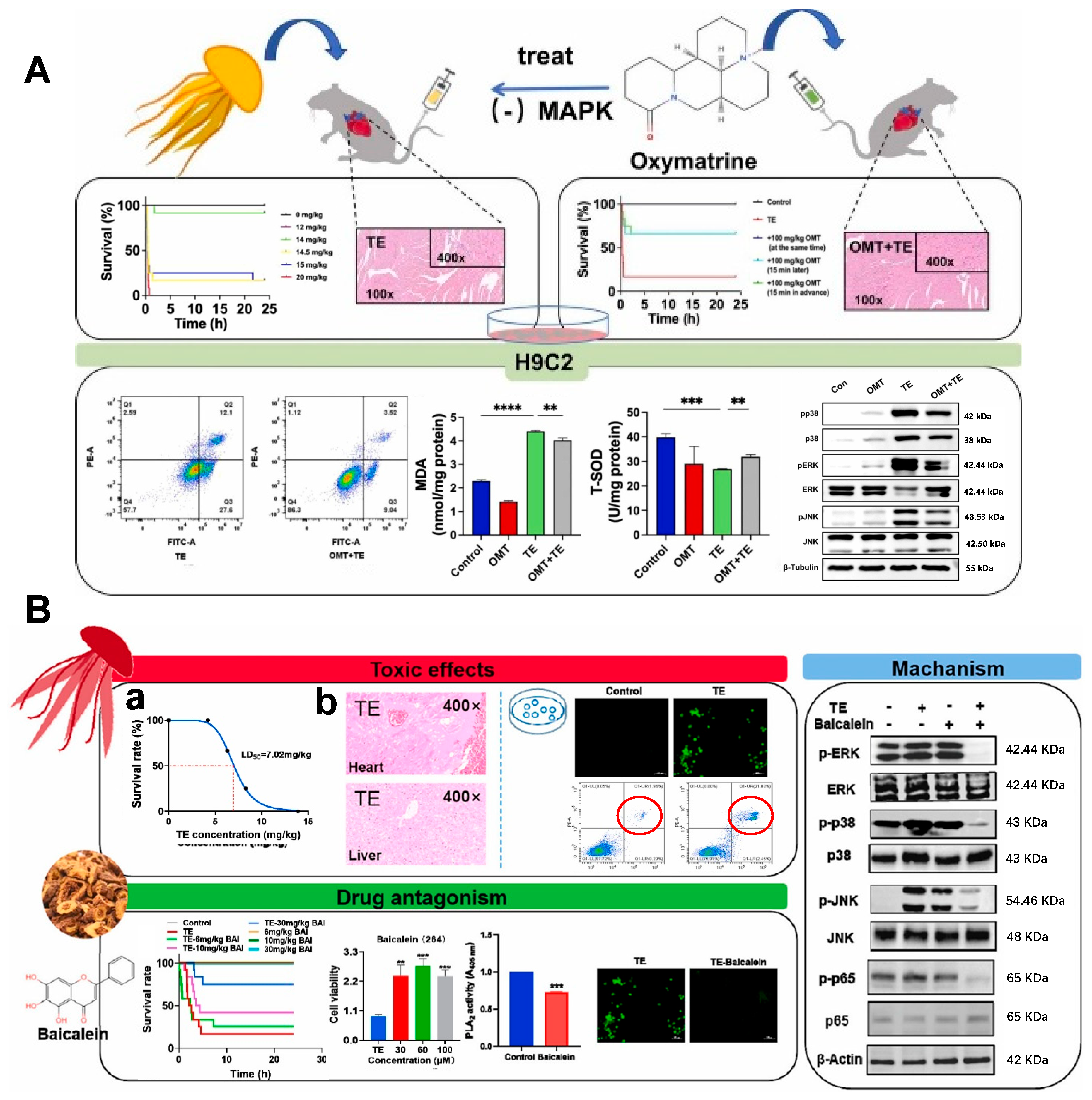
Oxidative stress plays a critical role in various cardiac pathological processes and contributes to numerous cardiovascular abnormalities. Accumulating evidence suggests that the cardiotoxicity induced by jellyfish envenomation may be associated with inflammatory responses and oxidative damage [15,18]. Therefore, targeting MAPK activation and oxidative stress represents a promising therapeutic strategy for mitigating jellyfish venom-induced cardiotoxicity. Dong et al. revealed that oxymatrine (OMT) effectively counteracted the lethal effects induced by tentacle extract (TE) of Nemopilema nomurai, reducing cardiac edema and myocardial fiber disruption. In vitro experiments further demonstrated that OMT attenuated TE-induced decreases in H9C2 cell viability and inhibited apoptosis [115]. Similarly, Wang et al. reported that pretreatment with baicalein significantly improved survival rates in mice exposed to tentacle extract from Rhopilema esculentum. Baicalein was shown to alleviate toxin-induced oxidative stress and cell death by modulating the ROS/MAPK/NF-κB pathway, concurrently ameliorating TE-triggered cardiac injury [116].
Moreover, cardiac glycogen synthesis capacity plays an essential role in normal heart development. Notably, as a central regulatory pathway in cardiac failure (CF) [123], glycogen metabolism demonstrates therapeutic potential for jellyfish venom-induced CF through maintaining storage capacity and enhancing energy production efficiency. Qin et al. [124] identified via single-cell transcriptome analysis that the energy-regulating protein α-1 acid glycoprotein (AAG), a key modulator of glycogen metabolism, was significantly upregulated in CF patients following jellyfish envenomation. These findings were further validated in animal studies using a toxin-induced CF mouse model, where AAG deficiency accelerated heart failure progression, while either exogenous supplementation or endogenous upregulation of AAG effectively reversed cardiac dysfunction. This discovery provides a novel targeted therapeutic strategy for jellyfish venom-associated CF.

7.3. Therapeutic Strategies for Immunotoxicity

The clinical management of immunodominant jellyfish envenomations poses distinct therapeutic challenges compared to cardiotoxicity-dominant cases. Nematocyst-derived components—such as chitin, collagen, and polysaccharides—can chronically stimulate innate immune cells (e.g., keratinocytes, dendritic cells, and mast cells) [27], leading to persistent vesiculobullous and pruritic skin reactions. In severe cases, such as envenomation by N. nomurai, the venom can trigger a deadly cytokine storm—a systemic and dysregulated inflammatory response that significantly contributes to multi-organ failure and mortality [18]. However, current research on immune-mediated jellyfish envenomation predominantly centers on cutaneous pathologies, especially jellyfish dermatitis.
As previously discussed, matrix metalloproteinases (MMPs) are pivotal mediators of both cutaneous damage and inflammatory responses following jellyfish envenomation. Mechanistic investigations indicate that venom-derived MMPs primarily exert their pathogenic effects by activating MAPK and NF-κB signaling pathways, which are known to be key regulators in the development of jellyfish dermatitis. Owing to this central role, MMP inhibitors have gained attention as potential therapeutics. Li et al. [122] recently demonstrated that batimastat and disodium EDTA, as MMP inhibitors, show marked efficacy against N. nomurai venom (NnNV)-induced dermatitis.
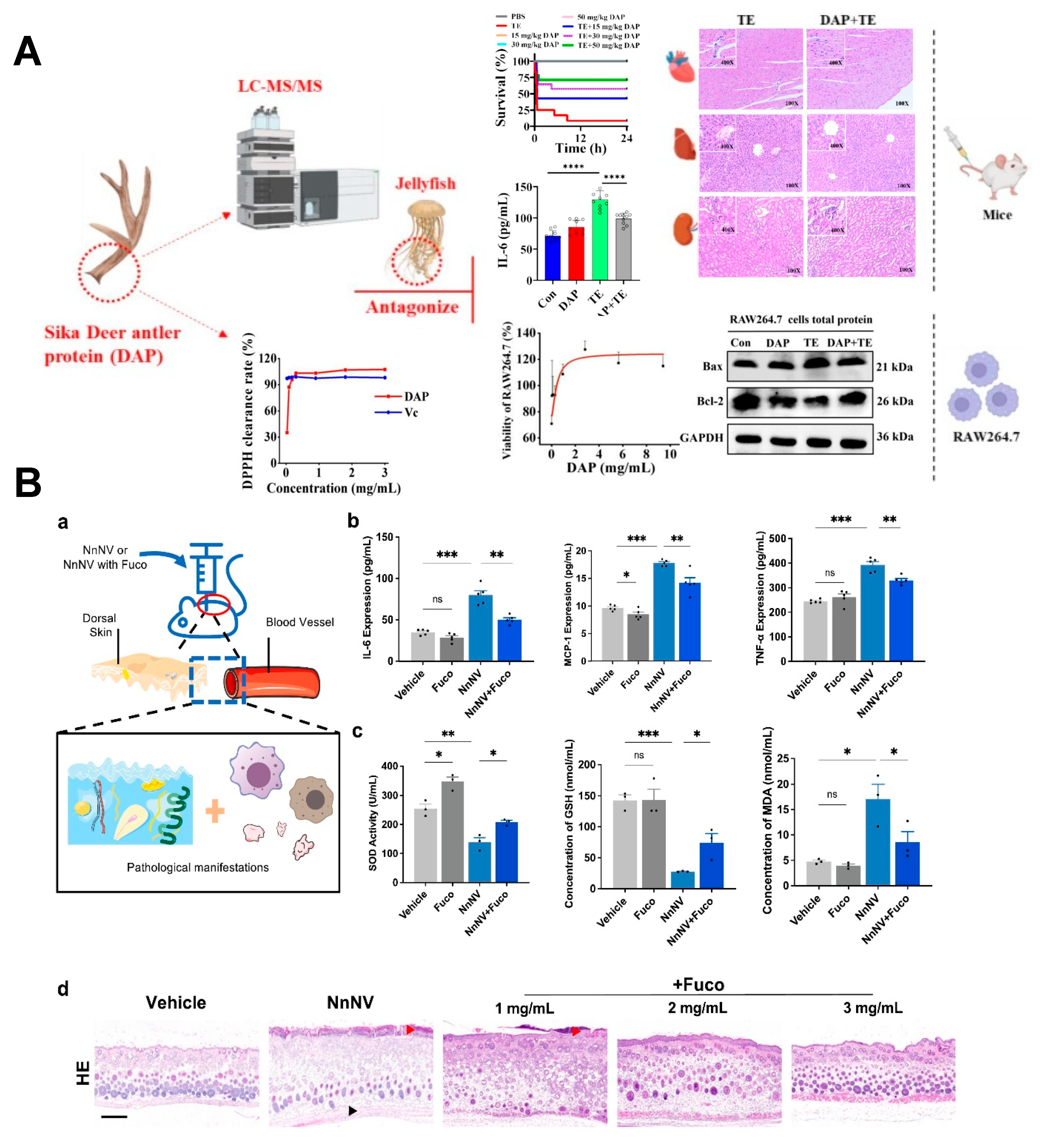
Oxidative stress may act synergistically with inflammatory responses [125], as intracellular ROS accumulation activates signaling pathways including MAPK, which subsequently exacerbates inflammatory cascades. Therefore, therapeutic interventions could target not only direct MMP inhibition but also MAPK/NF-κB signaling blockade and oxidative stress reduction (Figure 5). In particular, directly targeting the NF-κB signaling pathway has been shown to mitigate harmful effects of jellyfish envenomation. Xiao et al. have demonstrated that dexamethasone, a known inhibitor of the NF-κB pathway, effectively suppresses cytokine storm and mitigates organ damage, thereby improving survival rates in a mouse model of delayed jellyfish envenomation syndrome [18]. Similarly, Liu et al. [90] showed that troxerutin effectively mitigates S. meleagris venom-induced cutaneous toxicity and inflammation through dual suppression of MAPK and NF-κB pathways. Compounds such as fucoidan [16] and deer antler protein (DAP) [126] have been demonstrated to decrease key inflammatory markers (IL-6, IL-1β, and TNF-α) in murine models by inhibiting these same pathways, ultimately enhancing survival rates after N. nomurai venom exposure.
While acute cutaneous reactions are common following jellyfish envenomation, certain cases exhibit delayed and recurrent dermatological manifestations [127]. For such persistent symptoms, immunomodulators—including pimecrolimus and tacrolimus—have shown therapeutic efficacy by specifically inhibiting T-lymphocyte activity.
Collectively, these findings underscore that anti-inflammatory drugs represent a promising strategy for treating jellyfish envenomation, warranting further clinical investigation. Future research should focus on identifying additional inflammatory signaling pathways and immune cell interactions to uncover novel therapeutic targets. This approach reflects a strategic shift from merely neutralizing venom toxins toward actively modulating the host immune response—a potentially more practical and universally applicable paradigm given the vast interspecies diversity of jellyfish and their venom compositions.

8. Summary and Outlook

Jellyfish envenomations constitute the most common marine biological injuries and have become a major public health issue worldwide. However, the pathogenic mechanisms and clinical treatment strategies for jellyfish envenomation remain insufficiently elucidated. Current therapeutic approaches frequently address individual symptoms rather than the comprehensive, multi-system effects of jellyfish venoms. Here, we propose that species-specific treatment protocols could improve clinical outcomes, as jellyfish venoms have diversified evolutionarily to meet distinct ecological demands—such as the potent cardiotoxins of predatory species like C. fleckeri for rapid immobilization, or the immunotoxins used by competition-inhibiting species like S. meleagris to elicit inflammatory deterrence [128]. It is important to note, however, that while tailored treatments show promise, a more pragmatic and impactful long-term strategy emphasizes prevention. Effective public health measures—including bloom monitoring, predictive risk modeling, protective netting, and public education on avoidance and first aid—can substantially reduce exposure and health burdens. Integrating prevention into coastal management and health policy frameworks offers a sustainable approach to mitigating the growing threat of jellyfish envenomations.
Furthermore, it is essential to acknowledge the limitations inherent in current research paradigms. Studies on cardiotoxicity have largely relied on isolated myocardial models, which may overlook systemic effects mediated by venom components. Similarly, investigations of immune responses have predominantly focused on localized cutaneous reactions, leaving the interplay between immune activation and cardiac injury poorly understood. The full pathological sequence—from venom penetration through the skin and hematogenous spread to end-organ damage—likely involves mechanisms far more complex than currently recognized.
While the majority of jellyfish stings are mild and self-limiting, a small yet clinically significant subset leads to severe complications that account for the majority of envenomation-related mortality. The underlying mechanisms—particularly the systemic immune processes driving cardiac injury and dysregulated inflammatory responses—remain poorly elucidated. Future research should employ an integrated multi-omics strategy—combining proteomic, transcriptomic, and metabolomic approaches—to comprehensively characterize key pathogenic venom constituents and elucidate the dynamic spatiotemporal interactions between immune activation and cardiac dysfunction. Translating these mechanistic insights into clinical applications will require a clear translational pathway. This includes the development of physiologically relevant preclinical models (e.g., murine cardiotoxicity models or human cell-based platforms) that accurately recapitulate human immune–cardiac interactions, alongside rigorous biomarker validation for early detection of severe envenomation. Furthermore, overcoming regulatory challenges for venom-based therapies—particularly for rare severe cases—will depend on international collaboration to standardize venom extraction, toxin characterization, and clinical endpoint definitions. Successfully bridging basic science and clinical practice will enable the development of mechanism-driven interventions, such as targeted antivenoms, tailored nanoparticle delivery systems for toxin-specific neutralization, and adjunctive immunomodulatory therapies. These advances may ultimately pave the way for precision medicine approaches in jellyfish envenomation management.

Author Contributions

Conceptualization and methodology, Y.L. (Yueyue Li), Z.Q. and B.L.; writing original draft preparation, Y.L. (Yueyue Li), Z.Q. and B.L.; writing—review and editing, X.Y., X.G. and Y.L. (Yue Li); visualization, X.Y. and X.G.; supervision, J.Y. and W.L.; funding acquisition, J.Y. and W.L. All authors have read and agreed to the published version of the manuscript.

Funding

This work was supported by the Shanghai Natural Science Foundation (25ZR1401400), Military Project (CHJ24C025), National Natural Science Foundation of China (NSFC) Youth Program (82302118), and National Postdoctoral Fellowship Program (B) (GZB20230991).

Conflicts of Interest

The authors declare no conflicts of interest.

References

  1. Cegolon, L.; Heymann, W.C.; Lange, J.H.; Mastrangelo, G. Jellyfish stings and their management: A review. Mar. Drugs 2013, 11, 523–550. [Google Scholar] [CrossRef] [PubMed]
  2. Burnett, J.W. Clinical manifestations of jellyfish envenomation. Hydrobiologia 1991, 216, 629–635. [Google Scholar] [CrossRef]
  3. Benedetti-Cecchi, L.; Canepa, A.; Fuentes, V.; Tamburello, L.; Purcell, J.E.; Piraino, S.; Roberts, J.; Boero, F.; Halpin, P. Deterministic Factors Overwhelm Stochastic Environmental Fluctuations as Drivers of Jellyfish Outbreaks. PLoS ONE 2015, 10, e0141060. [Google Scholar] [CrossRef] [PubMed]
  4. Zhang, C.; Lyu, Z.; Wu, J.; Cheng, C.; Wang, Y.; Liu, Z.; Du, B.; Yang, Y.; Li, F.; Chen, Q. Epidemiological analysis of jellyfish stings in coastal bathing beaches in Qinhuangdao City from 2017 to 2019. Zhonghua Wei Zhong Bing Ji Jiu Yi Xue 2021, 33, 593–595. [Google Scholar] [CrossRef] [PubMed]
  5. Dobson, J.Y.; Fonfría, E.S.; Flores-García, A.; Bordehore, C. Epidemiology of jellyfish stings using the Sting Index to identify trends and support proactive management. Ocean Coast. Manag. 2024, 256, 107308. [Google Scholar] [CrossRef]
  6. Boulware, D.R. A randomized, controlled field trial for the prevention of jellyfish stings with a topical sting inhibitor. J. Travel Med. 2006, 13, 166–171. [Google Scholar] [CrossRef]
  7. Li, R.; Yu, H.; Li, A.; Yu, C.; Li, P. Preparation and Neutralization Efficacy of Novel Jellyfish Antivenoms against Cyanea nozakii Toxins. Toxins 2021, 13, 165. [Google Scholar] [CrossRef]
  8. Curatolo, R.; Madanchi, M.; Juratli, H.A. Delayed-type hypersensitivity reaction to jellyfish. Int. J. Dermatol. 2024, 63, 818–819. [Google Scholar] [CrossRef]
  9. Amato, G.; Vita, F.; Gemelli, F.; Tigano, V.; Minciullo, P.L.; Gangemi, S. Jellyfish anaphylaxis: A wide spectrum of sensitization routes. Allergy Asthma Proc. 2020, 41, 158–166. [Google Scholar] [CrossRef]
  10. Peng, X.; Liu, K.T.; Chen, J.B.; Yan, Z.H.; Danso, B.; Wang, M.K.; Peng, Z.Y.; Xiao, L. Jellyfish Stings: A Review of Skin Symptoms, Pathophysiology, and Management. Med. Sci. Monit. 2024, 30, e944265. [Google Scholar] [CrossRef]
  11. Srinivasan, M.T.; Varadharajan, A.; Logamoorthy, R.; Karthikeyan, K. The touch of the tentacles—Dermoscopy of jellyfish dermatitis. QJM Int. J. Med. 2024, 118, 355–356. [Google Scholar] [CrossRef]
  12. Li, R.; Yu, H.; Yue, Y.; Li, P. Combined Proteome and Toxicology Approach Reveals the Lethality of Venom Toxins from Jellyfish Cyanea nozakii. J. Proteome Res. 2018, 17, 3904–3913. [Google Scholar] [CrossRef]
  13. Beilei, W.; Lin, Z.; Qian, H.; Qianqian, W.; Tao, W.; Jia, L.; Xiaojuan, W.; Xuting, Y.; Liang, X.; Liming, Z. Direct cardiac toxicity of the tentacle-only extract from the jellyfish Cyanea capillata demonstrated in isolated rat heart. J. Cardiovasc. Pharmacol. 2012, 59, 331–338. [Google Scholar] [CrossRef]
  14. Brinkman, D.; Burnell, J. Identification, cloning and sequencing of two major venom proteins from the box jellyfish, Chironex fleckeri. Toxicon 2007, 50, 850–860. [Google Scholar] [CrossRef]
  15. Choudhary, I.; Hwang, D.H.; Lee, H.; Yoon, W.D.; Chae, J.; Han, C.H.; Yum, S.; Kang, C.; Kim, E. Proteomic analysis of novel components of Nemopilema nomurai jellyfish venom: Deciphering the mode of action. Toxins 2019, 11, 153. [Google Scholar] [CrossRef]
  16. Li, A.; Yue, Y.; Li, R.; Yu, C.; Wang, X.; Liu, S.; Xing, R.; Li, P.; Zhang, Q.; Yu, H. Fucoidan may treat jellyfish dermatitis by inhibiting the inflammatory effect of jellyfish venom. Int. J. Biol. Macromol. 2023, 253, 127449. [Google Scholar] [CrossRef] [PubMed]
  17. Thaikruea, L. The dermatological effects of box jellyfish envenomation in stinging victims in Thailand: Underestimated severity. Wilderness Environ. Med. 2023, 34, 462–472. [Google Scholar] [CrossRef] [PubMed]
  18. Wang, Y.; Wang, Y.; Yang, F.; Chen, J.; Geng, X.; Sun, Q.; Zhang, J.; Liu, C.; Lv, J.; Hou, X. Cytokine Storm Induction Linked to Multi-Organ Failure in Fatal Jellyfish Stings. Adv. Sci. 2025, e01104. [Google Scholar] [CrossRef] [PubMed]
  19. Wang, Y.; Zhang, P.; Huang, H.; Zhu, J. Bio-inspired transparent soft jellyfish robot. Soft Robot. 2023, 10, 590–600. [Google Scholar] [CrossRef]
  20. Miles, J.G.; Battista, N.A. Naut your everyday jellyfish model: Exploring how tentacles and oral arms impact locomotion. Fluids 2019, 4, 169. [Google Scholar] [CrossRef]
  21. Askew, G.; Neil, T.R. Jet-paddling jellies: Swimming performance in the Rhizostomeae jellyfish Catostylus mosaicus. J. Exp. Biol. 2018, 221, jeb191148. [Google Scholar] [CrossRef]
  22. Beckmann, A.; Özbek, S. The nematocyst: A molecular map of the cnidarian stinging organelle. Int. J. Dev. Biol. 2012, 56, 577–582. [Google Scholar] [CrossRef]
  23. Anderson, P.A.; Bouchard, C. The regulation of cnidocyte discharge. Toxicon 2009, 54, 1046–1053. [Google Scholar] [CrossRef]
  24. Garm, A.; Lebouvier, M.; Tolunay, D. Mating in the box jellyfish Copula sivickisi--Novel function of cnidocytes. J. Morphol. 2015, 276, 1055–1064. [Google Scholar] [CrossRef]
  25. Slautterback, D.B.; Fawcett, D.W. The development of the cnidoblasts of Hydra; an electron microscope study of cell differentiation. J. Cell Biol. 1959, 5, 441–452. [Google Scholar] [CrossRef] [PubMed]
  26. Holstein, T. The morphogenesis of nematocytes in Hydra and Forskålia: An ultrastructural study. J. Ultrastruct. Res. 1981, 75, 276–290. [Google Scholar] [CrossRef] [PubMed]
  27. Tibballs, J.; Yanagihara, A.A.; Turner, H.C.; Winkel, K. Immunological and toxinological responses to jellyfish stings. Inflamm. Allergy Drug Targets 2011, 10, 438–446. [Google Scholar] [CrossRef]
  28. Mariottini, G.L.; Giacco, E.; Pane, L. The Mauve Stinger Pelagia noctiluca (Forsskål, 1775). Distribution, Ecology, Toxicity and Epidemiology of Stings. Mar. Drugs 2008, 6, 496–513. [Google Scholar] [CrossRef] [PubMed]
  29. Pantin, C. The excitation of nematocysts. J. Exp. Biol. 1942, 19, 294–310. [Google Scholar] [CrossRef]
  30. Holstein, T.; Tardent, P. An ultrahigh-speed analysis of exocytosis: Nematocyst discharge. Science 1984, 223, 830–833. [Google Scholar] [CrossRef]
  31. Ozbek, S.; Balasubramanian, P.G.; Holstein, T.W. Cnidocyst structure and the biomechanics of discharge. Toxicon 2009, 54, 1038–1045. [Google Scholar] [CrossRef] [PubMed]
  32. Khalturin, K.; Shinzato, C.; Khalturina, M.; Hamada, M.; Fujie, M.; Koyanagi, R.; Kanda, M.; Goto, H.; Anton-Erxleben, F.; Toyokawa, M. Medusozoan genomes inform the evolution of the jellyfish body plan. Nat. Ecol. Evol. 2019, 3, 811–822. [Google Scholar] [CrossRef] [PubMed]
  33. Nüchter, T.; Benoit, M.; Engel, U.; Özbek, S.; Holstein, T.W. Nanosecond-scale kinetics of nematocyst discharge. Curr. Biol. 2006, 16, R316–R318. [Google Scholar] [CrossRef]
  34. D’Ambra, I.; Lauritano, C. A Review of Toxins from Cnidaria. Mar. Drugs 2020, 18, 507. [Google Scholar] [CrossRef]
  35. Lane, A.N.; Nash, P.D.; Ellsworth, S.A.; Nystrom, G.S.; Rokyta, D.R. The arylsulfatase- and phospholipase-rich venom of the plutoniumid centipede Theatops posticus. Toxicon 2023, 233, 107231. [Google Scholar] [CrossRef]
  36. Takeda, S. ADAM and ADAMTS Family Proteins and Snake Venom Metalloproteinases: A Structural Overview. Toxins 2016, 8, 155. [Google Scholar] [CrossRef] [PubMed]
  37. Nagaraju, S.; Girish, K.S.; Fox, J.W.; Kemparaju, K. ‘Partitagin’ a hemorrhagic metalloprotease from Hippasa partita spider venom: Role in tissue necrosis. Biochimie 2007, 89, 1322–1331. [Google Scholar] [CrossRef]
  38. Sharma, A.; Balde, A.; Nazeer, R.A. A review on animal venom-based matrix metalloproteinase modulators and their therapeutic implications. Int. Immunopharmacol. 2025, 157, 114703. [Google Scholar] [CrossRef]
  39. Kang, C.; Han, D.Y.; Park, K.I.; Pyo, M.J.; Heo, Y.; Lee, H.; Kim, G.S.; Kim, E. Characterization and neutralization of Nemopilema nomurai (Scyphozoa: Rhizostomeae) jellyfish venom using polyclonal antibody. Toxicon 2014, 86, 116–125. [Google Scholar] [CrossRef]
  40. Li, R.; Yu, H.; Xue, W.; Yue, Y.; Liu, S.; Xing, R.; Li, P. Jellyfish venomics and venom gland transcriptomics analysis of Stomolophus meleagris to reveal the toxins associated with sting. J. Proteom. 2014, 106, 17–29. [Google Scholar] [CrossRef]
  41. Leung, T.C.N.; Qu, Z.; Nong, W.; Hui, J.H.L.; Ngai, S.M. Proteomic Analysis of the Venom of Jellyfishes Rhopilema esculentum and Sanderia malayensis. Mar. Drugs 2020, 18, 655. [Google Scholar] [CrossRef] [PubMed]
  42. Li, R.; Yu, H.; Li, A.; Yu, C.; Li, P. Identification and characterization of the key lethal toxin from jellyfish Cyanea nozakii. Int. J. Biol. Macromol. 2023, 230, 123176. [Google Scholar] [CrossRef]
  43. Mohan Prakash, R.L.; Hwang, D.H.; Asirvatham, R.D.; Hong, I.H.; Kang, C.; Kim, E. Identification of cardiorespiratory toxic components of Nemopilema nomurai jellyfish venom using sequential chromatography methods. Toxicon 2023, 229, 107126. [Google Scholar] [CrossRef]
  44. Hernández, R.; Cabalceta, C.; Saravia-Otten, P.; Chaves, A.; Gutiérrez, J.M.; Rucavado, A. Poor regenerative outcome after skeletal muscle necrosis induced by Bothrops asper venom: Alterations in microvasculature and nerves. PLoS ONE 2011, 6, e19834. [Google Scholar] [CrossRef]
  45. Yu, C.; Yin, X.; Li, A.; Li, R.; Yu, H.; Xing, R.; Liu, S.; Li, P. Toxin metalloproteinases exert a dominant influence on pro-inflammatory response and anti-inflammatory regulation in jellyfish sting dermatitis. J. Proteom. 2024, 292, 105048. [Google Scholar] [CrossRef]
  46. Helmholz, H.; Ruhnau, C.; Schütt, C.; Prange, A. Comparative study on the cell toxicity and enzymatic activity of two northern scyphozoan species Cyanea capillata (L.) and Cyanea lamarckii (Péron & Léslieur). Toxicon 2007, 50, 53–64. [Google Scholar] [CrossRef]
  47. Feng, J.; Yu, H.; Xing, R.; Liu, S.; Wang, L.; Cai, S.; Li, P. Partial characterization of the hemolytic activity of the nematocyst venom from the jellyfish Cyanea nozakii Kishinouye. Toxicol 2010, 24, 1750–1756. [Google Scholar] [CrossRef]
  48. Heo, Y.; Kwon, Y.C.; Shin, K.; Yoon, W.D.; Han, C.H.; Yum, S.; Kim, E. cDNA and gene structures of two phospholipase A2 isoforms, acidic PLA2 PA4 and PLA2 PA3A/PA3B/PA5, in Nemopilema nomurai jellyfish venom. Toxicon 2016, 122, 160–166. [Google Scholar] [CrossRef]
  49. Dennis, E.A.; Cao, J.; Hsu, Y.H.; Magrioti, V.; Kokotos, G. Phospholipase A2 enzymes: Physical structure, biological function, disease implication, chemical inhibition, and therapeutic intervention. Chem. Rev. 2011, 111, 6130–6185. [Google Scholar] [CrossRef] [PubMed]
  50. Nevalainen, T.J.; Peuravuori, H.J.; Quinn, R.J.; Llewellyn, L.E.; Benzie, J.A.; Fenner, P.J.; Winkel, K.D. Phospholipase A2 in cnidaria. Comp. Biochem. Physiol. Part B Biochem. Mol. Biol. 2004, 139, 731–735. [Google Scholar] [CrossRef] [PubMed]
  51. Srinivasan, K.; Nampoothiri, M.; Khandibharad, S.; Singh, S.; Nayak, A.G.; Hariharapura, R.C. Proteomic diversity of Russell’s viper venom: Exploring PLA2 isoforms, pharmacological effects, and inhibitory approaches. Arch. Toxicol. 2024, 98, 3569–3584. [Google Scholar] [CrossRef]
  52. Urs, N.A.; Yariswamy, M.; Joshi, V.; Nataraju, A.; Gowda, T.; Vishwanath, B. Implications of phytochemicals in snakebite management: Present status and future prospective. Toxin Rev. 2014, 33, 60–83. [Google Scholar]
  53. Hessinger, D.A.; Lenhoff, H.M. Membrane structure and function. Mechanism of hemolysis induced by nematocyst venom: Roles of phospholipase A and direct lytic factor. Arch. Biochem. Biophys. 1976, 173, 603–613. [Google Scholar] [CrossRef]
  54. Swenson, S.; Markland, F.S., Jr. Snake venom fibrin(ogen)olytic enzymes. Toxicon 2005, 45, 1021–1039. [Google Scholar] [CrossRef]
  55. Heo, Y.; Kwon, Y.C.; Bae, S.K.; Hwang, D.; Yang, H.R.; Choudhary, I.; Lee, H.; Yum, S.; Shin, K.; Yoon, W.D.; et al. Cloning a Chymotrypsin-Like 1 (CTRL-1) Protease cDNA from the Jellyfish Nemopilema nomurai. Toxins 2016, 8, 205. [Google Scholar] [CrossRef] [PubMed]
  56. Bae, S.K.; Lee, H.; Heo, Y.; Pyo, M.J.; Choudhary, I.; Han, C.H.; Yoon, W.D.; Kang, C.; Kim, E. In vitro characterization of jellyfish venom fibrin(ogen)olytic enzymes from Nemopilema nomurai. J. Venom. Anim. Toxins Incl. Trop. Dis. 2017, 23, 35. [Google Scholar] [CrossRef]
  57. Anderluh, G.; Macek, P. Cytolytic peptide and protein toxins from sea anemones (Anthozoa: Actiniaria). Toxicon 2002, 40, 111–124. [Google Scholar] [CrossRef] [PubMed]
  58. Nagai, H.; Takuwa, K.; Nakao, M.; Ito, E.; Miyake, M.; Noda, M.; Nakajima, T. Novel proteinaceous toxins from the box jellyfish (sea wasp) Carybdea rastoni. Biochem. Biophys. Res. Commun. 2000, 275, 582–588. [Google Scholar] [CrossRef]
  59. Nagai, H.; Takuwa, K.; Nakao, M.; Sakamoto, B.; Crow, G.L.; Nakajima, T. Isolation and characterization of a novel protein toxin from the Hawaiian box jellyfish (sea wasp) Carybdea alata. Biochem. Biophys. Res. Commun. 2000, 275, 589–594. [Google Scholar] [CrossRef]
  60. Nagai, H.; Takuwa-Kuroda, K.; Nakao, M.; Oshiro, N.; Iwanaga, S.; Nakajima, T. A novel protein toxin from the deadly box jellyfish (Sea Wasp, Habu-kurage) Chiropsalmus quadrigatus. Biosci. Biotechnol. Biochem. 2002, 66, 97–102. [Google Scholar] [CrossRef] [PubMed]
  61. Wang, C.; Wang, B.; Wang, B.; Wang, Q.; Liu, G.; Wang, T.; He, Q.; Zhang, L. Unique Diversity of Sting-Related Toxins Based on Transcriptomic and Proteomic Analysis of the Jellyfish Cyanea capillata and Nemopilema nomurai (Cnidaria: Scyphozoa). J. Proteome Res. 2019, 18, 436–448. [Google Scholar] [CrossRef]
  62. Yap, W.Y.; Hwang, J.S. Response of Cellular Innate Immunity to Cnidarian Pore-Forming Toxins. Molecules 2018, 23, 2537. [Google Scholar] [CrossRef]
  63. Gold, D.A.; Katsuki, T.; Li, Y.; Yan, X.; Regulski, M.; Ibberson, D.; Holstein, T.; Steele, R.E.; Jacobs, D.K.; Greenspan, R.J. The genome of the jellyfish Aurelia and the evolution of animal complexity. Nat. Ecol. Evol. 2019, 3, 96–104. [Google Scholar] [CrossRef]
  64. Klompen, A.M.L.; Macrander, J.; Reitzel, A.M.; Stampar, S.N. Transcriptomic Analysis of Four Cerianthid (Cnidaria, Ceriantharia) Venoms. Mar. Drugs 2020, 18, 413. [Google Scholar] [CrossRef]
  65. Rachamim, T.; Morgenstern, D.; Aharonovich, D.; Brekhman, V.; Lotan, T.; Sher, D. The dynamically evolving nematocyst content of an anthozoan, a scyphozoan, and a hydrozoan. Mol. Biol. Evol. 2015, 32, 740–753. [Google Scholar] [CrossRef]
  66. Hodgson, W.C. Pharmacological action of Australian animal venoms. Clin. Exp. Pharmacol. Physiol. 1997, 24, 10–17. [Google Scholar] [CrossRef] [PubMed]
  67. Burnett, J.W.; Weinrich, D.; Williamson, J.A.; Fenner, P.J.; Lutz, L.L.; Bloom, D.A. Autonomic neurotoxicity of jellyfish and marine animal venoms. Clin. Auton. Res. 1998, 8, 125–130. [Google Scholar] [CrossRef]
  68. Winkel, K.D.; Tibballs, J.; Molenaar, P.; Lambert, G.; Coles, P.; Ross-Smith, M.; Wiltshire, C.; Fenner, P.J.; Gershwin, L.A.; Hawdon, G.M.; et al. Cardiovascular actions of the venom from the Irukandji (Carukia barnesi) jellyfish: Effects in human, rat and guinea-pig tissues in vitro and in pigs in vitro. Clin. Exp. Pharmacol. Physiol. 2005, 32, 777–788. [Google Scholar] [CrossRef]
  69. Balamurugan, E.; Menon, V.P. In vitro radical scavanging activities of Chrysaora quinquecirrha nematocyst venom. Drug Discov. Ther. 2009, 3, 56–61. [Google Scholar] [CrossRef] [PubMed]
  70. Cobbs, C.S.; Gaur, P.K.; Russo, A.J.; Warnick, J.E.; Calton, G.J.; Burnett, J.W. Immunosorbent chromatography of sea nettle (Chrysaora quinquecirrha) venom and characterization of toxins. Toxicon 1983, 21, 385–391. [Google Scholar] [CrossRef] [PubMed]
  71. Tamkun, M.M.; Hessinger, D.A. Isolation and partial characterization of a hemolytic and toxic protein from the nematocyst venom of the Portuguese Man-of-War, Physalia physalis. Biochim. Biophys. Acta Protein Struct. 1981, 667, 87–98. [Google Scholar] [CrossRef]
  72. Li, R.; Yu, H.; Xing, R.; Liu, S.; Qing, Y.; Li, K.; Li, B.; Meng, X.; Cui, J.; Li, P. Isolation and in vitro partial characterization of hemolytic proteins from the nematocyst venom of the jellyfish Stomolophus meleagris. Toxicol. Vitr. 2013, 27, 1620–1625. [Google Scholar] [CrossRef]
  73. Maisano, M.; Trapani, M.; Parrino, V.; Parisi, M.; Cappello, T.; D’Agata, A.; Benenati, G.; Natalotto, A.; Mauceri, A.; Cammarata, M. Haemolytic activity and characterization of nematocyst venom from Pelagia noctiluca (Cnidaria: Scyphozoa). Ital. J. Zool. 2013, 80, 168–176. [Google Scholar] [CrossRef]
  74. Brinkman, D.L.; Konstantakopoulos, N.; McInerney, B.V.; Mulvenna, J.; Seymour, J.E.; Isbister, G.K.; Hodgson, W.C. Chironex fleckeri (box jellyfish) venom proteins: Expansion of a cnidarian toxin family that elicits variable cytolytic and cardiovascular effects. J. Biol. Chem. 2014, 289, 4798–4812. [Google Scholar] [CrossRef] [PubMed]
  75. Rowley, O.C.; Courtney, R.; Northfield, T.; Seymour, J. Environmental drivers of the occurrence and abundance of the Irukandji jellyfish (Carukia barnesi). PLoS ONE 2022, 17, e0272359. [Google Scholar] [CrossRef] [PubMed]
  76. Lassen, S.; Helmholz, H.; Ruhnau, C.; Prange, A. A novel proteinaceous cytotoxin from the northern Scyphozoa Cyanea capillata (L.) with structural homology to cubozoan haemolysins. Toxicon 2011, 57, 721–729. [Google Scholar] [CrossRef]
  77. Lassen, S.; Wiebring, A.; Helmholz, H.; Ruhnau, C.; Prange, A. Isolation of a Nav channel blocking polypeptide from Cyanea capillata medusae—A neurotoxin contained in fishing tentacle isorhizas. Toxicon 2012, 59, 610–616. [Google Scholar] [CrossRef]
  78. Tibballs, J.; Li, R.; Tibballs, H.A.; Gershwin, L.A.; Winkel, K.D. Australian carybdeid jellyfish causing “Irukandji syndrome”. Toxicon 2012, 59, 617–625. [Google Scholar] [CrossRef]
  79. Barnes, J.H. Cause and Effect in Irukandji Stingings. Med. J. Aust. 1964, 1, 897–904. [Google Scholar] [CrossRef]
  80. Pereañez, J.A.; Granados, J.; Agudelo, R. Tako-tsubo cardiomyopathy in clinical toxinology: A systematic review. Toxicon 2022, 219, 106929. [Google Scholar] [CrossRef] [PubMed]
  81. Underwood, A.H.; Seymour, J.E. Venom ontogeny, diet and morphology in Carukia barnesi, a species of Australian box jellyfish that causes Irukandji syndrome. Toxicon 2007, 49, 1073–1082. [Google Scholar] [CrossRef]
  82. Houck, H.E.; Lipsky, M.M.; Marzella, L.; Burnett, J.V. Toxicity of sea nettle (Chrysaora quinquecirrha) fishing tentacle nematocyst venom in cultured rat hepatocytes. Toxicon 1996, 34, 771–778. [Google Scholar] [CrossRef]
  83. Kelman, S.N.; Calton, G.J.; Burnett, J.W. Isolation and partial characterization of a lethal sea nettle (Chrysaora quinquecirrha) mesenteric toxin. Toxicon 1984, 22, 139–144. [Google Scholar] [CrossRef]
  84. Lin, W.W.; Lee, C.Y.; Burnett, J.W. Effect of sea nettle (Chrysaora quinquecirrha) venom on isolated rat aorta. Toxicon 1988, 26, 1209–1212. [Google Scholar] [CrossRef] [PubMed]
  85. Munro, C.; Vue, Z.; Behringer, R.R.; Dunn, C.W. Morphology and development of the Portuguese man of war, Physalia physalis. Sci. Rep. 2019, 9, 15522. [Google Scholar] [CrossRef] [PubMed]
  86. Morishige, H.; Sugahara, T.; Nishimoto, S.; Muranaka, A.; Ohno, F.; Shiraishi, R.; Doi, M. Immunostimulatory effects of collagen from jellyfish in vivo. Cytotechnology 2011, 63, 481–492. [Google Scholar] [CrossRef]
  87. Olson, C.E.; Cargo, D.G.; Calton, G.J.; Burnett, J.W. Immunochromatography and cardiotoxicity of sea nettle (Chrysaora quinquecirrha) polyps and cysts. Toxicon 1985, 23, 127–133. [Google Scholar] [CrossRef] [PubMed]
  88. Edwards, L.; Hessinger, D.A. Portuguese Man-of-war (Physalia physalis) venom induces calcium influx into cells by permeabilizing plasma membranes. Toxicon 2000, 38, 1015–1028. [Google Scholar] [CrossRef]
  89. Tomkielska, Z.; Frias, J.; Simões, N.; de Bastos, B.P.; Fidalgo, J.; Casas, A.; Almeida, H.; Toubarro, D. Revealing the Bioactivities of Physalia physalis Venom Using Drosophila as a Model. Toxins 2024, 16, 491. [Google Scholar] [CrossRef]
  90. Liu, R.; Wang, Y.; Kuai, W.; Li, W.; Wang, Z.; Xiao, L.; Wu, J. Troxerutin suppress inflammation response and oxidative stress in jellyfish dermatitis by activating Nrf2/HO-1 signaling pathway. Front. Immunol. 2024, 15, 1369849. [Google Scholar] [CrossRef]
  91. Frazão, B.; Campos, A.; Osório, H.; Thomas, B.; Leandro, S.; Teixeira, A.; Vasconcelos, V.; Antunes, A. Analysis of Pelagia noctiluca proteome Reveals a Red Fluorescent Protein, a Zinc Metalloproteinase and a Peroxiredoxin. Protein J. 2017, 36, 77–97. [Google Scholar] [CrossRef] [PubMed]
  92. Mariottini, G.L.; Pane, L. Mediterranean jellyfish venoms: A review on scyphomedusae. Mar. Drugs 2010, 8, 1122–1152. [Google Scholar] [CrossRef]
  93. Del Negro, P.; Sciancalepore, M.; Mulas, G. Studi preliminari sulla dermotossicità da pelagia noctiluca nell’animale da esperimento. Atti Dell’Accademia Delle Scienze Dell’Istituto di Bologna. Classe di Scienze Fisiche. Rendi-Conti. 1987, 86, 147. [Google Scholar]
  94. Bruschetta, G.; Impellizzeri, D.; Morabito, R.; Marino, A.; Ahmad, A.; Spanò, N.; Spada, G.L.; Cuzzocrea, S.; Esposito, E. Pelagia noctiluca (Scyphozoa) crude venom injection elicits oxidative stress and inflammatory response in rats. Mar. Drugs 2014, 12, 2182–2204. [Google Scholar] [CrossRef]
  95. Freeman, S.E.; Turner, R.J. A pharmacological study of the toxin in a Cnidarian, Chironex fleckeri Southcott. Br. J. Pharmacol. 1969, 35, 510–520. [Google Scholar] [CrossRef] [PubMed]
  96. Andreosso, A.; Bansal, P.S.; Smout, M.J.; Wilson, D.; Seymour, J.E.; Daly, N.L. Structural Characterisation of Predicted Helical Regions in the Chironex fleckeri CfTX-1 Toxin. Mar. Drugs 2018, 16, 201. [Google Scholar] [CrossRef] [PubMed]
  97. Jafari, H.; Tamadoni Jahromi, S.; Zargan, J.; Zamani, E.; Ranjbar, R.; Honari, H. Cloning and Expression of N-CFTX-1 Antigen from Chironex fleckeri in Escherichia coli and Determination of Immunogenicity in Mice. Iran. J. Public Health. 2021, 50, 376–383. [Google Scholar] [CrossRef]
  98. Li, R.; Yu, H.; Li, T.; Li, P. Comprehensive Proteome Reveals the Key Lethal Toxins in the Venom of Jellyfish Nemopilema nomurai. J. Proteome Res. 2020, 19, 2491–2500. [Google Scholar] [CrossRef]
  99. Li, A.; Yu, H.; Li, R.; Liu, S.; Xing, R.; Li, P. Inhibitory Effect of Metalloproteinase Inhibitors on Skin Cell Inflammation Induced by Jellyfish Nemopilema nomurai Nematocyst Venom. Toxins 2019, 11, 156. [Google Scholar] [CrossRef]
  100. Kim, E.; Lee, S.; Kim, J.S.; Yoon, W.D.; Lim, D.; Hart, A.J.; Hodgson, W.C. Cardiovascular effects of Nemopilema nomurai (Scyphozoa: Rhizostomeae) jellyfish venom in rats. Toxicol. Lett. 2006, 167, 205–211. [Google Scholar] [CrossRef]
  101. Choudhary, I.; Lee, H.; Pyo, M.J.; Heo, Y.; Bae, S.K.; Kwon, Y.C.; Yoon, W.D.; Kang, C.; Kim, E. Proteomics approach to examine the cardiotoxic effects of Nemopilema nomurai Jellyfish venom. J. Proteom. 2015, 128, 123–131. [Google Scholar] [CrossRef]
  102. Kang, C.; Munawir, A.; Cha, M.; Sohn, E.T.; Lee, H.; Kim, J.S.; Yoon, W.D.; Lim, D.; Kim, E. Cytotoxicity and hemolytic activity of jellyfish Nemopilema nomurai (Scyphozoa: Rhizostomeae) venom. Comp. Biochem. Physiol. Part C Toxicol. Pharmacol. 2009, 150, 85–90. [Google Scholar] [CrossRef] [PubMed]
  103. Powell, M.D.; Åtland, Å.; Dale, T. Acute lion’s mane jellyfish, Cyanea capillata (Cnideria: Scyphozoa), exposure to Atlantic salmon (Salmo salar L.). J. Fish Dis. 2018, 41, 751–759. [Google Scholar] [CrossRef]
  104. Xiao, L.; Liu, G.S.; Wang, Q.Q.; He, Q.; Liu, S.H.; Li, Y.; Zhang, J.; Zhang, L.M. The lethality of tentacle-only extract from jellyfish Cyanea capillata is primarily attributed to cardiotoxicity in anaesthetized SD rats. Toxicon 2010, 55, 838–845. [Google Scholar] [CrossRef]
  105. Wilcox, C.L.; Yanagihara, A.A. Heated Debates: Hot-Water Immersion or Ice Packs as First Aid for Cnidarian Envenomations? Toxins 2016, 8, 97. [Google Scholar] [CrossRef]
  106. Remigante, A.; Costa, R.; Morabito, R.; La Spada, G.; Marino, A.; Dossena, S. Impact of Scyphozoan Venoms on Human Health and Current First Aid Options for Stings. Toxins 2018, 10, 133. [Google Scholar] [CrossRef]
  107. Morabito, R.; Marino, A.; Dossena, S.; La Spada, G. Nematocyst discharge in Pelagia noctiluca (Cnidaria, Scyphozoa) oral arms can be affected by lidocaine, ethanol, ammonia and acetic acid. Toxicon 2014, 83, 52–58. [Google Scholar] [CrossRef] [PubMed]
  108. Burnett, J.W. Treatment of Atlantic cnidarian envenomations. Toxicon 2009, 54, 1201–1205. [Google Scholar] [CrossRef]
  109. Exton, D.R.; Fenner, P.J.; Williamson, J.A. Cold packs: Effective topical analgesia in the treatment of painful stings by Physalia and other jellyfish. Med. J. Aust. 1989, 151, 625–626. [Google Scholar] [CrossRef] [PubMed]
  110. Ishikawa, T.; Vucenik, I.; Shamsuddin, A.; Niculescu, F.; Burnett, J.W. Two new actions of sea nettle (Chrysaora quinquecirrha) nematocyst venom: Studies on the mechanism of actions on complement activation and on the central nervous system. Toxicon 2004, 44, 895–899. [Google Scholar] [CrossRef]
  111. Ward, N.T.; Darracq, M.A.; Tomaszewski, C.; Clark, R.F. Evidence-based treatment of jellyfish stings in North America and Hawaii. Ann. Emerg. Med. 2012, 60, 399–414. [Google Scholar] [CrossRef]
  112. Pyo, M.J.; Lee, H.; Bae, S.K.; Heo, Y.; Choudhary, I.; Yoon, W.D.; Kang, C.; Kim, E. Modulation of jellyfish nematocyst discharges and management of human skin stings in Nemopilema nomurai and Carybdea mora. Toxicon 2016, 109, 26–32. [Google Scholar] [CrossRef]
  113. Montgomery, L.; Seys, J.; Mees, J. To Pee, or Not to Pee: A Review on Envenomation and Treatment in European Jellyfish Species. Mar. Drugs 2016, 14, 127. [Google Scholar] [CrossRef]
  114. Wang, Q.; Zhang, H.; Wang, B.; Wang, C.; Xiao, L.; Zhang, L. β adrenergic receptor/cAMP/PKA signaling contributes to the intracellular Ca2+ release by tentacle extract from the jellyfish Cyanea capillata. BMC Pharmacol. Toxicol. 2017, 18, 60. [Google Scholar] [CrossRef]
  115. Wang, X.; Wang, Y.; Geng, X.; Wang, Z.; Zhang, J.; Liu, T.; Chen, W.; Yang, J.; Xiao, L.; Dong, W. Oxymatrine antagonises oxidative stress and apoptosis in Nemopilema nomurai toxin-induced cardiotoxicity by inhibiting mitogen-activated protein kinase. Toxicol. Lett. 2025, 403, 120–131. [Google Scholar] [CrossRef] [PubMed]
  116. Wang, Z.; Yang, F.; Wang, Y.; Geng, X.; Zhang, J.; Wang, X.; Liu, C.; Danso, B.; Chen, J.; Pozzolini, M. Baicalein antagonises Rhopilema esculentum toxin-induced oxidative stress and apoptosis by modulating ROS-MAPK-NF-κB and inhibiting PLA2 activity. Toxicon 2025, 256, 108266. [Google Scholar] [CrossRef] [PubMed]
  117. Morciano, G.; Rimessi, A.; Patergnani, S.; Vitto, V.A.; Danese, A.; Kahsay, A.; Palumbo, L.; Bonora, M.; Wieckowski, M.R.; Giorgi, C. Calcium dysregulation in heart diseases: Targeting calcium channels to achieve a correct calcium homeostasis. Pharmacol. Res. 2022, 177, 106119. [Google Scholar] [CrossRef] [PubMed]
  118. Endean, R.; Sizemore, D.J. The effectiveness of antivenom in countering the actions of box-jellyfish (Chironex fleckeri) nematocyst toxins in mice. Toxicon 1988, 26, 425–431. [Google Scholar] [CrossRef]
  119. Ramasamy, S.; Isbister, G.K.; Seymour, J.E.; Hodgson, W.C. The in vivo cardiovascular effects of box jellyfish Chironex fleckeri venom in rats: Efficacy of pre-treatment with antivenom, verapamil and magnesium sulphate. Toxicon 2004, 43, 685–690. [Google Scholar] [CrossRef]
  120. Winter, K.L.; Isbister, G.K.; Jacoby, T.; Seymour, J.E.; Hodgson, W.C. An in vivo comparison of the efficacy of CSL box jellyfish antivenom with antibodies raised against nematocyst-derived Chironex fleckeri venom. Toxicol. Lett. 2009, 187, 94–98. [Google Scholar] [CrossRef]
  121. Andreosso, A.; Smout, M.J.; Seymour, J.E. Dose and time dependence of box jellyfish antivenom. J. Venom. Anim. Toxins Incl. Trop. Dis. 2014, 20, 34. [Google Scholar] [CrossRef]
  122. Bloom, D.A.; Burnett, J.W.; Hebel, J.R.; Alderslade, P. Effects of verapamil and CSL antivenom on Chironex fleckeri (box-jellyfish) induced mortality. Toxicon 1999, 37, 1621–1626. [Google Scholar] [CrossRef]
  123. Masoud, W.G.; Ussher, J.R.; Wang, W.; Jaswal, J.S.; Wagg, C.S.; Dyck, J.R.; Lygate, C.A.; Neubauer, S.; Clanachan, A.S.; Lopaschuk, G.D. Failing mouse hearts utilize energy inefficiently and benefit from improved coupling of glycolysis and glucose oxidation. Cardiovasc. Res. 2014, 101, 30–38. [Google Scholar] [CrossRef] [PubMed]
  124. Qin, Z.; Chen, J.; Liu, F.; Li, B.; Zhang, C.; Wang, X.; Liu, L.; Wang, M.; Wang, T.; Wang, S.; et al. Jellyfish stings-induced cardiac failure was ameliorated through AAG-mediated glycogen-driven ATP production. Exploration 2025, 5, 20230089. [Google Scholar] [CrossRef] [PubMed]
  125. Geetha, R.; Sathiya Priya, C.; Anuradha, C.V. Troxerutin abrogates mitochondrial oxidative stress and myocardial apoptosis in mice fed calorie-rich diet. Chem. Biol. Interact. 2017, 278, 74–83. [Google Scholar] [CrossRef] [PubMed]
  126. Zhang, J.; Yang, F.; Tang, D.; Wang, Z.; He, K.; Chen, J.; Danso, B.; Wei, D.; Höfer, J.; Sun, Y.; et al. Sika Deer antler protein antagonizes the inflammatory response and oxidative damage induced by jellyfish venom. Int. Immunopharmacol. 2024, 143, 113343. [Google Scholar] [CrossRef]
  127. Loredana Asztalos, M.; Rubin, A.I.; Elenitsas, R.; Groft MacFarlane, C.; Castelo-Soccio, L. Recurrent dermatitis and dermal hypersensitivity following a jellyfish sting: A case report and review of literature. Pediatr. Dermatol. 2014, 31, 217–219. [Google Scholar] [CrossRef]
  128. Xie, B.; Dashevsky, D.; Rokyta, D.; Ghezellou, P.; Fathinia, B.; Shi, Q.; Richardson, M.K.; Fry, B.G. Dynamic genetic differentiation drives the widespread structural and functional convergent evolution of snake venom proteinaceous toxins. BMC Biol. 2022, 20, 4. [Google Scholar] [CrossRef]
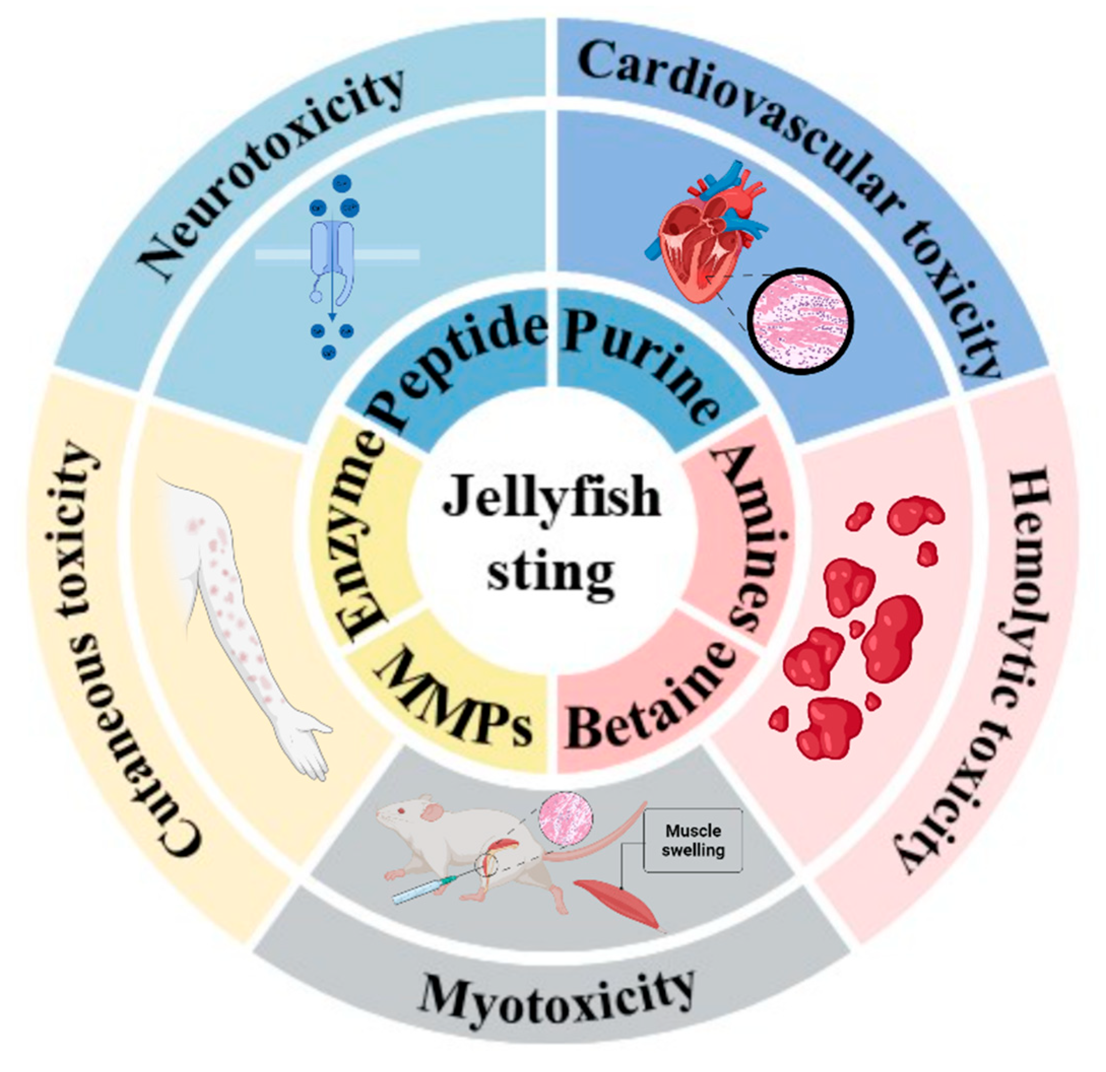
Figure 1. Schematic diagram of major toxic components and pathogenic effects of jellyfish venom. Jellyfish venom contains various proteins—including peptides, enzymes, and matrix metalloproteinases (MMPs)—and non-protein compounds such as purines, biogenic amines, and betaines. These toxins can induce multiple toxic effects, including cardiovascular toxicity, hemolytic toxicity, dermal toxicity, neurotoxicity, and myotoxicity. Created with BioRender.com.
Marinedrugs 23 00369 g001
Figure 2. Nematocyst discharge characterization: (A) Ultrastructural and minicollagen distribution analyses of Hydra nematocytes (stinging cells). Scale bars: 1 µm in (a,d); 5 µm in (b,c) [32]. (B) Schematic mechanism. Each nematocyte (blue) harbors one cyst (pink) with stylets that punch a hole into prey. (C) Framing-mode sequence @1,430,000 fps (Hamamatsu C4187); arrows indicate progress of discharge [33].
Marinedrugs 23 00369 g002
Figure 3. Pathological and ultrastructural changes in rat heart following TOE administration (5 mg/kg i.v., 60 min). (A,B) Hematoxylin-eosin (HE) staining of cardiac tissue (400× magnification): (A) Normal cardiac tissue architecture in control group. (B) Severe hemorrhage observed in cardiac tissue after TOE administration. (C,D) Transmission electron microscopy (TEM) of rat cardiomyocytes (scale bars: 1 μm): (C) Normal ultrastructure of control cardiomyocyte, showing intact sarcomeres with distinct Z-lines, and mitochondria with well-defined cristae. (D) TOE-treated group exhibiting disrupted and dissolved sarcomeric filaments and partial mitochondrial swelling, while mitochondrial cristae remained visible [104].
Marinedrugs 23 00369 g003
Figure 4. (A) Oxymatrine antagonizes oxidative stress and apoptosis in N.nomurai toxin-induced cardiotoxicity by inhibiting mitogen-activated protein kinase. ** p < 0.01, *** p < 0.001, **** p < 0.001, ns means no significance [115]. (B) Baicalein reduces mortality in mice and alleviates cardiomyocyte injury induced by R.esculentum toxin through regulation of the ROS/MAPK/NF-κB pathway. (a) LD50 curve of TE (n = 12). (b) Pathological section examination (n = 3). The scale bar in the reactive oxygen spices fluorescence imaging represents 100 μm. ** p < 0.01, *** p < 0.001, ns means no significance [116].
Marinedrugs 23 00369 g004
Figure 5. (A) Sika deer antler protein antagonizes the inflammatory response and oxidative damage induced by jellyfish venom [126]. **** p < 0.001. (B) Fucoidan alleviated NnNV-induced skin inflammatory stimulation effect in mice. (a) Graphical depiction of the inflammatory effects of NnNV. (b,c) Protein expression levels of inflammatory factors (IL-6, TNF-α, and MCP-1) and oxidative stress indicators in mouse blood (3 mg/mL). * p < 0.05, ** p < 0.01, *** p < 0.001, ns means no significance. (d) Pathological section of dermal toxicity. The scale bars of histopathological section are 100 μm. Red arrowheads indicate deepening of epidermal staining. Black arrowheads indicate damaged subcutaneous muscle tissue [16].
Marinedrugs 23 00369 g005
Disclaimer/Publisher’s Note: The statements, opinions and data contained in all publications are solely those of the individual author(s) and contributor(s) and not of MDPI and/or the editor(s). MDPI and/or the editor(s) disclaim responsibility for any injury to people or property resulting from any ideas, methods, instructions or products referred to in the content.