Article

Inhibition of Soluble Epoxide Hydrolase by Cembranoid Diterpenes from Soft Coral Sinularia maxima: Enzyme Kinetics, Molecular Docking, and Molecular Dynamics

1
Department of Biology Education, Teachers College and Institute for Phylogenomics and Evolution, Kyungpook National University, Daegu 41566, Republic of Korea
2
Institute of Marine Biochemistry, Vietnam Academy of Science and Technology (VAST), 18 Hoang Quoc Viet, Cau Giay, Hanoi 10072, Vietnam
3
Faculty of Pharmaceutical Chemistry and Technology, Hanoi University of Pharmacy, 13–15 Le Thanh Tong, Hoan Kiem, Hanoi 11021, Vietnam
*
Author to whom correspondence should be addressed.
Mar. Drugs 2024, 22(8), 373; https://doi.org/10.3390/md22080373
Submission received: 16 July 2024 / Revised: 16 August 2024 / Accepted: 16 August 2024 / Published: 17 August 2024
(This article belongs to the Special Issue Marine Drug Discovery through Molecular Docking)

Abstract

:
Soluble epoxide hydrolase (sEH) is essential for converting epoxy fatty acids, such as epoxyeicosatrienoic acids (EETs), into their dihydroxy forms. EETs play a crucial role in regulating blood pressure, mediating anti-inflammatory responses, and modulating pain, making sEH a key target for therapeutic interventions. Current research is increasingly focused on identifying sEH inhibitors from natural sources, particularly marine environments, which are rich in bioactive compounds due to their unique metabolic adaptations. In this study, the sEH inhibitory activities of ten cembranoid diterpenes (110) isolated from the soft coral Sinularia maxima were evaluated. Among them, compounds 3 and 9 exhibited considerable sEH inhibition, with IC50 values of 70.68 μM and 78.83 μM, respectively. Enzyme kinetics analysis revealed that these two active compounds inhibit sEH through a non-competitive mode. Additionally, in silico approaches, including molecular docking and molecular dynamics simulations, confirmed their stability and interactions with sEH, highlighting their potential as natural therapeutic agents for managing cardiovascular and inflammatory diseases.

1. Introduction

Soluble epoxide hydrolase (sEH) is a crucial enzyme that metabolizes epoxy fatty acids, such as epoxyeicosatrienoic acids (EETs), into their corresponding diols, dihydroxyeicosatrienoic acids (DHETs) [1,2]. EETs are important signaling molecules derived from arachidonic acid and play significant roles in various physiological processes, including blood pressure regulation, anti-inflammatory response, and pain modulation [3]. The role of sEH in converting EETs into less active DHETs positions it as a compelling target for therapeutic interventions [4]. sEH is predominantly expressed in the liver, kidneys, brain, and endothelium, where it influences several vital functions [5]. Given its significant role in inflammation, pain regulation, and vascular homeostasis maintenance, inhibiting sEH can elevate epoxy fatty acid levels, leading to considerable therapeutic benefits in treating conditions such as cardiovascular diseases, hypertension, and inflammatory diseases [6,7].
In recent years, there has been growing interest in identifying sEH inhibitors from natural sources, particularly marine environments [8]. Marine organisms represent a rich reservoir of bioactive compounds due to their unique metabolic pathways, which have evolved to adapt to the extreme conditions of their habitats [9]. Notably, marine-derived secondary metabolites exhibit promising sEH inhibitory activities. For instance, specific peptides and protein hydrolysates derived from marine fish have been studied for their sEH inhibitory properties, demonstrating the potential of these substances in developing nutraceuticals and pharmaceuticals targeting sEH [10,11]. Marine sponges and their associated microorganisms have also been explored for their capacity to produce sEH inhibitory compounds. These marine organisms produce secondary metabolites with diverse chemical structures, some of which exhibit significant sEH inhibitory effects [12].
Several classes of compounds have been identified as potent sEH inhibitors. Among these, terpenoids have garnered significant attention. For example, 2-oxopomolic acid, a triterpenoid isolated from Malus domestica, has demonstrated sEH inhibitory activity with an IC50 value of 11.4 ± 2.7 μM [13]. In addition, two triterpenoids with a protostane-type skeleton, 3β-hydroxy-25-anhydro-alisol F and 3β-hydroxy-alisol G, isolated from Alisma orientale, exhibited potential sEH inhibition with IC50 values of 10.06 ± 1.12 μM and 30.45 ± 2.65 μM, respectively [14]. Other protostane-type triterpenoids, such as 11-deoxy-25-anhydro alisol E and 11-deoxy alisol B, have shown sEH inhibitory effects with IC50 values of 5.94 ± 0.54 μM and 3.40 ± 0.57 μM, respectively [15]. These findings underscore the diversity of sEH inhibitors and emphasize the significant potential of natural products, particularly terpenoids, in this area of research.
The genus Sinularia comprises a diverse group of soft corals belonging to the family Alcyoniidae, within the class Octocorallia of the phylum Cnidaria [16,17]. Commonly referred to as soft corals [18], Sinularia species are characterized by their flexible structures and are typically found in shallow tropical marine environments [17,19,20]. The genus Sinularia includes over 90 described species, each exhibiting distinct morphological and chemical characteristics [20,21]. These corals serve as integral components of coral reef ecosystems, providing habitat and protection for various marine organisms. Additionally, they produce a wide range of secondary metabolites, particularly diterpenoids, which play crucial roles in chemical defense against predators and pathogens [20,21].
One notable species within this genus is Sinularia maxima, which has garnered attention for its distinctive chemical composition and ecological significance. S. maxima produces several bioactive compounds, including terpenoids such as 11β-acetoxypukalide and pukalide [22]. These compounds are not only crucial for the coral’s defense mechanisms but also have potential applications in pharmacology due to their biological activities. Studies have shown that extracts from S. maxima can deter feeding by reef fishes, highlighting the importance of these metabolites in the coral’s natural defense strategies [23]. Additionally, the chemical profile of S. maxima varies temporally, potentially influencing its susceptibility to predation and environmental stressors [23,24]. While cembranoid diterpenes from Sinularia species have not been previously reported as sEH inhibitors, the exploration of these compounds could reveal potential sEH inhibitory activities, enhancing our understanding of natural sEH inhibitors.
As part of our ongoing research to identify active compounds from the soft coral S. maxima [16,25], we recently reported the isolation, structural elucidation, and anti-inflammatory activities of 10 cembranoid diterpenes, including sinumaximols A–H (18), sethukarailin (9), and 5-epinorcembrene (10) (Figure 1). The current study offers new insights into the inhibitory activities and mechanisms of these diterpenes against sEH through a combination of in vitro assays and in silico simulations, including molecular docking and molecular dynamics (MD) studies.

2. Results

2.1. sEH Inhibitory Effects of the Isolated Cembranoid Diterpenes 110

The inhibitory effects of cembranoid diterpenes (110) on recombinant human sEH were evaluated using 3-phenyl-cyano(6-methoxy-2-naphthalenyl)methyl ester-2-oxiraneacetic acid (PHOME) as a substrate, with AUDA serving as a positive control (14.69 ± 0.07 nM) [26,27,28]. IC50 values indicate the concentration of each compound required to inhibit 50% of the enzyme (sEH) activity. The data revealed that most compounds, including 1, 2, 4, 5, 6, 7, 8, and 10, exhibit IC50 values greater than 100 μM, suggesting that they are weak inhibitors or ineffective against sEH under the tested conditions (Table 1).
In contrast, compound 3, which contains an additional methoxy group at C-3 compared to compounds 1 and 2, demonstrated a marked inhibitory effect against sEH, with an IC50 value of 70.68 ± 1.44 μM. The presence of multiple polar groups in compound 3 may enhance its binding affinity through hydrogen bonding and electrostatic interactions. Similarly, compound 9, which also features a methoxy group at C-3, exhibited sEH inhibition with an IC50 value of 78.83 ± 2.26 μM. These findings suggest that the methoxy group at C-3 plays a critical role in binding to sEH.
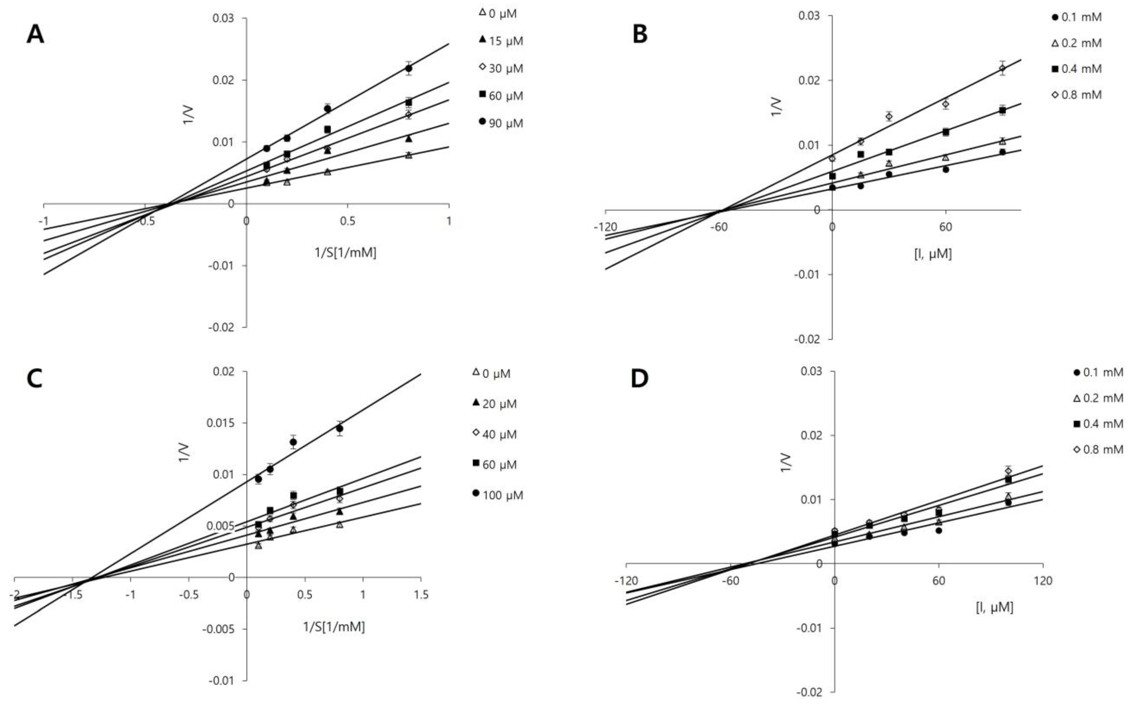
The inhibition type of a compound provides insight into how it interacts with the enzyme. To further investigate the inhibition types of compounds 3 and 9, the most potent compounds identified in this study, enzyme kinetics were analyzed using Lineweaver-Burk [29] and Dixon plots [30,31]. PHOME concentrations of 0.1, 0.2, 0.4, and 0.8 mM were used, along with compound 3 concentrations of 0, 15, 30, 60, and 90 µM, and compound 9 concentrations of 0, 20, 40, 60, and 100 µM. In the Lineweaver–Burk method, competitive and non-competitive inhibition is indicated by a series of straight lines intersecting at the same point on the y- and x-axes, respectively. As shown in Figure 2A,C, both compounds 3 and 9 exhibit non-competitive inhibition. In most cases, this implies that these inhibitors bind to an allosteric site on the enzyme rather than the active site, thereby preventing the enzyme from functioning regardless of the substrate concentration [32].
The inhibition constant (Ki) is a measure of the binding affinity of the inhibitor for the enzyme, with a lower value indicating a stronger affinity. Dixon plots were used to determine the Ki values for the compounds. Compounds 3 and 9 exhibited Ki values of 59.44 μM and 55.03 μM, respectively (Figure 2B,D).

2.2. Molecular Docking

Molecular docking simulations were performed using AutoDock 4.2.6 [33] to investigate the binding interactions between sEH and compounds 3 and 9. The structure of human sEH (entry ID: 3ANS) was obtained from the RCSB PDB database [34].
Human sEH consists of two distinct domains with separate functions: the N-terminal domain exhibits phosphatase activity, while the C-terminal domain, which encompasses both the core and cap domains, is responsible for hydrolase activity [35,36,37]. The active site of human sEH, an L-shaped hydrophobic pocket located within the protein’s core, features a narrow constriction that houses the catalytic triad (Asp335, Asp496, and His524) and stabilizing residues (Tyr383 and Tyr466) [38,39,40,41]. Additionally, the sEH enzyme possesses important allosteric sites that can be targeted for inhibition. These allosteric sites are situated outside the active site, in regions around the amino acid residues Cys423 and Cys522 [42,43]. Previous studies have demonstrated that non-competitive inhibitors can bind to these allosteric sites, disrupting enzyme function without directly competing with the substrate at the active site [42,44].
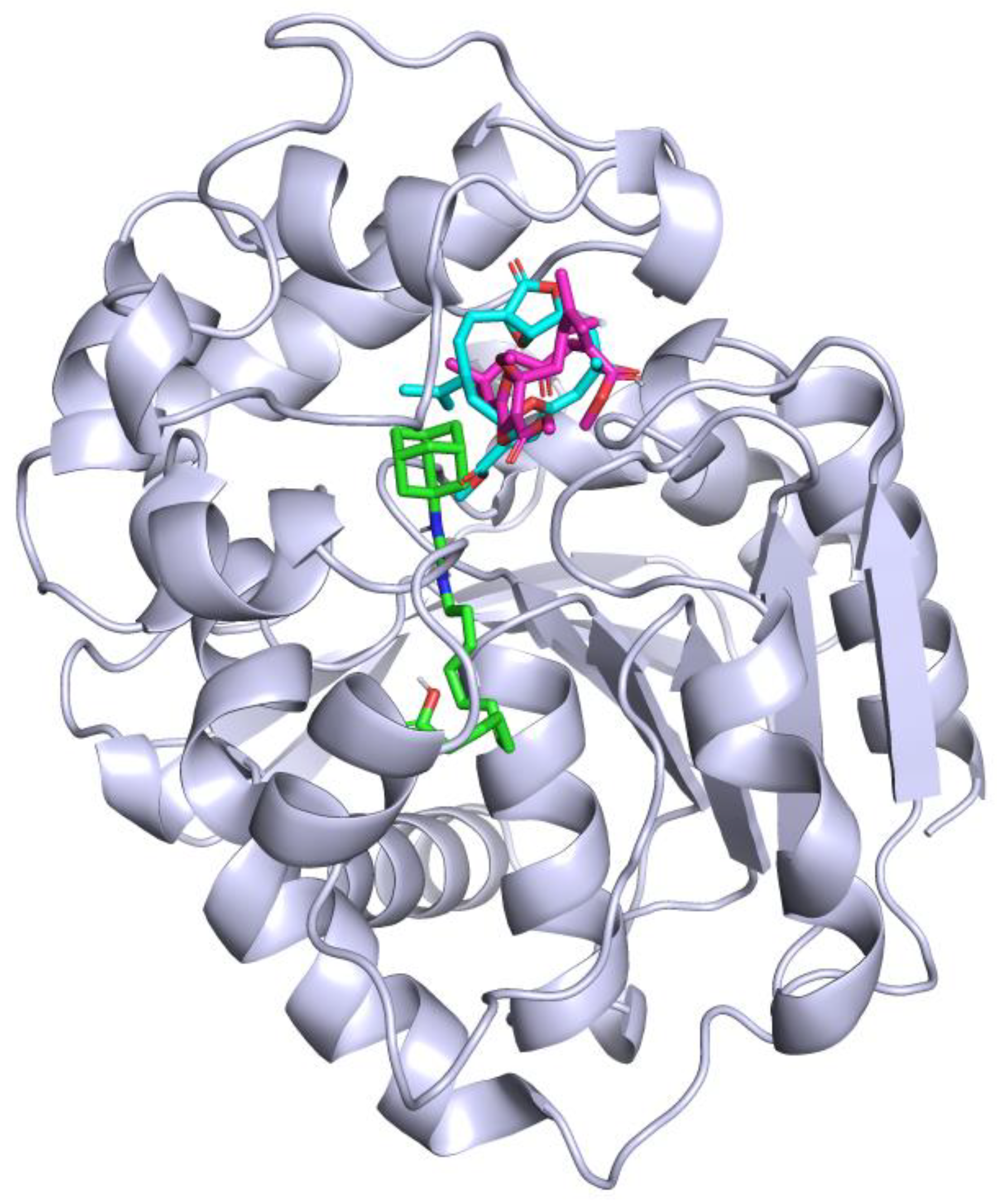
To optimize and validate the docking simulations, the positive control (AUDA), a known sEH inhibitor [45,46], was docked into the enzyme as a native ligand, revealing the active site. Subsequently, compounds 3 and 9 were individually docked into sEH under identical conditions. The top docking poses for each compound were clustered based on their binding energies. The primary docking site was selected from the cluster with the lowest binding energy, and alternative clusters with higher binding energies were ruled out. The binding sites of AUDA and compounds 3 and 9 on the sEH enzyme are illustrated in Figure 3.
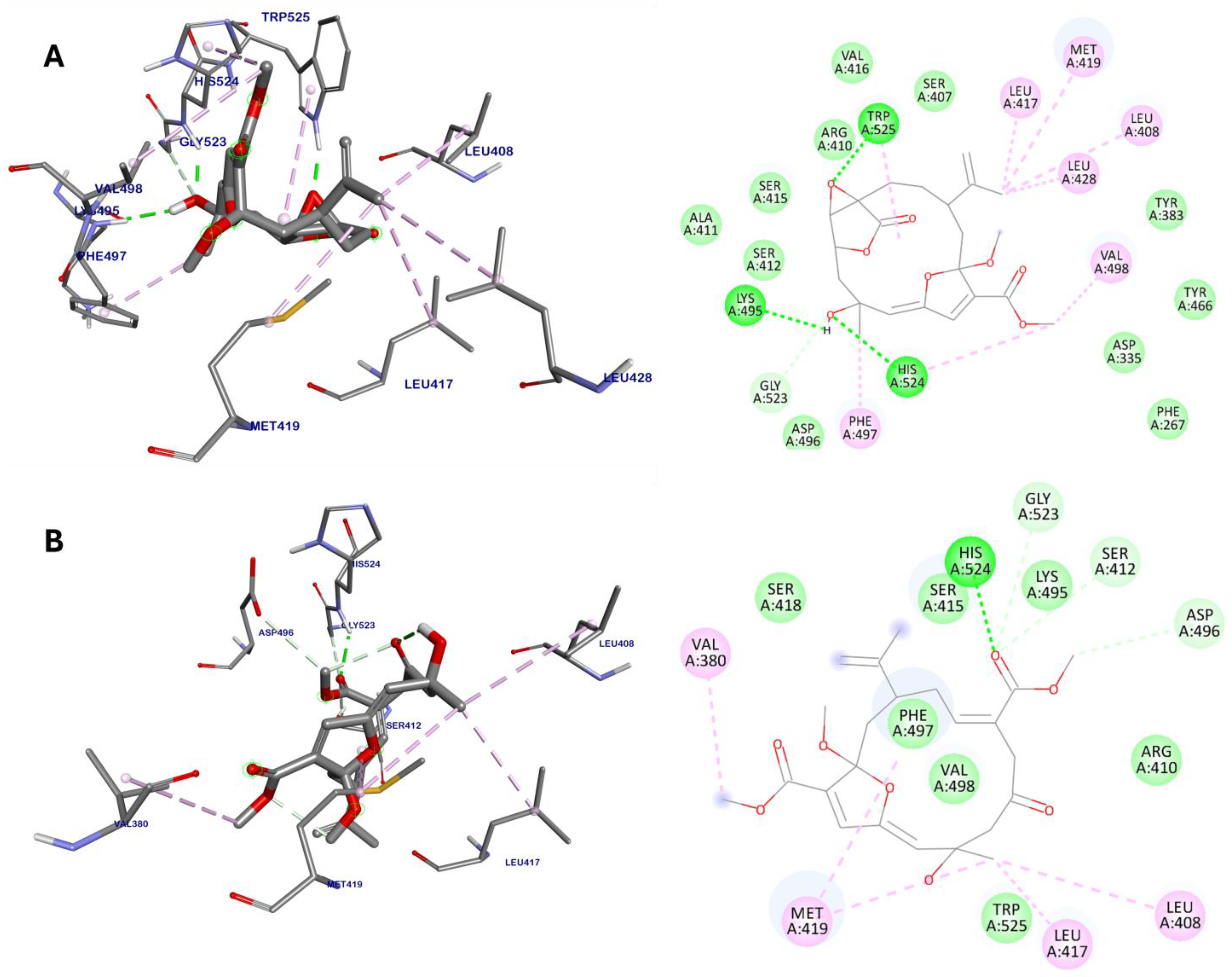
The docking results revealed that compound 3 exhibited a notably lower binding energy of −9.59 kcal/mol (Table 2), suggesting a strong interaction with sEH outside its active site, which may effectively disrupt enzyme function. Compound 3 formed hydrogen bonds with Lys495, Gly523, His524, and Trp525, and engaged in extensive van der Waals interactions with several residues, including Phe267, Asp335, Tyr383, Ser407, Arg410, Ala411, Ser412, Ser415, Val416, Tyr466, Asp496 (Figure 4A). Additionally, it established hydrophobic interactions with Leu408, Leu417, Met419, Leu428, Phe497, Val498, His524, and Trp525. These interactions strategically align with the hydrophobic and catalytic regions adjacent to the allosteric sites of sEH, which are essential for modulating enzyme activity and substrate binding.
On the other hand, compound 9 exhibited a slightly weaker binding energy of −8.94 kcal/mol compared to compound 3. It demonstrated interactions with residues Ser412, Asp496, Gly523, and His524 through hydrogen bonds (Figure 4B). Additionally, it engaged in van der Waals interactions with residues Arg410, Ser415, Ser418, Lys495, Phe497, Val498, and Trp525. Moreover, compound 9 participated in alkyl interactions with Val380, Leu408, Leu417, and Met419. Overall, both compounds 3 and 9 exhibited similar interaction profiles, targeting key residues involved in the allosteric regulation of sEH activity. However, the differences in binding energies and specific interactions indicate that compound 3 is the more potent inhibitor, aligning with the outcomes of the in vitro studies.
Interestingly, although the methoxy group at position C-3 of the cembranoid diterpene structures was identified as an important feature for sEH inhibition in vitro, no direct interactions between the methoxy group and the sEH protein were observed for either compound 3 or compound 9. This finding suggests that while the methoxy group may be important for enzyme inhibition, its role might be more indirect, potentially influencing the overall binding affinity or conformation of the compounds rather than directly interacting with the sEH allosteric site.

2.3. Molecular Dynamics

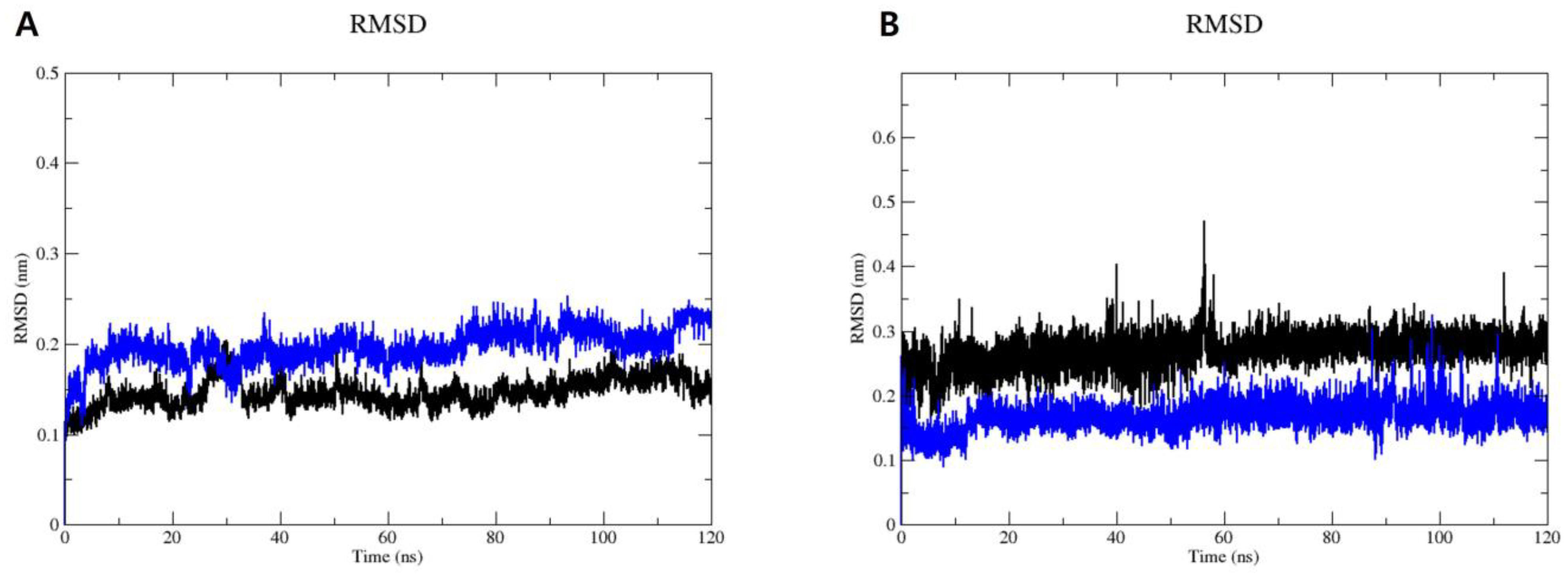
To analyze the dynamic behavior of sEH in complexes with compounds 3 and 9, MD simulations were conducted for 120 ns using GROMACS 2022.1 software. The root mean square deviation (RMSD) and root mean square fluctuation (RMSF) plots (Figure 5) provided further insights into the stability and flexibility of the systems. The RMSD plot of the protein backbone (Figure 5A) showed that the RMSD values for both systems fluctuated between 0.10 nm and 0.20 nm during the initial 40 ns of the MD simulations. Subsequently, both complexes maintained RMSD values of approximately 0.15 nm for the sEH–compound 3 complex and 0.20 nm for the sEH–compound 9 system until the end of the simulations. Following this, the RMSD of ligands after least-squares fitting to the protein backbone was assessed to better understand ligand binding and its impact on protein structure (Figure 5B). Throughout 120 ns of the MD simulations, compound 3 displayed an RMSD of approximately 0.23 nm, while compound 9 exhibited a slightly lower RMSD of around 0.16 nm. The low RMSD values observed for both compounds suggest that they maintain a consistent binding pose with minimal fluctuations.
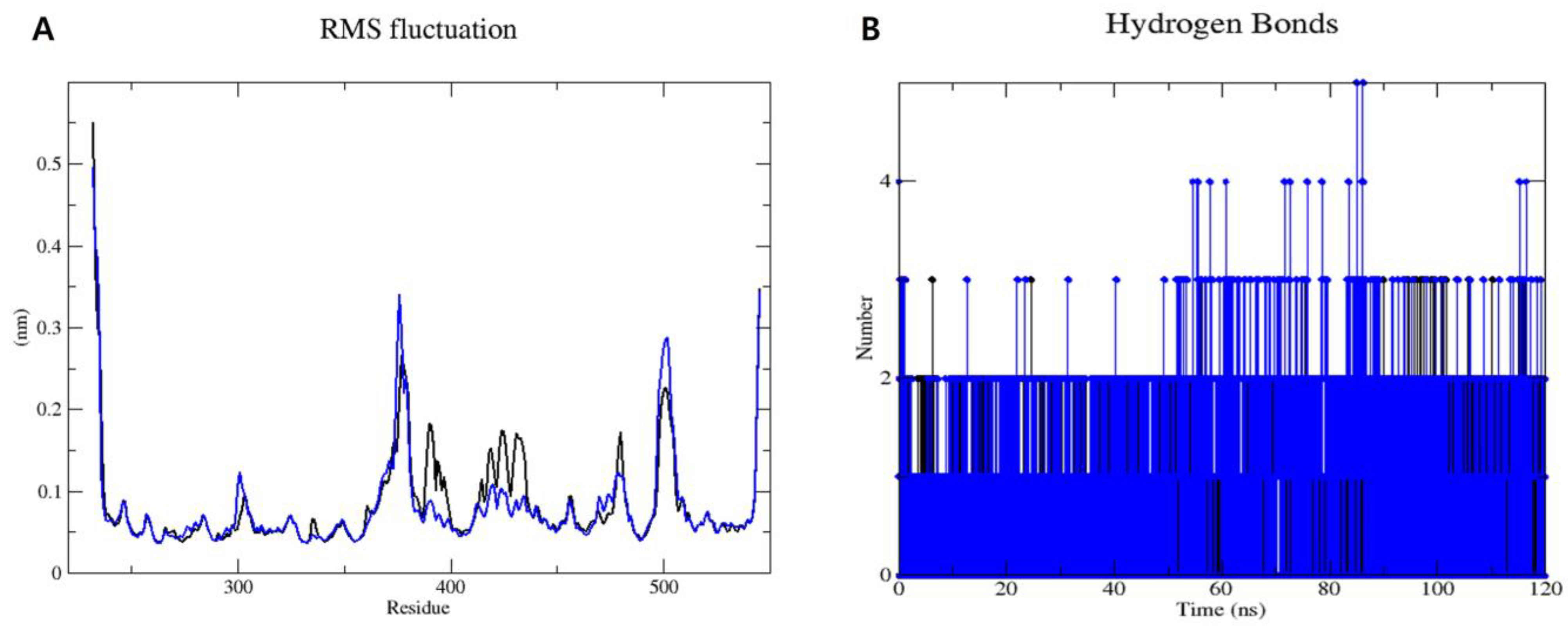
Furthermore, the RMSF plot (Figure 6A) displayed consistent flexibility patterns among individual residues in both systems. Notably, most residues exhibited fluctuations below 0.2 nm, indicating a high level of stability throughout the simulations. However, higher fluctuations were observed at the beginning and end of the MD simulation periods. These variations can be attributed to specific residues experiencing fewer constraints within the sEH–compound 3 and sEH–compound 9 complexes, resulting in increased flexibility. Importantly, these flexible residues are located far from the binding sites of compounds 3 and 9 on sEH. Therefore, while these residues exhibit greater movement, they do not impact the binding interactions between sEH and compounds 3 and 9.
Hydrogen bonding plays a crucial role in stabilizing protein-ligand complexes [47]. Figure 6B illustrates the hydrogen bonds formed between sEH and compounds 3 and 9 over a 120 ns MD trajectory. Both compounds 3 and 9 formed 2 to 5 hydrogen bonds with the sEH enzyme.
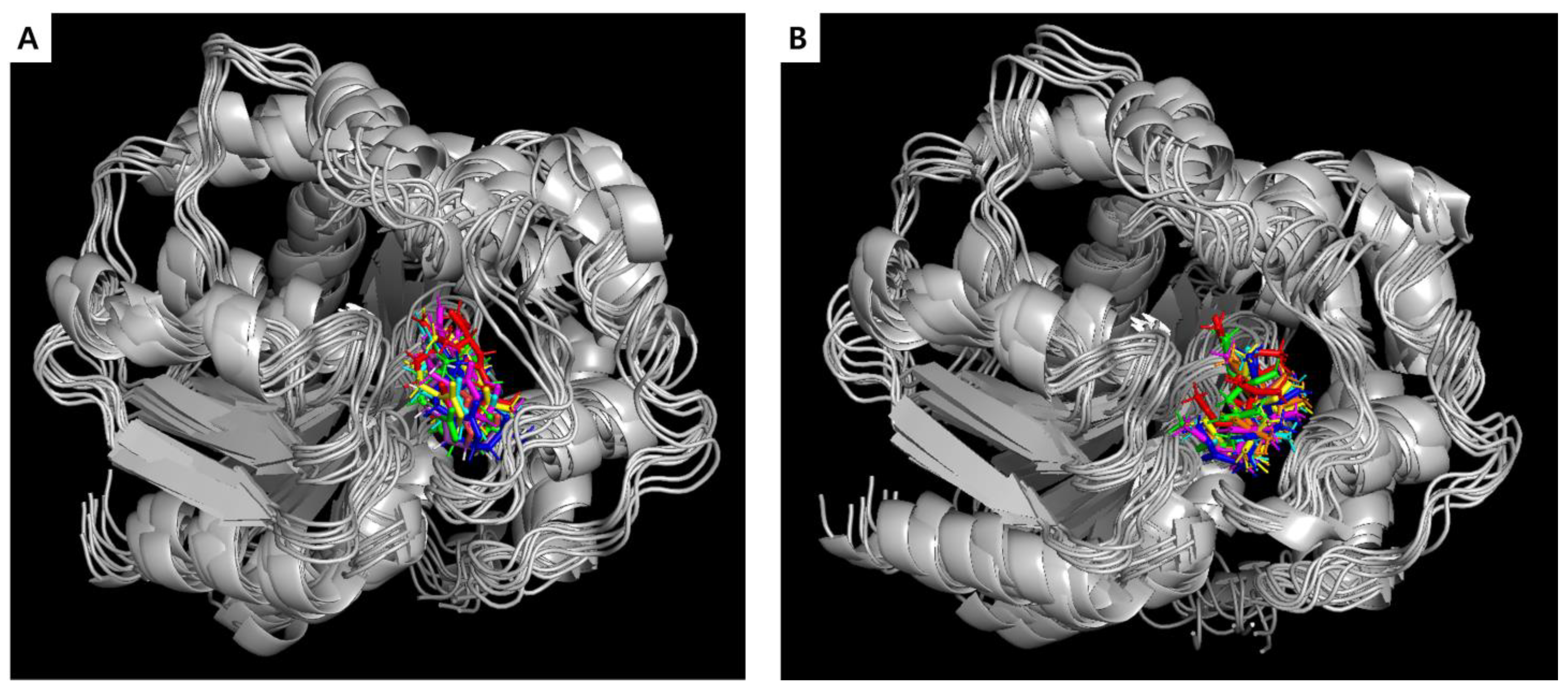
The superposition of compounds 3 and 9 within the binding site of sEH was monitored every 20 ns; the results are presented in Figure 7. Throughout the 120 ns MD simulation, there were no significant alterations in the binding sites of these two compounds within the target enzyme sEH, indicating a stable interaction between sEH and the active compounds 3 and 9. Comparison of these results with those from the docking studies revealed that the MD simulation consistently showed a similar binding mode throughout the 120 ns trajectory.

3. Discussion

Marine-derived compounds, renowned for their distinctive chemical structure and rich bioactivity, are increasingly the focus of research on enzyme inhibition [48,49,50]. Notably, soft corals, such as S. maxima, produce a wide array of secondary metabolites, particularly cembranoid diterpenes, which are well-known for their biological activities and potential pharmacological applications [20,51]. Our previous studies have described the isolation and structural elucidation of cembranoid diterpenes from S. maxima, including sinumaximols A–H (18), sethukarailin (9), and 5-epinorcembrene (10) [16,25]. These compounds have also been reported to possess potential anti-inflammatory activity. In this study, we evaluated the sEH inhibitory activities of these compounds. Notably, in vitro assays revealed that compounds 3 and 9 exhibited IC50 values of 70.68 ± 1.44 μM and 78.83 ± 2.26 μM, respectively. While these values are higher compared to some well-established sEH inhibitors [13,14,15], they still highlight the potential of compounds 3 and 9 as sEH inhibitors, especially considering their unique binding sites. Enzyme kinetics revealed that these compounds exhibit non-competitive inhibition of sEH, indicating they bind to allosteric sites distinct from the enzyme’s active site [52,53].
Virtual screening, employing molecular docking and MD simulations, is a potent strategy for the discovery and development of sEH inhibitors [54]. Molecular docking identifies potential inhibitors by assessing their binding energies and interaction patterns within binding sites [55]. In this study, molecular docking simulations were conducted to investigate the binding interactions between sEH and compounds 3 and 9. The docking studies revealed that compounds 3 and 9 exhibited notably lower binding energies of −9.18 and −8.94 kcal/mol, respectively. Unlike well-known sEH inhibitors like AUDA, which target the active site [46], compounds 3 and 9 showed interactions with key residues at the allosteric sites of sEH. Compound 3 interacted with Lys495, Gly523, His524, and Trp525 through hydrogen bonds and engaged in van der Waals interactions with a range of residues, such as Phe267, Asp335, Tyr383, Ser407, Arg410, Ala411, Ser412, Ser415, Val416, Tyr466, and Asp496. Additionally, it formed hydrophobic interactions with Leu408, Leu417, Met419, Leu428, Phe497, Val498, His524, and Trp525. Similar interactions with key amino acid residues in the allosteric sites were also observed in the complex between compound 9 and sEH. These findings suggest that compounds 3 and 9 may inhibit sEH through a new mechanism, distinct from that of active site-binding inhibitors, potentially offering unique therapeutic benefits.
The active compounds 3 and 9, which showed promising interactions, were further validated through MD simulations using GROMACS 2022.1 software [56] to assess their stability and conformational changes over a 120 ns period. The simulations aimed to analyze the dynamic behaviors and conformational stabilities of compounds 3 and 9 within the sEH binding site. Throughout the 120 ns simulation, both compounds exhibited minor conformational changes, as indicated by the RMSD plot. Moreover, the simulations confirmed the consistent binding modes of compounds 3 and 9 for sEH, as depicted in the RMSF plot and hydrogen bonding analysis over the simulation trajectory. Throughout the simulation period, both complexes remained robustly bound, with hydrogen bonds and van der Waals interactions contributing to the observed stability.
Investigating diterpenoids as sEH inhibitors is a relatively new approach. While other chemical classes have been extensively explored for sEH inhibition [1,27,57,58,59], marine-derived diterpenoids, particularly cembranoid diterpenes from Sinularia maxima, have not been studied in this context before. Our study is the first to evaluate the sEH inhibitory activity of these cembranoid diterpenes. The findings suggest that these compounds offer a new perspective on sEH inhibition, potentially expanding the collection of natural products with therapeutic applications.
In conclusion, these results underscore compounds 3 and 9 from S. maxima as promising sEH inhibitors, demonstrating stable interactions with the enzyme over prolonged simulations. The combination of molecular docking, MD simulations, and in vitro biochemical assays, including sEH inhibition and enzyme kinetics analysis, offers comprehensive insights into the molecular mechanisms underlying their inhibitory activity against sEH. This integrative approach provides a robust framework for the identification and characterization of marine-derived compounds with potential therapeutic applications in cardiovascular and inflammatory diseases.
However, to further substantiate these findings, additional in vivo experiments are essential to validate the inhibitory effects of compounds 3 and 9 in a physiological context. Such experimental evaluations are crucial for confirming their efficacy and safety, extremely facilitating the development and optimization of these marine-derived compounds for therapeutic use.

4. Materials and Methods

4.1. Biological Material

The soft coral S. maxima was collected from Nha Trang, Khanh Hoa, Vietnam, in November 2010 and authenticated by Professor Do Cong Thung (Institute of Marine Environment and Resources, VAST). The voucher specimen of S. maxima (accession code: SM112010_01) was deposited at the Department of Marine Medicinal Materials, Institute of Marine Biochemistry, VAST.

4.2. Chemicals and Reagents

sEH (human, recombinant; 10011669), PHOME (10009134), and AUDA (10007927) were purchased from Cayman Chemical (Ann Arbor, MI, USA). Bis-Tris (B9754) was purchased from Sigma-Aldrich (St. Louis, MO, USA).

4.3. sEH Inhibition Assay

The sEH inhibition assay was performed as described in a previous study [36], with slight modifications. In 96-well plates, separate mixtures were prepared for each test compound, containing 130 µL of sEH in Bis-Tris buffer with HCl (pH 7.0, containing 0.1% bovine serum albumin) and 20 µL of the test compound (10 mM, diluted in DMSO). Subsequently, 50 µL of PHOME, which serves as a substrate, was added to each well. The mixtures were then incubated at 37 °C, and fluorescence was monitored using the VICTOR Nivo multimode plate reader (PerkinElmer, Waltham, MA, USA) for 60 min, with excitation and emission wavelengths of 330 nm and 460 nm, respectively. AUDA, an sEH inhibitor, was used as the positive control. The inhibitory activity was calculated using the following equation:
Inhibitory activity (%) = [1 − (ΔI/ΔC)] × 100%,
where ΔI and ΔC are the fluorescence readings of the inhibitor and the control after 60 min, respectively. IC50 values were calculated using SigmaPlot 10.0 (Systat Software, CA, USA). Data are presented as means ± standard deviations from three independent experiments (n = 3, p < 0.05).
Enzyme kinetics were analyzed using Lineweaver–Burk and Dixon plots [29,30]. This approach helped determine the mode of inhibition and the inhibition constant (Ki) at various substrate concentrations, both in the absence (0 μM) and presence of inhibitors. Data were plotted using Microsoft Excel 365.

4.4. Molecular Docking Simulation

Molecular docking simulations were conducted to investigate the interactions between ligand molecules and the sEH enzyme. AutoDock 4.2 (The Scripps Research Institute, La Jolla, CA, USA) [33] was employed for these simulations, following the developer’s protocol [60]. The structure of human sEH (PDB ID: 3ANS) was retrieved from the RCSB Protein Data Bank (resolution: 1.98 Å) [34]. Protein preparation, which involved adding Kollman charges (−7.0) and exporting the structure to a “.pdbqt” file, was performed using AutoDockTools 1.5.6. The 3D structures of the test compounds were constructed using Spartan’24 (Wavefunction, CA, USA).
Due to the flexibility of the macrocycles (compounds 3 and 9), a conformational analysis was conducted to identify the most stable conformers before introducing them into the docking process. First, the conformer distribution was analyzed using Spartan’24 software with the MMFF force field and an energy cutoff of 5 kcal/mol. The obtained conformers were further optimized using the PM6 semi-empirical method, followed by the density functional theory (DFT) method at the B3LYP/6-31G(d) level in the vacuum phase (Supplementary Material). The most stable conformers were then assigned Gasteiger charges (0.0001 for compound 3 and −0.0002 for compound 9) and exported in “.pdbqt” format for subsequent docking simulations using AutoDockTools 1.5.6.
Blind docking techniques were applied with the following parameters: grid box dimensions (x = 96, y = 96, z = 124) and grid position (x = 27.058, y = 28.677, z = 108.904). Protein-ligand interactions were investigated using a molecular docking approach with an exhaustiveness value of 24 and a maximum of 1000 binding modes. The resulting docking poses and interaction diagrams were analyzed and visualized using BIOVIA Discovery Studio Visualizer 21.1 (Dassault Systèmes, San Francisco, CA, USA) and PyMOL 2.5.4 (Schrödinger, New York, NY, USA).

4.5. Molecular Dynamics Simulation

MD simulations were conducted using GROMACS 2022.1 [56] with the CHARMM36 force field [61], following previously described procedures [62,63]. The system setup included periodic boundary conditions and explicit solvation in a cubic box with TIP3P water molecules. Sodium (Na+) and/or chloride (Cl) ions were added for stability. Energy minimization was performed with a maximum force threshold of 10 kJ/mol. NVT equilibration was conducted at 300 K, followed by NPT ensemble relaxation at 1.013 bar. MD simulations were run for 120 ns, and trajectory data were analyzed using PyMOL 2.5.4 and Grace software version 5.1.22 (https://plasma-gate.weizmann.ac.il/Grace/, accessed on 10 July 2024).

Supplementary Materials

The following supporting information can be downloaded at: https://www.mdpi.com/article/10.3390/md22080373/s1, Figure S1: The most stable conformers of compound 3 calculated at B3LYP/6-31G(d) level; Figure S2: The most stable conformers of compound 9 calculated at B3LYP/6-31G(d) level; Figure S3: Energy profiles, total energy, and potential energy plots for the sEH complexes with compounds 3 and 9; Table S1: Electronic energies and Boltzmann populations for the most stable conformers of compound 3; Table S2: Electronic energies and Boltzmann populations for the most stable conformers of compound 9.

Author Contributions

Conceptualization, S.Y.Y.; methodology, S.Y.Y.; formal analysis, N.V.P.; investigation, N.V.P., N.P.T., L.B.V. and B.T.T.L.; resources, C.V.M.; writing—original draft preparation, N.V.P.; writing—review and editing, S.Y.Y.; supervision, S.Y.Y. All authors have read and agreed to the published version of the manuscript.

Funding

This research was supported by the Global—Learning & Academic research institution for Master’s·PhD students, and Postdocs(LAMP) Program of the National Research Foundation of Korea (NRF) grant funded by the Ministry of Education (No. RS-2023-00301914).

Institutional Review Board Statement

Not applicable.

Data Availability Statement

Data that support the findings of this study are available in the Article.

Conflicts of Interest

The authors declare no conflicts of interest.

References

  1. Kitamura, S.; Morisseau, C.; Inceoglu, B.; Kamita, S.G.; De Nicola, G.R.; Nyegue, M.; Hammock, B.D. Potent natural soluble epoxide hydrolase inhibitors from Pentadiplandra brazzeana Baillon: Synthesis, quantification, and measurement of biological activities in vitro and in vivo. PLoS ONE 2015, 10, e0117438. [Google Scholar] [CrossRef] [PubMed]
  2. Shan, J.; Hashimoto, K. Soluble epoxide hydrolase as a therapeutic target for neuropsychiatric disorders. Int. J. Mol. Sci. 2022, 23, 4951. [Google Scholar] [CrossRef] [PubMed]
  3. Li, N.; Liu, J.-Y.; Qiu, H.; Harris, T.R.; Sirish, P.; Hammock, B.D.; Chiamvimonvat, N. Use of metabolomic profiling in the study of arachidonic acid metabolism in cardiovascular disease. Congest. Heart Fail. 2011, 17, 42–46. [Google Scholar] [CrossRef]
  4. Zarriello, S.; Tuazon, J.P.; Corey, S.; Schimmel, S.; Rajani, M.; Gorsky, A.; Incontri, D.; Hammock, B.D.; Borlongan, C.V. Humble beginnings with big goals: Small molecule soluble epoxide hydrolase inhibitors for treating CNS disorders. Prog. Neurobiol. 2019, 172, 23–39. [Google Scholar] [CrossRef] [PubMed]
  5. Sura, P.; Sura, R.; EnayetAllah, A.E.; Grant, D.F. Distribution and expression of soluble epoxide hydrolase in human brain. J. Histochem. Cytochem. 2008, 56, 551–559. [Google Scholar] [CrossRef]
  6. Lee, G.H.; Oh, S.J.; Lee, S.Y.; Lee, J.-Y.; Ma, J.Y.; Kim, Y.H.; Kim, S.K. Discovery of soluble epoxide hydrolase inhibitors from natural products. Food Chem. Toxicol. 2014, 64, 225–230. [Google Scholar] [CrossRef]
  7. Buscató, E.l.; Büttner, D.; Brüggerhoff, A.; Klingler, F.-M.; Weber, J.; Scholz, B.; Živković, A.; Marschalek, R.; Stark, H.; Steinhilber, D.; et al. From a multipotent stilbene to soluble epoxide hydrolase inhibitors with antiproliferative properties. ChemMedChem 2013, 8, 919–923. [Google Scholar] [CrossRef] [PubMed]
  8. Obeme-Nmom, J.I.; Udenigwe, C.C. Soluble epoxide hydrolase: An emerging target for nutraceuticals against inflammation and oxidative stress. Curr. Opin. Food Sci. 2024, 57, 101174. [Google Scholar] [CrossRef]
  9. Barzkar, N.; Sukhikh, S.; Babich, O. Study of marine microorganism metabolites: New resources for bioactive natural products. Front. Microbiol. 2024, 14, 1285902. [Google Scholar] [CrossRef]
  10. Mavrommatis, Y.; Ross, K.; Rucklidge, G.; Reid, M.; Duncan, G.; Gordon, M.-J.; Thies, F.; Sneddon, A.; de Roos, B. Intervention with fish oil, but not with docosahexaenoic acid, results in lower levels of hepatic soluble epoxide hydrolase with time in apoE knockout mice. Br. J. Nutr. 2010, 103, 16–24. [Google Scholar] [CrossRef]
  11. Harris, T.R.; Hammock, B.D. Soluble epoxide hydrolase: Gene structure, expression and deletion. Gene 2013, 526, 61–74. [Google Scholar] [CrossRef] [PubMed]
  12. Kulkarni, R.R.; Jo, A.R.; Kim, Y.H.; Na, M. Epi-leptosphaerin: A new L-isoascorbic acid derivative from marine sponges. Nat. Prod. Sci. 2015, 21, 293–296. [Google Scholar] [CrossRef]
  13. Cho, I.S.; Kim, J.H.; Lin, Y.; Su, X.D.; Kang, J.S.; Yang, S.Y.; Kim, Y.H. Inhibitory activity of quercetin 3-O-arabinofuranoside and 2-oxopomolic acid derived from Malus domestica on soluble epoxide hydrolase. Molecules 2020, 25, 4352. [Google Scholar] [CrossRef]
  14. Zhao, W.-Y.; Zhang, X.-Y.; Zhou, M.-R.; Tian, X.-G.; Lv, X.; Zhang, H.-L.; Deng, S.; Zhang, B.-J.; Sun, C.-P.; Ma, X.-C. Natural soluble epoxide hydrolase inhibitors from Alisma orientale and their potential mechanism with soluble epoxide hydrolase. Int. J. Biol. Macromol. 2021, 183, 811–817. [Google Scholar] [CrossRef]
  15. Sun, C.-P.; Zhang, J.; Zhao, W.-Y.; Yi, J.; Yan, J.-K.; Wang, Y.-L.; Morisseau, C.; Liu, Z.-B.; Hammock, B.D.; Ma, X.-C. Protostane-type triterpenoids as natural soluble epoxide hydrolase inhibitors: Inhibition potentials and molecular dynamics. Bioorg. Chem. 2020, 96, 103637. [Google Scholar] [CrossRef]
  16. Thao, N.P.; Nam, N.H.; Cuong, N.X.; Quang, T.H.; Tung, P.T.; Tai, B.H.; Luyen, B.T.T.; Chae, D.; Kim, S.; Koh, Y.-S.; et al. Diterpenoids from the soft coral Sinularia maxima and their inhibitory effects on lipopolysaccharide-stimulated production of pro-inflammatory cytokines in bone marrow-derived dendritic cells. Chem. Pharm. Bull. 2012, 60, 1581–1589. [Google Scholar] [CrossRef] [PubMed]
  17. Yan, X.; Liu, J.; Leng, X.; Ouyang, H. Chemical diversity and biological activity of secondary metabolites from soft coral genus Sinularia since 2013. Mar. Drugs 2021, 19, 335. [Google Scholar] [CrossRef]
  18. Tran, H.H.T.; Nguyen Viet, P.; Nguyen Van, T.; Tran, H.T.; Nguyen Xuan, C.; Nguyen Hoai, N.; Do Cong, T.; Phan Van, K.; Chau Van, M. Cytotoxic steroid derivatives from the Vietnamese soft coral Sinularia brassica. J. Asian Nat. Prod. Res. 2017, 19, 1183–1190. [Google Scholar] [CrossRef] [PubMed]
  19. Khalesi, M.K.; Beeftink, H.H.; Wijffels, R.H. Flow-dependent growth in the zooxanthellate soft coral Sinularia flexibilis. J. Exp. Mar. Biol. Ecol. 2007, 351, 106–113. [Google Scholar] [CrossRef]
  20. Chen, W.-T.; Li, Y.; Guo, Y.-W. Terpenoids of Sinularia soft corals: Chemistry and bioactivity. Acta Pharm. Sin. B 2012, 2, 227–237. [Google Scholar] [CrossRef]
  21. Elkhouly, B.H.; Attia, Z.E.; Khedr, I.M.A.; Samy, N.M.; Fouad, A.M. Recent updates on Sinularia soft coral. Mini-Rev. Med. Chem. 2022, 22, 1152–1196. [Google Scholar] [CrossRef] [PubMed]
  22. Slattery, M.; Starmer, J.; Paul, V. Temporal and spatial variation in defensive metabolites of the tropical Pacific soft corals Sinularia maxima and S. polydactyla. Mar. Biol. 2001, 138, 1183–1193. [Google Scholar]
  23. Slattery, M.; Paul, V.J. Indirect effects of bleaching on predator deterrence in the tropical Pacific soft coral Sinularia maxima. Mar. Ecol. Prog. Ser. 2008, 354, 169–179. [Google Scholar] [CrossRef]
  24. Wylie, C.R.; Paul, V.J. Chemical defenses in three species of Sinularia (Coelenterata, Alcyonacea): Effects against generalist predators and the butterflyfish Chaetodon unimaculatus Bloch. J. Exp. Mar. Biol. Ecol. 1989, 129, 141–160. [Google Scholar] [CrossRef]
  25. Thao, N.P.; Nam, N.H.; Cuong, N.X.; Quang, T.H.; Tung, P.T.; Dat, L.D.; Chae, D.; Kim, S.; Koh, Y.-S.; Kiem, P.V.; et al. Anti-inflammatory norditerpenoids from the soft coral Sinularia maxima. Bioorg. Med. Chem. Lett. 2013, 23, 228–231. [Google Scholar] [CrossRef] [PubMed]
  26. Morisseau, C.; Goodrow, M.H.; Newman, J.W.; Wheelock, C.E.; Dowdy, D.L.; Hammock, B.D. Structural refinement of inhibitors of urea-based soluble epoxide hydrolases. Biochem. Pharmacol. 2002, 63, 1599–1608. [Google Scholar] [CrossRef] [PubMed]
  27. Kim, J.H.; Han, K.-S.; Lee, E.-S.; Kim, Y.-G.; Kim, Y.-I.; Cho, B.O.; Lee, I.S. The inhibition activity of natural methoxyflavonoid from Inula britannica on soluble epoxide hydrolase and NO production in RAW264.7 cells. Int. J. Mol. Sci. 2024, 25, 4357. [Google Scholar] [CrossRef]
  28. Shi, D.-H.; Xu, C.; Guo, B.-X.; Wang, X.-T.; Chen, Y.-X.; Tan, R.-X. Inhibition of soluble epoxide hydrolase by extracts derived from inflammation-treating Chinese medicinal herbs. Phytother. Res. 2008, 22, 1264–1268. [Google Scholar] [CrossRef]
  29. Lineweaver, H.; Burk, D. The determination of enzyme dissociation constants. J. Am. Chem. Soc. 1934, 56, 658–666. [Google Scholar] [CrossRef]
  30. Butterworth, P.J. The use of dixon plots to study enzyme inhibition. Biochim. Biophys. Acta Enzymol. 1972, 289, 251–253. [Google Scholar] [CrossRef]
  31. Burlingham, B.T.; Widlanski, T.S. An intuitive look at the relationship of Ki and IC50: A more general use for the Dixon plot. J. Chem. Educ. 2003, 80, 214. [Google Scholar] [CrossRef]
  32. Palmer, T.; Bonner, P.L. 8—Enzyme Inhibition. In Enzymes, 2nd ed.; Palmer, T., Bonner, P.L., Eds.; Woodhead Publishing: Sawston, UK, 2011; pp. 126–152. [Google Scholar]
  33. Morris, G.M.; Huey, R.; Lindstrom, W.; Sanner, M.F.; Belew, R.K.; Goodsell, D.S.; Olson, A.J. AutoDock4 and AutoDockTools4: Automated docking with selective receptor flexibility. J. Comput. Chem. 2009, 30, 2785–2791. [Google Scholar] [CrossRef]
  34. Tanaka, D.; Tsuda, Y.; Shiyama, T.; Nishimura, T.; Chiyo, N.; Tominaga, Y.; Sawada, N.; Mimoto, T.; Kusunose, N. A practical use of ligand efficiency indices out of the fragment-based approach: Ligand efficiency-guided lead identification of soluble epoxide hydrolase inhibitors. J. Med. Chem. 2011, 54, 851–857. [Google Scholar] [CrossRef] [PubMed]
  35. Hou, H.-H.; Liao, Y.-J.; Hsiao, S.-H.; Shyue, S.-K.; Lee, T.-S. Role of phosphatase activity of soluble epoxide hydrolase in regulating simvastatin-activated endothelial nitric oxide synthase. Sci. Rep. 2015, 5, 13524. [Google Scholar] [CrossRef] [PubMed]
  36. Phong, N.V.; Van Cong, P.; Min, B.S.; Yang, S.Y.; Kim, J.A. Inhibitory effects of phenylpropanoids and lignans isolated from the bark of Cinnamomum cassia L. on soluble epoxide hydrolase: Spectroscopic, kinetic, molecular docking, and molecular dynamics studies. J. Mol. Struct. 2024, 1301, 137376. [Google Scholar] [CrossRef]
  37. Kramer, J.; Proschak, E. Phosphatase activity of soluble epoxide hydrolase. Prostaglandins Other Lipid Mediat. 2017, 133, 88–92. [Google Scholar] [CrossRef]
  38. Liu, Z.-B.; Sun, C.-P.; Xu, J.-X.; Morisseau, C.; Hammock, B.D.; Qiu, F. Phytochemical constituents from Scutellaria baicalensis in soluble epoxide hydrolase inhibition: Kinetics and interaction mechanism merged with simulations. Int. J. Biol. Macromol. 2019, 133, 1187–1193. [Google Scholar] [CrossRef]
  39. Gurung, A.B.; Mayengbam, B.; Bhattacharjee, A. Discovery of novel drug candidates for inhibition of soluble epoxide hydrolase of arachidonic acid cascade pathway implicated in atherosclerosis. Comput. Biol. Chem. 2018, 74, 1–11. [Google Scholar] [CrossRef]
  40. Barbosa-Sicard, E.; Frömel, T.; Keserü, B.; Brandes, R.P.; Morisseau, C.; Hammock, B.D.; Braun, T.; Krüger, M.; Fleming, I. Inhibition of the soluble epoxide hydrolase by tyrosine nitration. J. Biol. Chem. 2009, 284, 28156–28163. [Google Scholar] [CrossRef]
  41. Dötsch, L.; Davies, C.; Hennes, E.; Schönfeld, J.; Kumar, A.; Guita, C.D.C.L.; Ehrler, J.H.M.; Hiesinger, K.; Thavam, S.; Janning, P.; et al. Discovery of the sEH inhibitor epoxykynin as a potent kynurenine pathway modulator. J. Med. Chem. 2024, 67, 4691–4706. [Google Scholar] [CrossRef] [PubMed]
  42. Qiu, Q.; Abis, G.; Mattingly-Peck, F.; Lynham, S.; Fraternali, F.; Conte, M.R. Allosteric regulation of the soluble epoxide hydrolase by nitro fatty acids: A combined experimental and computational approach. J. Mol. Biol. 2022, 434, 167600. [Google Scholar] [CrossRef]
  43. Oanh, V.T.; Phong, N.V.; Min, B.S.; Yang, S.Y.; Kim, J.A. Insights into the inhibitory activities of neolignans and diarylnonanoid derivatives from nutmeg (Myristica fragrans Houtt.) seeds on soluble epoxide hydrolase using in vitro and in silico approaches. J. Enzym. Inhib. Med. Chem. 2023, 38, 2251099. [Google Scholar] [CrossRef] [PubMed]
  44. Das Mahapatra, A.; Choubey, R.; Datta, B. Small molecule soluble epoxide hydrolase inhibitors in multitarget and combination therapies for inflammation and cancer. Molecules 2020, 25, 5488. [Google Scholar] [CrossRef] [PubMed]
  45. Tripathi, N.; Paliwal, S.; Sharma, S.; Verma, K.; Gururani, R.; Tiwari, A.; Verma, A.; Chauhan, M.; Singh, A.; Kumar, D.; et al. Discovery of novel soluble epoxide hydrolase inhibitors as potent vasodilators. Sci. Rep. 2018, 8, 14604. [Google Scholar] [CrossRef]
  46. Han, Y.K.; Lee, J.S.; Yang, S.Y.; Lee, K.Y.; Kim, Y.H. In vitro and in silico studies of soluble epoxide hydrolase inhibitors from the roots of Lycopus lucidus. Plants 2021, 10, 356. [Google Scholar] [CrossRef]
  47. Ahamad, S.; Gupta, D.; Kumar, V. Targeting SARS-CoV-2 nucleocapsid oligomerization: Insights from molecular docking and molecular dynamics simulations. J. Biomol. Struct. Dyn. 2022, 40, 2430–2443. [Google Scholar] [CrossRef]
  48. Alves, A.; Sousa, E.; Kijjoa, A.; Pinto, M. Marine-derived compounds with potential use as cosmeceuticals and nutricosmetics. Molecules 2020, 25, 2536. [Google Scholar] [CrossRef] [PubMed]
  49. Nair, D.G.; Weiskirchen, R.; Al-Musharafi, S.K. The use of marine-derived bioactive compounds as potential hepatoprotective agents. Acta Pharmacol. Sin. 2015, 36, 158–170. [Google Scholar] [CrossRef]
  50. Tian, Y.; Li, Y. A review on bioactive compounds from marine-derived Chaetomium species. J. Microbiol. Biotechnol. 2022, 32, 541–550. [Google Scholar] [CrossRef]
  51. Rodrigues, I.G.; Miguel, M.G.; Mnif, W. A brief review on new naturally occurring cembranoid diterpene derivatives from the soft corals of the genera Sarcophyton, Sinularia, and Lobophytum since 2016. Molecules 2019, 24, 781. [Google Scholar] [CrossRef] [PubMed]
  52. Congreve, M.; Oswald, C.; Marshall, F.H. Applying structure-based drug design approaches to allosteric modulators of GPCRs. Trends Pharmacol. Sci. 2017, 38, 837–847. [Google Scholar] [CrossRef]
  53. Zhang, M.; Dai, X.; Xiang, Y.; Xie, L.; Sun, M.; Shi, J. Advances in CD73 inhibitors for immunotherapy: Antibodies, synthetic small molecule compounds, and natural compounds. Eur. J. Med. Chem. 2023, 258, 115546. [Google Scholar] [CrossRef]
  54. Waltenberger, B.; Garscha, U.; Temml, V.; Liers, J.; Werz, O.; Schuster, D.; Stuppner, H. Discovery of potent soluble epoxide hydrolase (sEH) inhibitors by pharmacophore-based virtual screening. J. Chem. Inf. Model. 2016, 56, 747–762. [Google Scholar] [CrossRef]
  55. Trang, N.M.; Kim, E.-N.; Pham, T.H.; Jeong, G.-S. Citropten ameliorates osteoclastogenesis related to MAPK and PLCγ/Ca2+ signaling pathways through the regulation of amyloid beta. J. Agric. Food Chem. 2023, 71, 10037–10049. [Google Scholar] [CrossRef]
  56. Abraham, M.J.; Murtola, T.; Schulz, R.; Páll, S.; Smith, J.C.; Hess, B.; Lindahl, E. GROMACS: High performance molecular simulations through multi-level parallelism from laptops to supercomputers. SoftwareX 2015, 1–2, 19–25. [Google Scholar] [CrossRef]
  57. Zhao, W.-Y.; Yan, J.-J.; Zhang, M.; Wang, C.; Feng, L.; Lv, X.; Huo, X.-K.; Sun, C.-P.; Chen, L.-X.; Ma, X.-C. Natural soluble epoxide hydrolase inhibitors from Inula britanica and their potential interactions with soluble epoxide hydrolase: Insight from inhibition kinetics and molecular dynamics. Chem. Biol. Interact. 2021, 345, 109571. [Google Scholar] [CrossRef] [PubMed]
  58. Sun, C.-P.; Zhang, X.-Y.; Morisseau, C.; Hwang, S.H.; Zhang, Z.-J.; Hammock, B.D.; Ma, X.-C. Discovery of soluble epoxide hydrolase inhibitors from chemical synthesis and natural products. J. Med. Chem. 2020, 64, 184–215. [Google Scholar] [CrossRef] [PubMed]
  59. Kim, J.H.; Jo, Y.D.; Kim, H.-Y.; Kim, B.-R.; Nam, B. In vitro and in silico insights into sEH inhibitors with amide-scaffold from the leaves of Capsicum chinense Jacq. Comput. Struct. Biotechnol. J. 2018, 16, 404–411. [Google Scholar] [CrossRef]
  60. Forli, S.; Huey, R.; Pique, M.E.; Sanner, M.F.; Goodsell, D.S.; Olson, A.J. Computational protein–ligand docking and virtual drug screening with the AutoDock suite. Nat. Protoc. 2016, 11, 905–919. [Google Scholar] [CrossRef]
  61. Huang, J.; MacKerell Jr, A.D. CHARMM36 all-atom additive protein force field: Validation based on comparison to NMR data. J. Comput. Chem. 2013, 34, 2135–2145. [Google Scholar] [CrossRef] [PubMed]
  62. Phong, N.V.; Heo, M.-S.; Vinh, L.B.; Kim, Y.H.; Yang, S.Y. Investigation of the inhibitory activity of triterpenoids isolated from Actinidia polygama stems against β-glucuronidase via enzyme kinetics, molecular docking, and molecular dynamics analyses. J. Mol. Struct. 2024, 1317, 139135. [Google Scholar] [CrossRef]
  63. Yang, S.Y.; Phong, N.V. Phenolic compounds from the stems of Zanthoxylum piperitum and their inhibition of β-glucuronidase: Enzyme kinetics, molecular docking, and molecular dynamics analysis. J. Mol. Struct. 2024, 1317, 139117. [Google Scholar] [CrossRef]
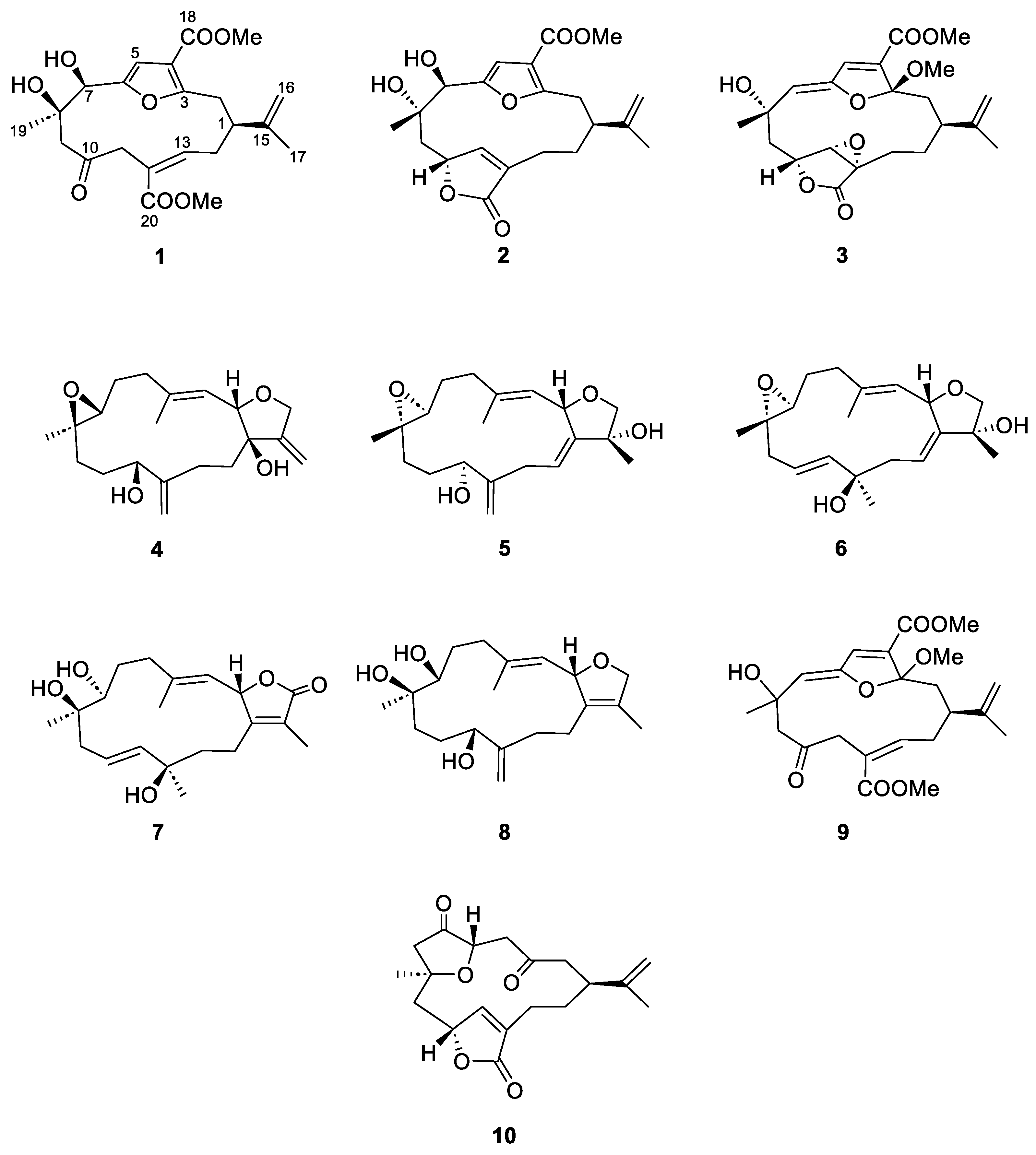
Figure 1. The structures of cembranoid diterpenes 110 isolated from the soft coral Sinularia maxima.
Marinedrugs 22 00373 g001
Figure 2. Lineweaver-Burk and Dixon plots for sEH inhibition by compounds 3 (A,B) and 9 (C,D), respectively.
Marinedrugs 22 00373 g002
Figure 3. Structure of sEH illustrating the binding sites of AUDA (green), compound 3 (cyan), and compound 9 (magenta).
Marinedrugs 22 00373 g003
Figure 4. Binding interactions between sEH and compounds 3 (A) and 9 (B), along with their respective interaction maps. Green lines indicate hydrogen bonds and pink lines represent alkyl and π–alkyl interactions.
Marinedrugs 22 00373 g004
Figure 5. RMSD of the protein backbone (A) and RMSD of the ligand after least-squares fitting to the protein (B) of the complexes formed between sEH and compounds 3 (black) and 9 (blue).
Marinedrugs 22 00373 g005
Figure 6. RMSF plots (A) and number of hydrogen bonds (B) for the sEH inhibition of compounds 3 (black) and 9 (blue), over the 120 ns MD trajectory.
Marinedrugs 22 00373 g006
Figure 7. Superposition for the complexes formed between sEH and compounds 3 (A) and 9 (B) over time. Colors represent different time points: 0 ns (red), 20 ns (green), 40 ns (yellow), 60 ns (pink), 80 ns (cyan), 100 ns (orange), and 120 ns (blue).
Marinedrugs 22 00373 g007
Table 1. sEH inhibitory effects, inhibition types, and inhibition constants (Ki) of compounds 110.
CompoundsIC50 (μM) 1Inhibition Type 2Ki (μM) 3
1>100
2>100
370.68 ± 1.44Non-competitive59.44
4>100
5>100
6>100
7>100
8>100
978.83 ± 2.26Non-competitive55.03
10>100
AUDA 414.69 ± 0.07 nM
1 The values (μM) indicate 50% inhibitory effects. Data are expressed as the mean ± SD (n = 3). 2 Determined using Lineweaver-Burk plots. 3 Determined using Dixon plots. 4 Positive control.
Table 2. Interactions between the enzyme sEH and compounds 3, 9, and AUDA: binding energy, hydrogen bonds, van der Waals interactions, and hydrophobic interactions.
CompoundBinding Energy
(kcal/mol)
Hydrogen Bondsvan der Waals
Interactions
Hydrophobic
Interactions
3−9.18Lys495
Gly523
His524
Trp525
Phe267
Asp335
Tyr383
Ser407
Arg410
Ala411
Ser412
Ser415
Val416
Tyr466
Asp496
Leu408 (alkyl)
Leu417 (alkyl)
Met419 (alkyl)
Leu428 (alkyl)
Phe497 (π−alkyl)
Val498 (alkyl)
His524 (π−alkyl)
Trp525 (π−alkyl)
9−8.94Ser412
Asp496
Gly523
His524
Arg410
Ser415
Ser418
Lys495
Phe497
Val498
Trp525
Val380 (alkyl)
Leu408 (alkyl)
Leu417 (alkyl)
Met419 (alkyl)
AUDA−8.53Asp335
Gln384
Tyr466
Phe267
Pro268
Thr360
Phe387
Leu428
Met469
Val498
Met503
Trp336 (π−alkyl)
Met339 (alkyl)
Phe381 (π−σ)
Tyr383 (π−alkyl)
Leu408 (alkyl)
Met419 (alkyl)
Leu499 (alkyl)
His524 (π−alkyl)
Trp525 (π−alkyl)
Disclaimer/Publisher’s Note: The statements, opinions and data contained in all publications are solely those of the individual author(s) and contributor(s) and not of MDPI and/or the editor(s). MDPI and/or the editor(s) disclaim responsibility for any injury to people or property resulting from any ideas, methods, instructions or products referred to in the content.