Article

Discovery of Prenyltransferase-Guided Hydroxyphenylacetic Acid Derivatives from Marine Fungus Penicillium sp. W21C371

College of Pharmaceutical Science & Collaborative Innovation Center of Yangtze River Delta Region Green Pharmaceuticals, Zhejiang University of Technology, Hangzhou 310014, China
*
Authors to whom correspondence should be addressed.
These authors contributed equally to this work.
Mar. Drugs 2024, 22(7), 296; https://doi.org/10.3390/md22070296
Submission received: 7 June 2024 / Revised: 21 June 2024 / Accepted: 24 June 2024 / Published: 26 June 2024

Abstract

:
Traditional isolation methods often lead to the rediscovery of known natural products. In contrast, genome mining strategies are considered effective for the continual discovery of new natural products. In this study, we discovered a unique prenyltransferase (PT) through genome mining, capable of catalyzing the transfer of a prenyl group to an aromatic nucleus to form C-C or C-O bonds. A pair of new hydroxyphenylacetic acid derivative enantiomers with prenyl units, (±)-peniprenydiol A (1), along with 16 known compounds (217), were isolated from a marine fungus, Penicillium sp. W21C371. The separation of 1 using chiral HPLC led to the isolation of the enantiomers 1a and 1b. Their structures were established on the basis of extensive spectroscopic analysis, including 1D, 2D NMR and HRESIMS. The absolute configurations of the new compounds were determined by a modified Mosher method. A plausible biosynthetic pathway for 1 was deduced, facilitated by PT catalysis. In the in vitro assay, 2 and 3 showed promising inhibitory activity against Escherichia coli β-glucuronidase (EcGUS), with IC50 values of 44.60 ± 0.84 μM and 21.60 ± 0.76 μM, respectively, compared to the positive control, D-saccharic acid 1,4-lactone hydrate (DSL). This study demonstrates the advantages of genome mining in the rational acquisition of new natural products.

1. Introduction

The secondary metabolites produced by filamentous fungi are crucial sources of valuable pharmaceuticals and agrochemicals. Penicillium, one of the most common fungal genera, consists of more than 350 species with a worldwide distribution [1]. These fungi colonize a wide range of habitats, including conventional environments such as soil, vegetation, food, and indoor spaces, as well as extreme environments on Earth [2]. The diverse lifestyles and habitats of Penicillium species equip them with robust metabolic capabilities. Since the discovery of penicillin from P. rubens (often identified as P. chrysogenum), the fungi of the genus Penicillium have been extensively studied for their production of bioactive secondary metabolites [3]. Over the past several decades, many secondary metabolites with diverse structures and intriguing biological activity have been discovered from Penicillium, making it a promising reservoir of novel drug leads. However, the metabolic potential of Penicillium is reported to be underestimated and remains to be fully exploited, despite being one of the most well-studied fungal genera. In a recent large-scale genome-based study, Nielsen and co-workers conducted the first genus-wide analysis of the genomic diversity of 24 Penicillium species and identified 1317 putative biosynthetic gene clusters (BGCs), highlighting the potential of these species as sources of new antibiotics and other pharmaceuticals [4].
As the largest biome on Earth, the marine ecosystem boasts extraordinarily rich biodiversity due to its extreme physical and chemical conditions compared to the terrestrial ecosystems [5]. This vast biodiversity is partly demonstrated by the unique groups of marine microorganisms that produce a variety of secondary metabolites [6]. A significant number of fungi inhabit the marine environment, found in sea water, sediments, or living organisms such as sponges, corals, and algae. Among these, marine fungi of the genus Penicillium have garnered considerable attention as an important source of secondary metabolites, featuring novel structures and remarkable bioactive properties [7]. To date, more than 500 novel natural products have been isolated and characterized from marine Penicillium fungi, exhibiting benefits such as antibacterial [8,9,10,11,12], anti-inflammatory [13], enzyme inhibition, and cytotoxic properties [14,15].
Escherichia coli β-glucuronidase (EcGUS) is one of the most abundant bacterial β-glucuronidases, catalyzing the hydrolysis of glucuronide conjugates to produce the corresponding aglycones [16]. Anticancer drug irinotecan and anti-inflammatory drug indomethacin produce glucuronidated metabolites that are rapidly hydrolyzed by EcGUS, resulting in highly toxic aglycones. The accumulation of these aglycones in the intestine can lead to severe gastrointestinal adverse reactions, such as fatal diarrhea [17]. Consequently, targeting EcGUS has emerged as a crucial strategy to mitigate the adverse gastrointestinal effects associated with these drugs.
The genome mining technique for the discovery of natural products, which leverages genomic sequence data to identify previously uncharacterized compounds, has proven effective and efficient for over a decade [18]. With the rapid accumulation of genomic sequences from various organisms, genome mining methods have led to the identification of an increasing number of natural products [19]. Traditional approaches often result in the rediscovery of known compounds, whereas genome-based strategies are now seen as a promising solution for the continual discovery of novel natural products. Prenyltransferases (PTs) are capable of catalyzing the transfer of prenyl groups from various prenyl donors. These prenylated secondary metabolites, including indole alkaloids, flavonoids, coumarins, ketones, quinones, and naphthalenes, are widely found in terrestrial and marine organisms and exhibit various types of biological activity, such as cytotoxicity, antioxidation, and antimicrobial effects [20]. For example, XptB, a ketone prenyltransferase from Aspergillus nidulans, catalyzes the O-prenylation of 1,7-dihydroxy-6-methyl-8-hydroxymethylxanthone, resulting in the production of variecoxanthone A [21]. Additionally, Streptomyces sp. CNQ-509 produces the rare O-prenylated phenazines, marinophenazines A and B, through the catalytic action of the enzyme CnqPT1 [22]. In our study, we analyzed the genomic data of PTs from our lab and the NCBI public databases, generated a sequence similarity network (SSN) of PTs, and discovered a previously uncharacterized branch. Phylogenetic tree analysis revealed that this enzyme belongs to a distinct branch of PTs, highlighting its unique characteristics. The secondary metabolites produced were investigated under different culture conditions using PT-guided isolation. A pair of new p-hydroxyphenylacetic acid derivative enantiomers, (±)-peniprenydiol A (1), containing prenyl units, and 16 known compounds (217) were isolated from Penicillium sp. W21C371 (Figure 1). Their structures were determined using thorough NMR analysis and a modified Mosher method. A plausible biosynthetic pathway for 1 was deduced, facilitated by PT catalysis. Additionally, their EcGUS inhibition activity was evaluated.

2. Results

2.1. Bioinformatics Analysis and Phylogenetic Tree Construction of PTs

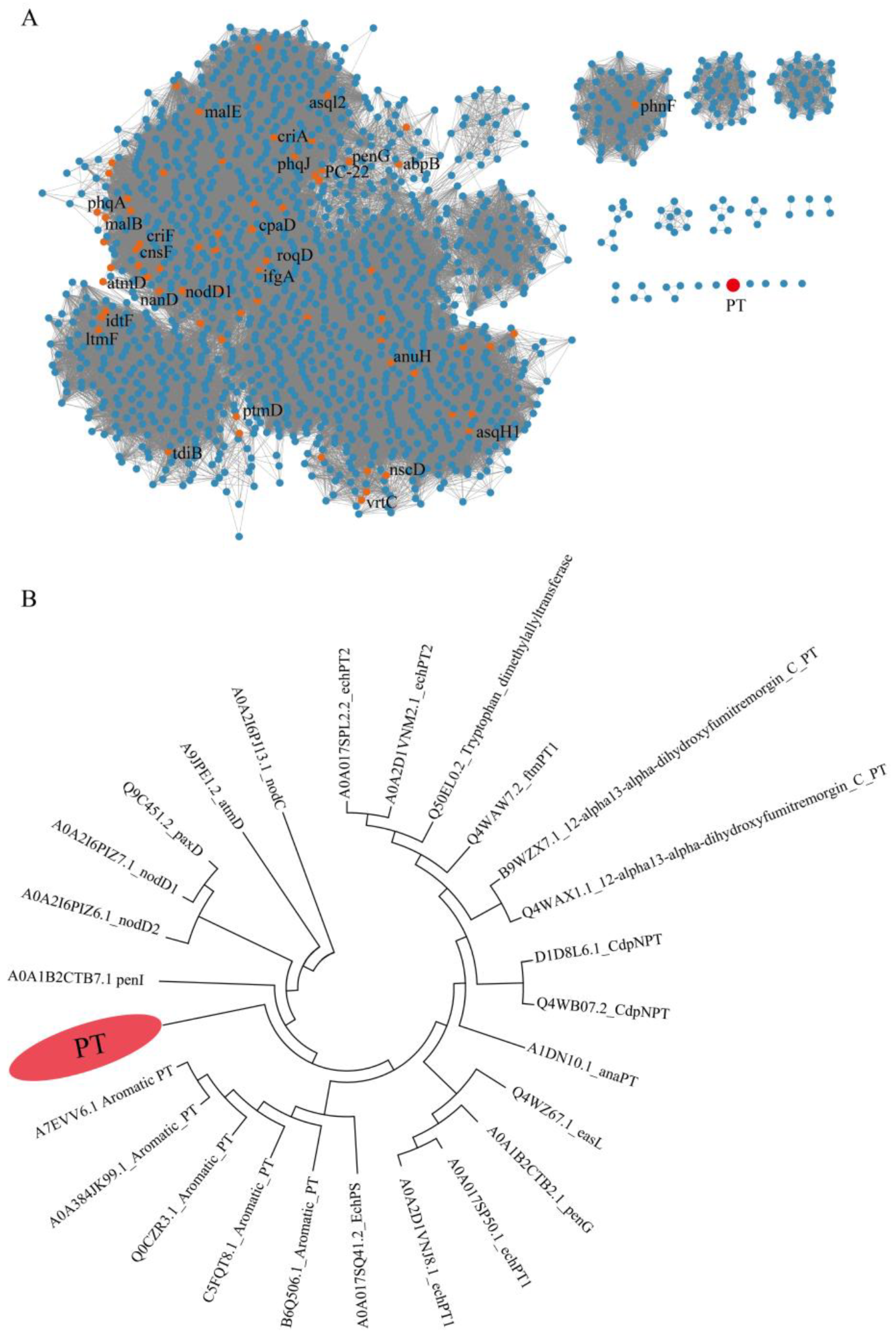
To explore PTs with different substrates, we conducted a comprehensive analysis of genomic data from our lab, the InterPro database, and the NCBI public database. Nearly 2000 prenyltransferases (PTs), including those from our lab and the InterPro database, were submitted to the Enzyme Function Initiative-Enzyme Similarity Tool (EFI-EST). Subsequently, an SSN of PTs was generated, where enzymes with similar functions clustered together. The SSN results showed that most known proteins clustered together (Figure 2A). A distinct evolutionary clade attracted our attention, as it did not cluster with any specific PTs, suggesting that this PT might be a novel enzyme catalyzing the transfer of prenyl units. In addition, 26 PTs were obtained from our lab and the NCBI public database, and phylogenetic trees were constructed using MEGA 7.0. The phylogenetic tree showed that the 26 PTs were divided into two large branches and five small branches. Among them, a unique small branch attracted our attention, suggesting that this enzyme may have structural novelty and the capability to catalyze the production of isoprenoids (Figure 2B). This unique PT originates from the marine fungus Penicillium sp. W21C371, which was sequenced in our laboratory. Comprehensive analysis indicates that this PT possesses distinctive properties, enabling it to catalyze the production of novel compounds. Therefore, further investigation into the secondary metabolites of Penicillium sp. W21C371 is warranted.

2.2. Structure Elucidation

Compound 1 (1a/1b) was obtained as a colorless oil. The molecular formula of 1 was determined as C14H20O5 by HRESIMS (m/z 291.1186 [M + Na]+, calcd. for C14H20O5Na+ 291.1203), indicating five degrees of unsaturation (Figures S14 and S15). The IR spectrum showed characteristic absorption bands for carbonyl (1732 cm−1) and hydroxy (3361 cm−1) groups (Figure S8). The 1H NMR spectrum (Figure S2 and Table 1) displayed proton resonances attributable to two methyls at δH 1.22 (3H, s, H3-10) and 1.24 (3H, s, H3-11), one methoxy at δH 3.62 (3H, s, H3-12), one oxygenated methine at δH 3.75 (dd, J = 7.8, 3.0 Hz, H-8), one oxygenated methylene at δH 3.92 (dd, J = 9.6, 7.8 Hz, Hb-7) and 4.26 (dd, J = 9.6, 3.0 Hz, Ha-7), and four aromatic protons at δH 6.90 (2H, d, J = 8.4 Hz, H-5 and 5′) and 7.19 (2H, d, J = 8.4 Hz, H-4 and 4′). The 13C NMR, DEPT, and HSQC spectra (Figures S3–S5) revealed the presence of fourteen carbon resonances, including two methyls at δC 25.6 (C-11) and 26.7 (C-10); one methoxy at δC 51.9 (C-12); two methylenes at δC 40.4 (C-2) and 70.6 (C-7); four aromatic methines at δC 115.4 (C-5), 115.4 (C-5′), 131.1 (C-4), and 131.1 (C-4′); one oxygenated methine at δC 77.1 (C-8); and four non-protonated carbons (one oxygenated at δC 71.9 (C-9), two aromatic at δC 127.4 (C-3) and 159.2 (C-6), and one carbonyl at δC 172.6 (C-1)). The NMR data of 1 were similar to those of westerdijkin A, a hydroxyphenylacetic acid derivative from the deep-sea fungus Aspergillus westerdijkiae SCSIO 05233 [23]. Detailed comparative analyses revealed that the signals for the Δ9 terminal double bond (δC 143.3 (C-9), δC 112.7 (C-10), δH 5.00 and 5.14 (H2-10)) in westerdijkin A were replaced by the newly emerging signals assignable for one methyl (δH 1.22 (H3-10) and δC 26.7 (C-10)) and one oxygenated quaternary carbon (δC 71.9 (C-9)) in 1. The 1H-1H COSY plots (Figure S6) of H-4/H-5 and H-4′/H-5′, combined with the HMBC (Figure S7) correlations from H-4, H-4′, H-5, and H-5′ to C-6, and from H-5 and H-5′ to C-3, established the para-substituted benzene ring moiety in 1. The HMBC correlations from H2-2 to C-1, C-3, C-4, and C-4′, as well as from H3-12 to C-1, suggested the presence of a methyl acetate residue that anchored at C-3 via C-2. In addition, the 1H-1H COSY plot of H2-7 (δH 3.92 and 4.26)/H-8 (δH 3.75) and the HMBC correlations from H2-7 (δH 3.92 and 4.26) and H-8 (δH 3.75) to the oxygenated quaternary carbon C-9 (δC 71.9) and from the two methyl singlets (δH 1.22 and 1.24) to C-8 (δC 77.1) and C-9 (δC 71.9), in combination with the molecular formula, constructed a 2,3-dihydroxyisopentane moiety that was linked to the benzene ring with an ether linkage between C-6 (δC 159.2) and C-7 (δC 70.6). The planar structure of 1 was thus established (Figure 3).
Compound 1 was initially assumed to be optically pure based on its specific rotation ([α ] D 20 : +14 (c 0.1, MeOH)). Hence, a modified Mosher experiment was performed to determine the absolute configuration of C-8. Unexpectedly, when the (R)- and (S)-MTPA esters of 1 were subjected to HPLC analysis on a routine ODS C-18 column, two pairs of diastereoisomers were obtained ((S)-MTPA-1a/(S)-MTPA-1b and (R)-MTPA-1a/(R)-MTPA-1b), suggesting that 1 was probably a partially racemic mixture. This was confirmed by the chiral resolution of 1 on a Chiralpak AD-H column, which afforded a pair of enantiomers 1a and 1b in a ratio of ca. 3:7. According to the shielding/deshielding effects of MTPA, the ΔδH(SR) values indicated an 8R configuration for (+)-1b (Figure 3 and Figures S16 and S17 and Table S2). Thus, 1a was deduced to be the enantiomer of 1b based on their identical 1H NMR and HRESIMS data (Supplementary Data, Figures S12–S15) and opposite specific rotations ([α ] D 20 : −32 (c 0.05, MeOH) for 1a and [α ] D 20 : +36 (c 0.1, MeOH) for 1b). Finally, (−)-1a and (+)-1b were named (−)-peniprenydiol A and (+)-peniprenydiol A, respectively.
Sixteen known compounds were identified as citridone A (2) [24], 5α,8α-epidioxy-23,24(R)-dimethylcholesta-6,9(11),22-trien-3β-ol (3) [23], 5α,8α-epidioxy-(23E,24R)-23-methylergosta-6,22-dien-3β-ol (4) [25], (22E)-23-methylergosta-5,7,22-trien-3β-ol (5) [26], ergosta-4,6,8(14),22-tetraen-3-one (6) [27], isocyathisterol (7) [28], ergosterol peroxide (8) [29], dankasterone B (9) [30], dankasterone (10) [31], curvularin (11) [32], 10,11-dedrocurvularin (12) [33], dehydrocurvularin (13) [34], sumalactone A (14) [35], morelsin D (15) [36], conocenolide A (16) [37], and p-hydroxy phenylacetic acid (17) [38] through the comparison of the spectroscopic data with those reported in the literature. The tremulane sesquiterpenoids, exemplified by morelsin D (15) and conocenolide A (16) in the present study, were obtained from Ascomycetes for the first time.

2.3. Plausible Biosynthetic Pathways of Peniprenydiol A

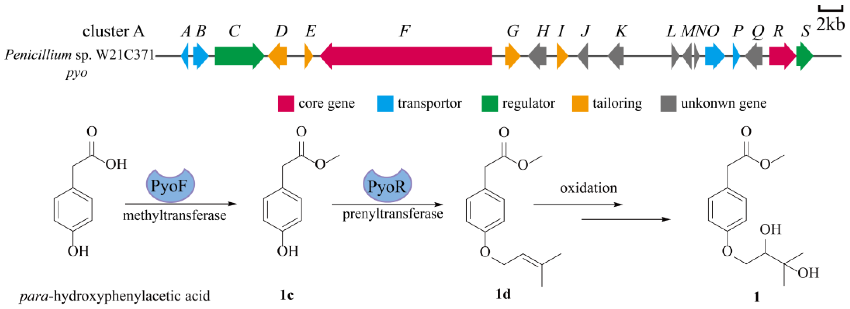
To investigate the connection between peniprenydiol A and its biosynthetic genes, we obtained the complete genome sequence of the marine fungus Penicillium sp. W21C371. The genome was analyzed and predicted using the online tool 2ndfind (https://biosyn.nih.go.jp/2ndfind/, accessed on 1 June 2024). Based on the analysis results, we propose that gene cluster A is responsible for the assembly and transport of peniprenydiol A. This gene cluster comprises multiple genes with diverse functions, including core genes, transporter proteins, regulatory proteins, tailoring enzymes, and genes of unknown function. Using peniprenydiol A as a case study to analyze its biosynthetic pathway, the core PyoF catalyzes the methylation reaction to produce 1c [39]. This intermediate is then prenylated by the PyoR enzyme to form 1d [40]. Finally, 1d is converted into the final product through a non-enzymatic oxidation reaction (Figure 4).

2.4. EcGUS Inhibition Assay

All isolates 117 were evaluated for their in vitro inhibitory activity against EcGUS, a potential target for the treatment of drug-induced gastrointestinal disorders, employing D-saccharic acid 1,4-lactone (DSL) as the positive control. In the preliminary screening, compounds 2 and 3 showed relative inhibitory rates of over 60% at a concentration of 100 μM and were subjected to further evaluation. As a result (Table 2), 2 and 3 were found to significantly inhibit the activity of EcGUS in a dose-dependent manner, comparable to that of DSL. In view of the potent inhibitory activity of 2 and 3 against EcGUS, their kinetic mechanisms of inhibition were determined using Lineweaver–Burk plots (Figure 5 and Table S1). The inhibition constants of the enzyme Ki and the enzyme–substrate complex Ki were obtained by secondary plots of “slope versus [I]” and “Y-intercept versus [I]”, respectively. As shown in Figure 5, the data lines of 2 intersected in the X axis. Meanwhile, the Vmax value decreased with the increased concentration of 2, while the Km value did not change. This suggested that 2 was a non-competitive inhibitor of EcGUS that bound with EcGUS and/or the EcGUS–substrate complex at a site other than the active site. The intersection of the data lines of 3 in the third quadrant demonstrated that 3 inhibited EcGUS in a mixed-type manner, which was verified by the changed Km and Vmax values with the increased concentration of 3. As a mixed-type inhibitor of EcGUS, 3 was supposed to bind either the free EcGUS or the EcGUS–substrate complex. The inhibition constant Ki (89.04 μM) for 3 was larger than Ki (48.80 μM), implying that it bound more easily and tightly to the EcGUS–substrate complex than the free EcGUS. To the best of our knowledge, 2 and 3 represent the first citridone and ergosterol derivatives with potent EcGUS-inhibitory activity. However, 2 and 3 were obtained in limited amount in the present study, preventing the in vivo evaluation of their EcGUS inhibition activity. The optimization of the fermentation and separation processes with the aim of increasing the yields of 2 and 3 is underway, which may help to accumulate greater amounts of the two compounds for further in-depth pharmacological studies.

3. Materials and Methods

3.1. General Experimental Procedures

Optical rotations were obtained with a Rudolph Research Autopol III automatic polarimeter. IR spectra were recorded on a Thermo Nicolet 6700 FT-IR microscope instrument (FT-IR microscope transmission) in KBr pellets. UV spectra were measured on a TU-1900 spectrometer (Persee, Beijing, China). Meanwhile, 1D and 2D-NMR spectra were obtained at 600 MHz for 1H and 150 MHz for 13C on a Bruker Avance 600 spectrometer, with solvent peaks used as references. HRESIMS data were measured on an Agilent-6210-LC/TOF mass spectrometer (Agilent Technologies, Inc., Santa Clara, CA, USA). EcGUS-inhibitory activity was measured spectrophotometrically using a SpectraMax Plus 384 microplate reader (Molecular Devices, San Jose, CA, USA). Column chromatography (CC) was performed on silica gel (SiO2; 200–300 mesh; Qingdao Marine Chemical Co., Ltd.), ODS C-18 gel (50 mm; YMC Co., Ltd., Kyoto, Japan), and MCI CHP20P gel (75–150 μm, Tokyo, Japan). Semi-preparative HPLC was performed on an Cosmosil 5C18-MS-II column (5 μm, 250 × 10 mm) with a Shimadzu LC-20AT system eluting with methanol/water or acetonitrile/water at a flow rate of 3 mL/min. Chiral separation was performed on a Chiralpak AD-H column (5 μm, 4.6 × 250 mm). Thin layer chromatography (TLC) was performed on precoated silica gel GF254 plates (Qingdao Marine Chemical Co., Ltd., Qingdao, China) and visualized by UV light and/or spraying with 10% H2SO4 in 95% EtOH, followed by heating. All solvents used were of analytical grade and obtained from commercially available sources. All reagents used for biological evaluation were obtained from Sigma-Aldrich (St. Louis, MO, USA).

3.2. Fungal Material

The fungus Penicillium sp. W21C371 was isolated from a seawater sample collected at Marceau Trench in 2017. It has been deposited in the China Typical Culture Preservation Centre, Wuhan University, Wuhan, China, with the deposit number of CCTCC NO: M2022063. For the identification of the fungus, the genomic DNA was extracted using the Trelief® Hi-Pure Plant Genomic DNA Kit (Tsingke, Beijing, China), according to the manufacturer’s instructions. The primers for the internal transcribed spacer 1 (ITS1) and 4 (ITS4) regions were TCCGTAGGTGAACCTGCGG (5′→3′) and TCCTCCGCTTATTGATATGC (5′→3′), respectively. The PCR amplification of the extracted DNA was performed in a 50 µL reaction mixture consisting of 1 µL gDNA template, 2 µL each of the forward and reverse primers, and 45 µL of Tsingke 1× TSE101 golden mix. The thermocycler was programed with the following PCR conditions: initial denaturation at 98 °C for 2 min, followed by 35 cycles of denaturation at 98 °C for 10 s, annealing at 55 °C for 10 s, and extension at 72 °C for 15 s, with a final extension at 72 °C for 5 min. Upon the completion of amplification, the PCR products were analyzed using gel electrophoresis with a 1% agarose gel (1 g of agarose in 100 mL of Tris buffer) stained with ethidium bromide. Sequencing was carried out by Sanger’s method [41] on a 3730xl DNA Analyzer (Thermo Fisher Scientific, Waltham, MA, USA) at Beijing Tsingke Biotech Co., Ltd. (Beijing, China). The ITS sequence was deposited in GeneBank under Accession No. PP733993.

3.3. Bioinformatics Analysis and Phylogenetic Tree Construction of PTs

The sequences of PTs were obtained from our lab, the InterPro database, and the NCBI database. Sequence similarity networks (SSNs) were constructed using the Enzyme Function Initiative (EFI; accessed at https://efi.igb.illinois.edu/, accessed on 1 June 2024) [42]. Cytoscape 3.5.1 was employed for network visualization. Multiple sequence alignments were performed using MUSCLE. Phylogenetic trees were constructed using the maximum likelihood method in MEGA6 and visualized using the Interactive Tree of Life (ITOL, http://itol.embl.de/, accessed on 1 June 2024).

3.4. Fermentation and Extraction

The fungus was cultured on potato dextrose agar (PDA) plates for 5 days at 28 °C. The spores were washed with 10 mL sterile water containing 2% tween 80 and aseptically inoculated into 45 conical flasks (1000 mL) containing rice medium (rice 100 g, distilled water 135 mL, sterilized at 121 °C for 20 min and cooled). The conical flasks were kept at 28 °C. After culturing for 20 days, the collected cultures were mashed and soaked in 95% ethanol for 4 days at room temperature and filtered (repeated 3 times). The filtrate was condensed in a vacuum to give a crude extract (550 g), which was suspended in distilled water (1.5 L) and partitioned with EtOAc (1.5 L) 3 times. The EtOAc layer was combined and concentrated in a vacuum to yield an EtOAc-soluble extract (64 g).

3.5. Isolation and Purification

The EtOAc-soluble extract (64 g) was subjected to MCI CHP20P CC eluting with a gradient of MeOH-H2O (20:80→100:0, v/v) to offer six fractions (Fr. A–F). Fr. B (2.26 g) was fractionated by silica gel CC eluting with a gradient of CH2Cl2-MeOH (60:1→40:1, v/v) to give five sub-fractions (Fr. B1–B5). Fr. B1 was separated by semi-preparative HPLC eluting with MeOH/H2O (35:65, v/v) to give 1 (21.1 mg, tR = 10.5 min). Fr. B2 was subjected to semi-preparative HPLC eluting with CH3CN-H2O (45:55, v/v) to afford 13 (1 mg, tR = 11.5 min). Fr. C (8.56 g) was subjected to silica gel CC eluting with a gradient of CH2Cl2-MeOH (40:1→5:1, v/v) to give three sub-fractions (Fr. C1–C3). Fr. C2 was separated by semi-preparative HPLC eluting with MeOH/H2O (40:60, v/v) to yield 14 (2 mg, tR = 16.5 min) and 17 (6.5 mg, tR = 14.5 min). Fr. D (14.7 g) was subjected to silica gel CC eluting with a gradient of CH2Cl2-MeOH (50:1→30:1, v/v) to give 11 (1.5 g) and four sub-fractions (Fr. D1–D4). Fr. D1 was chromatographed over ODS C-18 gel eluting with a gradient of MeOH-H2O (60:40→80:20, v/v) to furnish 12 (37.7 mg), 15 (19.5 mg), and 16 (5.4 g). The purification of Fr. E (12.5 g) by silica gel CC eluting with a gradient of CH2Cl2-MeOH (50:1→20:1, v/v) yielded four sub-fractions (Fr. E1-Fr. E4). Fr. E2 was subjected to silica gel CC eluting with a gradient of MeOH-H2O (80:20→90:10, v/v) to afford 9 (17 mg). Moreover, 2 (15.4 mg) was obtained from Fr. E4 by silica gel CC eluting with a gradient of MeOH-H2O (80:20→90:10, v/v). Fr. F (12.5 g) was first separated on a silica gel column eluting with a gradient of CH2Cl2-MeOH (50:1→20:1, v/v) to yield four sub-fractions (Fr. F1–F4). Fr. F1 was subjected to ODS C-18 CC and eluted with a gradient of MeOH-H2O (75:25→100:0, v/v) to offer 6 (10 mg). Fr. F2 was separated on an ODS C-18 column eluting with a gradient of MeOH-H2O (80:20→100:0, v/v) to furnish 5 (55.0 mg), 4 (4 mg), and 7 (15.3 mg). Fr. F3 was purified by semi-preparative HPLC eluting with MeOH-H2O (95:5, v/v) to give 10 (3 mg, tR = 15.5 min). Fr. F4 was separated by semi-preparative HPLC eluting with MeOH/H2O (95:5, v/v) to furnish 3 (1.7 mg, tR = 10.0 min) and 8 (15.2 mg, tR = 12.0 min). Finally, 1 (5.0 mg) was separated by chiral HPLC eluting with isopropanol/n-hexane (18:82, v/v) at a flow rate of 1 mL/min to yield 1a (0.8 mg, tR = 16.7 min) and 1b (3.2 mg, tR = 22.0 min) (Figure S11).

3.6. Spectroscopic Data

Peniprenydiol A (1), colorless oil, [α ] D 20 : +14 (c 0.1, MeOH), IR (KBr) (Figure S8): 3361, 2921, 2850, 1732, 1659, 1632, 1612, 1584, 1513, 1462, 1435, 1297, 1245, 1157, 1094, 1030, 805 cm−1. UV λmax (MeOH) nm (log ɛ): 207 (3.91) (Figure S10); HR-ESI-MS m/z: 291.1186 [M + Na]+ (calcd. for C14H20O5Na+, 291.1203). For 1H- and 13C-NMR data, see Table 1.
(−)-Peniprenydiol A (1a), colorless oil, [α ] D 20 : −32 (c 0.05, MeOH), HR-ESI-MS m/z 291.1202 [M + Na]+ (calcd. for C14H20O5Na+, 291.1203). For 1H-NMR data, see Figure S12.
(+)-Peniprenydiol A (1b), colorless oil, [α ] D 20 : +36 (c 0.1, MeOH), HR-ESI-MS m/z 291.1202 [M + Na]+ (calcd. for C14H20O5Na+, 291.1203). For 1H-NMR data, see Figure S13.

3.7. Preparation of (R)- and (S)-MTPA Esters of 1b

Compound 1 (1 mg, 3.73 μmol) was dissolved in 0.5 mL of anhydrous pyridine-d5, and (S)-MTPA chloride (5 μL, 26.5 μmol) was added under N2. The mixture was allowed to react for 36 h at room temperature. Further purification of the (R)-MTPA ester of 1 using semi-preparative HPLC with an ODS C18 column afforded the (R)-MTPA esters of 1a and 1b. The (S)-MTPA esters of 1a and 1b were prepared with (R)-MTPA chloride and were purified in the same manner [43].

3.8. Bioassay

The EcGUS inhibition assay was performed according to the method previously reported, employing DSL as the positive control [44]. The results are presented as means ± SD for three independent experiments.

4. Discussion

With the rapid development of bioinformatics, genome mining strategies have been widely applied to discover new natural products. By combining SSN analysis and phylogenetic tree construction, we can identify enzymes and genes with potential new functions. Using this approach, we discovered a unique PT that can transfer an isopropyl group to an aromatic nucleus, forming C-C or C-O bonds. Utilizing PT-guided separation techniques, we isolated a pair of novel hydroxyphenylacetic acid derivative enantiomers with prenyl units, (±)-peniprenydiol A, from the rice extracts of Penicillium sp. W21C371. The structure was formed through a crucial step involving PT catalysis, based on the known structure of p-hydroxy phenylacetic acid. In natural product studies, these compounds can appear in different enantiomeric forms. One form is a racemate, an equal mixture of two enantiomers, while the other form consists of non-racemic enantiomers in an unequal mixture. Currently, researchers focus more on racemic natural products, while non-racemic enantiomers are largely overlooked. This is because the optical purity of new natural products is rarely reported upon publication. It is usually assumed that when new chiral natural products exhibit optical activity, they are also optically pure. We measured the optical rotation of 1 ] D 20 : +14 (c 0.1, MeOH) and considered it optically pure. Subsequently, using a modified Mosher method to determine the configuration of its 8-OH group, we found that the compound exists as non-racemic enantiomers in unequal proportions. This compound was separated by chiral chromatography to obtain a pair of non-racemic enantiomers, (−)-1 and (+)-1.
Citridone A (2), featuring a unique phenyl-R-furopyridone skeleton (6-6/5/5 ring system), belongs to the citridone family of fungal pyridines and was first isolated by Ōmura and co-workers from Penicillium sp. FKI-1938. It was identified as a potentiator of miconazole against Candida albicans [45]. Additionally, it could inhibit the biosynthesis of staphyloxanthin, a key virulence factor in methicillin-resistant Staphylococcus aureus (MRSA), rendering it a promising antibiotic lead with a new mode of action [46]. As a result, citridone A (2) has drawn attention from both chemists and biochemists. The total synthesis of citridone A (2) was accomplished in 2011 [47], while the biosynthetic pathway was unveiled in 2020 [48]. Compounds 310 are derivatives of ergosterol, a representative fungisterol that is a component of the fungal cell membrane and determines the fluidity, permeability, and activity of membrane-associated proteins in fungi [49]. Compounds 35 are notable in this series of fungal sterols for harboring an additional C-23 methyl group, which was proposed to be formed by S-adenosyl-L-methine (SAM) [50]. Preliminary structure–activity relationship studies suggest that both the C-23 methyl group and Δ9(11) double bond play important roles in retaining the EcGUS-inhibitory activity, as seen in the activity of 3, 4, and 8. To the best of our knowledge, 2 and 3 represent the first citridone and ergosterol derivatives with potent EcGUS-inhibitory activity. Morelsin D (15) and conocenolide A (16) are tremulane sesquiterpenoids that have been only reported from Basidiomycetes so far. It is worth noting that our work represents the first report of tremulane sesquiterpenoids from Ascomycetes. The well-established genetic manipulation systems in Ascomycetes, particularly within the genus Penicillium, may facilitate biosynthetic studies and the metabolic engineering of this type of sesquiterpenoid.

5. Conclusions

In summary, we conducted a comprehensive genome mining analysis using genomic data from our lab, the InterPro database, and the publicly available NCBI database. Through this analysis, we identified a unique PT capable of catalyzing the transfer of a prenyl group to an aromatic nucleus, forming C-C or C-O bonds. From the marine fungus Penicillium sp. W21C371, a pair of novel hydroxyphenylacetic acid derivative enantiomers, (±)-peniprenydiol A (1) with prenyl units, along with sixteen known compounds 217, were isolated through PT-guided isolation. The plausible biosynthetic pathways of compound 1 were deduced. This study also marks the first time that tremulane sesquiterpenoids, exemplified by morelsin D (15) and conocenolide A (16), have been obtained from Ascomycetes. Additionally, compounds 2 and 3 were identified as potent EcGUS inhibitors for the first time, exhibiting non-competitive and mixed-type inhibition, respectively.

Supplementary Materials

The following supporting information can be downloaded at: https://www.mdpi.com/article/10.3390/md22070296/s1, Table S1: Inhibition kinetics of 2 and 3 against EcGUS; Table S2: Selected 1H (600 MHz) NMR data of 1b and its MTPA esters in Pyridine-d5 (δH in ppm, J in Hz); Table S3: Putative functions of selected genes from peniprenydiol A gene cluster. Figure S1: HPLC-DAD chromatogram of twenty-one extracts of Penicillium sp. W21C371; Figure S2: 1H NMR spectrum of 1 in Acetone-d6 (600 MHz); Figure S3: 13C NMR spectrum of 1 in Acetone-d6 (150 MHz); Figure S4: DEPT spectrum of 1 in Acetone-d6; Figure S5: HSQC spectrum of 1 in Acetone-d6; Figure S6: 1H-1H COSY spectrum of 1 in Acetone-d6; Figure S7: HMBC spectrum of 1 in Acetone-d6; Figure S8: The IR spectrum of 1; Figure S9: The HRESIMS spectroscopic data of 1; Figure S10: UV spectrum of 1; Figure S11: Chiral HPLC separation profile of 1a/1b; Figure S12: 1H NMR spectrum of 1a in Pyridine-d5 (600 MHz); Figure S13: 1H NMR spectrum of 1b in Pyridine-d5 (600 MHz); Figure S14: The HRESIMS spectrum of compound 1a; Figure S15: The HRESIMS spectrum of compound 1b; Figure S16: 1H NMR spectrum of (R)-MTPA ester of 1b in Pyridine-d5 (600 MHz); Figure S17: 1H NMR spectrum of (S)-MTPA ester of 1b in Pyridine-d5 (600 MHz).

Author Contributions

Conceptualization, project administration, and funding acquisition, Y.Y. and H.W.; methodology, C.W. (Cancan Wang) and Y.F.; software, J.T. and Y.Q.; formal analysis, K.X. and Y.D.; investigation, C.W. (Chenjie Wang) and Y.L.; resources, C.W. (Cancan Wang); data curation, Y.Y.; writing—original draft preparation, C.W. (Cancan Wang) and Y.F.; writing—review and editing, Y.Y. and H.W. All authors have read and agreed to the published version of the manuscript.

Funding

This research was funded by the National Key Research and Development Program of China (No. 2022YFC2804205 and 2022YFC2804104), the Natural Foundation of Zhejiang Province (LGF21H300003), the National Natural Science Foundation of China (No. 42276137), and the Key Research and Development Program of Zhejiang Province (No. 2021C03084).

Institutional Review Board Statement

Not applicable.

Data Availability Statement

Data are contained within the article or Supplementary Materials.

Conflicts of Interest

The authors declare no conflicts of interest.

References

  1. Perrone, G.; Susca, A. Penicillium species and their associated mycotoxins. Methods Mol. Biol. 2017, 1542, 107–119. [Google Scholar]
  2. Petersen, C.; Sørensen, T.; Nielsen, M.R.; Sondergaard, T.E.; Sørensen, J.L.; Fitzpatrick, D.A.; Frisvad, J.C.; Nielsen, K.L. Comparative genomic study of the Penicillium genus elucidates a diverse pangenome and 15 lateral gene transfer events. IMA Fungus 2023, 14, 3. [Google Scholar] [CrossRef]
  3. Houbraken, J.; Frisvad, J.C.; Samson, R.A. Fleming’s penicillin producing strain is not Penicillium chrysogenum but P. rubens. IMA Fungus 2011, 2, 87–95. [Google Scholar] [CrossRef]
  4. Nielsen, J.C.; Grijseels, S.; Prigent, S.; Ji, B.; Dainat, J.; Nielsen, K.F.; Frisvad, J.C.; Workman, M.; Nielsen, J. Global analysis of biosynthetic gene clusters reveals vast potential of secondary metabolite production in Penicillium species. Nat. Microbiol. 2017, 2, 17044–17053. [Google Scholar] [CrossRef]
  5. Shu, W.S.; Huang, L.N. Microbial diversity in extreme environments. Nat. Rev. Microbiol. 2022, 20, 219–235. [Google Scholar] [CrossRef]
  6. König, G.M.; Kehraus, S.; Seibert, S.F.; Abdel-Lateff, A.; Müller, D. Natural products from marine organisms and their associated microbes. Chembiochem. 2006, 7, 229–238. [Google Scholar] [CrossRef]
  7. Ma, H.G.; Liu, Q.; Zhu, G.L.; Liu, H.S.; Zhu, W.M. Marine natural products sourced from marine-derived Penicillium fungi. J. Asian Nat. Prod. Res. 2016, 18, 92–115. [Google Scholar] [CrossRef]
  8. Liu, S.; Su, M.; Song, S.J.; Jung, J.H. Marine-derived Penicillium species as producers of cytotoxic metabolites. Mar. Drugs 2017, 15, 329. [Google Scholar] [CrossRef]
  9. Yang, X.; Liu, J.; Mei, J.; Jiang, R.; Tu, S.; Deng, H.; Liu, J.; Yang, S.; Li, J. Origins, structures, and bioactivities of secondary metabolites from marine-derived Penicillium fungi. Mini-Rev. Med. Chem. 2021, 21, 2000–2019. [Google Scholar] [CrossRef]
  10. Hu, X.Y.; Li, X.M.; Yang, S.Q.; Liu, H.; Meng, L.H.; Wang, B.G. Three new sesquiterpenoids from the algal-derived fungus Penicillium chermesinum EN-480. Mar. Drugs 2020, 18, 194. [Google Scholar] [CrossRef]
  11. Shah, M.; Sun, C.; Sun, Z.; Zhang, G.; Che, Q.; Gu, Q.; Zhu, T.; Li, D. Antibacterial polyketides from antarctica sponge-derived fungus Penicillium sp. HDN151272. Mar. Drugs 2020, 18, 71. [Google Scholar] [CrossRef]
  12. Chen, J.; Wang, W.; Hu, X.; Yue, Y.; Lu, X.; Wang, C.; Wei, B.; Zhang, H.; Wang, H. Medium-sized peptides from microbial sources with potential for antibacterial drug development. Nat. Prod. Rep. 2024; ahead of print. [Google Scholar] [CrossRef]
  13. Li, F.L.; Sun, W.G.; Zhang, S.T.; Gao, W.X.; Lin, S.; Yang, B.Y.; Chai, C.W.; Li, H.Q.; Wang, J.P.; Hu, Z.X.; et al. New cyclopiane diterpenes with anti-inflammatory activity from the sea sediment-derived fungus Penicillium sp. TJ403-2. Chin. Chem. Lett. 2020, 31, 197–201. [Google Scholar] [CrossRef]
  14. Dai, L.T.; Yang, L.; Kong, F.D.; Ma, Q.Y.; Xie, Q.Y.; Dai, H.F.; Yu, Z.F.; Zhao, Y.X. Cytotoxic indole-diterpenoids from the marine-derived fungus Penicillium sp. KFD28. Mar. Drugs 2021, 19, 613. [Google Scholar] [CrossRef]
  15. Pang, X.; Zhou, X.; Lin, X.; Yang, B.; Tian, X.; Wang, J.; Xu, S.; Liu, Y. Structurally various sorbicillinoids from the deep-sea sediment derived fungus Penicillium sp. SCSIO06871. Bioorg. Chem. 2021, 107, 104600. [Google Scholar] [CrossRef]
  16. Wang, P.; Jia, Y.; Wu, R.; Chen, Z.; Yan, R. Human gut bacterial β-glucuronidase inhibition: An emerging approach to manage medication therapy. Biochem. Pharmacol. 2021, 190, 114566. [Google Scholar] [CrossRef]
  17. Chen, S.; Yueh, M.F.; Bigo, C.; Barbier, O.; Wang, K.; Karin, M.; Nguyen, N.; Tukey, R.H. Intestinal glucuronidation protects against chemotherapy-induced toxicity by irinotecan (CPT-11). Proc. Natl. Acad. Sci. USA 2013, 110, 19143–19148. [Google Scholar] [CrossRef]
  18. Bauman, K.D.; Butler, K.S.; Moore, B.S.; Chekan, J.R. Genome mining methods to discover bioactive natural products. Nat. Prod. Rep. 2021, 38, 2100–2129. [Google Scholar] [CrossRef]
  19. Chevrette, M.G.; Gavrilidou, A.; Mantri, S.; Selem-Mojica, N.; Ziemert, N.; Barona-Gómez, F. The confluence of big data and evolutionary genome mining for the discovery of natural products. Nat. Prod. Rep. 2021, 38, 2024–2040. [Google Scholar] [CrossRef]
  20. Fredimoses, M.; Zhou, X.; Ai, W.; Tian, X.; Yang, B.; Lin, X.; Xian, J.Y.; Liu, Y. Westerdijkin A, a new hydroxyphenylacetic acid derivative from deep sea fungus Aspergillus westerdijkiae SCSIO 05233. Nat. Prod. Res. 2015, 29, 158–162. [Google Scholar] [CrossRef]
  21. Pockrandt, D.; Ludwig, L.; Fan, A.; König, G.M.; Li, S.M. New insights into the biosynthesis of prenylated xanthones: Xptb from Aspergillus nidulans catalyses an O-prenylation of xanthones. ChemBioChem 2012, 13, 2764–2771. [Google Scholar] [CrossRef]
  22. Zeyhle, P.; Bauer, J.S.; Steimle, M.; Leipoldt, F.; Rösch, M.; Kalinowski, J.; Gross, H.; Heide, L. A membrane-bound prenyltransferase catalyzes the O-Prenylation of 1,6-dihydroxyphenazine in the marine bacterium Streptomyces sp. CNQ-509. ChemBioChem 2014, 15, 2385–2392. [Google Scholar] [CrossRef]
  23. Wang, F.; Fang, Y.; Zhang, M.; Lin, A.; Zhu, T.; Gu, Q.; Zhu, W. Six new ergosterols from the marine-derived fungus Rhizopus sp. Steroids 2008, 73, 19–26. [Google Scholar] [CrossRef]
  24. Fukuda, T.; Tomoda, H.; Omura, S. Citridones, new potentiators of antifungal miconazole activity, produced by Penicillium sp. FKH938. II. Structure elucidation. J. Antibiot. 2005, 58, 315–321. [Google Scholar] [CrossRef]
  25. Yaoita, Y.; Amemiya, K.; Ohnuma, H.; Furumura, K.; Masaki, A.; Matsuki, T.; Kikuchi, M. Sterol constituents from five edible mushrooms. Chem. Pharm. Bull. 1998, 46, 944–950. [Google Scholar] [CrossRef]
  26. Ohnuma, N.; Amemiya, K.; Kakuda, R.; Yaoita, Y.; Machida, K.; Kikuchi, M. Sterol constituents from two edible mushrooms, Lentinula edodes and Tricholoma matsutake. Chem. Pharm. Bull. 2000, 48, 749–751. [Google Scholar] [CrossRef]
  27. Lee, W.Y.; Park, Y.; Ahn, J.K.; Park, S. Cytotoxic activity of ergosta-4,6,8(14),22-tetraen-3-one from the sclerotia of Polyporus umbellatus. Bull. Korean Chem. Soc. 2005, 26, 1464–1466. [Google Scholar]
  28. Liu, X.H.; Miao, F.P.; Liang, X.R. Ergosteroid derivatives from an algicolous strain of Aspergillus ustus. Nat. Prod. Res. 2014, 28, 1182–1186. [Google Scholar] [CrossRef]
  29. Wang, X.H.; Hou, Y.Z.; Pan, X.H.; Wang, Q. Sterol compounds and their anti-complementary activities of Cordia dichotoma. Chem. Nat. Compd. 2020, 56, 759–760. [Google Scholar] [CrossRef]
  30. Amagata, T.; Tanaka, M.; Yamada, T.; Doi, M.; Minoura, K.; Ohishi, H.; Yamori, T.; Numata, A. Variation in cytostatic constituents of a sponge-derived Gymnascella dankaliensis by manipulating the carbon source. J. Nat. Prod. 2007, 70, 1731–1740. [Google Scholar] [CrossRef]
  31. Amagata, T.; Doi, M.; Tohgo, M.; Minoura, K.; Numata, A. Dankasterone, a new class of cytotoxic steroid produced by a Gymnascella species from a marine sponge. Chem. Commun. 1999, 30, 1321–1322. [Google Scholar] [CrossRef]
  32. Ghisalberti, E.; Hockless, D.; Rowland, C. Structural study of curvularin, a cell division inhibitor. Aust. J. Chem. 1993, 46, 571–577. [Google Scholar] [CrossRef]
  33. Zhao, L.; Zhang, H. Isolation of Secondary. Metabolites of 9F series marine fungi and their bioactivities against Pyricularia oryzae. Nat. Prod. Res. Dev. 2005, 17, 677–680. [Google Scholar]
  34. Kusano, M.; Nakagami, K.; Fujioka, S.; Kawano, T.; Shimada, A.; Kimura, Y. βγ-dehydrocurvularin and related compounds as nematicides of Pratylenchus penetrans from the fungus Aspergillus sp. Biosci. Biotechnol. Biochem. 2003, 67, 1413–1416. [Google Scholar] [CrossRef]
  35. Wu, Y.H.; Zhang, Z.H.; Zhong, Y.; Huang, J.J.; Li, X.X.; Jiang, J.Y.; Deng, Y.Y.; Zhang, L.H.; He, F. Sumalactones A–D, four new curvularin-type macrolides from a marine deep sea fungus Penicillium sumatrense. RSC Adv. 2017, 7, 40015–40019. [Google Scholar] [CrossRef]
  36. Yang, C.; Meng, Q.; Zhang, Y.; Hu, Y.; Xiao, S.J.; Zhang, Y.Q.; Ju, J.H.; Fu, S.B. Morelsins A–F, six sesquiterpenoids from the liquid culture of Morchella importuna. Tetrahedron 2020, 76, 131356. [Google Scholar] [CrossRef]
  37. Liu, D.Z.; Wang, F.; Liu, J.K. Sesquiterpenes from cultures of the basidiomycete Conocybe siliginea. J. Nat. Prod. 2007, 70, 1503–1506. [Google Scholar] [CrossRef]
  38. Wu, B.; Wu, L.J.; Zhang, L. Studies on the antibacterial chemical components of Senecio cannabifolius less (I). J. Shenyang Pharm. Univ. 2004, 21, 341–345. (In Chinese) [Google Scholar]
  39. Ward, L.C.; McCue, H.V.; Carnell, A.J. Carboxyl methyltransferases: Natural functions and potential applications in industrial biotechnology. ChemCatChem 2021, 13, 121–128. [Google Scholar] [CrossRef]
  40. Munakata, R.; Olry, A.; Takemura, T.; Tatsumi, K.; Ichino, T.; Villard, C.; Kageyama, J.; Kurata, T.; Nakayasu, M.; Jacob, F.; et al. Parallel evolution of UbiA superfamily proteins into aromatic O-prenyltransferases in plants. Proc. Natl. Acad. Sci. USA 2021, 118, e2022294118. [Google Scholar] [CrossRef]
  41. Sanger, F.; Nicklen, S.; Coulson, A.R. DNA sequencing with chain-terminating inhibitors. Proc. Natl. Acad. Sci. USA 1977, 74, 5463–5467. [Google Scholar] [CrossRef]
  42. Oberg, N.; Zallot, R.; John, A. EFI-EST, EFI-GNT, and EFI-CGFP: Enzyme Function Initiative (EFI) web resource for genomic enzymology tools. J. Mol. Biol. 2023, 435, 168018. [Google Scholar] [CrossRef]
  43. Hoye, T.R.; Jeffrey, C.S.; Shao, F. Mosher ester analysis for the determination of absolute configuration of stereogenic (chiral) carbinol carbons. Nat. Protoc. 2007, 2, 2451–2458. [Google Scholar] [CrossRef]
  44. Zhou, T.S.; Wei, B.; He, M.; Li, Y.S.; Wang, Y.K.; Wang, S.J.; Chen, J.W.; Zhang, H.W.; Cui, Z.N.; Wang, H. Thiazolidin-2-cyanamides derivatives as novel potent Escherichia coli β-glucuronidase inhibitors and their structure–inhibitory activity relationships. J. Enzyme Inhib. Med. Chem. 2020, 35, 1736–1742. [Google Scholar] [CrossRef]
  45. Fukuda, T.; Hasegawa, Y.; Sakabe, Y.; Tomoda, H.; Omura, S. Citrinamides, new potentiators of antifungal miconazole activity, produced by Penicillium sp. FKI-1938. J. Antibiot. 2008, 61, 550–555. [Google Scholar] [CrossRef]
  46. Fukuda, T.; Shimoyama, K.; Nagamitsu, T.; Tomoda, H. Synthesis and biological activity of citridone A and its derivatives. J. Antibiot. 2014, 67, 445–450. [Google Scholar] [CrossRef]
  47. Zhang, Z.; Qiao, T.; Watanabe, K.; Tang, Y. Concise biosynthesis of phenylfuropyridones in fungi. Angew. Chem. Int. Ed. 2020, 59, 19889–19893. [Google Scholar] [CrossRef]
  48. Miyagawa, T.; Nagai, K.; Yamada, A.; Sugihara, Y.; Fukuda, T.; Fukuda, T.; Uchida, R.; Tomoda, H.; Omura, S.; Nagamitsu, T. Total synthesis of citridone A. Org. Lett. 2011, 13, 1158–1161. [Google Scholar] [CrossRef]
  49. Ermakova, E.; Zuev, Y. Effect of ergosterol on the fungal membrane properties. All-atom and coarse-grained molecular dynamics study. Chem. Phys. Lipids 2017, 209, 45–53. [Google Scholar] [CrossRef]
  50. Nes, W.D. Enzyme mechanisms for sterol C-methylations. Phytochemistry 2003, 64, 75–95. [Google Scholar] [CrossRef]
Figure 1. Chemical structures of compounds 117.
Marinedrugs 22 00296 g001
Figure 2. (A) Sequence similarity network (SSN) analysis of PTs and their homologous proteins (e-value: 10). (B) Phylogenetic tree analysis shows PTs and its homologues.
Marinedrugs 22 00296 g002
Figure 3. (A) Key 1H-1H COSY and HMBC correlations of (±)-peniprenydiol A (1). (B) Δδ = δSδR values 171 in ppm obtained from the MTPA esters of 1b.
Marinedrugs 22 00296 g003
Figure 4. Proposed biosynthetic pathway for 1.
Marinedrugs 22 00296 g004
Figure 5. Lineweaver–Burk plots of (A) 2, (B) 3 against DSL. All data are expressed as mean ± SD of triplicate reactions.
Marinedrugs 22 00296 g005
Table 1. 1H (600 MHz) and 13C NMR (150 MHz) data of 1 in Acetone-d6 (δH in ppm, J in Hz).
PositionδC, TypeδH
1172.6, C-
240.4, CH23.56, s
3127.4, C-
4131.1, CH7.19, d (8.4)
4′131.1, CH7.19, d (8.4)
5115.4, CH6.90, d (8.4)
5′115.4, CH6.90, d (8.4)
6159.2, C-
770.6, CH23.92, dd, (9.6, 7.8)
4.26, dd, (9.6, 3.0)
877.1, CH3.75, dd (7.8, 3.0)
971.9, C-
1026.7, CH31.22, s
1125.6, CH31.24, s
1251.9, CH33.62, s
Table 2. Inhibitory activity of 2 and 3 against EcUGS.
CompoundIC50 (μM)
244.60 ± 0.84
321.60 ± 0.76
DSL47.94 ± 0.89
Disclaimer/Publisher’s Note: The statements, opinions and data contained in all publications are solely those of the individual author(s) and contributor(s) and not of MDPI and/or the editor(s). MDPI and/or the editor(s) disclaim responsibility for any injury to people or property resulting from any ideas, methods, instructions or products referred to in the content.