
Phyllofenones F–M, Scalarane Sesterterpenes from the Marine Sponge Phyllospongia foliascens

 , , and
Abstract
:1. Introduction
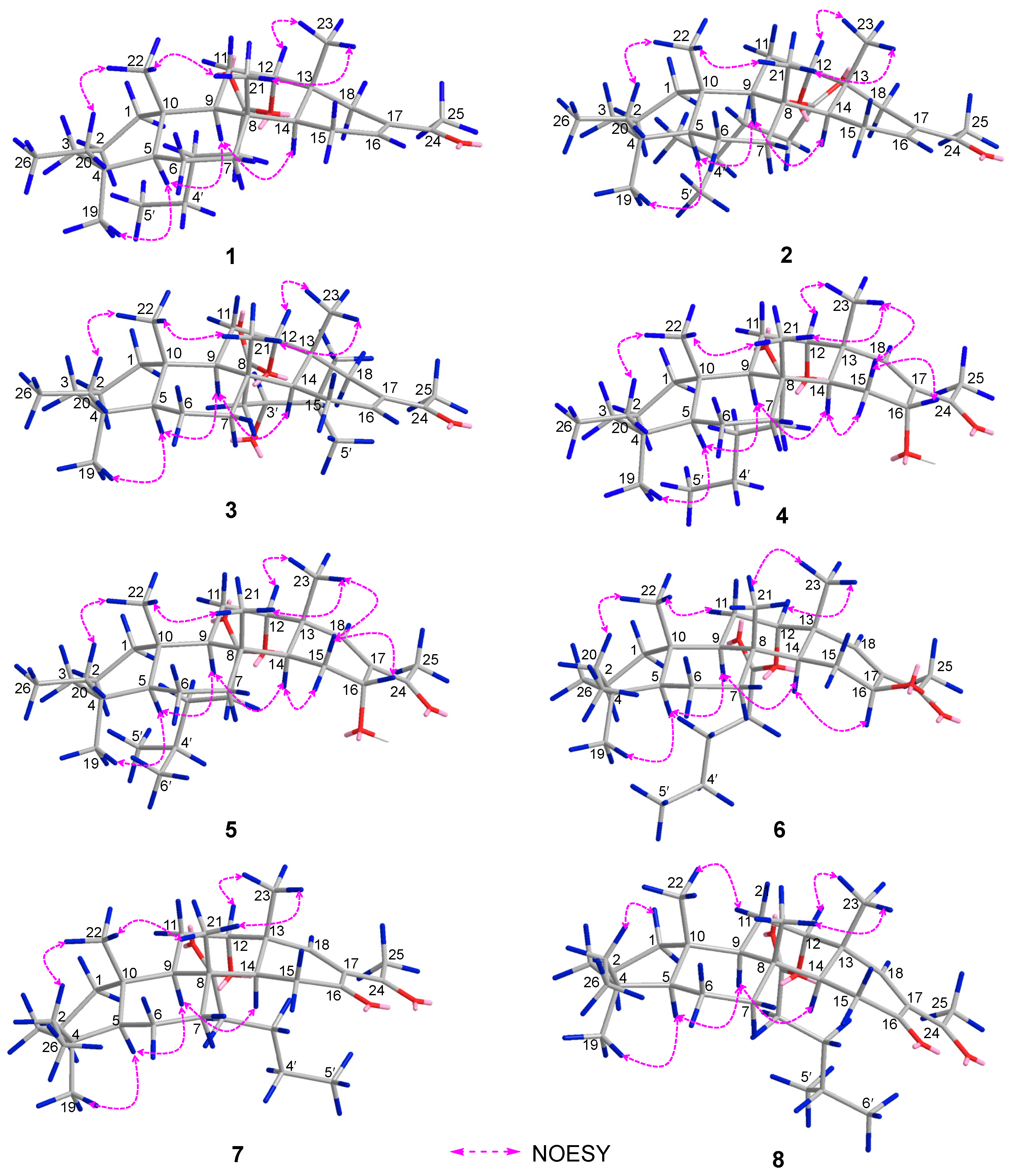
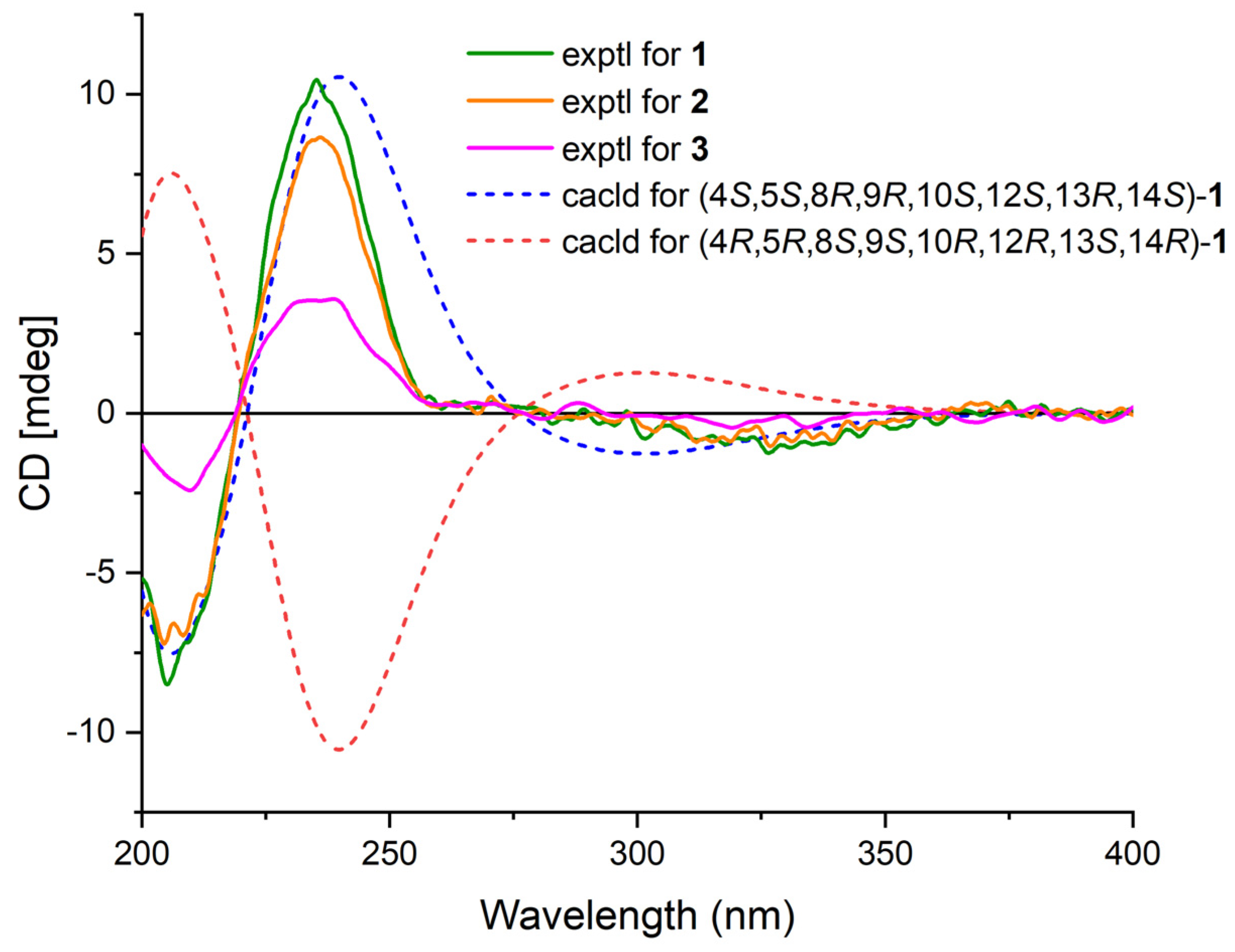
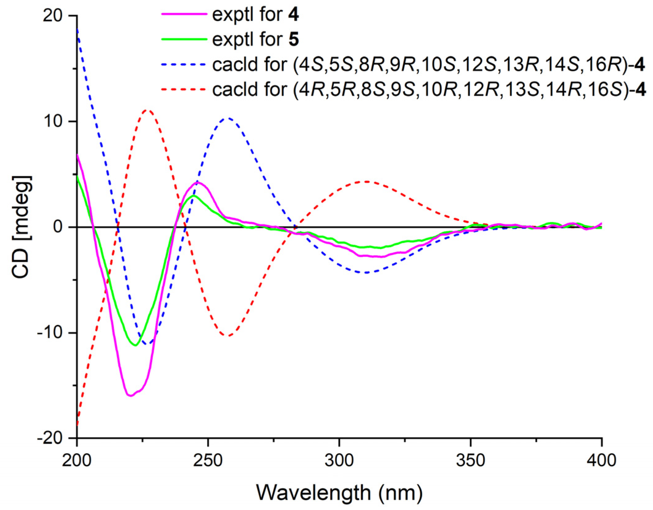
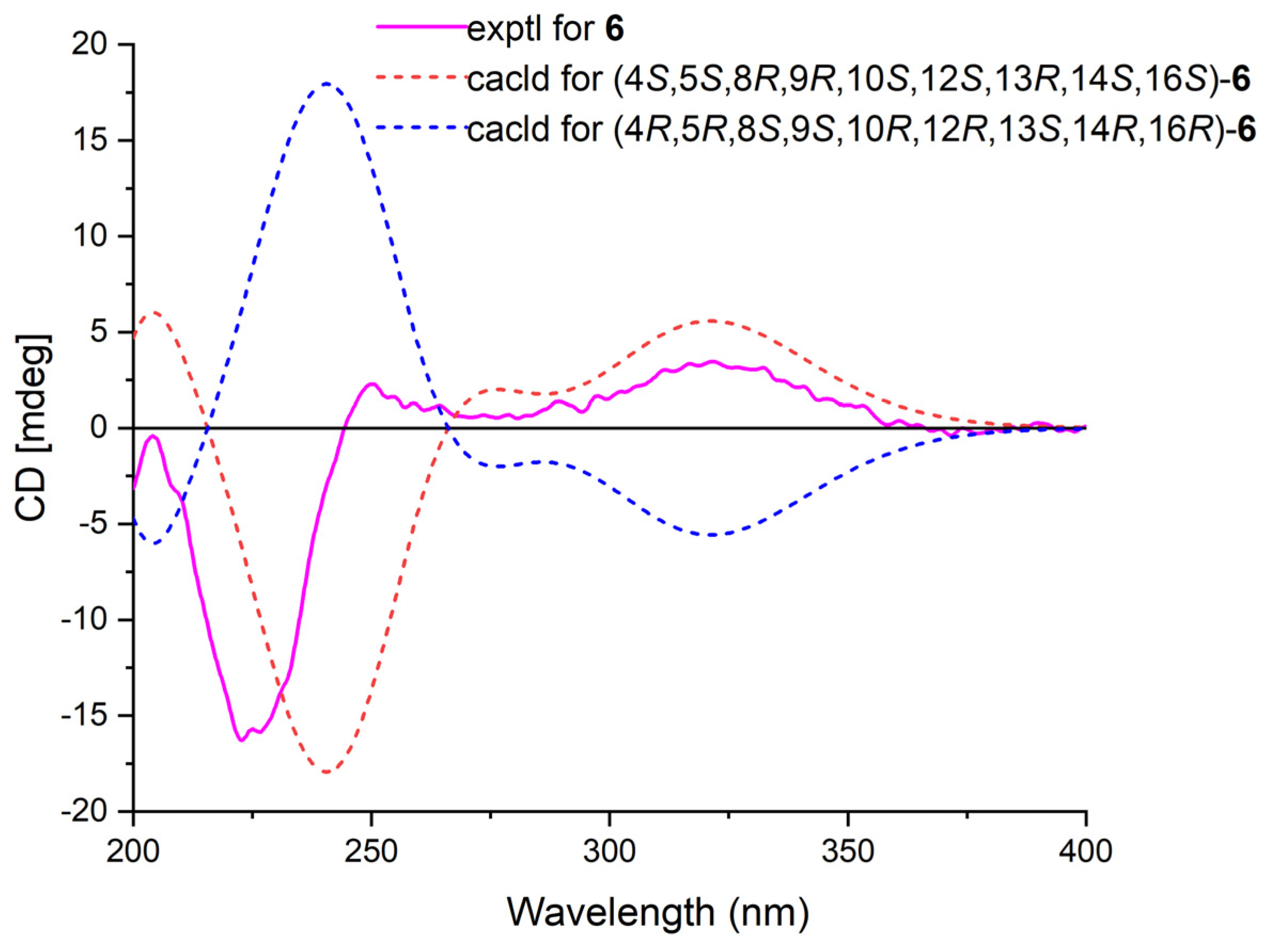
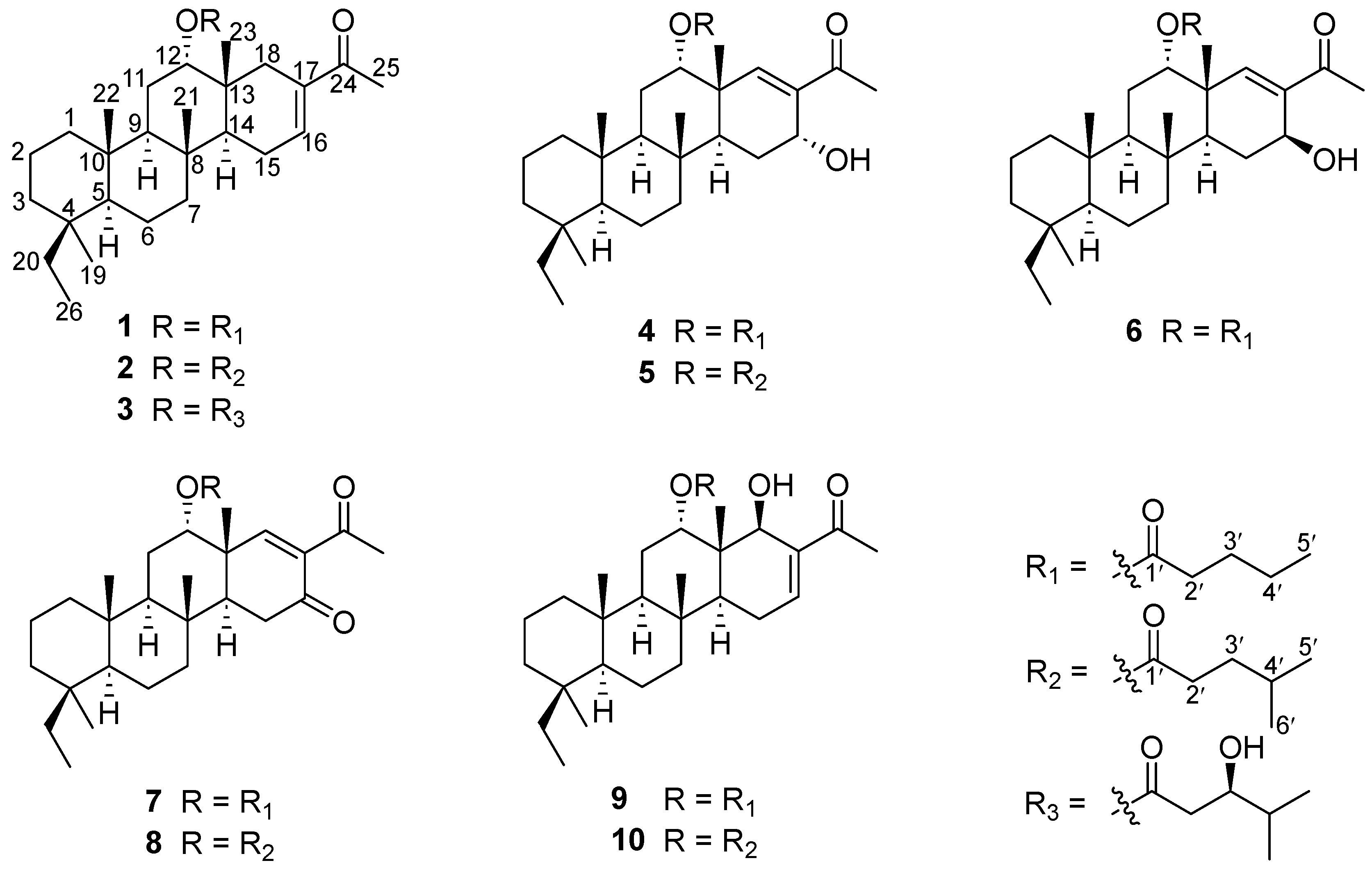
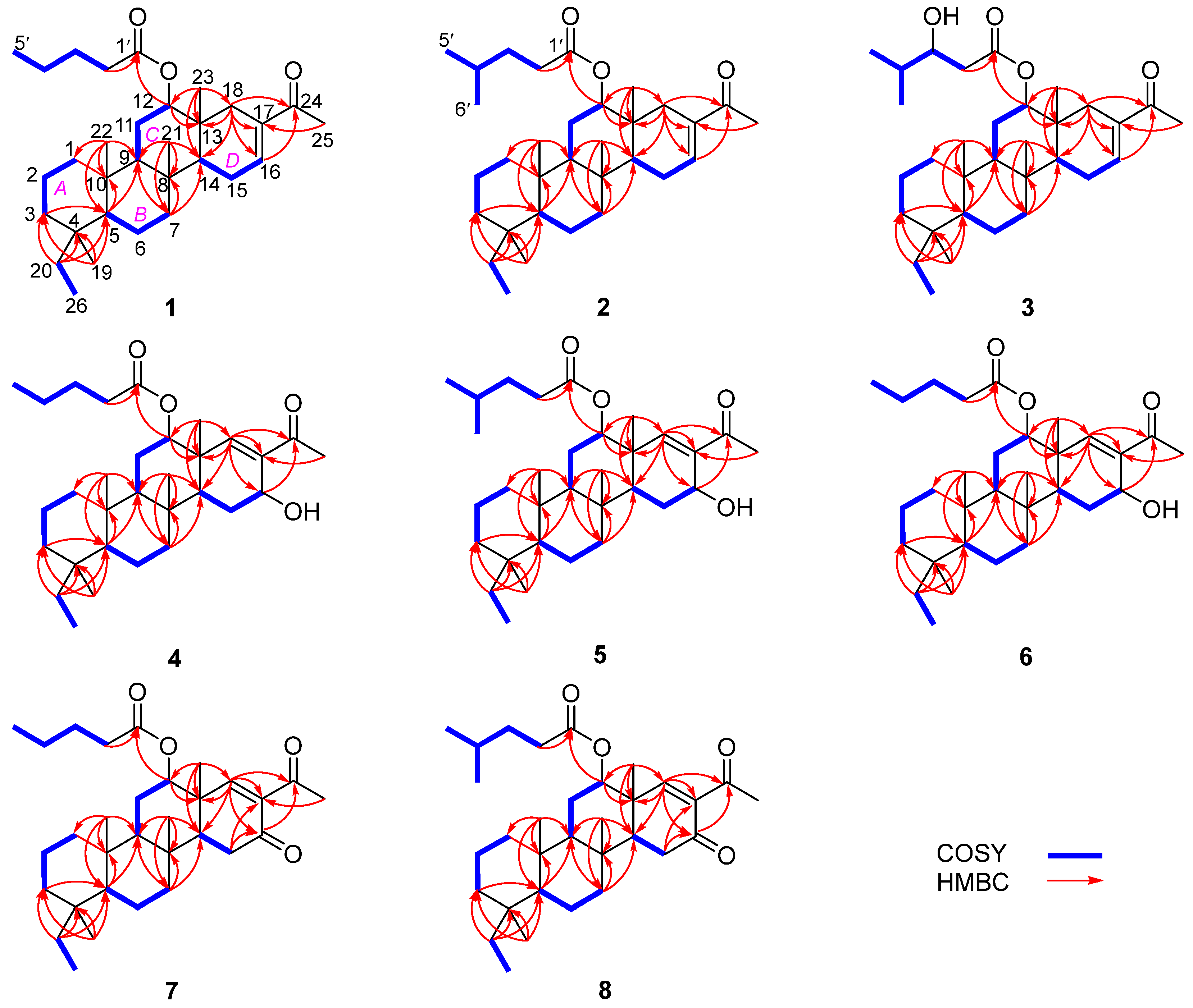
2. Results
3. Materials and Methods
3.1. General Experimental Procedures
3.2. Sponge Material
3.3. Extraction and Isolation
3.4. Acid Hydrolysis of Phyllofenone H (3)
3.5. Biological Assays
4. Conclusions
Supplementary Materials
Author Contributions
Funding
Institutional Review Board Statement
Data Availability Statement
Conflicts of Interest
References
- Gaudêncio, S.P.; Bayram, E.; Lukić Bilela, L.; Cueto, M.; Díaz-Marrero, A.R.; Haznedaroglu, B.Z.; Jimenez, C.; Mandalakis, M.; Pereira, F.; Reyes, F.; et al. Advanced Methods for Natural Products Discovery: Bioactivity Screening, Dereplication, Metabolomics Profiling, Genomic Sequencing, Databases and Informatic Tools, and Structure Elucidation. Mar. Drugs 2023, 21, 308–373. [Google Scholar] [CrossRef] [PubMed]
- Gonzalez, M.A. Scalarane sesterterpenoids. Curr. Bioact. Compd. 2010, 6, 178–206. [Google Scholar] [CrossRef]
- Yu, H.-B.; Chen, H.-Y.; Duan, S.; Zhu, Y.-P.; Hu, B.; He, Y.; Cheng, S.-T.; Jiao, B.-H.; Liu, X.-Y. Bioactive Scalarane-type Sesterterpenoids from Marine Sources. Chem. Biodivers. 2022, 19, e202200049. [Google Scholar] [CrossRef] [PubMed]
- Yu, H.-B.; Hu, B.; Wu, G.-F.; Ning, Z.; He, Y.; Jiao, B.-H.; Liu, X.-Y.; Lin, H.-W. Phyllospongianes A–E, Dinorscalarane Sesterterpenes from the Marine Sponge Phyllospongia foliascens. J. Nat. Prod. 2023, 86, 1754–1760. [Google Scholar] [CrossRef] [PubMed]
- Fattorusso, E.; Magno, S.; Santacroce, C.; Sica, D. Scalarin, a new pentacyclic C-25 terpenoid from the sponge Cacospongia scalaris. Tetrahedron 1972, 28, 5993–5997. [Google Scholar] [CrossRef]
- Zhou, M.; Peng, B.-R.; Tian, W.; Su, J.-H.; Wang, G.; Lin, T.; Zeng, D.; Sheu, J.-H.; Chen, H. Deacetyl-12-epi-scalaradial, a scalarane sesterterpenoid from a marine sponge Hippospongia sp., induces HeLa cells apoptosis via MAPK/ERK pathway and modulates nuclear receptor Nur77. Mar. Drugs 2020, 18, 375–387. [Google Scholar] [CrossRef] [PubMed]
- Sun, J.-B.; Hong, L.-L.; Shang, R.-Y.; Liu, H.-Y.; Zhang, L.; Liu, L.-Y.; Zhao, L.; Zhang, W.; Sun, F.; Jiao, W.-H.; et al. Dysiscalarones A–E, scalarane sesterterpenoids with nitric oxide production inhibitory activity from marine sponge Dysidea granulosa. Bioorg. Chem. 2021, 111, 104791–104799. [Google Scholar] [CrossRef] [PubMed]
- Hertzer, C.; Kehraus, S.; Boehringer, N.; Kaligis, F.; Bara, R.; Erpenbeck, D.; Woerheide, G.; Schaeberle, T.F.; Waegele, H.; Koenig, G.M. Antibacterial scalarane from Doriprismatica stellata nudibranchs (Gastropoda, Nudibranchia), egg ribbons, and their dietary sponge Spongia cf. agaricina (Demospongiae, Dictyoceratida). Beilstein J. Org. Chem. 2020, 16, 1596–1605. [Google Scholar] [CrossRef] [PubMed]
- Abdjul, D.B.; Takahashi, O.; Kirikoshi, R.; Namikoshi, M.; Yamazaki, H.; Mangindaan, R.E.P. Two new protein tyrosine phosphatase 1B inhibitors, hyattellactones A and B, from the Indonesian marine sponge Hyattella sp. Bioorg. Med. Chem. Lett. 2015, 25, 904–907. [Google Scholar] [CrossRef] [PubMed]
- Tian, X.-H.; Hong, L.-L.; Jiao, W.-H.; Lin, H.-W. Natural sesquiterpene quinone/quinols: Chemistry, biological activity, and synthesis. Nat. Prod. Rep. 2023, 40, 718–749. [Google Scholar] [CrossRef] [PubMed]
- Zeng, L.; Fu, X.; Su, J.; Chen, S.; Snyder, J.K. Phyllofenone A, a New Scalarane Sesterterpene from the Sponge Phyllospongia Foliascens (Pallas). Chem. Res. Chin. Univ. 1991, 7, 100–106. [Google Scholar]
- Hassan, M.H.A.; Rateb, M.E.; Hetta, M.; Abdelaziz, T.A.; Sleim, M.A.; Jaspars, M.; Mohammed, R. Scalarane sesterterpenes from the Egyptian Red Sea sponge Phyllospongia lamellosa. Tetrahedron 2015, 71, 577–583. [Google Scholar] [CrossRef]
- Pellicena, M.; Solsona, J.G.; Romea, P.; Urpí, F. Stereoselective titanium-mediated aldol reactions of α-benzyloxy methyl ketones. Tetrahedron 2012, 68, 10338–10350. [Google Scholar] [CrossRef]
- Lorente, A.; Pellicena, M.; Romea, P.; Urpí, F. Stereoselective acetate aldol reactions of α-silyloxy ketones. Tetrahedron 2015, 71, 1023–1035. [Google Scholar] [CrossRef]
- Orsini, F.; Sello, G.; Manzo, A.M.; Lucci, E.M. (1S,2S)-1-Amino-2-hydroxy-1,2,3,4-tetrahydronaphthalene: A new chiral auxiliary for asymmetric Reformatsky reactions. Tetrahedron Asymmetr. 2005, 16, 1913–1918. [Google Scholar] [CrossRef]
- Lan, W.-J.; Li, H.-J. New sesterterpenoids from the marine sponge Phyllospongia papyracea. Helv. Chim. Acta 2007, 90, 1218–1222. [Google Scholar] [CrossRef]
- Wang, Q.; Sun, Y.; Yang, L.; Luo, X.; de Voogd, N.J.; Tang, X.; Li, P.; Li, G. Bishomoscalarane Sesterterpenoids from the Sponge Dysidea granulosa Collected in the South China Sea. J. Nat. Prod. 2020, 83, 516–523. [Google Scholar] [CrossRef] [PubMed]
- Cao, F.; Wu, Z.-H.; Shao, C.-L.; Pang, S.; Liang, X.-Y.; de Voogd, N.J.; Wang, C.-Y. Cytotoxic scalarane sesterterpenoids from the South China Sea sponge Carteriospongia foliascens. Org. Biomol. Chem. 2015, 13, 4016–4024. [Google Scholar] [CrossRef] [PubMed]
- Yu, H.-B.; Glukhov, E.; Li, Y.; Iwasaki, A.; Gerwick, L.; Dorrestein, P.C.; Jiao, B.-H.; Gerwick, W.H. Cytotoxic Microcolin Lipopeptides from the Marine Cyanobacterium Moorea producens. J. Nat. Prod. 2019, 82, 2608–2619. [Google Scholar] [CrossRef] [PubMed]
- Yu, H.-B.; Gu, B.-B.; Iwasaki, A.; Jiang, W.-L.; Ecker, A.; Wang, S.-P.; Yang, F.; Lin, H.-W. Dactylospenes A–E, Sesterterpenes from the Marine Sponge Dactylospongia elegans. Mar. Drugs 2020, 18, 491–501. [Google Scholar] [CrossRef] [PubMed]


| Position | 1 | 2 | 3 | |||
|---|---|---|---|---|---|---|
| δC | δH, Mult. (J in Hz) | δC | δH, Mult. (J in Hz) | δC | δH, Mult. (J in Hz) | |
| 1α | 40.1, CH2 | 0.63, td (12.0, 6.0) | 40.1, CH2 | 0.64, td (12.0, 6.0) | 40.0, CH2 | 0.64, td (12.5, 4.0) |
| 1β | 1.59, m | 1.59, m | 1.57, m | |||
| 2α | 18.2, CH2 | 1.43, m | 18.2, CH2 | 1.43, m | 18.1, CH2 | 1.42, m |
| 2β | 1.36, m | 1.37, m | 1.36, m | |||
| 3α | 36.7, CH2 | 0.83, m | 36.7, CH2 | 0.84, m | 36.6, CH2 | 0.84, m |
| 3β | 1.66, m | 1.66, m | 1.65, m | |||
| 4 | 36.1, C | 36.1, C | 36.1, C | |||
| 5 | 58.7, CH | 0.88, m | 58.7, CH | 0.86, m | 58.6, CH | 1.90, m |
| 6 | 18.0, CH2 | 1.54, m | 18.0, CH2 | 1.55, m | 17.9, CH2 | 1.52, m |
| 7α | 41.7, CH2 | 1.73, m | 41.7, CH2 | 1.74, m | 41.6, CH2 | 1.73, m |
| 7β | 0.99, td (13.0, 4.0) | 0.99, m | 0.98, m | |||
| 8 | 37.4, C | 37.4, C | 37.4, C | |||
| 9 | 53.0, CH | 1.28, dd (12.5, 2.5) | 53.0, CH | 1.28, dd (12.0, 2.5) | 53.0, CH | 1.26, dd (12.0, 2.5) |
| 10 | 36.7, C | 36.9, C | 36.9, C | |||
| 11α | 22.3, CH2 | 1.74, m | 22.2, CH2 | 1.75, m | 22.3, CH2 | 1.70, m |
| 11β | 1.62, m | 1.63, m | 1.77, dt (8.5, 3.5) | |||
| 12 | 76.7, CH | 4.79, t (2.5) | 76.8, CH | 4.80, t (2.5) | 77.6, CH | 4.85, t (2.5) |
| 13 | 35.9, C | 35.9, C | 35.8, C | |||
| 14 | 47.9, CH | 1.53, m | 47.8, CH | 1.52, m | 47.8, CH | 1.52, m |
| 15α | 23.9, CH2 | 2.16, m | 23.9, CH2 | 2.18, m | 23.9, CH2 | 2.18, m |
| 15β | 2.28, m | 2.29, m | 2.27, m | |||
| 16 | 139.5, CH | 6.85, m | 139.5, CH | 6.85, m | 139.7, CH | 6.86, q (2.5) |
| 17 | 137.7, C | 137.7, C | 137.5, C | |||
| 18α | 35.5, CH2 | 2.21, m | 35.5, CH2 | 2.21, m | 35.3, CH | 2.20, m |
| 18β | 1.95, d (18.0) | 1.95, d (18.5) | 1.95, d (16.5) | |||
| 19 | 28.5, CH3 | 0.80, s | 28.6, CH3 | 0.79, s | 28.5, CH3 | 0.79, s |
| 20a | 24.5, CH2 | 1.16, m | 24.5, CH2 | 1.15, m | 24.5, CH2 | 1.15, m |
| 20b | 1.54, m | 1.54, m | 1.51, m | |||
| 21 | 15.9, CH3 | 0.90, s | 15.9, CH3 | 0.90, s | 15.9, CH3 | 0.90, s |
| 22 | 17.1, CH3 | 0.83, s | 17.1, CH3 | 0.83, s | 17.1, CH3 | 0.84, s |
| 23 | 19.9, CH3 | 0.85, s | 19.9, CH3 | 0.85, s | 19.9, CH3 | 0.86, s |
| 24 | 199.0, C | 199.1, C | 199.1, C | |||
| 25 | 25.3, CH3 | 2.28, s | 25.3, CH3 | 2.23, s | 25.2, CH3 | 2.29, s |
| 26 | 8.7, CH3 | 0.74, t (7.5) | 8.7, CH3 | 0.74, s | 8.6, CH3 | 0.74, t (7.5) |
| 1′ | 172.9, C | 173.1, C | 172.9, C | |||
| 2′a | 34.6, CH2 | 2.33, t (7.5) | 32.9, CH2 | 2.32, t (7.5) | 38.7, CH2 | 2.41, dd (16.5, 9.5) |
| 2′b | 2.56, dd (16.5, 2.5) | |||||
| 3′ | 27.3, CH2 | 1.62, m | 34.0, CH2 | 1.54, m | 72.7, CH | 3.79, t (7.5) |
| 4′ | 22.4, CH2 | 1.38, m | 27.7, CH | 1.60, m | 33.1, CH | 1.71, m |
| 5′ | 13.8, CH3 | 0.92, t (7.5) | 22.3, CH3 | 0.90, d 2.0 | 17.8, CH3 | 0.97, d (6.5) |
| 6′ | 22.3, CH3 | 0.90, d 2.0 | 18.6, CH3 | 0.93, d (6.5) | ||
| Position | 4 | 5 | 6 | |||
|---|---|---|---|---|---|---|
| δC | δH, Mult. (J in Hz) | δC | δH, Mult. (J in Hz) | δC | δH, Mult. (J in Hz) | |
| 1α | 40.1, CH2 | 0.69, td (10.5, 3.0) | 40.1, CH2 | 0.67, td (13.0, 4.5) | 40.0, CH2 | 0.65, td (11.0, 3.5) |
| 1β | 1.56, m | 1.55, m | 1.56, m | |||
| 2α | 18.3, CH2 | 1.50, m | 18.3, CH2 | 1.47, m | 17.9, CH2 | 1.47, m |
| 2β | 1.36, m | 1.34, m | 1.34, m | |||
| 3α | 36.6, CH2 | 0.86, m | 36.6, CH2 | 0.83, m | 36.6, CH2 | 0.84, m |
| 3β | 1.67, m | 1.65, m | 1.66, m | |||
| 4 | 36.1, C | 36.1, C | 36.1, C | |||
| 5 | 58.7, CH | 0.95, dd (10.5, 2.0) | 58.6, CH | 0.93, dd (12.0, 2.0) | 58.5, CH | 0.90, m |
| 6 | 17.9, CH2 | 1.59, m | 17.9, CH2 | 1.55, m | 18.2, CH2 | 1.55, m |
| 7α | 41.1, CH2 | 1.84, dt (10.5, 2.5) | 41.1, CH2 | 1.81, m | 41.2, CH2 | 1.84, dt (10.5, 2.5) |
| 7β | 1.14, td (10.5, 2.5) | 1.11, td (13.0, 4.0) | 0.97, m | |||
| 8 | 36.9, C | 37.0, C | 37.0, C | |||
| 9 | 53.5, CH | 1.38, m | 53.5, CH | 1.38, m | 53.4, CH | 1.24, m |
| 10 | 37.0, C | 36.9, C | 37.2, C | |||
| 11α | 22.3, CH2 | 1.75, m | 22.3, CH2 | 1.74, m | 22.1, CH2 | 1.72, m |
| 11β | 1.58, m | 1.37, m | 1.75, m | |||
| 12 | 76.1, CH | 5.06, t (2.0) | 76.2, CH | 5.05, t (3.0) | 76.1, CH | 5.03, t (2.5) |
| 13 | 41.4, C | 41.4, C | 41.5, C | |||
| 14 | 43.8, CH | 1.88, m | 43.7, CH | 1.88, m | 47.3, CH | 1.48, m |
| 15α | 25.2, CH2 | 1.58, m | 25.2, CH2 | 1.58, m | 25.5, CH2 | 1.49, m |
| 15β | 1.87, m | 1.86, m | 2.12, m | |||
| 16 | 63.3, CH | 4.56, d (4.0) | 63.3, CH | 4.54, d (4.5) | 68.0, CH | 4.59, m |
| 17 | 138.2, C | 138.2, C | 138.8, C | |||
| 18 | 152.2, CH | 6.60, s | 152.2, CH | 6.59, s | 152.4, CH | 6.57, s |
| 19 | 28.5, CH3 | 0.81, s | 28.5, CH3 | 0.79, br.s | 28.5, CH3 | 0.80, s |
| 20a | 24.5, CH2 | 1.19, m | 24.4, CH2 | 1.17, m | 24.4, CH2 | 1.19, m |
| 20b | 1.53, m | 1.59, m | 1.52, d (6.0) | |||
| 21 | 17.1, CH3 | 0.88, s | 17.1, CH3 | 0.87, s | 17.1, CH3 | 0.89, s |
| 22 | 16.8, CH3 | 0.86, s | 16.8, CH3 | 0.84, s | 16.7, CH3 | 0.84, s |
| 23 | 19.7, CH3 | 1.07, s | 19.7, CH3 | 1.05, s | 21.1, CH3 | 1.18, s |
| 24 | 201.5, C | 201.5, C | 202.1, C | |||
| 25 | 25.4, CH3 | 2.26, s | 25.4, CH3 | 2.24, s | 25.6, CH3 | 2.23, s |
| 26 | 8.6, CH3 | 0.76, t (6.5) | 8.7, CH3 | 0.74, t (4.5) | 8.6, CH3 | 0.74, t (6.0) |
| 1′ | 173.6, C | 173.8, C | 173.4, C | |||
| 2′ | 34.6, CH2 | 2.31, td (6.0, 1.0) | 32.9, CH2 | 2.30, t (7.5) | 34.5, CH2 | 2.28, dd (6.0, 1.5) |
| 3′ | 27.5, CH2 | 1.58, m | 34.2, CH2 | 1.48, m | 27.4, CH2 | 1.57, m |
| 4′ | 22.3, CH2 | 1.35, m | 27.6, CH | 1.55, m | 22.3, CH2 | 1.34, dd (12.5, 6.0) |
| 5′ | 13.8, CH3 | 0.90, t (6.0) | 22.3, CH3 | 0.87, d (3.5) | 13.8, CH3 | 0.90, t (6.0) |
| 6′ | 22.2, CH3 | 0.88, d (3.5) | ||||
| Position | 7 | 8 | ||
|---|---|---|---|---|
| δC | δH, Mult. (J in Hz) | δC | δH, Mult. (J in Hz) | |
| 1α | 40.0, CH2 | 0.65, td (11.0, 3.5) | 40.0, CH2 | 0.63, td (13.5, 4.0) |
| 1β | 1.58, m | 1.56, m | ||
| 2α | 17.8, CH2 | 1.47, m | 18.2, CH2 | 1.47, m |
| 2β | 1.36, m | 1.35, m | ||
| 3α | 36.6, CH2 | 0.85, m | 36.6, CH2 | 0.82, m |
| 3β | 1.68, m | 1.67, m | ||
| 4 | 36.1, C | 36.1, C | ||
| 5 | 58.8, CH | 0.90, m | 58.8, CH | 0.88, m |
| 6α | 17.8, CH2 | 1.59, m | 17.8, CH2 | 1.57, m |
| 6β | ||||
| 7α | 40.8, CH2 | 1.75, m | 40.8, CH2 | 1.70, m |
| 7β | 1.04, td (10.5, 3.0) | 1.01, td (12.0, 3.5) | ||
| 8 | 37.3, C | 37.3, C | ||
| 9 | 53.0, CH | 1.27, m | 53.0, CH | 1.25, m |
| 10 | 37.0, C | 37.0, C | ||
| 11α | 22.0, CH2 | 1.87, dt (11.0, 1.5) | 22.0, CH2 | 1.85, dt (15.0, 3.0) |
| 11β | 1.73, m | 1.73, m | ||
| 12 | 75.8, CH | 5.08, t (2.5) | 75.8, CH | 5.04, t (3.0) |
| 13 | 41.3, C | 41.3, C | ||
| 14 | 48.7, CH | 2.13, dd (12.0, 9.0) | 48.7, CH | 2.09, dd (14.5, 4.0) |
| 15α | 34.9, CH2 | 2.43, m | 34.9, CH2 | 2.40, m |
| 15β | 2.52, dd (14.5, 3.5) | 2.50, dd (17.5, 4.0) | ||
| 16 | 197.8, C | 197.1, C | ||
| 17 | 136.5, C | 136.5, C | ||
| 18 | 163.9, CH | 7.31, s | 163.9, CH | 7.29, s |
| 19 | 28.5, CH3 | 0.82, s | 28.5, CH3 | 0.82, s |
| 20a | 24.5, CH2 | 1.18, m | 24.5, CH2 | 1.15, m |
| 20b | 1.54, m | 1.51, m | ||
| 21 | 16.4, CH3 | 0.96, s | 16.4, CH3 | 0.93, s |
| 22 | 16.9, CH3 | 0.87, s | 16.9, CH3 | 0.84, s |
| 23 | 18.5, CH3 | 1.17, s | 18.5, CH3 | 1.15, s |
| 24 | 197.9, C | 197.8, C | ||
| 25 | 30.6, CH3 | 2.44, s | 30.7, CH3 | 2.42, s |
| 26 | 8.7, CH3 | 0.76, t (6.0) | 8.6, CH3 | 0.80, t (7.5) |
| 1′ | 173.0, C | 173.2, C | ||
| 2′ | 34.4, CH2 | 2.31, t (6.5) | 32.7 CH2 | 2.29, t (8.0) |
| 3′ | 27.3, CH2 | 1.58, m | 34.1, CH2 | 1.48, m |
| 4′ | 22.3, CH2 | 1.36, m | 27.6, CH | 1.56, m |
| 5′ | 13.8, CH3 | 0.92, t (4.0) | 22.2, CH3 | 0.88, d (5.0) |
| 6′ | 22.3, CH3 | 0.89, d (5.0) | ||
| Compd | HeLa | HCT-116 | H460 | SW1990 |
|---|---|---|---|---|
| 1 | >20 | >20 | >20 | >20 |
| 2 | 11.8 | 19.8 | 18.5 | 9.8 |
| 3 | NT | NT | NT | NT |
| 4 | 15.3 | 17.2 | 15.3 | 13.2 |
| 5 | 3.4 | 7.3 | 5.9 | 3.5 |
| 6 | >20 | >20 | >20 | >20 |
| 7 | 18.8 | 16.2 | 12.3 | 11.5 |
| 8 | 6.9 | 7.2 | 10.1 | 9.2 |
| 9 | >20 | >20 | >20 | >20 |
| 10 | 14.4 | 16.2 | 17.7 | 14.6 |
| Cisplatin | 0.5 | 2.6 | 2.8 | 1.1 |
Disclaimer/Publisher’s Note: The statements, opinions and data contained in all publications are solely those of the individual author(s) and contributor(s) and not of MDPI and/or the editor(s). MDPI and/or the editor(s) disclaim responsibility for any injury to people or property resulting from any ideas, methods, instructions or products referred to in the content. |
© 2023 by the authors. Licensee MDPI, Basel, Switzerland. This article is an open access article distributed under the terms and conditions of the Creative Commons Attribution (CC BY) license (https://creativecommons.org/licenses/by/4.0/).
Share and Cite
Yu, H.-B.; Hu, B.; Ning, Z.; He, Y.; Men, X.-L.; Yin, Z.-F.; Jiao, B.-H.; Liu, X.-Y.; Lin, H.-W. Phyllofenones F–M, Scalarane Sesterterpenes from the Marine Sponge Phyllospongia foliascens. Mar. Drugs 2023, 21, 507. https://doi.org/10.3390/md21100507
Yu H-B, Hu B, Ning Z, He Y, Men X-L, Yin Z-F, Jiao B-H, Liu X-Y, Lin H-W. Phyllofenones F–M, Scalarane Sesterterpenes from the Marine Sponge Phyllospongia foliascens. Marine Drugs. 2023; 21(10):507. https://doi.org/10.3390/md21100507
Chicago/Turabian StyleYu, Hao-Bing, Bo Hu, Zhe Ning, Ying He, Xiao-Ling Men, Zi-Fei Yin, Bing-Hua Jiao, Xiao-Yu Liu, and Hou-Wen Lin. 2023. "Phyllofenones F–M, Scalarane Sesterterpenes from the Marine Sponge Phyllospongia foliascens" Marine Drugs 21, no. 10: 507. https://doi.org/10.3390/md21100507
APA StyleYu, H.-B., Hu, B., Ning, Z., He, Y., Men, X.-L., Yin, Z.-F., Jiao, B.-H., Liu, X.-Y., & Lin, H.-W. (2023). Phyllofenones F–M, Scalarane Sesterterpenes from the Marine Sponge Phyllospongia foliascens. Marine Drugs, 21(10), 507. https://doi.org/10.3390/md21100507