Article

Coral Holobionts Possess Distinct Lipid Profiles That May Be Shaped by Symbiodiniaceae Taxonomy

1
A.V. Zhirmunsky National Scientific Center of Marine Biology, Far Eastern Branch, Russian Academy of Sciences, ul. Palchevskogo 17, 690041 Vladivostok, Russia
2
Institute of Natural Products Chemistry, Vietnam Academy of Science and Technology, 18 Hoang Quoc Viet, Hanoi 100000, Vietnam
3
Graduate University of Science and Technology, Vietnam Academy of Science and Technology, Hanoi 100000, Vietnam
*
Author to whom correspondence should be addressed.
Mar. Drugs 2022, 20(8), 485; https://doi.org/10.3390/md20080485
Submission received: 11 May 2022 / Revised: 25 July 2022 / Accepted: 25 July 2022 / Published: 28 July 2022
(This article belongs to the Special Issue Reef Ecology and Marine Drug Discovery)

Abstract

:
Symbiotic relationships are very important for corals. Abiotic stressors cause the acclimatization of cell membranes in symbionts, which possess different membrane acclimatization strategies. Membrane stability is determined by a unique lipid composition and, thus, the profile of thylakoid lipids can depend on coral symbiont species. We have analyzed and compared thylakoid lipidomes (mono- and digalactosyldiacylglycerols (MGDG and DGDG), sulfoquinovosyldiacylglycerols (SQDG), and phosphatidylglycerols (PG)) of crude extracts from symbiotic reef-building coral Acropora sp., the hydrocoral Millepora platyphylla, and the octocoral Sinularia flexibilis. S. flexibilis crude extracts were characterized by a very high SQDG/PG ratio, a DGDG/MGDG ratio < 1, a lower degree of galactolipid unsaturation, a higher content of SQDG with polyunsaturated fatty acids, and a thinner thylakoid membrane which may be explained by the presence of thermosensitive dinoflagellates Cladocopium C3. In contrast, crude extracts of M. platyphylla and Acropora sp. exhibited the lipidome features of thermotolerant Symbiodiniaceae. M. platyphylla and Acropora sp. colonies contained Cladocopium C3u and Cladocopium C71/C71a symbionts, respectively, and their lipidome profiles showed features that indicate thermotolerance. We suggest that an association with symbionts that exhibit the thermotolerant thylakoid lipidome features, combined with a high Symbiodiniaceae diversity, may facilitate further acclimatization/adaptation of M. platyphylla and Acropora sp. holobionts in the South China Sea.

1. Introduction

Coral, as a holobiont, forms complex interactions with a wide range of microorganisms that constitute an important part of the entire metaorganism [1]. The best-known coral symbionts are intracellular microalgae (dinoflagellates of the family Symbiodiniaceae) [1]. In response to an increase in sea surface temperature and solar radiation, coral symbionts can be completely or partially expelled, which results in coral bleaching. This process is a major cause of coral reef destruction [2] and, therefore, a subject of intensive research worldwide. Indo-Pacific coral reefs are spread over a vast ocean area. The South China Sea (SCS) is a region in the central Indo-Pacific [3] that, despite being adjacent to the western border of the Coral Triangle, a recognized center of maximum marine biodiversity, has received much less scientific and conservation attention [4]. Nevertheless, SCS coral reefs also exhibit an unexpectedly high biodiversity [5].
Reef-building scleractinian corals (Anthozoa), producing calcium carbonate (CaCO3) skeletons, are a key habitat-forming group in tropical reefs that provides shelter for many other marine organisms [6]. Approximately 757 species of reef-building corals are currently reported for shallow-water reefs of the Indo-Pacific [7]. Symbiotic taxa from the family Milleporidae (Hydrozoa) are also important members of reef-building cnidarians. The genus Millepora comprises around 16 species distributed over tropical/subtropical seas worldwide [8]. Octocorals are considered the second most common group of macrobenthic animals after the reef-building stony corals in many shallow-water Indo-Pacific reefs [9]. Octocorallia (Anthozoa) can be recognized by their distinctive morphological features: polyps with eight-fold symmetry and an internal skeleton composed of microscopic calcareous sclerites [10]. Alcyonacea is the most speciose order of Octocorallia, encompassing the vast majority of octocoral taxa in tropical coral reefs [9].
On the basis of nuclear ribosomal DNA (rDNA) (the ribosomal large subunit (LSU) 28S rDNA and small subunit (SSU) 18S rDNA, and the internal transcribed spacer (ITS) regions) and plastid 23S rDNA marker regions, the family Symbiodiniaceae was divided into nine phylogenetic clades (A to I) and then further subdivided into 15 genus-level lineages [11,12,13,14,15,16]. Symbiotic corals are most commonly associated with Symbiodiniaceae of the genera Symbiodinium (clade A), Breviolum (clade B), Cladocopium (clade C), Durusdinium (clade D), Effrenium (clade E), Fugacium (clade F), and Gerakladium (clade G) [16]. Various Symbiodiniaceae genera differ in their degree of tolerance to increased light and temperature conditions [2,17,18,19]. For example, Symbiodinium thermophilum is a locally abundant Symbiodiniaceae species in reef-building corals of the southern Persian Gulf (Arabian Sea), a habitat distinguished by a high water temperature [20]. The thermotolerant Durusdinium trenchii is widely distributed in ocean waters with abnormally high temperatures [21], while common symbionts of the genus Cladocopium are known to be more thermosensitive [21]. Nevertheless, a culture of Cladocopium C1 or Breviolum psygmophilum (1046) in laboratory experiments exhibited better photophysiology compared to other Symbiodiniaceae species [22,23]. These adaptations translate into different properties for the coral host [24,25].
Another photosynthesizing coral symbiont is an endolithic alga of the genus Ostreobium (Bryopsidales, Ulvophyceae) [26]. It absorbs light of a wavelength not used by symbiotic dinoflagellates. Ostreobium carries out oxygenic photosynthesis even under extreme low light conditions [27]. It has been assumed to replace the function of symbiotic dinoflagellate during bleaching events [28]. An environmental genome survey has shown a possible co-evolution between Ostreobium and both coral and dinoflagellates [26]. In contrast to that of Symbiodiniaceae, the relationship between the Ostreobium and a cnidarian host are far from being completely understood.
Lipids play an essential role in metabolism and maintaining health of corals [29,30]. The lipid composition of each taxon largely depends on its genetic capability of synthesizing certain fatty acids (FA). Therefore, FAs are successfully used in chemical systematics of various taxonomic groups including bacteria, fungi, macro- and microalgae, and corals [31,32,33]. Several classes of glycolipids such as sulfoquinovosyldiacylglycerol (SQDG), monogalactosyldiacylglycerol and digalactosyldiacylglycerol (MGDG and DGDG), and phosphorus-containing class phosphatidylglycerol (PG) are essential components of biomembranes of the photosynthetic apparatus in plants and form a basis of symbiotic dinoflagellate lipids [34,35,36]. Their relative proportions are stable, indicating the presence of mechanisms responsible for and controlling a steady state (homeostasis) of the membrane lipidome in thylakoids, in certain environmental and physiological contexts [37]. These lipid classes, as well as some polyunsaturated fatty acids (PUFA), which comprise acyl groups of MGDG and DGDG, are considered the major lipid biomarkers of coral endosymbionts [38,39,40]. There are few studies on the molecular species profile of certain thylakoid lipid classes in coral endosymbionts [37,38]. Studies of thylakoid membranes are mainly based on the FA composition of total thylakoid lipids (SQDG, MGDG, and DGDG) [38,39,40]. SQDG mainly contains saturated acyl chains [37], while galactolipids are highly unsaturated [37], and their combined analysis may be challenging.
Since it was previously shown that the composition of marker lipid classes (PG, MGDG, DGDG, and SQDG) of the Symbiodiniaceae genera with different thermal sensitivities has distinctive features [41,42], the profile of thylakoid lipids can depend on species of photosynthetic coral symbionts. In order to test this hypothesis, we collected colonies of the major taxonomic groups of SCS corals: the reef-building coral Acropora sp., the hydrocoral Millepora platyphylla, and the octocoral Sinullaria flexibilis. We investigated the molecular genetic diversity of symbiotic dinoflagellates and Ostreobium communities associated with colonies of this coral species on the basis of the nuclear ITS1-5.8S-ITS2 rRNA region, ITS2 region, 28S rRNA gene, and plastid 23S rRNA (pDNA) sequences. Using chromatography with high resolution mass-spectrometry, we identified lipidome profiles of the thylakoid membrane of symbiotic coral dinoflagellates (PG, MGDG, DGDG, and SQDG). Then, we compared the obtained genetic and lipidome data.

2. Results

2.1. Genetic Identification of Coral Endosymbionts

To identify the intragenomic variants for the internal transcribed spacer-2 (ITS2) region, we determined the SymPortal [43] profiles for the Symbiodiniaceae community in corals. Our study shows the presence of multiple Symbiodiniaceae types in all of the Acropora sp. and M. platyphylla colonies and in three colonies of S. flexibilis (Table 1).
The most common types among Acropora sp. colonies were Breviolum B1 (B. minutum) and Cladocopium C3u, occurring together in the same colonies. Symbiodinium A1a type was found in two colonies of Acropora, with Breviolum and Cladocopium occurring in the same colonies. Some of Symbiodinaceae taxa were found in a single colony, for example, C66 and C3sg in A2 colony. The rare C50bn type was found in two (A4 and A5) of five Acropora sp. colonies. We also found a sequence of green algae, Ostreobium sp. (Bryopsidales, Ulvophyceae), in the A4 colony. The 23S rRNA gene sequence shared a 99% similarity to the published sequence of Ostreobium sp. HV05042 [44] with only a 1-bp difference.
The Cladocopium C71/C71a type was common across M. platyphylla colonies. Durusdinium D1 type (D. trenchii) was found in four colonies of M. platyphylla, with Cladocopium in the same colony. Type C3 was found only in the M5 colony mixed with C3sg, C71/C71a, and D1. We also found sequences of green algae Ostreobium sp. (Bryopsidales, Ulvophyceae) in the M2–M5 colonies. The 23S rRNA gene sequences in the M2–M5 colonies shared a 100% identity with the same sequence from the Acropora colony A4 and a 99% similarity to the published sequence of Ostreobium sp. HV05042.
Most Symbiodiniaceae species found in the S. flexibilis colonies were of two main types, C3 and C71/C71a. The Breviolum B1 type (B. minutum) was found only in the S1 colony, and the C3sg type was in the S4 coral colony. Some of the colonies were associated with one Symbiodiniaceae type. S2 and S3 harbored the C3 type, whereas colonies of S1, S4, and S5 were associated with multiple types: B1 + C3 + C71/C71a, C3 + C3sg + C71/C71a, and C3 + C71/C71a, respectively.

2.2. Thylakoid Membrane Lipidome of Coral Endosymbionts

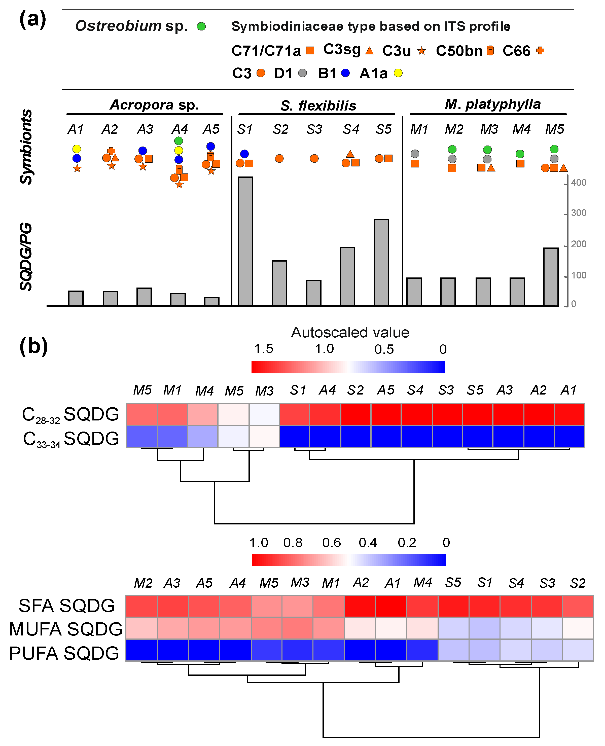
2.2.1. Phospholipid PG and Glycolipid SQDG

Coral lipid extracts contained small amounts of PG (0.003–0.147% of lipid extracts), especially in S. flexibilis, where only a single molecular species (PG 16:2/20:2) was detected (Table S1). The Acropora sp., S. flexibilis, and M. platyphylla extracts contained SQDG at 0.50–13.86% of lipid extracts (Table S1). The SQDG/PG ratio varied between corals. The highest SQDG/PG ratio was observed in S. flexibilis (from 415.95 to 82.32). In Acropora sp. and M. platyphylla, the SQDG/PG ratios were 25.71–46.85 and 40.92–186.68, respectively (Figure 1a, Table S1).
In this study, 14 molecular species of SQDG were identified (Figure 1b, Table S1). They were grouped on the basis of chain length and unsaturation degree of FA residues: C28–32 SQDG (with C14,16 FAs), C33–34 SQDG (with C17,18 FAs), SFA SQDG (with saturated FAs), MUFA SQDG (with monounsaturated FAs), and PUFA SQDG. The total C28–32 SQDG content was higher in the Acropora sp. and S. flexibilis lipid extracts than in the M. platyphylla extracts. A higher content of C33–34 FA SQDG species (16:0/17:0, 16:0/18:3, 16:0/18:2, 16:0/18:1, and 16:0/18:0) was observed in the M. platyphylla extracts. These components were not detected in the Acropora sp. and S. flexibilis samples (Table S1). The content of MUFA SQDG in S. flexibilis lipids was significantly lower than that in M. platyphylla and Acropora sp. lipids (HSD test, p < 0.1). Moreover, a high content of PUFA SQDG was observed in S. flexibilis (16.35 ± 1.85% of total SQDG). These molecular species were absent in Acropora sp. and were present in M. platyphylla only in small amounts (0.82 ± 0.51% of total SQDG) (Table S1). A cluster analysis of SQDG with different chain lengths split the coral species into two clusters: (1) M. platyphylla and (2) S. flexibilis and Acropora sp. colonies. A cluster analysis of SQDG with different unsaturation degrees split the coral species into two clusters: (1) S. flexibilis and (2) Acropora sp. and M. platyphylla colonies (Figure 1b).

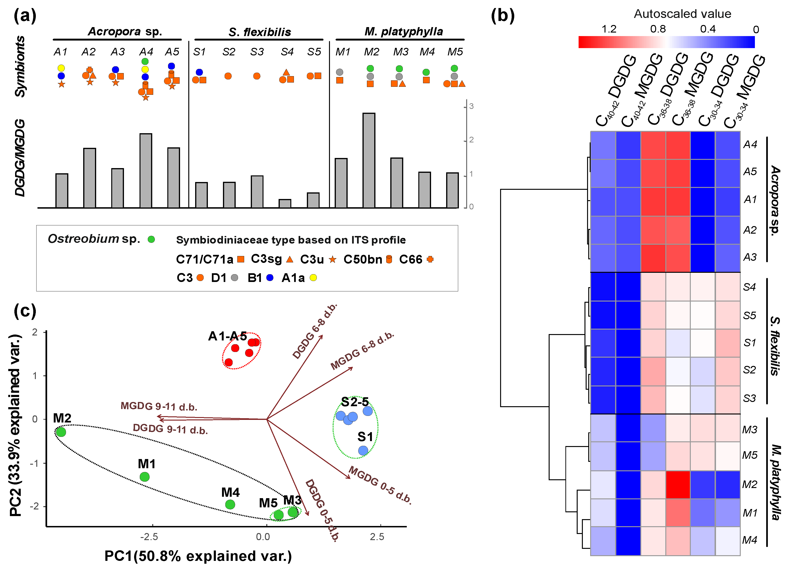
2.2.2. Galactolipids MGDG and DGDG

The Acropora, S. flexibilis, and M. platyphylla extracts contained MGDG at 1.23–5.32% and DGDG at 0.86–6.48% of lipid extracts, respectively. The DGDG/MGDG ratio varied between corals. The lowest DGDG/MGDG ratio was observed in S. flexibilis (0.25–0.95). In Acropora sp. and M. platyphylla, the DGDG/MGDG ratios were 1.01–2.19 and 1.04–2.95, respectively (Figure 2a, Table S1).
A total of 26 molecular species of MGDG and 24 molecular species of DGDG were identified (Table S1). These were grouped on the basis of chain length of FA residues: C30–34 MGDG and DGDG (with C16,18 FAs), C36–38 MGDG and DGDG (with C18,20 FAs), and C40–42 MGDG and DGDG (with C18,20,22 FAs) (Figure 2b, Table 2). C36–38 MGDG and DGDG were most abundant in the Acropora sp. colonies (89.08 ± 2.87 and 89.77 ± 1.98% of total MGDG and DGDG, respectively). In the S. flexibilis and M. platyphylla colonies, C30–34 and C36–38 MGDG and DGDG were most abundant in comparable proportions: in S. flexibilis, MGDG accounted for 56.18 ± 4.60 and 43.61 ± 4.37%, and DGDG accounted for 40.51 ± 6.02 and 58.71 ± 5.83%, respectively. In the M. platyphylla colonies M3-M5, MGDG accounted for 45.73 ± 5.71 and 54.27 ± 6.17%, DGDG accounted for 43.78 ± 14.10 and 29.29 ± 17.44% of total DGDG, respectively. C40–42 MGDG were detected only in the Acropora sp. colonies (4.43 ± 0.96% of total MGDG). C40–42 DGDG were detected in all of the coral colonies studied. Their content was significantly higher in M. platyphylla colonies (29.86 ± 5.62% of total DGDG) relative to the S. flexibilis and Acropora sp. colonies (0.98 ± 0.94 and 8.83 ± 2.29% of total DGDG, respectively) (Table S1). A cluster analysis of MGDG and DGDG with different FA lengths split the coral colonies into two clusters: (1) Acropora sp. colonies and (2) S. flexibilis and M. platyphylla colonies (Figure 2b).
The MGDG and DGDG molecular species were grouped on the basis of the unsaturation degree of molecules: MGDG and DGDG with 0–5 double bonds, MGDG and DGDG with 6–8 double bonds, and MGDG and DGDG with 9–11 double bonds (Figure 2c; Table S1). A PCA analysis of MGDG and DGDG molecular species with different unsaturation degrees (with PC1 constituting 58% of variations; PC2, 33%; and eigenvalue > 1) showed a shift of S. flexibilis colonies along the PC1 axis (PC1 axis correlated with MGDG and DGDG with 9–11 double bonds (r = −0.53 and r = −0.51, respectively) and MGDG with 0–5 and 6–8 double bonds (r = 0.40 and r = 0.42, respectively)) (Figure 2c and Figure S1; Table S2). This shift was driven by a higher content of MGDG with 0–5 and 6–8 double bonds (34.83 ± 5.31 and 47.68 ± 5.06% of total MGDG, respectively) than that in M. platyphylla colonies, and lower contents of MGDG and DGDG with 9–11 double bonds than these in M. platyphylla and Acropora sp. colonies. The shift of the Acropora sp. colonies along the PC2 axis (PC2 axis correlated with DGDG with 0–5 and 6–8 double bonds (r = −0.64 and r = 0.56, respectively) and MGDG with 0–5 and 6–8 double bonds (r = −0.39 and r = 0.34, respectively)) was driven by a higher content of MGDG and DGDG with 6–8 double bonds (49.01 ± 2.17 and 45.15 ± 6.89% of total MGDG and DGDG, respectively), a lower content of MGDG with 0–5 double bonds (5.14 ± 1.48% of total MGDG), and the lack of DGDG with 0–5 double bonds. On the basis of these parameters, the Acropora, M. platyphylla, and S. flexibilis colonies formed separate clusters (Figure 2c and Figure S2). The M. platyphylla colonies were not grouped into common clusters with Acropora or S. flexibilis. The contents of MGDG and DGDG with different unsaturation degrees were not similar between the M. platyphylla colonies (Table 2). M3 and M5 were similar in contents of MGDG and DGDG with different unsaturation degrees. MGDG with 6–8 double bonds (41.28 ± 1.65% of total MGDG) and DGDG with 0–5 double bonds (57.82 ± 1.30% of total DGDG) were most abundant. M1, M2, and M4 contained a higher percentage of MGDG (90.18, 82.82, and 43.90% of total MGDG, respectively) and DGDG (90.18, 82.82, and 63.21% of total DGDG, respectively) with 9–11 double bonds. The lowest percentage of MGDG and DGDG with 0–5 double bonds (0.32 and 2.61% of total MGDG and DGDG, respectively) was recorded from M1.

3. Discussion

Symbiotic relationships are of crucial importance for a coral holobiont and its adaptation to changing environmental conditions. Various corals have different degrees of specificity to certain types of Symbiodiniaceae. The species-specific interactions between algal endosymbionts and coral hosts are determined by the glycan–lectin interactions [45]. Thus, corals contain one main Symbiodiniaceae genus and several additional as a way of adaptation [46]. According to a genetic analysis, Acropora sp. (A1–A5), M. platyphylla (M1–M5), and S. flexibilis (S1, S4, S5) contained several species of symbionts (symbiotic dinoflagellates and Ostreobium sp.). A cluster analysis of thylakoid lipids showed that colonies were clustered depending on the coral species. This probably indicates that each coral species hosted one main Symbiodiniaceae species that contributed more to the lipidome profile of thylakoid membrane. All of the studied Acropora sp. colonies hosted Cladocopium C3u. As was shown earlier, in the corals A. formosa, A. corymbosa, and A. digitifera, the Cladocopium C3u is one of the main symbionts [47,48,49]. The S. flexibilis colonies contained Cladocopium C3, which is a common species for corals and was previously found in corals of this genus [42,50]. Only Cladocopium C71/C71a was found in all the examined colonies of M. platyphylla.

3.1. Thylakoid Lipidome Features of Coral Endosymbionts

The cell membrane acclimatization in biological systems occurs under abiotic stressors such as elevated temperature and high solar irradiance [51,52]. The membrane stability is determined by a unique lipid composition, or lipidome. The lipidome of thylakoid membranes of each photosynthetic symbiont species is characterized by its distinguishing features [41,42]. Lipidome remodeling is a way of acclimatization of the photosynthesis apparatus exposed to adverse factors. Different species may have their own strategies for membrane acclimatization, with their efficiency depending on the initial lipidome. In the present work, it was shown that Acropora, M. platyphylla, and S. flexibilis colonies formed distinct clusters based on various parameters such as degree of unsaturation and chain length of SQDG and galactolipids. These parameters can determine the membrane properties [53]. The lipid unsaturation degree influences membrane viscosity during the acclimatization to stressful temperatures, while the lipid head groups can impart curvature or surface charge [54].

3.1.1. Lipid Head Groups

In an organism under stable environmental conditions, the membrane lipidome in thylakoids is in a steady state (homeostasis), which is manifested as a constant ratio of thylakoid lipids. Anionic phosphoglycerolipid PG is considered a vital lipid, mainly for its role as a cofactor of photosystems. The anionic glycerolipid SQDG with a sulfur-containing polar head interacts with photosynthetic proteins and some annexins [55]. Under normal conditions, SQDG is dispensable [56] and SQDG appears as a sort of stand-in actor performing the PG functions when phosphate is limited [37]. The SQDG/PG ratio has a constant value for a particular photosynthetic organism and can be considered as its characteristic feature. It is probable that such a feature as the very high SQDG/PG ratio that distinguishes the octocoral S. flexibilis may be caused by the presence of the thermosensitive Cladocopium C3. Nonetheless, Cladocopium C3 was also present in Acropora (A2, A3, A4 and A5) and M. platyphylla (M5) colonies that were characterized by a low SQDG/PG ratio. Since our work only established the presence or absence, but not the relative abundance, of certain algae symbionts, we can only hypothesize, but not proof, that Cladocopium C3 was the main symbiont species in S. flexibilis, while it was an additional, possibly less abundant symbiont type in Acropora and M. platyphylla.
Galactoglycerolipids are the most abundant lipids in photosynthetic membranes. Two neutral major classes, MGDG and DGDG, contain one or two galactose residues in their polar head, respectively [37]. These lipids have contrasting biophysical properties, with DGDG being bilayer-forming and MGDG non-bilayer lipids [57]. The DGDG/MGDG ratio is crucial for proper physiological functioning of thylakoid membrane [58]; it remains stable as plants grow under favorable controlled conditions [37]. Recently, it has been shown that the DGDG/MGDG ratio recorded from thermosensitive Cladocopium C3 is significantly lower compared to that of thermotolerant D. trenchii (both hosted by A. valida) [41]. In the thermotolerant symbiotic dinoflagellate Symbiodinium microadriaticum, the DGDG/MGDG ratio increases under heat stress [35]. In our work, the DGDG/MGDG ratio in S. flexibilis was lower, in contrast to Acropora sp. and M. platyphylla, where this ratio was higher. Thus, such a feature of S. flexibilis as DGDG/MGDG < 1 is likely to be caused by the presence of the thermosensitive Cladocopium C3. Having a higher DGDG/MGDG ratio (>1), symbionts of Acropora sp. and M. platyphylla may exhibit features of thermotolerant symbiotic dinoflagellates.

3.1.2. Lipid Acyl Chains: Degree of Unsaturation

The most obvious basic role of polar glycerolipids in thylakoids is to constitute a lipophilic matrix in which photosystems are embedded. The lipid matrix should allow the lateral diffusion of the photosystems. Due to the overcrowding of protein complexes in photosynthetic membranes, the lateral mobility might be relatively limited. The presence of multiple double bonds in FAs of galactoglycerolipids may ease the lateral dynamics in this packed environment [37].
The separation of S. flexibilis colonies as a single cluster associated with the lowest degree of MGDG and DGDG unsaturation may be caused by the presence of Cladocopium C3 type. Earlier, this galactolipid feature was demonstrated for Cladocopium C3 host-associated by A. valida and S. heterospiculata [41,42]. A high degree of unsaturation of MGDG and DGDG is reported to be more typical of the thermotolerant D. trenchii hosted by A. valida and Palythoa tuberculosa [41,42]. A genetic analysis did not show the presence of D. trenchii in the Acropora symbiotic community. Apparently, this galactolipid feature may be determined by the presence of such symbionts as Cladocopium C3u that were found in all five Acropora colonies. A large variation in the degree of unsaturation of MGDG and DGDG was observed among the colonies of M. platyphylla. The presence of Cladocopium C3sg in the M3 and M5 colonies of M. platyphylla may result in a lower galactolipid unsaturation in contrast with M1, M2, and M4 colonies with an increased degree of galactolipid unsaturation. Probably, in addition to Cladocopium C71/C71a, this species of symbionts can significantly contribute to the thylakoid lipidome of the Millepora colonies M3 and M5. On the other hand, such symbiont species as Cladocopium C3, C71/C71a and C3sg were found simultaneously in Acropora, S. flexibilis and M. platyphylla. Therefore, the pattern of glycolipid composition differences between corals may be more complex.
SQDG of free-living and hosted dinoflagellates were previously reported to be saturated or monounsaturated [34,39,42,59]. Rosset et al. [41] suggested the central role of the SQDG saturation in response to the temperature stress, as the unsaturation degree was already reduced in D. trenchii at ambient temperature, and the difference from Cladocopium C3 further increased under temperature stress. In our work, the presence of Cladocopium C3 in the octocoral S. flexibilis correlated with an increased content of SQDG with PUFAs. Previously, the same feature was shown for Cladocopium C3 hosted by A. valida [41]. In contrast, SQDG with PUFAs were either absent or present in trace amounts in Acropora sp. and M. platyphylla. This is a characteristic feature of the thermotolerant D. trenchii [41,42].

3.1.3. Lipid Acyl Chains: Chain Length

The composition of bilayer-forming DGDG in the corals under study varied. Both short- and medium-length DGDG were present in the S. flexibilis colonies. In contrast to the Acropora sp. colonies that contained medium-chain DGDG, the M. platyphylla colonies, in addition to short- and medium-chain DGDG, also contained long-chain DGDG. The length of the acyl group of membrane lipid molecules strongly influences the thickness of lipid bilayer [60]. Thus, the thylakoid membrane of S. flexibilis symbionts is probably thinner, in contrast to that of symbionts of Acropora sp. and M. platyphylla, whose lipidome features are similar to those of the thermotolerant D. trenchii. Numerous studies support a growing consensus that lipid bilayers can play an essential role in determining the function of membrane proteins [61], whose behavior can be severely compromised depending on the thickness and curvature of the lipid bilayer [62,63]. The bilayer thickness has been associated with other membrane functions such as pore formation [64] and passive permeation [65]. Since the DGDG molecule is known to be formed through the addition of a galactose unit to the MGDG molecule [36], patterns of the DGDG and MGDG compositions may be similar, as we observed in S. flexibilis and Acropora sp. On the contrary, MGDG and DGDG compositions were different in M. platyphylla. Long-chain MGDG were absent and the content of long-chain DGDG was above 25%. Probably, the MGDG chain length can play a different function in M. platyphylla. With forming reverse micelles, MGDG may be beneficial in highly curved membrane domains or in the vicinity of some large protein complexes [58].
The chain length of SQDG also varies between corals containing different symbionts. SQDG with longer chains were a characteristic feature of the symbionts hosted by M. platyphylla. The symbionts of S. flexibilis and Acropora sp. typically had SQDG with shorter chains. The chain lengths of MGDG, DGDG, and SQDG varied even within the same coral species. Differences between SQDG and other GL can probably be explained by the different locations of these GL in the thylakoid membrane. As has recently been shown for diatoms, the thylakoid membrane has a characteristic arrangement where six membranes compose a band of three thylakoids that span all along the plastid length [66]. The segregation is assumed to be based on differences in the lipid composition between the outer and inner membranes, with the outer ones specifically enriched in SQDG, and the inner membranes containing more MGDG [67].

3.2. Symbionts with Thermotolerant Features of Lipidome May Help Corals Adapt to Climate Change in the SCS

The Symbiodiniaceae exhibit a high genetic diversity with different physiological properties of thylakoid membrane within and between species, resulting in the acclimation of their photosynthetic performance to various temperature and light conditions. The functional differences between symbiotic dinoflagellates are also observed at lower taxonomic levels, e.g., among species [68]. To survive adverse environmental conditions, the flexibility of a coral host to associate with different Symbiodiniaceae endosymbionts can be critical [18,24,47]. For example, a wide biodiversity of symbionts has been previously shown for corals of the genus Acropora [48,69]. Symbiodinium, Breviolum [70,71,72,73,74], Cladocopium also with Durusdinium and Geracladium were found in corals of the Acropora genus [71,72,73,75]. In contrast to S. flexibilis and M. platyphylla, a high diversity of Symbiodiniaceae and complex “mosaic” patterns of associations with different symbiont types were observed across the studied Acropora colonies. Recently, it has been shown that this resistance/adaption strategy is characteristic of branched corals (Acropora and Pocillopora) that exhibit a high intra- and inter-species flexibility in their Symbiodiniaceae assemblages, while massive corals (Porites) exhibit a low flexibility and are environmentally highly resistant corals [76]. The enhanced symbiotic diversity observed in the Acropora colonies, represented by multiple genera, Symbiodinium, Breviolum, and Cladocopium (C3u, C71/C71a, C50bn, C3, and C3sg), is most likely an advantage in survival which leads to the acquisition of various tolerances and plasticity to environmental changes. The high diversity of Symbiodiniaceae, combined with the association with symbionts which exhibit thylakoid lipidome properties similar to those of the thermotolerant D. trenchii, can provide an opportunity for further acclimatization/adaptation of the Acropora sp. in the SCS.
Previous studies demonstrated that the Cnidaria–Symbiodiniaceae association is not stochastic, but mostly determined by host phylogeny and geography [77,78]. The coral genera that were found to maintain only a single Symbiodiniaceae species begin hosting more species with increase in latitude [48,79]. In our work, the coral colonies were collected in the SCS region at a lower latitude. Accordingly, these waters should be inhabited by corals with a lower symbiotic diversity. In our work, the colonies of the octocoral S. flexibilis and the hydrocoral M. platyphylla also showed a low biodiversity. Five colonies of M. platyphylla contained symbionts with thermotolerant lipidome features and, moreover, the colonies M1, M2, M3 and M5 additionally contained thermotolerant Durusdinium. Type D1, found in the M. platyphylla colonies, had not been recorded from Millepora before. Mutation rates in Symbiodiniaceae can be high, resulting in new variants of individual genotypes [80,81]. Natural selection leads to higher temperature tolerance inherited in symbiont populations [23]. By enhancing this selection in its symbiont population, the coral host can survive periods of warming [82]. Thus, the content of symbionts with thermotolerant features, which are most likely Cladocopium C71/C71a, as well as the ability to establish symbiosis with the thermotolerant Durusdinium can facilitate the survival of the hydrocoral M. platyphylla in the SCS. In contrast to the hydrocoral colonies, five colonies of S. flexibilis contained thermosensitive symbionts Cladocopium C3, while the colonies S1, S4, and S5 additionally contained C71/C71a, C3sg, and Breviolum. In the future, this specificity of S. flexibilis to the thermosensitive Cladocopium C3 type and the low symbiont diversity may pose a threat to the survival of this species in the SCS.

4. Conclusions

Genetic methods have become a standard, widely accepted approach to identifying organisms. However, lipid analysis is an additional independent method providing important results that are comparable with similar works based exclusively on analysis of FA profiles and occurrence of FAs in different lipid classes (lipidome). The use of a lipid profile as a chemotaxonomic biomarker alongside molecular genetics analyses can provide additional information on specific lipid compositions. Such polyphasic analysis can reveal new reliable chemotaxonomic markers for differentiating symbionts on a low taxonomic scale. Most likely, the thylakoid membrane lipidome is determined by the major symbiont species. Features of S. flexibilis such as a very high SQDG/PG ratio, a DGDG/MGDG ratio < 1, the lowest degree of galactolipid unsaturation, a higher content of SQDG with PUFAs, and a thinner thylakoid membrane may be explained by the presence of the thermosensitive dinoflagellate species Cladocopium C3. In contrast, symbionts of M. platyphylla and Acropora sp. exhibited thermotolerant lipidome features. Since each colony of M. platyphylla and Acropora sp. contained Cladocopium C3u and Cladocopium C71/C71a, respectively, these species could be responsible for the expression of thermotolerant lipidome features. Thus, there may be significant differences in thylakoid lipidome among the Symbiodiniaceae species within the same genus, likely associated with light affinity and temperature tolerance. In the future, a comprehensive comparative study of the thylakoid membrane structure and properties in symbionts at the intraspecific taxonomic level is necessary to fully understand the thylakoid lipidome features responsible for variations in thermotolerance.

5. Materials and Methods

5.1. Specimen Collection

Colonies of three tropical coral species, Sinularia flexibilis (class Anthozoa, subclass Octocorallia, order Alcyonacea, family Alcyoniidae), Millepora platyphylla (class Hydrozoa, subclass Hydroidolina, order Anthoathecata, family Milleporidae), and Acropora sp. (class Anthozoa, subclass Hexacorallia, order Scleractinia, family Acroporidae), were collected in the South China Sea. Specimens were sampled by SCUBA divers off three islands (Hon Mot: 12.181341° N, 109.276905° E; Hon Tre: 12.180763° N, 109.283778° E; and Hon Tam: 12.174820° N, 109.238961° E) at a depth of 3–8 m in April 2021 (Table S3). Environmental conditions at the sampling site are presented in Supplementary Materials. Colonies of the same coral species were sampled at a distance of at least three meters. The species were identified on the basis of arrangement of sclerites using a microscope. The colonies were carefully cleaned of non-coral debris. The specimens collected were placed immediately into tanks with seawater at the collection site and transported to the laboratory. Five different colonies of each coral species were used for lipid and molecular genetics analyses.

5.2. Molecular Genetics Analysis

A total of 15 coral colonies representing three coral species (n = 5 per species) were collected. Extraction of DNA from ethanol-fixed tissue samples was carried out according to the guanidine thiocyanate-based protocol published previously [52]. Amplification reactions were performed on a thermal cycler (Applied Biosystems Veriti 96 Well, Waltham, MA, USA) using an iProof high-fidelity PCR master mix (Bio-Rad, München, Germany) according to the manufacturer’s protocol.
The presence of symbiont types was verified by PCR screening using Symbiodinium clade-specific primers (A to F) [83,84,85] and universal primers for Symbiodiniaceae [69,86]. Additionally, the DNA extracts were also amplified using primers designed by Palumbi et al. [87] to obtain partial plastid 23S rRNA gene sequences of Ostreobium sp. The amplification of ITS1-5.8S-ITS2 rRNA was performed with universal primer zITSf [86] and primer zITSr [88]. Amplification of symbiodiniaceae-specific ITS2 region was performed with primer ITSintfor2 [89] and primer ITSreverse [90], or ITS2clamp [90]. PCR of genus-specific ITS2 region was performed with seven primer pairs by [84], and genus-specific ITS1-5.8S-ITS2 rRNA for Symbiodinium (A) was carried out with primers designed by [83]. Amplification of 28S rRNA was performed with symbiodiniaceae-specific primers for the D1-D2 domain [69], with specific for Breviolum, Cladocopium, Durusdinium (B, C and D clades) designed by Correa et al. [83], with six Symbiodiniaceae genus-specific primer pairs (A to F) [85]. The primer pairs used in the reported analysis and the PCR conditions are listed in Table S4.
Amplified PCR products were electrophoresed on a 1.5–2% agarose gel and then visualized under UV illumination. The non-specific amplicons of ITS-rDNA and partial 28S rRNA were excised from the gel and then purified and cloned using a pCR4-TOPO TA cloning kit (Invitrogen, Carlsbad, CA, USA). PCR products were purified using ExoSAP-IT (Applied Biosystems, Thermo Fisher Scientific Corp., Waltham, MA, USA). Sequencing was performed using a Big Dye Terminator v3.1 Cycle Sequencing Kit (Perkin-Elmer, Foster City, CA, USA) according to the manufacturer’s protocol. After the sequencing reaction, primers and unreacted dye were removed using Centri-Sep™ columns on Sephadex G-50 (Princeton Separations, Inc. Adelphia, NJ, USA). Sequencing products were analyzed on an ABI PRISM 3500 Genetic Analyzer (Applied Biosystems, Foster City, CA, USA). Sequence chromatograms were screened for quality, edited, and assembled in the Geneious Prime software (2022.0.1, Biomatters Limited, Auckland, New Zealand). ITS2 type profiling was carried out by comparing the obtained sequences with the file containing 2564 sequences, used to define ‘defining intragenomic variants’ (DIVs) [43], and the file containing 8409 sequences from published samples, used through the SymPortal quality control pipeline and Minimum Entropy Decomposition from the SymPortal database [43]. In addition, the similarity of the obtained sequences to those of the available sequences in GenBank was determined by a BLASTn search [91]. The sequence data were deposited in GenBank under the following accession numbers: ON077166-ON077187, ON114155-ON114176, and ON142063-ON142067. The alignment was carried out using the MAFFT [92] with default settings implemented in Geneious Prime and checked by eye to exclude ambiguous regions that were difficult to align. Sequences of coral-associated organisms were identified only by their similarity with sequences from GenBank and the SymPortal database; no further phylogenetic or networks analyses were performed.

5.3. Lipid Analysis

Chloroform and methanol of analytical grade were used for lipid extraction. Hexane, 2-propanol, formic acid, and triethylamine of LC-MS grade (Sigma-Aldrich) were used for high-performance liquid chromatography (HPLC). The phosphatidylcholine (PC) internal standard 12:0/12:0 (850335), PG 18:0/18:1 (840503) and MGDG mixture (840523), DGDG mixture (840524) and SQDG mixture (840525) were purchased from Avanti Polar Lipids (Alabaster, AL, USA).
Total lipid extracts were obtained according to Folch et al. [93] with some modifications. A sample (~5 g) of fresh coral colonies was homogenized on a disperser (IKA T25 digital Ultra-Turrax, Staufen im Breisgau, Germany) in 10 mL of a chloroform/methanol mixture (1:2 v/v) and filtered. The residues were extracted twice with 10 mL of a chloroform/methanol mixture (2:1 v/v). The extracts were combined, mixed with chloroform (10 mL) and water (15 mL), and then left overnight for phase separation at 4 °C. Then the near-bottom layer was separated and evaporated; total lipids were dissolved in a small volume of chloroform under argon and stored at −40 °C.
Contents and structures of GL molecular species were analyzed on a HPLC system coupled with a high-resolution tandem mass spectrometer [94]. Total lipids were separated on a Shim-Pack diol column (4.6 mm × 50 mm, particle size 5 μm) (Shimadzu, Kyoto, Japan) using a Nexera-e chromatography system (Shimadzu, Kyoto, Japan). Solvent system A (2-propanol/hexane/H2O/HCOOH/(28%)NH4OH/Et3N, 28/72/1.5/0.1/0.05/0.02, v/v) and solvent system B (2-propanol/H2O/HCOOH/(28%)NH4OH/Et3N, 100/1.5/0.1/0.05/0.02, v/v) were used as eluents. The percentage of system B in the total solvent flow was programmed as follows: 0 to 20% (7 min), 20 to 100% (5 min), 100% (5 min), 100 to 0% (0.1 min), and 0% (10 min). The elution rate was 0.2 mL/min. Lipids were detected on a high-resolution tandem mass spectrometer LCMS-IT-TOF (Shimadzu, Japan). Analysis was performed in the electrospray ionization (ESI) mode with simultaneous registration of signals of positive and negative ions. Scanning was performed in an m/z range of 100–1200. Source voltage was −3.5 kV in the case of negative ion formed and 4.5 kV in the case of positive ions formed. The temperature of the ion source was 250 °C; the dry gas (N2) pressure, 200 kPa; and the flow rate of nebulizing gas (N2), 1.5 L/ min. Argon (0.003 Pa) was used in the collision chamber of the mass spectrometer. Percentages of certain molecular species of SQDG, MGDG, and PG were calculated on the basis of the peak area of negative ions [M–H], except for DGDG, which were determined using the peak area of positive ions [M+Na]+ or [M+H]+. Molecular species were identified as described earlier [94]. For quantitative analysis, the internal PC standard 12:0/12:0 was added to aliquots of total lipid extracts. The difference in the PC, GL, and PG ionizations was taken into account using a correction coefficient. The coefficients were calculated as the ratio of the sum of the areas of individual molecular species in the standard GL mixtures (MGDG, DGDG and SQDG, separately) or the areas of individual molecular species of PG 18:0/18:1 to the area of the internal standard (PC 12:0/12:0). The peak areas of molecular species of analyzed coral lipids were calculated using the obtained coefficients. The MS/MS spectra of individual GL molecular species are represented in Figures S3 and S4.

5.4. Statistical Analysis

The raw data were used after being tested for normality of distribution (Shapiro–Wilk’s test). Significant differences between levels within the factors were determined by the analysis of variance (ANOVA) with the post hoc Tukey’s HSD test. A probability level of p < 0.05 was considered statistically significant. Values are presented as mean ± standard deviation. The preliminary data was arcsine-transformed prior to the principal component analysis (PCA) and building heat maps with cluster (tree clustering, wards method, and Euclidean distances) analyses [95]. Heat maps were composed, and all of the statistical analyses were performed using the R statistical software (“rstatix” package for ANOVA and the post hoc Tukey’s HSD test, “pheatmap” package for heat maps, “ggbiplot” package for PCA, and “cluster” and “factoextra” packages for clustering algorithms and visualization).

Supplementary Materials

The following supporting information can be downloaded at https://www.mdpi.com/article/10.3390/md20080485/s1. Table S1: Profiles of the thylakoid lipid molecular species (% of total in lipid class) and content in lipid extract of the corals Acropora sp., Sinularia flexibilis, and Millepora platyphylla. Abbreviations are as follows: sulfoquinovosyldiacylglycerol (SQDG), mono- and digalactosyldiacylglycerol (MGDG and DGDG), and phosphatidylglycerol (PG); Figure S1: PCA analysis (additional data) of coral glycolipid molecular species grouped on the basis of double bonds: MGDG and DGDG with 0–5 double bonds, MGDG and DGDG with 6–8 double bonds, and MGDG and DGDG with 9–11 double bonds. Eigenvalues scree plot; Table S2: PCA analysis (additional data) of coral glycolipid molecular species grouped on the basis of double bonds: MGDG and DGDG with 0–5 double bonds, MGDG and DGDG with 6–8 double bonds, and MGDG and DGDG with 9–11 double bonds; Figure S2: Cluster analysis (Wards method and Euclidean distances) of coral glycolipid molecular species grouped on the basis of double bonds: MGDG and DGDG with 0–5 double bonds, MGDG and DGDG with 6–8 double bonds, and MGDG and DGDG with 9–11 double bonds. Acropora sp. colonies A1–A5; Sinularia flexibilis colonies S1–S5, and Millepora platyphylla colonies M1-M5; Table S3: Colonies of three tropical coral species and conditions of sample collection; Table S4: Primers and amplification conditions used in this study; Figure S3 and S4: Electrospray ionization mass spectra of 16:0/16:3,16:1/16:2, and 18:4/18:4, respectively.

Author Contributions

Conceptualization, T.V.S., E.V.E. and K.V.E.; Methodology, T.V.S., E.V.E., L.T.P.D. and K.V.E.; Software, T.V.S. and K.V.E.; Writing—original draft preparation, T.V.S. and K.V.E.; writing—review and editing, T.V.S. and K.V.E. All authors have read and agreed to the published version of the manuscript.

Funding

This study was supported by the Russian Foundation for Basic Research (Grant 21-54-54002) and the Vietnam Academy of Science and Technology (Grant QTRU01.10/21-22).

Institutional Review Board Statement

Not applicable.

Data Availability Statement

Not applicable.

Acknowledgments

We are grateful to Hoang Xuan Ben (Museum of Oceanography, Institute of Oceanography, Nha Trang, Khanh Hoa, Vietnam) for the division of marine specimen management. We also thank Evgeny P. Shvetsov for his help in proofreading the English.

Conflicts of Interest

The authors declare no conflict of interest.

References

  1. Bourne, D.G.; Morrow, K.M.; Webster, N.S. Insights into the coral microbiome: Underpinning the health and resilience of reef ecosystems. Annu. Rev. Microbiol. 2016, 70, 317–340. [Google Scholar] [CrossRef] [PubMed]
  2. Baird, A.H.; Bhagooli, R.; Ralph, P.J.; Takahashi, S. Coral bleaching: The role of the host. Trends Ecol. Evol. 2008, 24, 16–20. [Google Scholar] [CrossRef] [PubMed]
  3. Spalding, M.D.; Fox, H.E.; Halpern, B.S.; McManus, M.A.; Molnar, J.; Allen, G.R.; Davidson, N.; Jorge, Z.A.; Lombana, A.L.; Lourie, S.A.; et al. Marine ecoregions of the world: A bioregionalization of coastal and shelf areas. Bioscience 2007, 57, 573–583. [Google Scholar] [CrossRef] [Green Version]
  4. Clifton, J. Science, funding and participation: Key issues for marine protected area networks and the Coral Triangle Initiative. Environ. Conserv. 2009, 36, 91–96. [Google Scholar] [CrossRef]
  5. Huang, D.W.; Licuanan, W.Y.; Hoeksema, B.W.; Chen, C.A.; Ang, P.O.; Huang, H.; Lane, D.J.W.; Vo, S.T.; Waheed, Z.; Affendi, Y.A.; et al. Extraordinary diversity of reef corals in the South China Sea. Mar. Biodivers. 2015, 45, 157–168. [Google Scholar] [CrossRef]
  6. Muir, P.R.; Pichon, M. Biodiversity of Reef-Building, Scleractinian Corals. Mesophotic Coral Ecosyst. 2019, 12, 589–620. [Google Scholar]
  7. Veron, J.E.N.; Stafford-Smith, M.G.; Turak, E.; De Vantier, L.M. Corals of the World. Available online: http://www.coralsoftheworld.org/coral_geographic/interactive_map/ (accessed on 27 July 2022).
  8. Lewis, J.B. Biology and Ecology of the Hydrocoral Millepora on Coral Reefs. In Advances in Marine Biology; Academic Press: Cambridge, MA, USA, 2006; Volume 50, pp. 1–55. [Google Scholar]
  9. Benayahu, Y.; Bridge, T.C.L.; Colin, P.L.; Liberman, R.; McFadden, C.S.; Pizarro, O.; Schleyer, M.H.; Shoham, E.; Reijnen, B.T.; Weis, M.; et al. Octocorals of the Indo-Pacific. Mesophotic Coral Ecosyst. 2019, 12, 709–728. [Google Scholar]
  10. Bayer, F.M. Key to the genera of Octocorallia exclusive of Pennatulacea (Coelenterata: Anthozoa), with diagnoses of new taxa. Proc. Biol. Soc. Wash. 1981, 94, 902–947. [Google Scholar]
  11. Coffroth, M.A.; Santos, S.R. Genetic diversity of symbiotic dinoflagellates in the genus Symbiodinium. Protist 2005, 156, 19–34. [Google Scholar] [CrossRef]
  12. Ebenezer, V.; Medlin, L.K.; Ki, J.S. Molecular detection, quantification, and diversity evaluation of microalgae. Mar. Biotechnol. 2012, 14, 129–142. [Google Scholar]
  13. LaJeunesse, T.C.; Parkinson, J.E.; Gabrielson, P.W.; Jeong, H.J.; Reimer, J.D.; Voolstra, C.R.; Santos, S.R. Systematic revision of symbiodiniaceae highlights the antiquity and diversity of coral endosymbionts. Curr. Biol. 2018, 28, 2570–2580.e6. [Google Scholar] [CrossRef] [Green Version]
  14. LaJeunesse, T.C.; Wiedenmann, J.; Casado-Amezúa, P.; D’Ambra, I.; Turnham, K.E.; Nitschke, M.R.; Oakley, C.A.; Goffredo, S.; Spano, C.A.; Cubillos, V.M.; et al. Revival of Philozoon Geddes for host-specialized dinoflagellates, ‘zooxanthellae’, in animals from coastal temperate zones of northern and southern hemispheres. Eur. J. Phycol. 2022, 57, 166–180. [Google Scholar] [CrossRef]
  15. Pochon, X.; Gates, R.D. A new Symbiodinium clade (Dinophyceae) from soritid foraminifera in Hawai’i. Mol. Phylogenet. Evol. 2010, 56, 492–497. [Google Scholar] [CrossRef] [PubMed]
  16. Pochon, X.; LaJeunesse, T.C. Miliolidium n. gen, a new symbiodiniacean genus whose members associate with Soritid Foraminifera or are free-living. J. Eukaryot. Microbiol. 2021, 68, e12856. [Google Scholar] [CrossRef]
  17. Ziegler, M.; Arif, C.; Voolstra, C.R. Symbiodiniaceae Diversity in Red Sea Coral Reefs & Coral Bleaching. Coral Reefs World 2019, 11, 69–89. [Google Scholar]
  18. Berkelmans, R.; van Oppen, M.J.H. The role of zooxanthellae in the thermal tolerance of corals: A ‘nugget of hope’ for coral reefs in an era of climate change. Proc. R. Soc. B-Biol. Sci. 2006, 273, 2305–2312. [Google Scholar]
  19. Silverstein, R.N.; Cunning, R.; Baker, A.C. Tenacious D: Symbiodinium in clade D remain in reef corals at both high and low temperature extremes despite impairment. J. Exp. Biol. 2017, 220, 1192–1196. [Google Scholar] [PubMed] [Green Version]
  20. Hume, B.C.C.; D’Angelo, C.; Smith, E.G.; Stevens, J.R.; Burt, J.A.; Wiedenmann, J. Validation of the binary designation Symbiodinium thermophilum (dinophyceae). J. Phycol. 2018, 54, 762–764. [Google Scholar] [CrossRef] [PubMed]
  21. LaJeunesse, T.C.; Wham, D.C.; Pettay, D.T.; Parkinson, J.E.; Keshavmurthy, S.; Chen, C.A. Ecologically differentiated stress-tolerant endosymbionts in the dinoflagellate genus Symbiodinium (Dinophyceae) Clade D are different species. Phycologia 2014, 53, 305–319. [Google Scholar]
  22. Chakravarti, L.J.; Beltran, V.H.; van Oppen, M.J.H. Rapid thermal adaptation in photosymbionts of reef-building corals. Glob. Chang. Biol. 2017, 23, 4675–4688. [Google Scholar]
  23. Russnak, V.; Rodriguez-Lanetty, M.; Karsten, U. Photophysiological tolerance and thermal plasticity of genetically different Symbiodiniaceae endosymbiont species of Cnidaria. Front. Mar. Sci. 2021, 8, 657348. [Google Scholar] [CrossRef]
  24. Abrego, D.; Ulstrup, K.E.; Willis, B.L.; van Oppen, M.J.H. Species-specific interactions between algal endosymbionts and coral hosts define their bleaching response to heat and light stress. Proc. R. Soc. B-Biol. Sci. 2008, 275, 2273–2282. [Google Scholar] [CrossRef] [PubMed] [Green Version]
  25. Howells, E.J.; Beltran, V.H.; Larsen, N.W.; Bay, L.K.; Willis, B.L.; van Oppen, M.J.H. Coral thermal tolerance shaped by local adaptation of photosymbionts. Nat. Clim. Chang. 2012, 2, 116–120. [Google Scholar] [CrossRef]
  26. del Campo, J.; Pombert, J.F.; Slapeta, J.; Larkum, A.; Keeling, P.J. The ‘other’ coral symbiont: Ostreobium diversity and distribution. ISME J. 2017, 11, 296–299. [Google Scholar] [CrossRef] [Green Version]
  27. Kuhl, M.; Holst, G.; Larkum, A.W.D.; Ralph, P.J. Imaging of oxygen dynamics within the endolithic algal community of the massive coral Porites lobata. J. Phycol. 2008, 44, 541–550. [Google Scholar] [CrossRef]
  28. Fine, M.; Loya, Y. Endolithic algae: An alternative source of photoassimilates during coral bleaching. Proc. R. Soc. B-Biol. Sci. 2002, 269, 1205–1210. [Google Scholar] [CrossRef] [Green Version]
  29. Lim, C.S.; Bachok, Z.; Hii, Y.S. Effects of supplementary polyunsaturated fatty acids on the health of the scleractinian coral Galaxea fascicularis (Linnaeus, 1767). J. Exp. Mar. Biol. Ecol. 2017, 491, 1–8. [Google Scholar] [CrossRef]
  30. Treignier, C.; Grover, R.; Ferrier-Pages, C.; Tolosa, I. Effect of light and feeding on the fatty acid and sterol composition of zooxanthellae and host tissue isolated from the scleractinian coral Turbinaria reniformis. Limnol. Oceanogr. 2008, 53, 2702–2710. [Google Scholar] [CrossRef] [Green Version]
  31. Dalsgaard, J.; John, M.S.; Kattner, G.; Muller-Navarra, D.; Hagen, W. Fatty acid trophic markers in the pelagic marine environment. Adv. Mar. Biol. 2003, 46, 225–340. [Google Scholar]
  32. Imbs, A.B.; Latyshev, N.A.; Dautova, T.N.; Latypov, Y.Y. Distribution of lipids and fatty acids in corals by their taxonomic position and presence of zooxanthellae. Mar. Ecol.-Prog. Ser. 2010, 409, 65–75. [Google Scholar] [CrossRef]
  33. Sikorskaya, T.V.; Ermolenko, E.V.; Efimova, K.V. Lipids of Indo-Pacific gorgonian corals are modified under the influence of microbial associations. Coral Reefs 2022, 41, 277–291. [Google Scholar] [CrossRef]
  34. Awai, K.; Matsuoka, R.; Shioi, Y. Lipid and fatty acid compositions of Symbiodinium strains. In Proceedings of the 12th International Coral Reef Symposium, Cairns, Australia, 9–13 July 2012. [Google Scholar]
  35. Leblond, J.D.; Khadka, M.; Duong, L.; Dahmen, J.L. Squishy lipids: Temperature effects on the betaine and galactolipid profiles of a C-18/C-18 peridinin-containing dinoflagellate, Symbiodinium microadriaticum (Dinophyceae), isolated from the mangrove jellyfish, Cassiopea xamachana. Phycol. Res. 2015, 63, 219–230. [Google Scholar] [CrossRef]
  36. Li-Beisson, Y.; Thelen, J.J.; Fedosejevs, E.; Harwood, J.L. The lipid biochemistry of eukaryotic algae. Prog. Lipid Res. 2019, 74, 31–68. [Google Scholar] [CrossRef] [PubMed]
  37. Boudiere, L.; Michaud, M.; Petroutsos, D.; Rebeille, F.; Falconet, D.; Bastien, O.; Roy, S.; Finazzi, G.; Rolland, N.; Jouhet, J.; et al. Glycerolipids in photosynthesis: Composition, synthesis and trafficking. Biochim. Biophys. Acta-Bioenerg. 2014, 1837, 470–480. [Google Scholar] [CrossRef]
  38. Bishop, D.G.; Kenrick, J.R. Fatty acid composition of symbiotic zooxanthellae in relation to their hosts. Lipids 1980, 15, 799–804. [Google Scholar] [CrossRef] [PubMed]
  39. Leblond, J.D.; Chapman, P.J. Lipid class distribution of highly unsaturated long chain fatty acids in marine dinoflagellates. J. Phycol. 2000, 36, 1103–1108. [Google Scholar] [CrossRef]
  40. Tchernov, D.; Gorbunov, M.Y.; de Vargas, C.; Yadav, S.N.; Milligan, A.J.; Haggblom, M.; Falkowski, P.G. Membrane lipids of symbiotic algae are diagnostic of sensitivity to thermal bleaching in corals. Proc. Natl. Acad. Sci. USA 2004, 101, 13531–13535. [Google Scholar] [PubMed] [Green Version]
  41. Rosset, S.; Koster, G.; Brandsma, J.; Hunt, A.N.; Postle, A.D.; D’Angelo, C. Lipidome analysis of Symbiodiniaceae reveals possible mechanisms of heat stress tolerance in reef coral symbionts. Coral Reefs 2019, 38, 1241–1253. [Google Scholar] [CrossRef] [Green Version]
  42. Sikorskaya, T.V.; Efimova, K.V.; Imbs, A.B. Lipidomes of phylogenetically different symbiotic dinoflagellates of corals. Phytochemistry 2021, 181, 112579. [Google Scholar] [PubMed]
  43. Hume, B.C.C.; Smith, E.G.; Ziegler, M.; Warrington, H.J.M.; Burt, J.A.; LaJeunesse, T.C.; Wiedenmann, J.; Voolstra, C.R. SymPortal: A novel analytical framework and platform for coral algal symbiont next-generation sequencing ITS2 profiling. Mol. Ecol. Resour. 2019, 19, 1063–1080. [Google Scholar] [CrossRef] [PubMed] [Green Version]
  44. Verbruggen, H.; Marcelino, V.R.; Guiry, M.D.; Cremen, M.C.M.; Jackson, C.J. Phylogenetic position of the coral symbiont Ostreobium (Ulvophyceae) inferred from chloroplast genome data. J. Phycol. 2017, 53, 790–803. [Google Scholar] [CrossRef] [PubMed] [Green Version]
  45. Tortorelli, G.; Rautengarten, C.; Bacic, A.; Segal, G.; Ebert, B.; Davy, S.K.; van Oppen, M.J.H.; McFadden, G.I. Cell surface carbohydrates of symbiotic dinoflagellates and their role in the establishment of cnidarian–dinoflagellate symbiosis. ISME J. 2022, 16, 190–199. [Google Scholar] [CrossRef] [PubMed]
  46. Thornhill, D.J.; Howells, E.J.; Wham, D.C.; Steury, T.D.; Santos, S.R. Population genetics of reef coral endosymbionts (Symbiodinium, Dinophyceae). Mol. Ecol. 2017, 26, 2640–2659. [Google Scholar] [CrossRef] [PubMed] [Green Version]
  47. Qin, Z.J.; Yu, K.F.; Chen, B.; Wang, Y.H.; Liang, J.Y.; Luo, W.W.; Xu, L.J.; Huang, X.Y. Diversity of Symbiodiniaceae in 15 coral species from the southern South China Sea: Potential relationship with coral thermal adaptability. Front. Microbiol. 2019, 10, 2343. [Google Scholar] [CrossRef]
  48. Chauka, L.J.; Steinert, G.; Mtolera, M.S.P. Influence of local environmental conditions and bleaching histories on the diversity and distribution of Symbiodinium in reef-building corals in Tanzania. Afr. J. Mar. Sci. 2016, 38, 57–64. [Google Scholar] [CrossRef]
  49. Qin, Z.J.; Yu, K.F.; Chen, S.C.; Chen, B.; Liang, J.Y.; Yao, Q.C.; Yu, X.P.; Liao, Z.H.; Deng, C.Q.; Liang, Y.T. Microbiome of juvenile corals in the outer reef slope and lagoon of the South China Sea: Insight into coral acclimatization to extreme thermal environments. Environ. Microbiol. 2021, 23, 4389–4404. [Google Scholar] [CrossRef]
  50. Wicks, L.C.; Sampayo, E.; Gardner, J.P.A.; Davy, S.K. Local endemicity and high diversity characterise high-latitude coral-Symbiodinium partnerships. Coral Reefs 2010, 29, 989–1003. [Google Scholar] [CrossRef]
  51. Chwastek, G.; Surma, M.A.; Rizk, S.; Grosser, D.; Lavrynenko, O.; Rucinska, M.; Jambor, H.; Saenz, J. Principles of membrane adaptation revealed through environmentally induced bacterial lipidome remodeling. Cell Rep. 2020, 32, 108165. [Google Scholar]
  52. Jeucken, A.; Molenaar, M.R.; van de Lest, C.; Jansen, J.W.A.; Helms, J.B.; Brouwers, J.F. A Comprehensive functional characterization of escherichia coli lipid genes. Cell Rep. 2019, 27, 1597–1606.e2. [Google Scholar] [CrossRef] [Green Version]
  53. Ernst, R.; Ejsing, C.S.; Antonny, B. Homeoviscous Adaptation and the regulation of membrane lipids. J. Mol. Biol. 2016, 428, 4776–4791. [Google Scholar] [CrossRef] [Green Version]
  54. Funakoshi, Y.; Iwao, Y.; Noguchi, S.; Itai, S. Effect of alkyl chain length and unsaturation of the phospholipid on the physicochemical properties of lipid nanoparticles. Chem. Pharm. Bull. 2015, 63, 731–736. [Google Scholar] [CrossRef] [PubMed] [Green Version]
  55. Shimojima, M. Biosynthesis and functions of the plant sulfolipid. Prog. Lipid Res. 2011, 50, 234–239. [Google Scholar] [CrossRef]
  56. Yu, B.; Xu, C.C.; Benning, C. Arabidopsis disrupted in SQD2 encoding sulfolipid synthase is impaired in phosphate-limited growth. Proc. Natl. Acad. Sci. USA 2002, 99, 5732–5737. [Google Scholar] [CrossRef] [Green Version]
  57. Dormann, P.; Benning, C. Galactolipids rule in seed plants. Trends Plant Sci. 2002, 7, 112–118. [Google Scholar] [CrossRef]
  58. Bruce, B.D. The role of lipids in plastid protein transport. Plant Mol. Biol. 1998, 38, 223–246. [Google Scholar] [CrossRef]
  59. Garrett, T.A.; Schmeitzel, J.L.; Klein, J.A.; Hwang, J.J.; Schwarz, J.A. Comparative lipid profiling of the cnidarian Aiptasia pallida and its dinoflagellate symbiont. PLoS ONE 2013, 8, e57975. [Google Scholar] [CrossRef] [PubMed] [Green Version]
  60. Imbs, A.B.; Grigorchuk, V.P. Lipidomic study of the influence of dietary fatty acids on structural lipids of cold-water nudibranch molluscs. Sci. Rep. 2019, 9, 20013. [Google Scholar] [CrossRef]
  61. Lingwood, D.; Simons, K. Lipid rafts as a membrane-organizing principle. Science 2010, 327, 46–50. [Google Scholar] [CrossRef] [Green Version]
  62. Jensen, M.O.; Mouritsen, O.G. Lipids do influence protein function—The hydrophobic matching hypothesis revisited. Biochim. Biophys. Acta (BBA)—Biomembr. 2004, 1666, 205–226. [Google Scholar] [CrossRef] [PubMed] [Green Version]
  63. Phillips, R.; Ursell, T.; Wiggins, P.; Sens, P. Emerging roles for lipids in shaping membrane-protein function. Nature 2009, 459, 379–385. [Google Scholar] [CrossRef] [PubMed] [Green Version]
  64. Bennett, W.F.D.; Sapay, N.; Tieleman, D.P. Atomistic simulations of pore formation and closure in lipid bilayers. Biophys. J. 2014, 106, 210–219. [Google Scholar] [CrossRef] [Green Version]
  65. Orsi, M.; Essex, J.W. Permeability of drugs and hormones through a lipid bilayer: Insights from dual-resolution molecular dynamics. Soft Matter 2010, 6, 3797–3808. [Google Scholar] [CrossRef]
  66. Lepetit, B.; Goss, R.; Jakob, T.; Wilhelm, C. Molecular dynamics of the diatom thylakoid membrane under different light conditions. Photosynth. Res. 2012, 111, 245–257. [Google Scholar] [CrossRef] [PubMed]
  67. Büchel, C. Light-Harvesting Complexes of diatoms: Fucoxanthin-chlorophyll proteins. In Photosynthesis in Algae: Biochemical and Physiological Mechanisms; Larkum, A.W.D., Grossman, A.R., Raven, J.A., Eds.; Springer International Publishing: Cham, Switzerland, 2020; pp. 441–457. [Google Scholar]
  68. Mansour, J.S.; Pollock, F.J.; Diaz-Almeyda, E.; Iglesias-Prieto, R.; Medina, M. Intra- and interspecific variation and phenotypic plasticity in thylakoid membrane properties across two Symbiodinium clades. Coral Reefs 2018, 37, 841–850. [Google Scholar] [CrossRef]
  69. Loh, W.K.W.; Loi, T.; Carter, D.; Hoegh-Guldberg, O. Genetic variability of the symbiotic dinoflagellates from the wide-ranging coral species Seriatopora hystrix and Acropora longicyathus in the Indo-West Pacific. Mar. Ecol.-Prog. Ser. 2001, 222, 97–107. [Google Scholar] [CrossRef]
  70. Crabbe, M.J.C.; Carlin, J.P. Multiple Symbiodinium clades in Acropora species scleractinian corals from the Ningaloo reef, Australia. Int. J. Integr. Biol. 2009, 5, 72–74. [Google Scholar]
  71. Rouzé, H.; Lecellier, G.J.; Saulnier, D.; Planes, S.; Gueguen, Y.; Wirshing, H.H.; Berteaux-Lecellier, V. An updated assessment of Symbiodinium spp. that associate with common scleractinian corals from Moorea (French Polynesia) reveals high diversity among background symbionts and a novel finding of clade B. PeerJ 2017, 5, e2856. [Google Scholar] [CrossRef] [Green Version]
  72. Abrego, D.; Van Oppen, M.J.H.; Willis, B.L. Highly infectious symbiont dominates initial uptake in coral juveniles. Mol. Ecol. 2009, 18, 3518–3531. [Google Scholar] [CrossRef]
  73. Yamashita, H.; Koike, K.; Shinzato, C.; Jimbo, M.; Suzuki, G. Can Acropora tenuis larvae attract native Symbiodiniaceae cells by green fluorescence at the initial establishment of symbiosis? PLoS ONE 2021, 16, e0252514. [Google Scholar] [CrossRef] [PubMed]
  74. Mies, M.; Francini-Filho, R.B.; Zilberberg, C.; Garrido, A.G.; Longo, G.O.; Laurentino, E.; Güth, A.Z.; Sumida, P.Y.G.; Banha, T.N.S. South atlantic coral reefs are major global warming refugia and less susceptible to bleaching. Front. Mar. Sci. 2020, 7, 514. [Google Scholar] [CrossRef]
  75. Thomas, L.; Kendrick, G.A.; Kennington, W.J.; Richards, Z.T.; Stat, M. Exploring Symbiodinium diversity and host specificity in Acropora corals from geographical extremes of Western Australia with 454 amplicon pyrosequencing. Mol. Ecol. 2014, 23, 3113–3126. [Google Scholar] [CrossRef]
  76. Stimson, J.; Sakai, K.; Sembali, H. Interspecific comparison of the symbiotic relationship in corals with high and low rates of bleaching-induced mortality. Coral Reefs 2002, 21, 409–421. [Google Scholar] [CrossRef]
  77. Davy, S.K.; Turner, J.R. Early development and acquisition of zooxanthellae in the temperate symbiotic sea anemone Anthopleura ballii (Cocks). Biol. Bull. 2003, 205, 66–72. [Google Scholar] [CrossRef]
  78. Chauka, L.J.; Macdonald, A.H.H. Status of coral-Symbiodiniaceae research in Western Indian Ocean. Symbiosis 2019, 77, 207–215. [Google Scholar] [CrossRef]
  79. Sebastian, C.R.; Sink, K.J.; McClanahan, T.R.; Cowan, D.A. Bleaching response of corals and their Symbiodinium communities in southern Africa. Mar. Biol. 2009, 156, 2049–2062. [Google Scholar] [CrossRef]
  80. Shoguchi, E.; Shinzato, C.; Kawashima, T.; Gyoja, F.; Mungpakdee, S.; Koyanagi, R.; Takeuchi, T.; Hisata, K.; Tanaka, M.; Fujiwara, M.; et al. Draft assembly of the Symbiodinium minutum nuclear genome reveals dinoflagellate gene structure. Curr. Biol. 2013, 23, 1399–1408. [Google Scholar] [CrossRef] [PubMed] [Green Version]
  81. van Oppen, M.J.H.; Bongaerts, P.; Underwood, J.N.; Peplow, L.M.; Cooper, T.F. The role of deep reefs in shallow reef recovery: An assessment of vertical connectivity in a brooding coral from west and east Australia. Mol. Ecol. 2011, 20, 1647–1660. [Google Scholar] [CrossRef]
  82. Chakravarti, L.J.; van Oppen, M.J.H. Experimental evolution in coral photosymbionts as a tool to increase thermal tolerance. Front. Mar. Sci. 2018, 5, 227. [Google Scholar] [CrossRef] [Green Version]
  83. Correa, A.M.S.; Brandt, M.E.; Smith, T.B.; Thornhill, D.J.; Baker, A.C. Symbiodinium associations with diseased and healthy scleractinian corals. Coral Reefs 2009, 28, 437–448. [Google Scholar] [CrossRef]
  84. Saad, O.S.; Lin, X.; Ng, T.Y.; Li, L.; Ang, P.; Lin, S. Species richness and generalists–specialists mosaicism of symbiodiniacean symbionts in corals from Hong Kong revealed by high-throughput ITS sequencing. Coral Reefs 2022, 41, 1–12. [Google Scholar] [CrossRef]
  85. Yamashita, H.; Suzuki, G.; Hayashibara, T.; Koike, K. Do corals select zooxanthellae by alternative discharge? Mar. Biol. 2010, 158, 87–100. [Google Scholar] [CrossRef]
  86. Hunter, C.L.; Morden, C.W.; Smith, C.M. The utility of ITS sequences in assessing relationships among zooxanthellae and corals. Proc. 8th Int. Coral Reef Symp. 1997, 2, 1599–1602. [Google Scholar]
  87. Palumbi, S.R. Nucleic Acids II: The polymerase chain reaction. In Molecular Systematics, 2nd ed.; Hillis, D.M., Moritz, C., Mable, B.K., Eds.; Sinauer: Sunderland, MA, USA, 1996. [Google Scholar]
  88. White, T.J.; Bruns, T.; Lee, S.; Taylor, J.W. Amplification and direct sequencing of fungal ribosomal RNA genes for phylogenetics. In PCR Protocols: A Guide to Methods and Applications; Innis, M.A., Gelfand, D.H., Snisky, J.J., White, T.J., Eds.; Academic Press Inc.: San Diego, CA, USA, 1990; pp. 315–322. [Google Scholar]
  89. LaJeunesse, T.; Trench, R. Biogeography of two species of Symbiodinium (Freudenthal) inhabiting the intertidal sea anemone Anthopleura elegantissima (Brandt). Biol. Bull. 2000, 199, 126–134. [Google Scholar] [CrossRef] [PubMed] [Green Version]
  90. Coleman, A.W.; Suarez, A.; Goff, L.J. Molecular delineation of species and syngens in volvocacean green algae (chlorophyta). J. Phycol. 1994, 30, 80–90. [Google Scholar] [CrossRef]
  91. Altschul, S.F.; Gish, W.; Miller, W.; Myers, E.W.; Lipman, D.J. Basic local alignment search tool. J. Mol. Biol. 1990, 215, 403–410. [Google Scholar] [CrossRef]
  92. Katoh, K.; Rozewicki, J.; Yamada, K.D. MAFFT online service: Multiple sequence alignment, interactive sequence choice and visualization. Brief. Bioinform. 2018, 20, 1160–1166. [Google Scholar] [CrossRef] [PubMed] [Green Version]
  93. Folch, J.; Lees, M.; Sloane-Stanley, G.A. A simple methods for the isolation and pyrification of total lipid extraction from animal tissue. J. Biol. Chem. 1957, 226, 497–509. [Google Scholar] [CrossRef]
  94. Sikorskaya, T.V.; Ermolenko, E.V.; Imbs, A.B. Effect of experimental thermal stress on lipidomes of the soft coral Sinularia sp. and its symbiotic dinoflagellates. J. Exp. Mar. Biol. Ecol. 2020, 524, 151295. [Google Scholar] [CrossRef]
  95. Zar, J. Biostatistical Analysis; Prentice Hall: Upper Saddle River, NJ, USA, 1999. [Google Scholar]
Figure 1. The lipidome features of the coral colonies Acropora sp. (A1–A5), Sinularia flexibilis (S1–S5), and Millepora platyphylla (M1–M5). (a) A ratio of total sulfoquinovosyldiacylglycerol (SQDG) to total phosphatidylglycerol (PG). (b) A heat map of SQDG molecular species grouped on the basis of chain length (C28–32 and C33–34) and fatty acids with different unsaturation degree (saturated fatty acids (SFA), monounsaturated fatty acids (MUFA), and polyunsaturated fatty acids (PUFA)) with a clustering (tree clustering, wards method, and Euclidean distances). The scale bar above the heatmap(s) represents the arcsine-transformed relative abundance of lipid content in the samples. Symbiodiniaceae community in corals was determined based on the internal transcribed spacer-2 (ITS2) region. Symbiodiniaceae type: “A1a”—Symbiodinium; “B1”—Breviolum; “C3”, “C3sg”, “C3u”, “C50bn”, “C66”, “C71/C71a”—Cladocopium; “D1”—Durusdinium. The green algae Ostreobium sp. (Bryopsidales, Ulvophyceae) in corals was determined based on the 23S rRNA gene sequences.
Marinedrugs 20 00485 g001
Figure 2. The lipidome features of the coral colonies Acropora sp. (A1–A5), Sinularia flexibilis (S1–S5), and Millepora platyphylla (M1–M5). (a) A ratio of total digalactosyldiacylglycerol (DGDG) to total monogalactosyldiacylglycerol (MGDG). (b) A heat map of galactolipid molecular species grouped on the basis of chain length (C30–34, C36–38, and C40–42) with a clustering (tree clustering, wards method, and Euclidean distances). The scale bar above the heatmap(s) represents the arcsine-transformed relative abundance of lipid content in the samples. (c) A principal component analysis of the composition of galactolipid molecular species with different unsaturation degrees (with 0–5 double bonds (MGDG 0–5 d.b. and DGDG 0–5 d.b.), 6–8 double bonds (MGDG 6–8 d.b. and DGDG 6–8 d.b.), and 9–11 double bonds (MGDG 9–11 d.b. and DGDG 9–11 d.b.)). The dotted line outlines clusters (tree clustering, wards method, and Euclidean distances). Symbiodiniaceae community in corals was determined based on the internal transcribed spacer-2 (ITS2) region. Symbiodiniaceae type: A1a”—Symbiodinium; “B1”—Breviolum; “C3”, “C3sg”, “C3u”, “C50bn”, “C66”, “C71/C71a”—Cladocopium; “D1”—Durusdinium. The green algae Ostreobium sp. (Bryopsidales, Ulvophyceae) in corals was determined based on the 23S rRNA gene sequences.
Marinedrugs 20 00485 g002
Table 1. Results of a symbiont genetic screening in the coral colonies of Acropora sp. (A1–A5), Sinularia flexibilis (S1–S5), and Millepora platyphylla (M1–M5). “A1a”—Symbiodinium; “B1”—Breviolum; “C3”, “C3sg”, “C3u”, “C50bn”, “C66”, “C71/C71a”—Cladocopium; “D1”—Durusdinium.
ColonySymbiodiniaceae Type Based on ITS ProfileOstreobium sp. (23S rRNA)Nucleotide Sequences per Coral Colony
A1aB1C3C3sgC3uC50bnC66C71/C71aD1
A1++ + 3
A2 +++ + 4
A3 ++ + + 4
A4+++ ++ + +7
A5 ++ ++ + 5
M1 ++ 2
M2 +++3
M3 + +++4
M4 + +2
M5 ++ +++5
S1 ++ + 3
S2 + 1
S3 + 1
S4 ++ + 3
S5 + + 2
Table 2. Thylakoid lipid molecular species (% of sum in lipid class) of the corals Acropora sp. (A1–A5), Sinularia flexibilis (S1–S5) and Millepora platyphylla (M1–M5). Abbreviations: mono- and digalactosyldiacylglycerol (MGDG and DGDG), and number of double bonds in glycolipid molecules (d.b.).
MGDGDGDG
Coral ColoniesC30–34C36–38C40–42With 0–5 d.b.With 6–8 d.b.With 9–11 d.b.C30–34C36–38C40–42With 0–5 d.b.With 6–8 d.b.With 9–11 d.b.
A1Content, % of sum in lipid class3.2691.574.723.2650.5245.770.0091.695.660.0040.8356.52
A25.3784.754.995.3745.3844.360.0087.328.290.0035.7559.85
A37.3787.565.407.3749.0743.880.0091.928.080.0046.0553.95
A44.6790.682.934.6749.2444.370.0088.9110.740.0051.6048.05
A55.0490.874.095.0450.8344.120.0089.0111.360.0051.5048.87
S163.0436.960.0042.3941.0116.6043.5155.432.4129.9745.4525.93
S257.9842.020.0037.9543.9518.0932.4266.591.2814.2152.8533.22
S355.3444.660.0031.1352.7816.0936.0463.010.9519.5853.6926.73
S450.9748.360.0029.3651.7218.2646.8552.610.1716.9444.2838.42
S553.5946.020.0033.3348.9317.3543.7455.950.1216.9850.3932.43
M117.0580.870.0016.2917.6963.9410.2950.6234.9911.263.7782.34
M21.5398.290.000.329.3290.182.6154.7336.472.619.3482.82
M351.5748.400.0023.7642.4533.7652.2417.7827.4558.741.3937.34
M440.1660.700.0032.4024.5643.9027.5049.3623.3131.695.2763.21
M545.4553.710.0023.1940.1135.8651.5920.7327.0856.900.7342.18
Publisher’s Note: MDPI stays neutral with regard to jurisdictional claims in published maps and institutional affiliations.

