Article

Antimicrobial Biosynthetic Potential and Phylogenetic Analysis of Culturable Bacteria Associated with the Sponge Ophlitaspongia sp. from the Yellow Sea, China

1
Department of Bioengineering, School of Marine Science and Technology, Harbin Institute of Technology at Weihai, Weihai 264209, China
2
Shanghai Center for Systems Biomedicine, Key Laboratory of Systems Biomedicine (Ministry of Education), Shanghai Jiao Tong University, Shanghai 200240, China
*
Authors to whom correspondence should be addressed.
Mar. Drugs 2022, 20(10), 588; https://doi.org/10.3390/md20100588
Submission received: 31 July 2022 / Revised: 13 September 2022 / Accepted: 16 September 2022 / Published: 21 September 2022
(This article belongs to the Special Issue Reef Ecology and Marine Drug Discovery)

Abstract

:
Sponge-derived bacteria are considered to be a promising source of novel drugs, owing to their abundant secondary metabolites that have diverse biological activities. In this study, we explored the antimicrobial biosynthetic potential and phylogenetics of culturable bacteria associated with the sponge Ophlitaspongia sp. from the Yellow Sea, China. Using culture-dependent methods, we obtained 151 bacterial strains, which were then analysed for their antimicrobial activities against seven indicator strains. The results indicate that 94 (62.3%) of the 151 isolated strains exhibited antimicrobial activities and inhibited at least one of the indicator strains. Fifty-two strains were selected for further phylogenetic analysis using 16S rRNA gene sequencing, as well as for the presence of polyketide synthase (PKS) and non-ribosomal peptide synthetase (NRPS) genes. These 52 strains belonged to 20 genera from 18 families in 4 phyla, including Actinobacteria, Bacteroidetes, Firmicutes, and Proteobacteria. Five strains with PKS genes and ten strains with NRPS genes were detected. Among them, two strains contained both PKS and NRPS genes. Notoacmeibacter sp. strain HMA008 (class Alphaproteobacteria) exhibited potent antimicrobial activity; thus, whole genome sequencing methods were used to analyse its secondary metabolite biosynthetic gene clusters. The genome of HMA008 contained 12 biosynthetic gene clusters that potentially encode secondary metabolites belonging to compound classes such as non-ribosomal peptides, prodigiosin, terpene, β-lactones, and siderophore, among others. This study indicates that the sponge Ophlitaspongia sp. harbours diverse bacterial strains with antimicrobial properties and may serve as a potential source of bioactive compounds.

1. Introduction

Marine ecological environments contain numerous symbiotic relationships established between microorganisms and a wide range of eukaryotic hosts [1]. Marine sponges are multi-cellular filter-feeding invertebrates that harbour various bacteria, representing approximately 41 known phyla [2,3]. These sponge-derived bacteria are responsible for a series of functional metabolic activities, including antibacterial, antifungal, anti-tumour, and anti-infective properties, and have been recognised as a promising source of pharmaceutically bioactive substances [4,5]. More than 5300 different metabolites have been isolated from sponges and their associated microorganisms, some of which are currently in use in clinical trials [6]. Both Cytosine arabinoside (AraC) [7] and Eribulin [8] are anti-cancer agents derived from sponge-derived natural products. Sorbicillactone-A, which is derived from Penicillium chrysogenum (associated with the marine sponge formerly known as Ircinia fasciculata, currently accepted as Sarcotragus fasciculatus), has also exhibited promising activities against the cytopathic effects of HIV-1 [9]. In addition, sponge-derived microorganisms belonging to at least 35 bacterial and 12 fungal genera have been identified as producing antimicrobials [10]. These natural products include kocurin (against methicillin resistant Staphylococcus aureus) [11], sydonic acid (against Escherichia coli) [12], trichoderin A (against Mycobacterium smegmatis) [13], and saadamycin (against Candida albicans) [14].
Microbial natural products are regarded as important components in the modern drug arsenal and are synthesised by a series of enzymes encoded by certain genes localised in biosynthetic gene clusters (BGCs). Most commonly, BGCs encode polyketide synthases (PKS), non-ribosomal peptide synthetase (NRPS), terpenecyclase, and ribosomally synthesised post-translationally modified peptides (RiPPs) [15]. Among these, PKS and NRPS are responsible for synthesising polyketides (PKs) and non-ribosomal peptides (NRPs), respectively. To date, more than 23,000 natural PK and NRP products have been identified and characterised, including antibiotics and agents with anti-tumour, anthelminthic, immunosuppressant, and other biological activities [16]. Thus, the search for genes that encode PKS and NRPS has proven useful for uncovering the biosynthesis potential of a specific organism. Screening for PKS and NRPS genes in sponge-associated bacteria could not only help detect new functional gene sequences but could also provide a basis for revealing the symbiotic relationships between sponges and bacteria and may offer insight into the origin of sponge-derived metabolites [17,18].
Culturing sponge-derived microorganisms that produce interesting metabolites has been deemed the most direct method for the large-scale production of bioactive compounds [19,20]. However, owing to their symbiotic relationships and specific environmental settings, most sponge-associated bacteria cannot be cultured in the laboratory, and their metabolic roles remain unknown [21]. The need to culture more sponge-associated bacteria has led to great efforts to improve culture-dependent methods. So far, multiple culturing condition modifications have been made, including adjustments to nutrients, pH, oxygen levels, and incubation temperatures. These improved conditions enable the recoverability of uncultured sponge-derived bacteria [22]. However, a large majority of BGCs in microorganisms are cryptic in conventional laboratory culturing methods but can be activated using other approaches. Genome sequencing and mining have paved the way for exposing potential secondary metabolites [23,24]. In addition, variations in culturing conditions (e.g., supplementing with antibiotics or specific chemicals, co-culturing, and nutrient screening) [25], mutations [26], and synthetic biology [27] can also trigger the expression of specific metabolite-producing pathways.
The sponge genus Ophlitaspongia Bowerbank, 1866 belongs to family Microcionidae of order Poecilosclerida, class Demospongiae, belonging to phylum Porifera. To our knowledge, the genus Ophlitaspongia has been reported in countries bordering the Atlantic Ocean including Portugal, France, Spain, and the British Isles and have not yet been reported from countries with a Pacific Ocean coastline, including China [28,29]. The sponge genus Ophlitaspongia may be a promising source of bioactive compounds. The extracts of six marine sponges belonging to genus Ophlitaspongia have previously exhibited strong cytotoxic activity, with inhibition of cell proliferation and high detachment rates in neuroblastoma cultures [30]. A genome-specific analysis performed on a O. papilla sponge-derived strain Endozoicomonas sp. OPT23 revealed the enrichment of gene clusters encoding for unique metabolic features and adaptive genomic signatures, which might facilitate the specific sponge–bacteria interaction [31]. However, to our knowledge, the antimicrobial activities of microorganisms isolated from Ophlitaspongia sp. have not been reported. In this study, we performed antimicrobial activity, biosynthetic potential (PKS and NRPS genes), and phylogenetic analyses of culturable bacteria associated with the sponge Ophlitaspongia sp. collected from the Yellow Sea, China.

2. Results

2.1. Antimicrobial Activity of Sponge-Associated Bacteria

A total of 151 sponge-associated bacteria were isolated from Ophlitaspongia sp. (Figure 1a). All 151 strains were screened for growth inhibition of seven indicator strains. Of the 151 strains, 94, 36, and 23 exhibited antibacterial activity against at least one, two, and three of the indicator strains, respectively (Table S1). The isolated strains HMA010 (Dietzia sp.) and HMA033 (Streptomyces sp.) exhibited antimicrobial activities against four indicator strains, while HMA011 (Streptomyces sp.) and HMA008 (Notoacmeibacter sp.) exhibited antibacterial activities against five of the indicator strains.
The isolated strains exhibited higher antimicrobial activities against Gram-positive bacteria (Bacillus subtilis and S. aureus) than Gram-negative bacteria (E. coli, Pseudomonas aeruginosa, Vibrio parahaemolyticus, and V. anguillarum), with 92 strains (60.93%) inhibiting Gram-positive bacteria, while only 8 strains (5.29%) inhibited Gram-negative bacteria. A total of 83 strains (57%) exhibited inhibitory activity against S. aureus, and 19 strains had inhibition zones larger than 12 mm. The isolated strain HMB057, which had a 99.7% similarity with Ruegeria atlantica strain CECT 4292, had the strongest inhibitory effects against S. aureus, with an inhibition zone diameter of 21.5 mm. A total of 60 strains inhibited B. subtilis, of which 10 had inhibition zones larger than 12 mm. The isolated strain HMA033, which was 100% similar to Streptomyces gougerotii NBRC 3198, was active against B. subtilis, with an inhibition zone of 15.1 mm. HMA033 also exhibited strong inhibitory effects against C. albicans, with an inhibition zone of 13.8 mm. HMA008 and HMB046 exhibited antimicrobial activities against P. aeruginosa and E. coli, respectively. In addition, two and six strains exhibited activities against V. anguillarum and V. parahaemolyticus, respectively. Twenty-eight strains (18.5%) exhibited antimicrobial activities against C. albicans, with strain HMA020 displaying the largest inhibition zone (17.5 mm). Strain HMA008 exhibited potent antimicrobial activities against E. coli, P. aeruginosa, S. aureus, V. anguillarum, and V. parahemolyticus, with inhibition zones >20 mm.

2.2. Phylogenetics of Sponge-Associated Bacteria

Based on their colonial and cellular morphologies, 52 strains were selected for molecular identification and phylogenetic analysis. The 16S rRNA gene-sequencing-based analyses indicate that these strains belonged to 20 genera from 18 families in four phyla, with similarities to type strains in the range of 98.77–100% (Figure 1b and S1). Nearly half (24 strains) belonged to the phylum Actinobacteria. They were distributed among nine genera (Dietzia, Kytococcus, Microbacterium, Kocuria, Rothia, Micromonospora, Saccharomonospora, Streptomyces, and Nocardiopsis) from eight families (Dietziaceae, Kytococcaceae, Microbacteriaceae, Micrococcaceae, Micromonosporaceae, Pseudonocardiaceae, Streptomycetaceae, and Nocardiopsaceae), with Streptomyces (12 strains) as the dominant genus. An additional 12 strains were from the phylum Firmicutes. These belonged to three genera (Bacillus, Exiguobacterium, and Melghirimyces) in three families (Bacillaceae, Exiguobacteriaceae, and Thermoactinomycetaceae), and most were Bacillus (10 strains). In addition, 14 strains were from the phylum Proteobacteria, with 5 strains from Alphaproteobacteria and 9 from Gammaproteobacteria. The Alphaproteobacteria strains were distributed among three genera (Notoacmeibacter, Ruegeria, and Erythrobacter) in three families (Notoacmeibacteraceae, Rhodobacteraceae, and Erythrobacteraceae), while the Gammaproteobacteria strains were distributed among three genera (Pseudoalteromonas, Photobacterium, and Vibrio) in three families (Pseudoalteromonadaceae, Photobacterium, and Vibrionaceae). Vibrio was the dominant Proteobacteria genus in this study. Finally, the phylum Bacteroidetes was represented in two strains, which were related to the genera Leeuwenhoekiella and Polaribacter of the family Flavobacteriaceae.

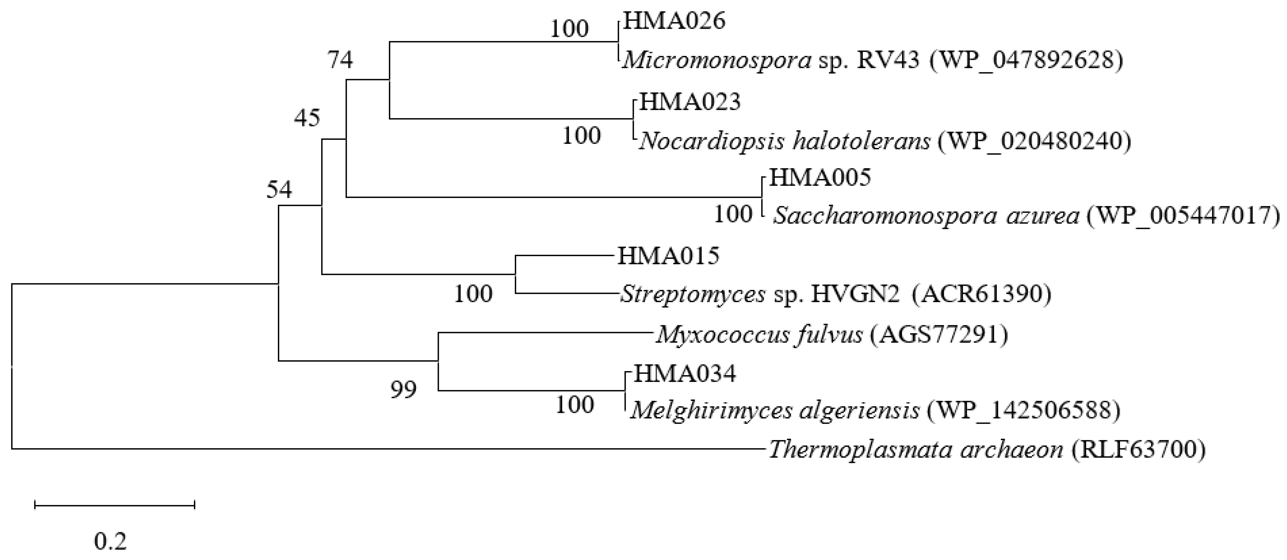
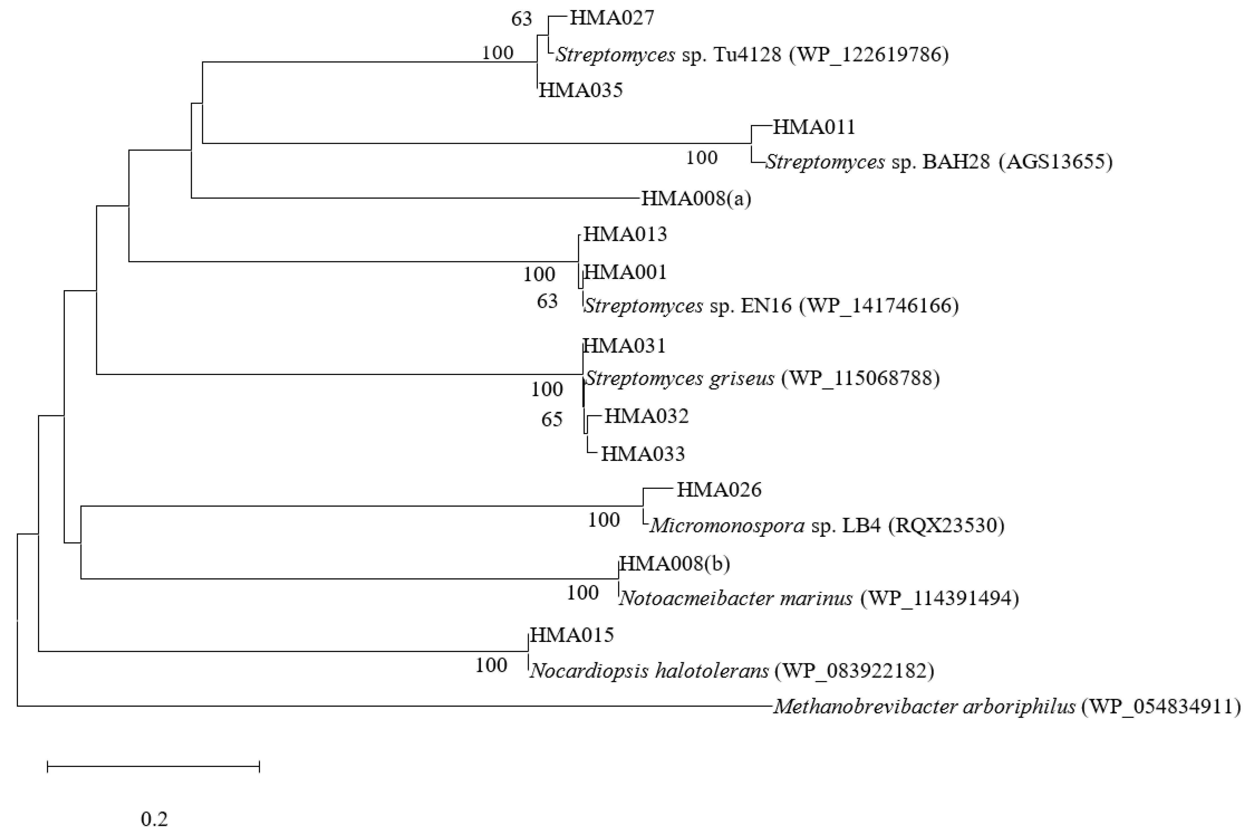
2.3. Phylogenetic Analyses of PKS and NRPS Encoding Genes

Of the 52 strains screened, 13 were positive for PKS or NRPS genes, indicating that these strains possessed at least one BGC encoding for either a polyketide or a non-ribosomal peptide compound (Table 1, Figure 2 and Figure 3). PKS genes were detected in five of the thirteen strains. The translated amino acid sequences of these five strains exhibited 60.27–99.56% similarity with their closest relatives based on BLASTP results. Eleven strains contained NRPS genes and had 95.51–100% amino acid sequence homology with their closest matches that manifested in the A domain fragments. Strains HMA015 (Nocardiopsis sp.) and HMA026 (Micromonospora sp.) were positive for both PKS and NRPS sequences and exhibited considerable antimicrobial activity. Of the 13 strains containing PKS or NRPS genes, 12 belonged to the phylum Actinobacteria and the dominant genus was Streptomyces. Furthermore, strain HMA008 (class Alphaproteobacteria) harboured two NRPS gene sequences.

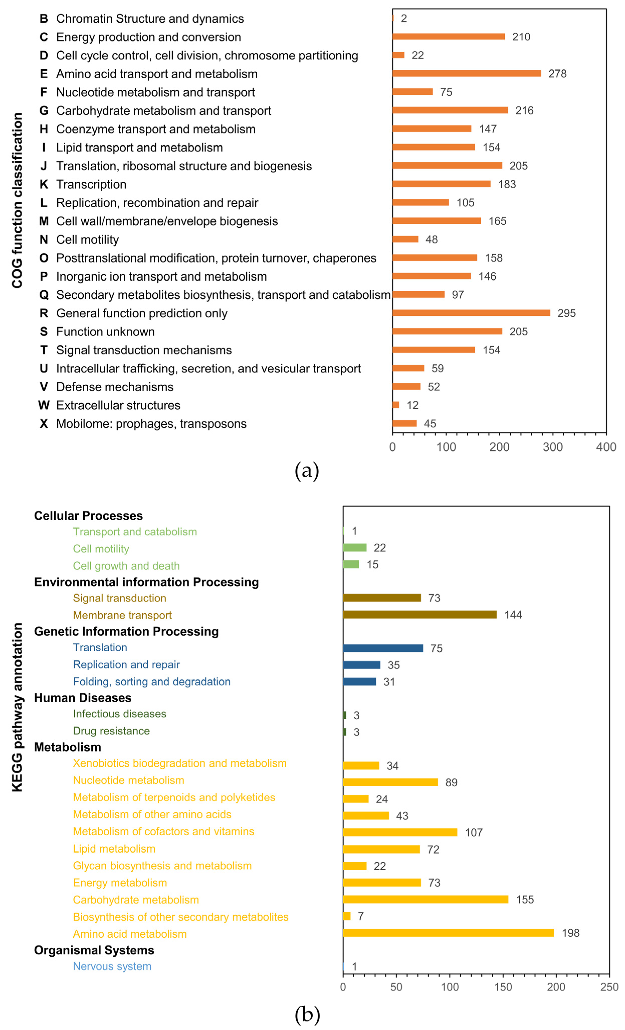
2.4. HMA008 Genome Sequence Annotation and Mining for Secondary Metabolite Biosynthetic Gene Clusters

Genome sequencing of strain HMA008 (Notoacmeibacter sp.) yielded a draft genome that was 3,531,348 bp in length after assembly, producing 22 contigs. The N50 value was 462,813, and the L50 value was 4. The G+C content of strain HMA008 was 61.5%. We investigated the gene functions and metabolic pathways of HMA008 using the Clusters of Orthologous Groups of Proteins (COG) and Kyoto Encyclopaedia of Genes and Genomes (KEGG) databases. A total of 3033 genes were assigned to 23 COG functional categories (Figure 4a). Among these, “general function prediction only” occupied the largest proportion (295 genes), followed by “amino acid transport and metabolism” (278 genes), “carbohydrate transport and metabolism” (216 genes), and “energy production and conversion” (210 genes). Ninety-seven genes were involved in secondary metabolite biosynthesis, transport, and catabolism. Of the KEGG annotated genes, 1227 were classified into six categories, including cellular processes, environmental information processing, genetic information processing, human diseases, metabolism, and organismal systems (Figure 4b). Among these, genes involved in amino acid metabolism (198 genes), carbohydrate metabolism (155 genes), and membrane transport (144 genes) were the most enriched classes. Some pathways were related to terpenoid and polyketide metabolism (24 genes) and the biosynthesis of other secondary metabolites (7 genes). Antibiotics Secondary Metabolites Analysis Shell (antiSMASH) analysis of the contigs detected 12 BGCs putatively encoding for diverse secondary metabolites, including clusters of β-lactone-containing protease inhibitor, butyrolactone, homoserine lactone, N-acetylglutaminylglutamine amide, NRPS, prodigiosin, siderophore, terpene, and other unspecified RiPPs (Table S2). Specifically, two NRPS clusters were present. One of the NRPS clusters exhibited a weak similarity (6%) to the known aurantimycin A BGC, while the other did not match putative gene clusters in the antiSMASH database. Strain HMA008 contained the siderophore cluster, which exhibited 42% similarity to the homologous gene cluster that produces ochrobactin. The β-lactone-containing protease inhibitor gene cluster in HMA008 had a 13% similarity to a homologous gene cluster that produces fengycin. Another BGC of HMA008 exhibited 38% similarity to a homologous gene cluster that produces prodigiosin.

3. Discussion

Sponges harbour highly diverse symbiotic microorganism communities and are considered to be one of the major contributors to marine microbial diversity [2]. Although culture-independent techniques, including DNA fingerprinting approaches, metagenomic libraries, and next-generation sequencing techniques, have determined that sponge microbial communities are dominated by >60 phyla, most of the sponge-associated microorganisms cannot be cultured without the host; thus, sponge resources remain untapped [22,32,33,34]. By applying a culture-dependent approach, we successfully isolated 151 strains from the sponge Ophlitaspongia sp., as well as their representative strains, which are distributed across 20 genera from 18 families in 4 phyla and include Actinobacteria, Bacteroidetes, Firmicutes, and Proteobacteria. Phylogenetic analyses confirmed that all 20 genera of the isolated strains overlapped with previously reported isolates associated with sponges and other marine hosts, such as corals, ascidians, and algae, from different geographical locations obtained using culture-dependent methods [35,36,37,38,39,40,41,42,43,44,45,46]. Genera such as Bacillus, Microbacterium, Nocardiopsis, Pseudoalteromonas, Saccharomonospora, and Streptomyces are distributed throughout aquatic environments and are well-known secondary metabolite producers that may protect host sponges from microbial infection and predators [47,48,49,50,51,52]. For example, at least 740 bioactive substances have been isolated from the genus Micromonospora, which produces the largest number of natural products among rare actinomycetes [53]. Chemically diverse products such as new alkaloids and α-pyrones have recently been isolated from Saccharomonospora [54,55]. Genera such as Dietzia, Rothia, Leeuwenhoekiella, and Polaribacter have served as specific degraders of chemically diverse carbohydrates and may play crucial roles in the marine food web and energy metabolism [56,57,58,59]. Specifically, the genus Dietzia is known for degrading hydrocarbons such as polycyclic aromatic compounds [56]. Members of the genus Rothia have diverse metabolic capacities, including carbohydrate and peptide fermentation, to primary metabolites, while Leeuwenhoekiella species have been found to hydrolyse esters, glucose, and amides, as well as utilise labile carbon sources [60,61]. These genera could serve as helper bacteria that provide growth substrates for specialised bacterial communities [60]. Pigmented strains of the genus Pseudoalteromonas, which possessed secondary bioactive metabolites such as bromoalterochromides and indolmycin, also harbour chitinolytic machinery that degrades chitin [62]. Novel species discovered in sponges continue to be added to Bacillus, Erythrobacter, Leeuwenhoekiella, Micromonospora, and Streptomyces [37,43,63,64,65]. Previously, Alex et al. [66] used classic isolation techniques with a marine agar 2216 medium (containing Amphotericin B) and observed a high incidence of Vibrio sp. (~58%) among the cultured bacterial isolates associated with O. papilla and two other sponge hosts collected from the Atlantic coast of Portugal. Moreover, high Vibrio sp. abundances were also reported among other host sponges, as Vibrio sp. are considered to play an important role in nitrogen fixation [67,68]. However, only a small proportion of the representative microbial community were grouped into Vibrionales in this study, and some of the cultured isolates belonged to actinomycetes instead. This difference can be attributed to many factors, including pre-treatment techniques, antibiotic supplements, and culturing media selection. Specifically, in addition to using 2216E, we used four media such as M1 and ISP-2, which have been deemed successful media for the isolation of actinomycetes associated with sponge samples [69]. Dilution samples were also heated prior to inoculation, which could have stimulated the germination of actinomycete spores and inhibited the growth of irrelative genera; however, some Vibrio sp. are susceptible to heat, and mild heat treatments might not be effective against their growth [69,70].
Antibiotic resistance poses a major ongoing threat to global health security. Therefore, it is crucial to develop novel antimicrobial agents with superior properties and specificity. The antimicrobial assays performed in this study detected a large number of bacterial extracts that were active against three of the indicator strains (S. aureus, B. subtilis, and C. albicans). The strains with antimicrobial activities against these three indicator strains were mainly Actinobacteria (genera Dietzia, Microbacterium, Rothia, and Streptomyces) and Proteobacteria (genera Ruegeria and Vibrio). These two phyla have been identified as the most dominant sponge-derived groups that produce bioactive metabolites [71]. Most active strains inhibited Gram-positive indicator bacteria, while fewer strains exhibited antimicrobial activity against Gram-negative bacteria. Only two strains (HMA008 and HMB04) were observed to inhibit E. coli. Similar results have been obtained previously in other sponge species, as well as in other marine invertebrates such as corals and ascidians [44,72]. It is generally considered that the existence of an outer membrane barrier and multi-drug efflux pumps make Gram-negative bacteria more resistant to antibiotics than Gram-positive bacteria [73]. However, the isolate Xp 4.2 associated with the sponge Xestospongia testudinaria exhibited a more powerful inhibitory activity against Gram-negative bacteria (E. coli and Klebsiella pneumoniae) than against Gram-positive bacteria (Bacillus subtilis) and displayed the most potent bioactive activity against E. coli [74].
Detection of genes that encode PKS and NRPS has generally been used to assess the biosynthetic potential of each microbial strain. Among the 52 strains sequenced for functional genes, 14 contained PKS and/or NRPS genes and exhibited antimicrobial activities. Most of these strains were Actinobacteria, particularly from the genera Streptomyces, which has exhibited extraordinary genetic potential to produce various secondary metabolites [75,76]. Moreover, the Actinobacteria Micromonospora sp. HMA026 and Nocardiopsis sp. HMA015 had both PKS and NRPS genes. The PKS gene sequence from HMA005 exhibited the closest amino acid similarity (99.11%) with sequences from Saccharomonospora azurea, which produces primycin [77]. The NRPS gene sequences of HMA035 and HMA027 exhibited 96.2% and 97.5% similarities at the amino acid level, respectively, with that of Streptomyces sp. Tu4128, which produces bagremycins [78]. Similarly, the genes from strains HMA031, HMA032, and HMA033 exhibited the closest BLASTP matches (96–98% amino acid similarities) and were grouped with sequences involved in the production of polycyclic tetramate macrolactams (PTMs), which have been found in bacteria such as Streptomyces species and possess antifungal, antibiotic, and antioxidant properties [79]. The remaining 38 strains were negative for PKS and/or NRPS genes, while 26 possessed a broad spectrum of antimicrobial activities. For example, the actinobacterial strain HMA010 (Dietzia sp.), which was negative for PKS and NRPS genes, exhibited inhibition against four indicator strains (B. subtilis, S. aureus, V. parahemolyticus, and C. albicans). Previous studies have shown that microorganisms have multiple and complex BGCs involved in the production of bioactive products [80,81]. Therefore, these strains with considerable antimicrobial activities may contain other known or potentially novel BGCs responsible for antibiotic biosynthesis.
Genus Notoacmeibacter, which belongs to the family Notoacmeibacteraceae in the class Alphaproteobacteria, was first reported by Huang et al. [82]. To date, Notoacmeibacter only comprises two species, N. marinus and N. ruber, which were isolated from the gut of a limpet and from a Rhizophorastylosa leaf, respectively [82,83]. The HMA008 strain was identified as N. marinus (99.93% similarity) according to the phylogenetic analysis of 16S rRNA gene sequences. To our knowledge, this is the first time that a strain of the rarely recovered genus Notoacmeibacter has been isolated from a marine sponge. We also tested the antimicrobial activity of Notoacmeibacter sp. HMA008 for the first time. The results indicate that HMA008 exhibited potent antimicrobial activities against five indicator strains, including both Gram-positive and Gram-negative bacteria.
The entire HMA008 genome was sequenced and annotated with COG and KEGG. The COG database assesses the phyletic evolution of protein families, while the KEGG database provides a platform to identify the represented metabolic pathways. According to the COG annotation, 6.76% of genes (205) had unknown functions, indicating that there are still many proteins in N. marinus that remain to be investigated. An enriched array of the gene sequences was involved in amino acid and carbon hydrate transport and metabolism functions, which might support higher levels of energy production and conversion by HMA008. The potential metabolic activities revealed by KEGG, as well as the membrane transport function, also suggest a complex secondary metabolism for strain HMA008. Secondary metabolite gene cluster analysis revealed 12 BGCs responsible for producing diverse bioactive metabolites, such as β-lactone natural products, NRPs, prodigiosin, and siderophores. One of the NRPS clusters displayed a weak similarity to the known clusters potentially encoding aurantimycin A, a depsipeptide antibiotic with cytotoxic properties. The other NRPS clusters, as well as RiPP-like clusters, did not match the antiSMASH database, which may due to the incomplete genome assemblies acquired by next-generation sequencing technologies. Siderophores are known to act effectively against drug-resistant pathogens such as S. aureus, P. aeruginosa, and A. baumannii [84]. β-lactone natural products have drawn significant attention for their potent bioactivity against bacteria, fungi, and human cancer cells [85]. Prodigiosin has also been described as a bacteriostatic agent on E. coli [86,87]. Collectively, the genome mining of the BGCs in HMA008 indicates that the strain produces many secondary metabolites that might make it capable of inhibiting a broad spectrum of indicator strains, including S. aureus, Escherichia coli, P. aeruginosa, V. anguillarum, and V. parahemolyticus. Our results provide preliminary evidence for the antimicrobial potential of N. marinus. Further studies are necessary to identify the bioactive substances produced by HMA008.
Furthermore, strain HMA034, which belongs to the phylum Firmicutes, is closely related to Melghirimyces algeriensis NariEX (99.65% similarity) of the family Thermoactinomycetaceae. At least 43 species belonging to 21 genera of the family Thermoactinomycetaceae have been discovered to date in a variety of natural environments, including hot springs, sediment, and the human gut [88,89,90]. Recent research on microorganisms associated with the sponge Scopalina hapalia observed that an unidentified genus related to the family Thermoactinomycetaceae had tyrosinase inhibitory activities, which indicated its potential to overcome skin aging [91]. In this study, we found potential antimicrobial activities of the Thermoactinomycetaceae species against B. subtilis, S. aureus, and C. albicans. In addition, biosynthetic potential mining indicates that it contains the PKS gene. Thus, the Thermoactinomycetaceae strains may represent a promising source for discovering new antimicrobial agents.
In conclusion, the results presented in this study suggest that the sponge Ophlitaspongia sp. from the Yellow Sea harbours diverse culturable bacteria. Many of the strains had antimicrobial activities, and a number of them were positive for the amplification of PKS and NRPS genes, suggesting that these sponge-derived strains may be promising sources of bioactive secondary metabolites. In addition, strain Notoacmeibacter sp. HMA008 had potent antimicrobial activities and several BGCs that encode secondary metabolites. Further studies are needed that focus on isolating and identifying the potential bioactive compounds produced by these strains.

4. Materials and Methods

4.1. Sample Collection and Preparation

Ophlitaspongia sp. (Figure 1a) samples (n = 10 specimens) were collected from the intertidal zone of Xiaoshi Island (37°31′49′′ N, 122°0′5′′ E) off the Weihai coast in the North Yellow Sea, China, in June 2017. After collection, fresh samples were immediately placed into sterile plastic containers in an icebox and transported to the laboratory within 1–2 h. The sponge samples were rinsed three times with filter-sterilised natural seawater to remove loosely attached microorganisms and other foreign bodies attached to the sponge surfaces. Following this, three sponge samples were cut with sterile tweezers and scissors into pieces of approximately 0.5 cm3, which were then ground into homogenates with filter-sterilised natural seawater.

4.2. Isolation of Culturable Bacteria

The sample homogenates were added to sterile flasks with 10 volumes of filter-sterilised natural seawater and glass beads, shaken on a rotary shaker (180 rpm) for 30 min, and mixed thoroughly. Following this, the supernatant was diluted across a 10-fold concentration series in filter-sterilised natural seawater ranging from 10−2 to 10−5 and then subsequently plated on Petri dishes containing one of the five kinds of culture media. The media used were 2216E, M1, Glucose-Yeast-Peptone (GPY), International Streptomyces Project Medium No. 2 (ISP-2, also referred to as Yeast Extract-Malt Extract), and Reasoner’s 2A (R2A) [92]. All of the media were prepared with 1.8% (w/v) agar. Inoculated plates were incubated at 28 °C for 7–30 d and were monitored daily throughout the cultivation period.
To obtain greater numbers of Actinobacteria, the sample homogenates were incubated in a 55 °C water bath for 10 min. The supernatant was then serially diluted and plated on Petri dishes containing one of the five kinds of culture media (2216E, M1, GPY, ISP2, and R2A). All of the media were prepared with 1.8% (w/v) agar and were supplemented to 20 μg mL−1 nalidixic acid and 100 μg mL−1 cycloheximide final concentrations, which were used to inhibit the growth of Gram-negative bacteria and fungi [90], respectively. The inoculated plates were incubated at 28 °C for at least 3 weeks and were monitored daily throughout the cultivation period.

4.3. Selection and Preservation of Collected Strains

Single colonies growing on the media plates were selected based on colony phenotypical characteristics, including growth rate, morphology, size, pigmentation, and margin characteristics. Following subculture and confirmation of strain purity, the collected bacteria were preserved in 15% glycerin (v/v) at −80 °C for further study.

4.4. Preparation of Crude Extracts and Antibacterial Activity Screening

All isolated bacteria (151 strains) were analysed for their antimicrobial activities against seven indicator strains. The indicator strains included Escherichia coli (ATCC 25922), Pseudomonas aeruginosa (ATCC 27853), Bacillus subtilis (ATCC 6633), Staphylococcus aureus (ATCC 6538), Candida albicans (ATCC 10231), Vibrio parahaemolyticus (ATCC 17802), and Vibrio anguillarum. Most of the indicator strains were purchased from the ATCC, except for V. anguillarum, which was donated by Associate Professor Yu-Xia Zou from the Institute of Oceanology, Chinese Academy of Sciences.
Prior to antimicrobial activity assays, the seven indicator strains were inoculated in Mueller–Hinton (MH) liquid medium (beef extract 3 g; soluble starch 1.5 g; acid hydrolysate of casein 17.5 g; distilled water 1 L; pH 7.3 ± 0.2) overnight at 37 °C and 100 µL of each indicator strain suspension adjusted to 0.5 McFarland standard was plated on MH agar (MH with 18 g of agar).
Antimicrobial activity was determined by observing the growth inhibition of bacteria or fungi according to previous protocols [93]. The test strains were inoculated into 50 mL of liquid medium (2216E medium for bacteria, M1 medium for Actinobacteria) and incubated at 28 °C for approximately 5–7 d (bacteria) and 7–10 d (Actinobacteria) on a rotary shaker. The culture supernatants were subsequently extracted three times with 50 mL ethyl acetate. The organic layers were separated, combined, dried over anhydrous sodium sulphate, decanted, and dried under vacuum to obtain a crude extract. The crude extracts of each of the strains were dissolved in dimethyl sulfoxide (DMSO) at a concentration of 25 mg mL−1, after which 10 μL of the crude extract solution was added to 6 mm diameter filter papers. The filter paper disks were then placed onto the surface of the MH agar plates with the indicator strains. The same amount of DMSO was used as a negative control, and crude extracts of Marine 2216E or M1 media were also used as negative controls. Chloramphenicol (30 μg), gentamicin (10 μg), and nystatin (10 μg) (Sigma-Aldrich, St Louis, MO, USA) were used as positive controls. The antimicrobial assay plates were then incubated at 37 °C for 24 h for the bacterial indicator strains, and at 37 °C for 48 h for C. albicans. The inhibition zones around the paper disks were measured to gauge the antimicrobial activities of the collected strains. Three or more replicates were performed for each isolate to establish average inhibition zone sizes. The inhibition degrees against indicator strains used the following symbols: (−), no inhibition; (+), 6 < inhibition zone < 8 mm; (++), 8 ≤ inhibition zone < 10 mm; (+++), 10 ≤ inhibition zone < 12 mm; (++++), inhibition zone ≥ 12 mm. The isolated strain for which each inhibition zone size was greater than 12 mm exhibited higher antimicrobial activities.

4.5. 16S rRNA Gene Sequencing and Phylogenetic Analyses

Fifty-two strains were selected based on their colony morphologies on Marine 2216E solid medium and the results of Gram staining, after which their 16S rRNA genes were sequenced to determine their phylogenetic positions. Bacteria were cultured in the 2216E liquid medium and Actinobacteria were cultured in the M1 liquid medium. Extraction of genomic DNA and PCR amplification were performed as described by a previous study [46]. Cells were harvested at stationary-phase by centrifugation (10,000× g for 1 min). Genomic DNA was extracted by using a Bacterial Genome DNA Extraction Kit (Sangon Biotech, Shanghai, China). The universal primers 27F (5’-AGAGTTTGATCCTGGCTCAG-3’) and 1492R (5’-GGTTACCTTGTTACGACTT-3’) were used in polymerase chain reaction (PCR) amplifcation of the 16S rRNA gene. Extracted genomic DNA was used as a PCR template. Genomic DNA from the bacterial strain E. coli was used as a positive control, and sterile distilled water was used as a negative control. DNA was denatured at 94 °C for 5 min, followed by 35 cycles of 94 °C for 1 min, 56 °C for 1 min, and 72 °C for 90 s, with a final 10 min extension at 72 °C. PCR products (1500 bp) were sequenced by Sangon Biotech, Shanghai, China.
Sequence identity searches and calculations of pairwise similarity values between collected strains and their closely related types were performed using the EzBioCloud Database [94]. 16S rRNA gene sequences of the related strains were downloaded from the National Center for Biotechnology Information (NCBI) GenBank database (http://www.ncbi.nlm.nih.gov (accessed on 10 May 2022). Sequences were further analysed by performing multiple sequence alignments using Clustal X [95]. A phylogenetic tree was constructed using the neighbour-joining algorithm, which was implemented using the MEGA (v.6.0) software package [96]. Phylogenetic analyses used Pyrococcus abyssi GE5T (L19921) as the outgroup. The 16S rRNA gene sequences of the cultured bacteria were submitted to the NCBI GenBank Database under Accession Numbers MG905366–MG905417.

4.6. Genome Sequencing and Secondary Metabolite Cluster Analysis

Genome sequencing of strain HMA008 was performed on an Illumina HiSeq PE150 platform by Novogene Bioinformatics Technology Co., Ltd. (Beijing, China). A-tailed, ligated to paired-end adaptors, and PCR amplified with a 350 bp insert was used for library construction. SOAP denovo (v.2.04) (SOAP, RRID:SCR_000689) was used to assemble genome sequences. The genome sequences were submitted to the NCBI GenBank Database under accession number SAMN09273368 and assembly accession number ASM333676v1. Functional annotation of genes was predicted using COG (http://www.ncbi.nlm.nih.gov/COG/ (accessed on 26 July 2022) [97] and KEGG (http://www.genome.jp/kegg/ (accessed on 26 July 2022) [98]. Secondary metabolite BGCs were predicted using antiSMASH (https://antismash.secondarymetabolites.org (accessed on 26 July 2022) [99].

4.7. Screening for PKS and NRPS Genes

The presence of genes (PKS and NRPS) involved in the production of secondary metabolites was screened from 52 strains that had undergone 16S rRNA gene sequencing. Genomic DNA was extracted as described above. DKF (5’GTGCCGGGTNCCRTGNGYYTC3’) and DKR (5’GCGATGGAYCCNCARCARYG3’) primers were used for PCR amplification of the gene clusters encoding the KS domain using the extracted genomic DNA as a template [100]. NRPS gene clusters that encode the adenylation (A) domain were amplified using A3F (5′GCSTACSYSATSTACACSTCSGG3′) and A7R (5′SASGTCVCCSGTSCGGTAS3′) primers [101]. DNA from the previously characterised strain Bacillus sp. HSA006, which contains PKS and NRPS systems, was used as the positive control and sterile distilled water was used as the negative control [102]. PCR cycles comprised an initial denaturation step of 5 min at 95 °C, 35 cycles of 30 s at 95 °C, 2 min at 57 °C, and 4 min at 72 °C, with a final extension of 10 min at 72 °C. PCR amplification products, corresponding to 650–700 bp for the KS domain of PKS and 700–800 bp for the A domain of NRPS, were purified using the SanPrep Column DNA Gel Extraction Kit (Sangon Biotech, Shanghai, China). The PCR products were ligated into pMD18-T (Takara Bio. Inc., Dalian, China). M13 universal primers were used for sequencing, which was performed by Sangon Biotech. Translated protein sequences were derived from the nucleotide sequences using the Open Reading Frame (ORF) Finder search program available on the NCBI website (https://www.ncbi.nlm.nih.gov/orffinder/ (accessed on 5 February 2022). The deduced amino acid sequences were used as queries to search for related proteins in the Non-redundant (NR) Protein Database using the BLASTP algorithm. Multiple sequence alignments of nucleotide sequences were performed using Clustal X (RRID:SCR_017055), and phylogenetic trees based on the amino acid sequences encoded by the PKS or NRPS genes were constructed using the neighbour-joining method in MEGA (v6.0) (MEGA Software, RRID:SCR_000667) combined with bootstrap analysis using 1000 replications. The phylogenetic analyses used Thermoplasmata archaeon (RLF63700) and Methanobrevibacter arboriphilus (WP_054834911) as outgroups, respectively. The PKS and NRPS gene sequences were submitted to the NCBI GenBank Database under Accession Numbers MG993566−MG993571, MH024360−MH024364, MH045857−MH045858, and MH050930−MH9050932.

Supplementary Materials

The following supporting information can be downloaded at: https://www.mdpi.com/article/10.3390/md20100588/s1, Table S1: Antimicrobial activities of the isolated strains associated with the sponge Ophlitaspongia sp.; Table S2: Secondary metabolite biosynthetic gene clusters (BGCs) in the genome of Notoacmeibacter sp. HMA008; Figure S1: Neighbour-joining phylogenetic tree based on comparisons of the 16S rRNA sequences of bacterial strains associated with the sponge Ophlitaspongia sp. and other related taxa.

Author Contributions

Conceptualization, L.C. and G.-Y.W.; formal analysis, X.-N.W., and H.-Y.B.; investigation, H.-Y.B.; writing—original draft preparation, L.C. and X.-N.W.; writing—review and editing, L.C. and G.-Y.W.; supervision, L.C.; project administration, G.-Y.W.; funding acquisition, L.C. and G.-Y.W. All authors have read and agreed to the published version of the manuscript.

Funding

This work was supported by Discipline Construction Guide Foundation at the Harbin Institute of Technology at Weihai [No. WH20150204 and No. WH20160205] and the Research Innovation Foundation at the Harbin Institute of Technology at Weihai [No. 2019KYCXJJYB15].

Data Availability Statement

The data used to support the findings of this study are included within the article. The genome sequences were submitted to the NCBI GenBank Database under accession number SAMN09273368 and assembly accession number ASM333676v1.

Acknowledgments

The authors thank Zhi-Yong Li (Shanghai Jiao Tong University) for the molecular identification of sponge.

Conflicts of Interest

The authors declare no conflict of interest.

References

  1. Zuppa, A.; Costantini, S.; Costantini, M. Comparative sequence analysis of bacterial symbionts from the marine sponges Geodia cydonium and Ircinia muscarum. Bioinformation 2014, 10, 196–200. [Google Scholar] [CrossRef] [PubMed]
  2. Thomas, T.; Moitinho-Silva, L.; Lurgi, M.; Björk, J.; Easson, C.; Garcia, M.D.C.A.; Olson, J.B.; Erwin, P.M.; Lopez-Legentil, S.; Luter, H.; et al. Diversity, structure and convergent evolution of the global sponge microbiome. Nat. Commun. 2016, 7, 11870. [Google Scholar] [CrossRef] [PubMed]
  3. Lackner, G.; Peters, E.E.; Helfrich, E.J.; Piel, J. Insights into the lifestyle of uncultured bacterial natural product factories associated with marine sponges. Proc. Natl. Acad. Sci. USA 2017, 114, E347–E356. [Google Scholar] [CrossRef] [PubMed]
  4. Liu, T.; Wu, S.; Zhang, R.; Wang, D.; Chen, J.; Zhao, J. Diversity and antimicrobial potential of Actinobacteria isolated from diverse marine sponges along the Beibu Gulf of the South China Sea. FEMS Microbiol. Ecol. 2019, 95, fiz089. [Google Scholar] [CrossRef] [PubMed]
  5. Dat, T.T.H.; Cuc, N.T.K.; Cuong, P.V.; Smidt, H.; Sipkema, D. Diversity and antimicrobial activity of Vietnamese sponge-associated bacteria. Mar. Drugs 2021, 19, 353. [Google Scholar] [CrossRef] [PubMed]
  6. Ye, J.; Zhou, F.; Al-Kareef, A.M.; Wang, H. Anticancer agents from marine sponges. J. Asian Nat. Prod. Res. 2015, 17, 64–88. [Google Scholar] [CrossRef]
  7. Jimenez, P.C.; Wilke, D.V.; Branco, P.C.; Bauermeister, A.; Rezende-Teixeira, P.; Gaudêncio, S.P.; Costa-Lotufo, L.V. Enriching cancer pharmacology with drugs of marine origin. Br. J. Pharmacol. 2020, 177, 3–27. [Google Scholar] [CrossRef]
  8. Swami, U.; Shah, U.; Goel, S. Eribulin in cancer treatment. Mar. Drugs 2015, 13, 5016–5058. [Google Scholar] [CrossRef]
  9. Bringmann, G.; Lang, G.; Mühlbacher, J.; Schaumann, K.; Steffens, S.; Rytik, P.G.; Hentschel, U.; Morschhäuser, J.; Müller, W.E. Sorbicillactone A: A structurally unprecedented bioactive novel-type alkaloid from a sponge-derived fungus. Prog. Mol. Subcell. Biol. 2003, 37, 231–253. [Google Scholar]
  10. Indraningrat, A.A.; Smidt, H.; Sipkema, D. Bioprospecting sponge-associated microbes for antimicrobial compounds. Mar. Drugs 2016, 14, 87. [Google Scholar] [CrossRef]
  11. Palomo, S.; González, I.; de la Cruz, M.; Martín, J.; Tormo, J.R.; Anderson, M.; Hill, R.T.; Vicente, F.; Reyes, F.; Genilloud, O. Sponge-derived Kocuria and Micrococcus spp. as sources of the new thiazolyl peptide antibiotic kocurin. Mar. Drugs 2013, 11, 1071–1086. [Google Scholar] [CrossRef] [PubMed] [Green Version]
  12. Li, D.; Xu, Y.; Shao, C.-L.; Yang, R.-Y.; Zheng, C.-J.; Chen, Y.-Y.; Fu, X.-M.; Qian, P.-Y.; She, Z.-G.; De Voogd, N.J.; et al. Antibacterial bisabolane-type sesquiterpenoids from the sponge-derived fungus Aspergillus sp. Mar. Drugs 2012, 10, 234–241. [Google Scholar] [CrossRef]
  13. Pruksakorn, P.; Arai, M.; Kotoku, N.; Vilchèze, C.; Baughn, A.D.; Moodley, P.; Jacobs, W.R., Jr.; Kobayashi, M. Trichoderins, novel aminolipopeptides from a marine sponge-derived Trichoderma sp., are active against dormant mycobacteria. Bioorganic Med. Chem. Lett. 2010, 20, 3658–3663. [Google Scholar] [CrossRef] [PubMed]
  14. El-Gendy, M.M.; El-Bondkly, A.M. Production and genetic improvement of a novel antimycotic agent, saadamycin, against dermatophytes and other clinical fungi from endophytic Streptomyces sp. Hedaya48. J. Ind. Microbiol. Biotechnol. 2010, 37, 831–841. [Google Scholar] [CrossRef] [PubMed]
  15. Kwon, M.J.; Steiniger, C.; Cairns, T.C.; Wisecaver, J.H.; Lind, A.L.; Pohl, C.; Regner, C.; Rokas, A.; Meyer, V. Beyond the biosynthetic gene cluster paradigm: Genome-wide coexpression networks connect clustered and unclustered transcription factors to secondary metabolic pathways. Microbiol. Spectr. 2021, 9, e0089821. [Google Scholar] [CrossRef] [PubMed]
  16. Katz, L.; Baltz, R.H. Natural product discovery: Past, present, and future. J. Ind. Microbiol. Biotechnol. 2016, 43, 155–176. [Google Scholar] [CrossRef]
  17. Pimentel-Elardo, S.M.; Grozdanov, L.; Proksch, S.; Hentschel, U. Diversity of nonribosomal peptide synthetase genes in the microbial metagenomes of marine sponges. Mar. Drugs 2012, 10, 1192–1202. [Google Scholar] [CrossRef]
  18. Liu, H.; Zhu, G.; Zhao, S.; Fu, P.; Zhu, W. Bioactive natural products from the marine sponge-derived Nocardiopsis dassonvillei OUCMDZ-4534. Chin. J. Org. Chem. 2019, 39, 507–514. [Google Scholar] [CrossRef]
  19. Ameen, F.; AlNadhari, S.; Al-Homaidan, A.A. Marine microorganisms as an untapped source of bioactive compounds. Saudi J. Biol. Sci. 2021, 28, 224–231. [Google Scholar] [CrossRef]
  20. Riyanti; Balansa, W.; Liu, Y.; Sharma, A.; Mihajlovic, S.; Hartwig, C.; Leis, B.; Rieuwpassa, F.J.; Ijong, F.G.; Wägele, H.; et al. Selection of sponge-associated bacteria with high potential for the production of antibacterial compounds. Sci. Rep. 2020, 10, 19614. [Google Scholar] [CrossRef]
  21. Dat, T.T.H.; Steinert, G.; Cuc, N.T.K.; Smidt, H.; Sipkema, D. Bacteria cultivated from sponges and bacteria not yet cultivated from sponges—A review. Front. Microbiol. 2021, 12, 737925. [Google Scholar] [CrossRef] [PubMed]
  22. Laport, M.S. Isolating bacteria from sponges: Why and how? Curr. Pharm. Biotechnol. 2017, 18, 1224–1236. [Google Scholar] [CrossRef] [PubMed]
  23. Arulprakasam, K.R.; Dharumadurai, D. Genome mining of biosynthetic gene clusters intended for secondary metabolites conservation in actinobacteria. Microb. Pathog. 2021, 161 Pt A, 105252. [Google Scholar] [CrossRef]
  24. Nicault, M.; Tidjani, A.R.; Gauthier, A.; Dumarcay, S.; Gelhaye, E.; Bontemps, C.; Leblond, P. Mining the biosynthetic potential for specialized metabolism of a Streptomyces soil community. Antibiotics 2020, 9, 271. [Google Scholar] [CrossRef] [PubMed]
  25. Pan, R.; Bai, X.; Chen, J.; Zhang, H.; Wang, H. Exploring structural diversity of microbe secondary metabolites using OSMAC strategy: A literature review. Front. Microbiol. 2019, 10, 294. [Google Scholar] [CrossRef]
  26. Wang, W.; Drott, M.; Greco, C.; Luciano-Rosario, D.; Wang, P.; Keller, N.P. Transcription factor repurposing offers insights into evolution of biosynthetic gene cluster regulation. mBio 2021, 12, e0139921. [Google Scholar] [CrossRef] [PubMed]
  27. Lee, N.; Hwang, S.; Lee, Y.; Cho, S.; Palsson, B.; Cho, B.K. Synthetic biology tools for novel secondary metabolite discovery in Streptomyces. J. Microbiol. Biotechnol. 2019, 29, 667–686. [Google Scholar] [CrossRef]
  28. Alex, A.; Antunes, A. Whole-genome comparisons among the genus Shewanella reveal the enrichment of genes encoding ankyrin-repeats containing proteins in sponge-associated bacteria. Front. Microbiol. 2019, 10, 5. [Google Scholar] [CrossRef]
  29. Howson, C.M.; Chambers, S.J. Ophlitaspongia and Ophlitaspongia papilla reinstated, and a new species of Ophlitaspongia described (Porifera: Demospongiae: Microcionidae). J. Mar. Biol. Assoc. 1999, 79, 609–620. [Google Scholar] [CrossRef]
  30. Ferreira, M.; Cabado, A.G.; Chapela, M.J.; Fajardo, P.; Atanassova, M.; Garrido, A.; Vieites, J.M.; Lago, J. Cytotoxic activity of extracts of marine sponges from NW Spain on a neuroblastoma cell line. Environ. Toxicol. Pharmacol. 2011, 32, 430–437. [Google Scholar] [CrossRef]
  31. Alex, A.; Antunes, A. Comparative genomics reveals metabolic specificity of endozoicomonas isolated from a marine sponge and the genomic repertoire for host-bacteria symbioses. Microorganisms 2019, 7, 635. [Google Scholar] [CrossRef] [Green Version]
  32. Moitinho-Silva, L.; Nielsen, S.; Amir, A.; Gonzalez, A.; Ackermann, G.L.; Cerrano, C.; Astudillo-Garcia, C.; Easson, C.; Sipkema, D.; Liu, F.; et al. The sponge microbiome project. Gigascience 2017, 6, gix077. [Google Scholar] [CrossRef]
  33. Su, J.; Zhang, F.; Sun, W.; Karuppiah, V.; Zhang, G.; Li, Z.; Jiang, Q. A new alkaline lipase obtained from the metagenome of marine sponge Ircinia sp. World J. Microbiol. Biotechnol. 2015, 31, 1093–1102. [Google Scholar] [CrossRef]
  34. Gaikwad, S.; Shouche, Y.S.; Gade, W.N. Microbial community structure of two freshwater sponges using Illumina MiSeq sequencing revealed high microbial diversity. AMB Express 2016, 6, 40. [Google Scholar] [CrossRef]
  35. Yang, S.; Sun, W.; Tang, C.; Jin, L.; Zhang, F.; Li, Z. Phylogenetic diversity of actinobacteria associated with soft coral Alcyonium gracllimum and stony coral Tubastraea coccinea in the East China Sea. Microb. Ecol. 2013, 66, 189–199. [Google Scholar] [CrossRef]
  36. Mitova, M.; Popov, S.; De Rosa, S. Cyclic peptides from a Ruegeria strain of bacteria associated with the sponge Suberites domuncula. J. Nat. Prod. 2004, 67, 1178–1181. [Google Scholar] [CrossRef]
  37. Mitra, S.; Matsuo, Y.; Haga, T.; Yasumoto-Hirose, M.; Yoon, J.; Kasai, H.; Yokota, A. Leptobacterium flavescens gen. nov., sp. nov., a marine member of the family Flavobacteriaceae, isolated from marine sponge and seawater. Int. J. Syst. Evol. Microbiol. 2009, 59 Pt 2, 207–212. [Google Scholar] [CrossRef]
  38. Selvin, J.; Shanmugha Priya, S.; Seghal Kiran, G.; Thangavelu, T.; Sapna Bai, N. Sponge-associated marine bacteria as indicators of heavy metal pollution. Microbiol. Res. 2009, 164, 352–363. [Google Scholar] [CrossRef]
  39. Abbamondi, G.R.; De Rosa, S.; Iodice, C.; Tommonaro, G. Cyclic dipeptides produced by marine sponge-associated bacteria as quorum sensing signals. Nat. Prod. Commun. 2014, 9, 229–232. [Google Scholar] [CrossRef]
  40. Sun, W.; Zhang, F.; He, L.; Karthik, L.; Li, Z. Actinomycetes from the South China Sea sponges: Isolation, diversity, and potential for aromatic polyketides discovery. Front. Microbiol. 2015, 6, 1048. [Google Scholar] [CrossRef]
  41. Amelia, T.S.M.; Amirul, A.A.; Saidin, J.; Bhubalan, K. Identification of cultivable bacteria from tropical marine sponges and their biotechnological potentials. Trop. Life Sci. Res. 2018, 29, 187–199. [Google Scholar] [CrossRef]
  42. Batista, D.; Costa, R.; Carvalho, A.P.; Batista, W.R.; Rua, C.; de Oliveira, L.; Leomil, L.; Fróes, A.M.; Thompson, F.L.; Coutinho, R.; et al. Environmental conditions affect activity and associated microorganisms of marine sponges. Mar. Environ. Res. 2018, 142, 59–68. [Google Scholar] [CrossRef]
  43. Zhuang, L.; Lin, B.; Xu, L.; Li, G.; Wu, C.J.; Luo, L. Erythrobacter spongiae sp. nov., isolated from marine sponge. Int. J. Syst. Evol. Microbiol. 2019, 69, 1111–1116. [Google Scholar] [CrossRef]
  44. Mahmoud, H.M.; Kalendar, A.A. Coral-associated actinobacteria: Diversity, abundance, and biotechnological potentials. Front. Microbiol. 2016, 7, 204. [Google Scholar] [CrossRef]
  45. Chen, J.; Xu, L.; Zhou, Y.; Han, B. Natural products from actinomycetes associated with marine organisms. Mar. Drugs 2021, 19, 629. [Google Scholar] [CrossRef]
  46. Chen, L.; Wang, X.N.; Fu, C.M.; Wang, G.Y. Phylogenetic analysis and screening of antimicrobial and antiproliferative activities of culturable bacteria associated with the ascidian Styela clava from the Yellow Sea, China. BioMed Res. Int. 2019, 2019, 7851251. [Google Scholar] [CrossRef]
  47. Francis, A.; Chakraborty, K. Marine macroalga-associated heterotroph Bacillus velezensis as prospective therapeutic agent. Arch. Microbiol. 2021, 203, 1671–1682. [Google Scholar] [CrossRef]
  48. Savi, D.C.; Shaaban, K.A.; Gos, F.M.W.; Thorson, J.S.; Glienke, C.; Rohr, J. Secondary metabolites produced by Microbacterium sp. LGMB471 with antifungal activity against the phytopathogen Phyllosticta citricarpa. Folia Microbiol. 2019, 64, 453–460. [Google Scholar] [CrossRef]
  49. Bennur, T.; Kumar, A.R.; Zinjarde, S.; Javdekar, V. Nocardiopsis species: Incidence, ecological roles and adaptations. Microbiol. Res. 2015, 174, 33–47. [Google Scholar] [CrossRef]
  50. Paulsen, S.S.; Strube, M.L.; Bech, P.K.; Gram, L.; Sonnenschein, E.C. Marine chitinolytic Pseudoalteromonas represents an untapped reservoir of bioactive potential. mSystems 2019, 4, e00060-19. [Google Scholar] [CrossRef]
  51. Le, T.C.; Lee, E.J.; Lee, J.; Hong, A.; Yim, C.-Y.; Yang, I.; Choi, H.; Chin, J.; Cho, S.J.; Ko, J.; et al. Saccharoquinoline, a cytotoxic alkaloidal meroterpenoid from marine-derived bacterium Saccharomonospora sp. Mar. Drugs 2019, 17, 98. [Google Scholar]
  52. Čihák, M.; Kameník, Z.; Šmídová, K.; Bergman, N.; Benada, O.; Kofroňová, O.; Petříčková, K.; Bobek, J. Secondary metabolites produced during the germination of Streptomyces coelicolor. Front. Microbiol. 2017, 8, 2495. [Google Scholar] [CrossRef] [Green Version]
  53. Bérdy, J. Bioactive microbial metabolites. J. Antibiot. 2005, 58, 1–26. [Google Scholar] [CrossRef]
  54. Maloney, K.N.; Macmillan, J.B.; Kauffman, C.A.; Jensen, P.R.; Dipasquale, A.G.; Rheingold, A.L.; Fenical, W. Lodopyridone, a structurally unprecedented alkaloid from a marine actinomycete. Org. Lett. 2009, 11, 5422–5424. [Google Scholar]
  55. Yim, C.Y.; Le, T.C.; Lee, T.G.; Yang, I.; Choi, H.; Lee, J.; Kang, K.Y.; Lee, J.S.; Lim, K.M.; Yee, S.T.; et al. Saccharomonopyrones A–C, new α-pyrones from a marine sediment-derived bacterium Saccharomonospora sp. CNQ-490. Mar. Drugs 2017, 15, 239. [Google Scholar] [CrossRef]
  56. Ausuri, J.; Vitale, G.A.; Coppola, D.; Palma Esposito, F.; Buonocore, C.; de Pascale, D. Assessment of the degradation potential and genomic insights towards phenanthrene by Dietzia psychralcaliphila JI1D. Microorganisms 2021, 9, 1327. [Google Scholar]
  57. Uranga, C.C.; Arroyo, P., Jr.; Duggan, B.M.; Gerwick, W.H.; Edlund, A. Commensal oral Rothia mucilaginosa produces enterobactin, a metal-chelating siderophore. Msystems 2020, 5, e00161-20. [Google Scholar] [CrossRef]
  58. Tahon, G.; Lebbe, L.; De Troch, M.; Sabbe, K.; Willems, A. Leeuwenhoekiella aestuarii sp. nov., isolated from salt-water sediment and first insights in the genomes of Leeuwenhoekiella species. Int. J. Syst. Evol. Microbiol. 2020, 70, 1706–1719. [Google Scholar] [CrossRef]
  59. Xing, P.; Hahnke, R.L.; Unfried, F.; Markert, S.; Huang, S.; Barbeyron, T.; Harder, J.; Becher, D.; Schweder, T.; Glöckner, F.O.; et al. Niches of two polysaccharide-degrading Polaribacter isolates from the North Sea during a spring diatom bloom. ISME J. 2015, 9, 1410–1422. [Google Scholar] [CrossRef]
  60. Gao, B.; Gallagher, T.; Zhang, Y.; Elbadawi-Sidhu, M.; Lai, Z.; Fiehn, O.; Whiteson, K.L. Tracking polymicrobial metabolism in cystic fibrosis airways: Pseudomonas aeruginosa metabolism and physiology are influenced by Rothia mucilaginosa-derived metabolites. mSphere 2018, 3, e00151-18. [Google Scholar] [CrossRef]
  61. Morris, J.J.; Kirkegaard, R.; Szul, M.J.; Johnson, Z.I.; Zinser, E.R. Facilitation of robust growth of Prochlorococcus colonies and dilute liquid cultures by “helper” heterotrophic bacteria. Appl. Environ. Microbiol. 2008, 74, 4530–4534. [Google Scholar] [CrossRef]
  62. Wang, X.; Isbrandt, T.; Strube, M.L.; Paulsen, S.S.; Nielsen, M.W.; Buijs, Y.; Schoof, E.M.; Larsen, T.O.; Gram, L.; Zhang, S.D. Chitin Degradation machinery and secondary metabolite profiles in the marine bacterium Pseudoalteromonas rubra S4059. Mar. Drugs 2021, 19, 108. [Google Scholar] [CrossRef]
  63. Risdian, C.; Landwehr, W.; Rohde, M.; Schumann, P.; Hahnke, R.L.; Spröer, C.; Bunk, B.; Kämpfer, P.; Schupp, P.J.; Wink, J. Streptomyces bathyalis sp. nov., an actinobacterium isolated from the sponge in a deep sea. Antonie Leeuwenhoek 2021, 114, 425–435. [Google Scholar] [CrossRef]
  64. Li, L.; Zhu, H.R.; Xu, Q.H.; Lin, H.W.; Lu, Y.H. Micromonospora craniellae sp. nov., isolated from a marine sponge, and reclassification of Jishengella endophytica as Micromonospora endophytica comb. nov. Int. J. Syst. Evol. Microbiol. 2019, 69, 715–720. [Google Scholar] [CrossRef]
  65. Lee, G.E.; Im, W.T.; Park, J.S. Bacillus spongiae sp. nov. isolated from sponge of Jeju Island. J. Microbiol. 2018, 56, 217–222. [Google Scholar] [CrossRef]
  66. Alex, A.; Silva, V.; Vasconcelos, V.; Antunes, A. Evidence of unique and generalist microbes in distantly related sympatric intertidal marine sponges (Porifera: Demospongiae). PLoS ONE 2013, 8, e80653. [Google Scholar] [CrossRef]
  67. Thompson, F.L.; Iida, T.; Swings, J. Biodiversity of Vibrios. Microbiol. Mol. Biol. Rev. 2004, 68, 403–431. [Google Scholar] [CrossRef]
  68. Fiore, C.L.; Jarett, J.K.; Olson, N.D.; Lesser, M.P. Nitrogen fixation and nitrogen transformations in marine symbioses. Trends Microbiol. 2010, 18, 455–463. [Google Scholar] [CrossRef]
  69. Hameş-Kocabaş, E.E.; Uzel, A. Isolation strategies of marine-derived actinomycetes from sponge and sediment samples. J. Microbiol. Methods 2012, 88, 342–347. [Google Scholar] [CrossRef]
  70. Froelich, B.A.; Daines, D.A. In hot water: Effects of climate change on Vibrio-human interactions. Environ. Microbiol. 2020, 22, 4101–4111. [Google Scholar] [CrossRef]
  71. Bibi, F.; Faheem, M.; Azhar, E.I.; Yasir, M.; Alvi, S.A.; Kamal, M.A.; Ullah, I.; Naseer, M.I. Bacteria from marine sponges: A source of new drugs. Curr. Drug Metab. 2017, 18, 11–15. [Google Scholar] [CrossRef] [PubMed]
  72. Chen, L.; Liu, C.; Liu, X.; Wang, G.Y. Phylogenetic analysis and screening of antimicrobial and cytotoxic activities of culturable bacteria associated with the ascidian Botryllus schlosseri. J. Appl. Microbiol. 2020, 129, 892–905. [Google Scholar] [CrossRef] [PubMed]
  73. Alibert, S.; N’Gompaza Diarra, J.; Hernandez, J.; Stutzmann, A.; Fouad, M.; Boyer, G.; Pagès, J.M. Multidrug efflux pumps and their role in antibiotic and antiseptic resistance: A pharmacodynamic perspective. Expert Opin. Drug Metab. Toxicol. 2017, 13, 301–309. [Google Scholar] [CrossRef] [PubMed]
  74. Cita, Y.P.; Suhermanto, A.; Radjasa, O.K.; Sudharmono, P. Antibacterial activity of marine bacteria isolated from sponge Xestospongia testudinaria from Sorong, Papua. Asian Pac. J. Trop. Biomed. 2017, 7, 450–454. [Google Scholar] [CrossRef]
  75. Jackson, S.A.; Crossman, L.; Almeida, E.L.; Margassery, L.M.; Kennedy, J.; Dobson, A.D.W. Diverse and abundant secondary metabolism biosynthetic gene clusters in the genomes of marine sponge derived Streptomyces spp. isolates. Mar. Drugs 2018, 16, 67. [Google Scholar] [CrossRef] [PubMed]
  76. Guerrero-Garzón, J.F.; Zehl, M.; Schneider, O.; Rückert, C.; Busche, T.; Kalinowski, J.; Bredholt, H.; Zotchev, S.B. Streptomyces spp. from the marine sponge Antho dichotoma: Analyses of secondary metabolite biosynthesis gene clusters and some of their products. Front. Microbiol. 2020, 11, 437. [Google Scholar] [CrossRef]
  77. Valasek, A.; Kiss, Í.; Fodor, I.; Kovács, M.; Urbán, P.; Jámbor, É.; Fekete, C.; Kerepesi, I. Proteomic insight into the primycin fermentation process of Saccharomonospora azurea. Acta Biol. Hung. 2016, 67, 424–430. [Google Scholar] [CrossRef]
  78. Zhang, Z.Q.; Zhang, J.L.; Song, R.T.; Guo, Z.X.; Wang, H.X.; Zhu, J.; Lu, C.H.; Shen, Y. Ansavaricins A–E: Five new streptovaricin derivatives from Streptomyces sp. S012. Rsc. Adv. 2017, 7, 5684–5693. [Google Scholar] [CrossRef]
  79. Luo, Y.; Huang, H.; Liang, J.; Wang, M.; Lu, L.; Shao, Z.; Cobb, R.E.; Zhao, H. Activation and characterization of a cryptic polycyclic tetramate macrolactam biosynthetic gene cluster. Nat. Commun. 2013, 4, 2894. [Google Scholar] [CrossRef]
  80. Liu, L.; Salam, N.; Jiao, J.Y.; Jiang, H.C.; Zhou, E.M.; Yin, Y.R.; Ming, H.; Li, W.J. Diversity of culturable thermophilic actinobacteria in hot springs in Tengchong, China and studies of their biosynthetic gene profiles. Microb. Ecol. 2016, 72, 150–162. [Google Scholar] [CrossRef]
  81. Bull, A.T.; Stach, J.E. Marine actinobacteria: New opportunities for natural product search and discovery. Trends Microbiol. 2007, 15, 491–499. [Google Scholar] [CrossRef] [PubMed]
  82. Huang, Z.; Guo, F.; Lai, Q.; Shao, Z. Notoacmeibacter marinus gen. nov., sp. nov., isolated from the gut of a limpet and proposal of Notoacmeibacteraceae fam. nov. in the order Rhizobiales of the class Alphaproteobacteria. Int. J. Syst. Evol. Microbiol. 2017, 67, 2527–2531. [Google Scholar] [CrossRef] [PubMed]
  83. Yan, X.R.; Tuo, L. Notoacmeibacter ruber sp. nov., a novel endophytic bacterium isolated from leaf of Rhizophora stylosa. Antonie Leeuwenhoek 2019, 112, 919–925. [Google Scholar] [CrossRef] [PubMed]
  84. Gokarn, K.; Pal, R.B. Activity of siderophores against drug-resistant Gram-positive and Gram-negative bacteria. Infect. Drug Resist. 2018, 11, 61–75. [Google Scholar] [CrossRef] [Green Version]
  85. Robinson, S.L.; Christenson, J.K.; Wackett, L.P. Biosynthesis and chemical diversity of β-lactone natural products. Nat. Prod. Rep. 2019, 36, 458–475. [Google Scholar] [CrossRef]
  86. Danevčič, T.; Borić Vezjak, M.; Zorec, M.; Stopar, D. Prodigiosin—A multifaceted Escherichia coli antimicrobial agent. PLoS ONE 2016, 11, e0162412. [Google Scholar] [CrossRef]
  87. Suryawanshi, R.K.; Patil, C.D.; Koli, S.H.; Hallsworth, J.E.; Patil, S.V. Antimicrobial activity of prodigiosin is attributable to plasma-membrane damage. Nat. Prod. Res. 2017, 31, 572–577. [Google Scholar] [CrossRef]
  88. Dubourg, G.; Lagier, J.C.; Robert, C.; Armstrong, N.; Couderc, C.; Fournier, P.E.; Raoult, D. Risungbinella massiliensis sp. nov., a new member of Thermoactinomycetaceae isolated from human gut. Antonie Leeuwenhoek 2016, 109, 773–784. [Google Scholar] [CrossRef]
  89. Frikha Dammak, D.; Zarai, Z.; Najah, S.; Abdennabi, R.; Belbahri, L.; Rateb, M.E.; Mejdoub, H.; Maalej, S. Antagonistic properties of some halophilic Thermoactinomycetes isolated from superficial sediment of a solar saltern and production of cyclic antimicrobial peptides by the novel isolate Paludifilum halophilum. Bio. Med. Res. Int. 2017, 2017, 1205258. [Google Scholar] [CrossRef]
  90. Jiang, Z.; Xiao, M.; Yang, L.L.; Zhi, X.Y.; Li, W.J. Genome-based taxonomic classification within the family Thermoactinomycetaceae. Int. J. Syst. Evol. Microbiol. 2019, 69, 2028–2036. [Google Scholar] [CrossRef]
  91. Hassane, C.S.; Fouillaud, M.; Le Goff, G.; Sklirou, A.D.; Boyer, J.B.; Trougakos, I.P.; Jerabek, M.; Bignon, J.; De Voogd, N.J.; Ouazzani, J.; et al. Microorganisms associated with the marine sponge Scopalina hapalia: A reservoir of bioactive molecules to slow down the aging process. Microorganisms 2020, 8, 1262. [Google Scholar] [CrossRef] [PubMed]
  92. Karuppiah, V.; Li, Y.; Sun, W.; Feng, G.; Li, Z. Functional gene-based discovery of phenazines from the actinobacteria associated with marine sponges in the South China Sea. Appl Microbiol Biotechnol. 2015, 99, 5939–5950. [Google Scholar] [CrossRef] [PubMed]
  93. Dhiman, A.; Nanda, A.; Ahmad, S.; Narasimhan, B. In vitro antimicrobial activity of methanolic leaf extract of Psidium guajava L. J. Pharm. Bioallied Sci. 2011, 3, 226–229. [Google Scholar] [CrossRef] [PubMed]
  94. Yoon, S.H.; Ha, S.M.; Kwon, S.; Lim, J.; Kim, Y.; Seo, H.; Chun, J. Introducing EzBioCloud: A taxonomically united database of 16S rRNA gene sequences and whole-genome assemblies. Int. J. Syst. Evol. Microbiol. 2017, 67, 1613–1617. [Google Scholar] [CrossRef] [PubMed]
  95. Thompson, J.D.; Gibson, T.J.; Plewniak, F.; Jeanmougin, F.; Higgins, D.G. The CLUSTAL_X windows interface: Flexible strategies for multiple sequence alignment aided by quality analysis tools. Nucleic Acids Res. 1997, 25, 4876–4882. [Google Scholar] [CrossRef]
  96. Tamura, K.; Stecher, G.; Peterson, D.; Filipski, A.; Kumar, S. MEGA6: Molecular evolutionary genetics analysis version 6.0. Mol. Biol. Evol. 2013, 30, 2725–2729. [Google Scholar] [CrossRef]
  97. Galperin, M.Y.; Kristensen, D.M.; Makarova, K.S.; Wolf, Y.I.; Koonin, E.V. Microbial genome analysis: The COG approach. Brief. Bioinform. 2017, 20, 1063–1070. [Google Scholar] [CrossRef] [PubMed]
  98. Kanehisa, M.; Goto, S. KEGG: Kyoto encyclopedia of genes and genomes. Nucleic Acids Res. 2000, 28, 27–30. [Google Scholar] [CrossRef]
  99. Medema, M.H.; Blin, K.; Cimermancic, P.; de Jager, V.; Zakrzewski, P.; Fischbach, M.A.; Weber, T.; Takano, E.; Breitling, R. antiSMASH: Rapid identification, annotation and analysis of secondary metabolite biosynthesis gene clusters in bacterial and fungal genome sequences. Nucleic Acids Res. 2011, 39, W339–W346. [Google Scholar] [CrossRef]
  100. Barrios-Llerena, M.E.; Burja, A.M.; Wright, P.C. Genetic analysis of polyketide synthase and peptide synthetase genes in cyanobacteria as a mining tool for secondary metabolites. J. Ind. Microbiol. Biotechnol. 2007, 34, 443–456. [Google Scholar] [CrossRef]
  101. Li, J.; Dong, J.D.; Yang, J.; Luo, X.M.; Zhang, S. Detection of polyketide synthase and nonribosomal peptide synthetase biosynthetic genes from antimicrobial coral-associated actinomycetes. Antonie Leeuwenhoek 2014, 106, 623–635. [Google Scholar] [CrossRef] [PubMed]
  102. Chen, L.; Du, S.; Qu, W.Y.; Guo, F.R.; Wang, G.Y. Biosynthetic potential of culturable bacteria associated with Apostichopus japonicus. J. Appl. Microbiol. 2019, 127, 1686–1697. [Google Scholar] [CrossRef] [PubMed]
Figure 1. Sponge Ophlitaspongia sp. and its associated bacteria. (a) Sponge Ophlitaspongia sp. sample collected from the Weihai coast of the North Yellow Sea, China. (b) A pie chart showing the proportion of the different bacterial genera isolated from the sponge.
Marinedrugs 20 00588 g001
Figure 2. Neighbour-joining phylogenetic tree based on comparisons of the amino acid sequences of the PKS genes of bacterial strains associated with the sponge Ophlitaspongia sp. Bootstrap values (>40%) based on 1000 replicates are shown at branch nodes. Bar represents 0.2 amino acid substitution per site.
Marinedrugs 20 00588 g002
Figure 3. Neighbour-joining phylogenetic tree based on comparisons of the amino acid sequences of the NRPS genes of bacterial strains associated with the sponge Ophlitaspongia sp. Bootstrap values (>50%) based on 1000 replicates are shown at branch nodes. Bar represents 0.2 amino acid substitution per site.
Marinedrugs 20 00588 g003
Figure 4. Clusters of Orthologous Groups (COG) and Kyoto Encyclopedia of Genes and Genomes (KEGG) classifications of the HMA008 genome: (a) COG analysis; (b) KEGG analysis.
Marinedrugs 20 00588 g004
Table 1. Comparison of 16S rRNA genes and amino acid sequences of polyketide synthase (PKS) and non-ribosomal peptide synthetase (NRPS) genes of bacterial strains associated with the sponge Ophlitaspongia sp.
Test Strain16S rRNA Gene Homology (%) with Type StrainsPKS Amino Acid Sequence Homology (%)NRPS Amino Acid Sequence Homology (%)
HMA001Streptomyces pratensis NRRLB 24916T/JQ806215/99.88-Streptomyces sp. EN16/WP_141746166/100
HMA005Saccharomonospora azurea DSM 44631T/Z38017/99.71Saccharomonospora azurea/WP_005447017/99.11-
HMA008Notoacmeibacter marinus MCCC 1A01882T/NR157752/99.93-NRPS a: Notoacmeibacter marinus/WP_114391483/100
NRPS b: Notoacmeibacter marinus/WP_114391494/100
HMA011Streptomyces iakyrus DSM 40482T/NR041231/100-Streptomyces sp. BAH28/AGS13655/95.51
HMA013Streptomyces griseolus NBRC 3415T/NR041207/99.87-Streptomyces sp. EN16 WP_141746166/98.72
HMA015Nocardiopsis halotolerans DSM 44410T/AJ290448/100Streptomyces sp. HVGN2/ACR61390/76.92Nocardiopsis halotolerans/WP_083922182/100
HMA023Nocardiopsis halotolerans DSM 44410T/AJ290448/99.71Nocardiopsis halotolerans DSM4410/WP_020480240/99-
HMA026Micromonospora aurantiaca ATCC 27029T/NR074415/100Micromonospora sp. RV43/WP_047892628/99.56Micromonospora sp. LB4/RQX23530/96.52
HMA027Streptomyces malachitospinus NBRC 101004T/AB249954/99.86-Streptomyces sp. Tu4128/WP_122619786/97.5
HMA031Streptomyces gougerotii NBRC 3198T/NR041201/100-Streptomyces griseus/WP_115068788/98.25
HMA032Streptomyces gougerotii NBRC 3198T/NR041201/100-Streptomyces griseus/WP_115068788/98.69
HMA033Streptomyces gougerotii NBRC 3198T/NR041201/100-Streptomyces griseus/SUP57408/98.69
HMA034Melghirimyces algeriensis DSM 45474T/HQ383683/99.65Melghirimyces algeriensis/WP_142506588/99.55-
HMA035Streptomyces malachitospinus NBRC 101004T/AB249954/99.87-Streptomyces sp. Tu4128/WP_122619786/96.2
“-” indicates the absence of amplicons for the targeted biosynthetic gene cluster (PKS or NRPS) being evaluated.
Publisher’s Note: MDPI stays neutral with regard to jurisdictional claims in published maps and institutional affiliations.

