Article

Amino Acid-Coupled Bromophenols and a Sulfated Dimethylsulfonium Lanosol from the Red Alga Vertebrata lanosa

by
Joshua Jacobtorweihen
1,
Marthe Schmitt
1,2 and
Verena Spiegler
1,*
1
Institute for Pharmaceutical Biology and Phytochemistry, University of Münster, Corrensstraße 48, 48149 Münster, Germany
2
SIGMA Clermont, 20 Avenue Blaise Pascal, TSA 62006, CEDEX, 63178 Aubière, France
*
Author to whom correspondence should be addressed.
Mar. Drugs 2022, 20(7), 420; https://doi.org/10.3390/md20070420
Submission received: 2 June 2022 / Revised: 23 June 2022 / Accepted: 24 June 2022 / Published: 27 June 2022
(This article belongs to the Section Structural Studies on Marine Natural Products)

Abstract

:
Vertebrata lanosa is a red alga that can commonly be found along the shores of Europe and North America. Its composition of bromophenols has been studied intensely. The aim of the current study was therefore to further investigate the phytochemistry of this alga, focusing more on the polar components. In total, 23 substances were isolated, including lanosol-4,7-disulfate (4) and the new compounds 3,5-dibromotyrosine (12), 3-bromo-5-sulfodihydroxyphenylalanine (13), 3-bromo-6-lanosyl dihydroxyphenylalanine (14), 3-(6′-lanosyl lanosyl) tyrosine (15) and 5-sulfovertebratol (16). In addition, 4-sulfo-7-dimethylsulfonium lanosol (7) was identified. While, in general, the dimethylsulfonium moiety is widespread in algae, its appearance in bromophenol is unique. Moreover, the major glycerogalactolipids, including the new ((5Z,8Z,11Z,14Z,17Z)-eicosapentaenoic acid 3′-[(6′’-O-α-galactopyranosyl-β-D-galactopyranosyl)]-1-glycerol ester (23), and mycosporine-like amino acids, porphyra-334 (17), aplysiapalythine A (18) and palythine (19), were identified.

Graphical Abstract

1. Introduction

Vertebrata lanosa (L.) T.A. Christensen, formerly known as Polysiphonia lanosa (L.) Tandy, is a red alga belonging to the family of Rhodomelaceae. It is distributed commonly around the Atlantic shores of Europe and North America and the Baltic Sea [1], where it grows as an epiphyte on Ascophyllum nodosum [2]. Due to its characteristic aroma, it can be used as a spice [2], but also, the application of extracts as cosmetic ingredients is increasingly popular [3]. According to its widespread occurrence and usage, several studies have been conducted to investigate the phytochemical composition of V. lanosa, most of which focused on bromophenols. A series of different hydroxylated bromophenol derivatives, particularly lanosol (2,3-dibromo-4,5-dihydroxybenzyl alcohol) and similar benzyl alcohols or benzaldehydes, has been isolated [4,5,6,7]. These also included dimeric [6,7] or even tetrameric structures [7], as well as bromophenols with less common scaffolds, such as rhodomelol and methylrhodomelol [6,8], or vertebratol, which is composed of a dibromodihydroxybenzyl moiety linked to ornithin via an ureido group [6].
Regarding their bioactivities, selected lanosol derivatives [5,6] and dimeric bromophenols [6] were assayed for their antimicrobial properties against different Gram-positive and Gram-negative bacteria, but the inhibition of bacterial growth was either rather weak or absent [5,6]. Cytotoxic effects have been observed for certain bromophenolic compounds enriched in the dichloromethane fraction of a methanolic extract [9] but not for all bromophenols from V. lanosa in general [7].
Despite the numerous phytochemical investigations, much less is known about the more polar secondary metabolites of the alga. For example, only one sulfated bromophenol, namely 2,3-dibromo-5-hydroxybenzyl-1′,4-disulfate, has been reported to be present in a hydroethanolic extract from V. lanosa [10] and was isolated as dipotassium salt [11,12]. Further, the galactan sulfate of the red alga has been characterized as a polysaccharide of the agar class, containing methylated and sulfated units of d- and l-galactose [13]. The high quantities of the polysaccharide [13] could possibly contribute to the use of V. lanosa extracts in cosmetics as a moisturizing component [3,14]. Moreover, the presence of mycosporine-like amino acids (MAA) [15], which are known for their very efficient absorbance of UV radiation [16,17], might have an additional value in the seaweed extract for cosmetical purposes. Similar to the bromophenols [18], seasonal variations of MAA patterns in red algae [15] have to be considered. The composition of MAA in V. lanosa has been investigated using LC-ESI-qTOF-MS, resulting in the identification of several known compounds, such as porphyra-334 or palythine. In addition, the presence of MAAs previously undescribed for this species was also indicated [15], however, no MAAs have been isolated from V. lanosa for structure elucidation until now.
The aim of the current study was, therefore, to further characterize the phytochemical composition of V. lanosa, with a focus on the more polar compounds in particular, including the major MAA of the alga. Partitioning of a methanol–water extract between water and ethyl acetate led to an enrichment of sulfated bromophenols and MAAs, with a high amount of polysaccharides. Thirteen compounds were isolated from the aqueous phase, including three new sulfated bromophenols and one bromophenol carrying a unique sulfonium moiety. Their structures were elucidated by means of mass spectrometry and NMR spectroscopy. Furthermore, one new galactolipid was obtained from the ethyl acetate partition, together with eleven known bromophenols and galactolipids.

2. Results

2.1. Sulfated Bromophenols and Amino Acid Derivatives

In order to characterize the more polar components in a methanol–water (8:2 v/v) extract from V. lanosa, the crude extract was partitioned between ethyl acetate and water. The aqueous phase W1 was then fractionated by RP18 medium pressure column chromatography, which led to the isolation of lanosol-4,7-disulfate 4 (107 mg, Aq III) together with subfractions Aq I–Aq V. Resorcinol sulfuric acid tests and UPLC-MS analysis revealed the presence of polysaccharides and mycosporine-like amino acids (MAAs) in fractions Aq I and II and a variety of bromophenols in fractions Aq IV and Aq V. Hence, subfractions Aq IV, Aq IVa and Aq IVb were further fractionated by preparative HPLC, affording lanosol methyl ether (2, 2.8 mg), 4-sulfo-7-dimethylsulfonium lanosol (7, 3.2 mg), L-phenylalanine (10, 11.5 mg), L-tyrosine (11, 11.8 mg), 3,5-dibromotyrosine (12, 5.1 mg), 3-bromo-5-sulfo-L-dihydroxyphenylalanine (13, 9.5 mg), 3-bromo-6-lanosyl dihydroxyphenylalanine (14, 3.7 mg), 3-(6′-lanosyl lanosyl)-tyrosine (15, 25.5 mg) and 5-sulfovertebratol (16, 1.1 mg), of which 7 and 1216 are new natural products. The known bromophenols 2 and 4 were identified by comparison of the NMR and MS data with the literature data [6,19]. Figure 1 shows the fractionation procedure.
The fragmentation of 7 in positive- and negative-mode HR-ESI-MS indicated the presence of a sulfate moiety (m/z 345/343/341 [M – SO3]+), a lanosol scaffold (m/z 283/281/279 [C7H5Br2O2]+) and, by the cleavage of two methyl groups (m/z 409/407/405 [M – CH3] and m/z 393/391/389 [M – C2H6]), also a heteroatom in the side chain carrying the methyl groups (m/z 361/359/357 [M – C2H6S]). This hypothesis was further corroborated by the calculated sum formula C9H1179Br2O5S2+ of the [M + H]+ adduct (m/z 420.8463), suggesting a sulfonium moiety in the side chain. The presence of two bromine atoms could also be deduced from the isotopic pattern in the intensity ratio of 1:2:1. An analysis of the 1H-, 13C- and 2D-NMR spectra (Table 1 and Figure 2b) also supported a sulfonium substructure by the shifts of the benzylic carbon (δC 50.16) and the methyl groups (δC 25.28, δH 2.97); as in the literature, similar shifts have been reported for sulfonium moieties [20]. HMBC correlations from the benzylic position to methyl groups C-8, C-9 and the aromatic carbons C-2 and C-6 confirmed the connection between the two subunits. The position of the sulfate moiety was assigned due to a stronger signal intensity of the aromatic proton to C-4 (δC 141.40) in the HMBC spectrum in comparison to C-5 (δC 151.20) and the shift similarity of the aromatic signals in the known metabolite 4 (Supplementary Materials) [19]. The structure of 7 was therefore established as 4-sulfo-7-dimethylsulfonium lanosol.
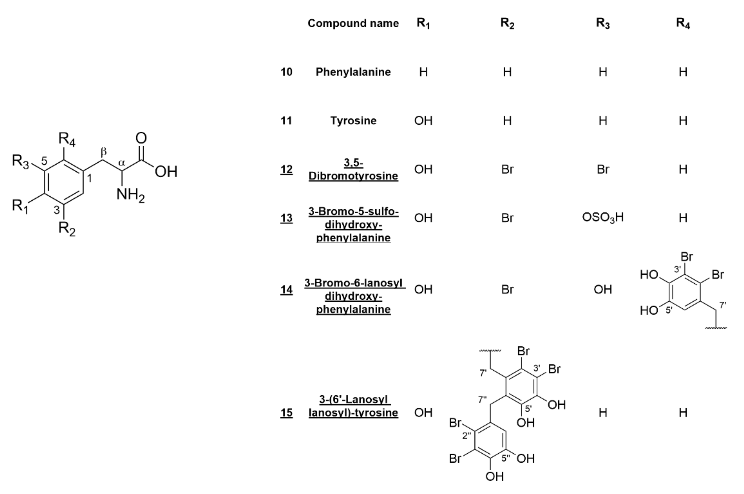
An analysis of the 1H-, 13C- and 2D-NMR data of 1215 showed that all of them possess carboxylic carbons (δC approx. 173) adjacent to a α-carbon with an amino function (δC approx. 55) and a benzylic β-carbon (δC approx. 35) (Table 2), indicating a phenylalanine or tyrosine scaffold. For 12, the (+)ESI-MS data also supported the amino acid substructure by the occurrence of [M – NH3]+ (m/z 325/323/321) and [M – HCOOH]+ (m/z 296/294/292) fragments. The calculated sum formula for the [M + H]+ adduct of 12 (m/z 337.8983, C9H1079Br2NO3+) suggested 12 to be a dibromotyrosine derivative, which was corroborated by the 1H-NMR data being consistent with the literature values for synthesized 3,5-dibromo-L-tyrosine [21]. The configuration of α-carbon was assigned by the optical rotation ([α]20D = −67.7) in comparison to the literature data of tyrosine [22]. This is the first report of this compound being isolated from its natural source.
For 13, the occurrence of a fragment resulting from the loss of a sulfate group (m/z 276/274 [M – SO3]) could be observed in (-)ESI-MS. The isotopes of the [M – H] ion cluster (m/z 355.9304/353.9326) showed relative intensities of 1:1 and gave a calculated sum formula of C9H979BrNO7S for the monoisotopic mass. HMBC correlations of the aromatic and the benzylic protons were used to elucidate the substitution of the phenyl ring: the β-protons showed intense HMBC correlations with the unsubstituted aromatic carbons (δC 132.12 (C-2) and 123.92 (C-6)). H-2, in turn, strongly correlated to β-carbon; C-3; C-4 and C-6 (δC 35.37, 111.84, 146.47 and 123.92, resp.) and H-6 to β-carbon; C-2; C-4 and C-5 (δC 35.37, 132.12, 146.47 and 140.47, resp.). Additionally, both protons showed weak four-bond correlations to the respective carbons. The sulfate moiety was determined to be attached to C-5 by the relative upfield shift of the respective carbon in comparison to unsubstituted phenolic carbons. The configuration of 13 was also determined by optical rotation ([α]20D = −10.4) and comparison to the literature data of tyrosine [22]. Hence, the chemical structure of 13 was found to be 3-bromo-5-sulfo-L-dihydroxyphenylalanine.
Both 14 and 15 showed substituted aromatic carbons with similar shifts (C-6, δC 128.62 and C-3, δC 126.47, resp.) in their 13C-NMR spectra. For both of these carbons, compared to unsubstituted tyrosine, the expected upfield shift in the ortho-position to the hydroxyl group was not apparent, suggesting aliphatic or benzylic substituents attached to the tyrosine scaffold. Further analysis of the HMBC correlations revealed the presence of either one (14) or two (15) lanosyl moieties attached to C-6 (14) or C-3 (15). The isotopic pattern of the [M – H] ion of 14 (m/z 557.8233/555.8243/553.8265/551.8285) in the relative abundance of 1:3:3:1 indicated the presence of three bromine atoms in the molecule, suggesting a bromine substitution in the dihydroxyphenylalanine scaffold in addition to the lanosyl moiety. The position of the third bromine-substituted carbon was deduced from the strong upfield shift and the HMBC correlations from H-2 to C-3 (δC 109.97) to be at position 3. Thus, the chemical structure of 14 was determined to be 3-bromo-6-lanosyl dihydroxyphenylalanine.
The second lanosol residue of 15 is connected to C-6′ via its benzylic carbon. For 15, four bromine atoms were confirmed by the relative isotopic abundance of the [M – H] ion cluster (m/z 743.7806/741.7758/739.7778/737.7803/735.7761) in the ratio of 1:4:6:4:1. The assignment of certain carbons (δC 113.94, C-3′ and δC 143.33, C-4′) was not possible from HMBC correlations, since there are no protons in the molecular vicinity. Based on the substitution of the 2′’,3′’-dibromo-4′’,5′’-dihydroxybenzyl moiety and since p-hydroxybenzaldehyde is the biogenetic origin of the lanosol scaffold [23], we propose the reported assignments (see Table 2) and, therefore, the structure of 15 to be 3-(6′-lanosyl lanosyl)-tyrosine. The structures of compounds 1015 are given in Figure 3.
The HR-ESI-MS of 16 showed an [M + H]+ adduct at m/z 533.9257, inferring the sum formula C13H1879Br2N3O8S+. The presence of sulfur was deduced from a fragment at m/z 453.9645 [M – SO3]+ (C13H1879Br2N3O5), which corresponds to vertebratol [6]. NMR data were mostly consistent with those of vertebratol [6], except for benzylic carbon (δC 75.85), which was shifted about 30 ppm downfield, and the brominated aromatic carbons (δC 114.34 and 112.70, see also Table 1). Once again, the position of the sulfate moiety was assigned by HMBC correlations of H-6 to C-4 and C-5 and, additionally, by comparison of the simulated spectra of the respective 4- and 5-sulfo derivatives. Thus, the structure of 16 was found to be 5-sulfovertebratol (Figure 2c). Due to the low yield, the configuration of 16 could not be determined.

2.2. Mycosporine-Like Amino Acids

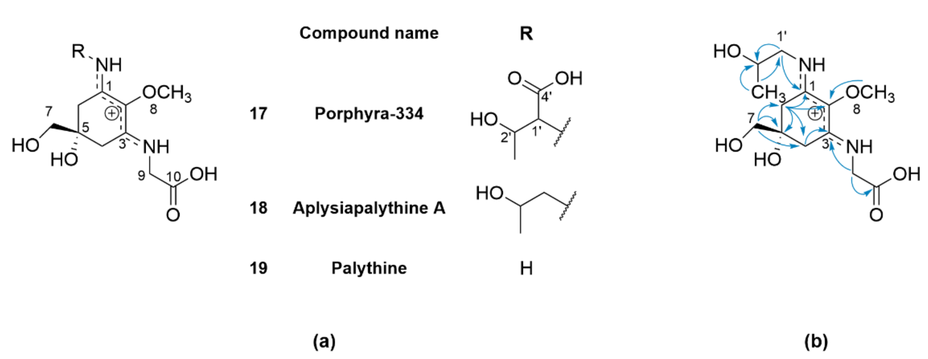
The presence of several MAAs in V. lanosa has been reported recently, however, not all MAAs could be unambiguously identified by LC-MS [15]. Therefore, the aqueous-phase W2 was further fractionated to obtain pure MAAs. Briefly, W2 was first depleted of polysaccharides by ethanolic precipitation and, subsequently, fractionated via cation exchange chromatography on Amberlite™ IR120 (H) resin. The fraction containing the highest amount of MAA (Amb9) was finally purified by preparative HPLC to obtain 17 (5 mg), 18 (1 mg) and 19 (1 mg). The structures of these MAAs are given in Figure 4a. To assess the purity of the compounds and effectiveness of the fractionation procedure, a HPLC method using a HILIC stationary phase was developed to facilitate the targeted isolation of the desired MAA. The isolated compounds correspond to porphyra-334 (17), palythine (18) and aplysiapalythine A (19), respectively, as determined by the comparison of HR-ESI-MS and NMR data (see Supplementary Materials) with the literature [24,25]. Compound 19 was distinguished from its isomer palythinol by the HMBC correlation of methylene protons H-1′ (δH 3.48 and 3.45) to C-1 (δC 162.72) in the cyclohexenimine ring [24].

2.3. Bromophenols and Glycerogalactolipids

In addition to the characterization of the aqueous phase of the methanolic extract, the composition of the ethyl acetate phase was investigated by initial fractionation via FCPC and subsequent preparative HPLC of the fractions obtained (Figure 1). For E1, this method yielded bromophenols lanosol methyl ether (2, 1.3 mg), 5 (7,7′-bis-lanosol ether, 4.6 mg) and 8 (rhodomelol, 0.4 mg). The application of the same method to E2 facilitated the isolation of 1 (lanosol, 5.2 mg), 3 (2,3-dibromo-4,5-dihydroxyphenyl acetic acid methyl ester, 1.0 mg), 5 (7,7′-bis-lanosol ether, 1.2 mg), 6 (2′-lanosyl-3′-bromo-5′,6′-dihydroxybenzyl alcohol, 2.8 mg), 20 ((4Z,7Z,10Z,13Z)-hexadecatetraenoic acid 3′-β-D-galactopyranosyl-1-glycerol ester, 0.6 mg), 21 ((6Z,9Z,12Z,15Z)-stearidonic acid 3′-β-D-galactopyranosyl-1-glycerol ester, 2.7 mg) and 22 ((5Z,8Z,11Z,14Z,17Z)-eicosapentaenoic acid 3′-β-D-galactopyranosyl-1-glycerol ester, 22.8 mg). Figure 5 shows the structures of the isolated glycerogalactolipids. The extruded stationary phase (G II) of E2 was subjected to further fractionation by FCPC with a different solvent system, which ultimately led to the isolation of 9 (methylrhodomelol, 5.7 mg), 20 (0.7 mg) and 23 ((5Z,8Z,11Z,14Z,17Z)-eicosapentaenoic acid 3′-[(6′’-O-α-galactopyranosyl-β-D-galactopyranosyl)]-1-glycerol ester, 1.6 mg) via preparative HPLC.
Compounds 1, 2, 3, 5, 6, 8 and 9 (Figure 2) represent the known bromophenols and were identified by comparison of the NMR and MS data to the literature data [6,26,27]. The structures of 2022 were verified by comparison of the literature data [28,29,30] to the measured MS and NMR spectra (Supplementary Data), followed by sugar hydrolysis and the identification by thin layer chromatography (TLC) and capillary zone electrophoresis (CZE). The NMR data for 23 were very similar to those of 22, suggesting the presence of an eicosapentaenoic acid moiety connected to a 3′-β-glucopyranosylglycerol moiety via a C-1′ ester. Moreover, six additional signals were present in the 1H- and 13C-NMR spectra, indicating a further galactosyl moiety. By the HMBC correlation of H-6″ (δH 3.70/3.56) to C-1′″ (δC 101.10) and the small coupling constant of H-1′″ (δH 4.84, d, J1,2 3.6 Hz), the α-1,6 glycosidic linkage was determined [31]. The identity of the digalactoside was confirmed by sugar hydrolysis and comparison to the reference compounds on TLC. The D-configuration was subsequently determined by CZE analysis after diastereomeric derivatization. This led to the identification of 23 as (5Z,8Z,11Z,14Z,17Z)-eicosapentaenoic acid 3′-(6″-O-α-D-galactopyranosyl-β-D-galactopyranosyl)-1-glycerol ester (Figure 5).

3. Discussion

Rhodomelaceae, especially the genus Vertebrata (syn. Polysiphonia), are known to frequently contain haloaryl derivatives [32], and a series of bromophenolic compounds was isolated from a hydroalcoholic extract of V. lanosa. Lanosol (1), representing the simplest structure, has been isolated from various red algae previously and is reported to possess cytotoxic [9], antiviral [33], antioxidant [34] and glucosidase-inhibiting [35] activity in vitro [32,36]. Similarly, the methyl ether of lanosol (2) showed cytotoxic [9] and antimicrobial [27] effects [32,36], although it might be an isolation artifact formed during methanolic extraction [5]. This might also be the case for 3, which has not been isolated from algae before, unlike the native phenyl acetic acid derivative found in Rhodomela confervoides [37]. Compared to simple lanosols, dimeric bromophenols, such as 5, seem to enhance the observed in vitro effects [6,7,38,39,40,41] and, additionally, showed antifungal [42] and anti-inflammatory [43] activity. Further, 5 and, to a lesser extent, also 6 have been reported to inhibit glucose-6-phosphate dehydrogenase [40,44]. It should, however, be mentioned that the observed biological activities of bromophenols were often relatively weak—in particular, the antimicrobial effects [6,32,36]—and, with the exception of some compounds such as 8, rather unselective [36]. Kurihara et al. pointed out that enzyme inhibition could be the result of an o-quinone addition to the respective proteins [35], and as Baell emphasized, compounds with a variety of reported moderate bioactivities should be considered as potential drug leads only with caution, as, particularly, molecules with catechol substructures are prone to interfering with multiple in vitro assays [45].
In addition to the more frequently occurring bromophenols, two new brominated tyrosine derivatives (12 and 13) and two amino acids coupled to lanosyl moieties (14 and 15) were also isolated. A bromotyrosine has previously been found in Rhodomela confervoides [46], as well as derivatives of lanosol coupled to pyroglutamate [47]. So far, L-tyrosine (11) has been considered the main precursor of bromophenols, with the respective hydroxy benzoic acid derivatives being brominated at later steps in biosynthesis [23]. On the other hand, as also suspected by Ma et al. [46], the presence of 12 and 13 suggests that bromination occurs at a much earlier stage of biosynthesis. Whether bromoperoxidases in V. lanosa, unlike other macroalgae [48], are capable of directly converting L-tyrosine remains a topic for further research.
Another intriguing finding with respect to the biosynthesis is that, for Vertebrata species, typically, 2,3-dibrominated benzyl moieties have been reported (Vertebrata decipiens (syn. Polysiphonia decipiens) [38] and V. lanosa [6]). This is well in line with most of the compounds isolated in this study (17 and 1416); however, 12 is an exception from this substitution pattern. The 3,5-dibrominated benzyl moiety, as in 12, is usually found in members of the Polysiphonia genus (Polysiphonia stricta (syn. P. urceolata) [49] and P. morrowii [50]), and the occurrence of 12 in V. lanosa might therefore be a hint for the presence of two regioselective bromoperoxidases in this alga.
Along with the amino acid-coupled compounds, sulfated bromophenols (4, 7, 14 and 16) were also isolated. Lanosol-4,7-disulfate (4) is one of the major secondary metabolites within the extract, and its occurrence in V. lanosa has been known for a long time [11,12]. Probably due to its high quantity, it has previously even been assumed to be the only sulfated compound in extracts from this alga [10]. On the other hand, the presence of sulfates could depend on parameters like the extraction procedure, as suggested by Weinstein et al., who suspected lanosol and its derivatives to be artifacts [51]. Despite this, Barreto and Meyer still found the lanosol disulfate ester in Osmundaria serrata after an extraction procedure of one week [52]. Taking into account that salt, unlike the unsulfated lanosols, was inactive as a feeding deterrent [53], they proposed bromophenols to be stored in algae as inactive salts and the release of lanosols upon injury [52]. On the other hand, Ma et al. did not find a significant difference in the cytotoxicity of a certain bromophenol versus its sulfate ester [46]. Within the current study, we also found the sulfate ester of vertebratol (16), a bromophenol that was just recently isolated from V. lanosa [6], but we cannot infer from our data which compound is genuine to the plant.
In contrast, compound 7 represents a unique structure containing a dimethyl sulfonium group. Generally, methyl sulfonium moieties, particularly dimethyl sulfonioproionate (DMSP), are widely distributed and also present in marine algae [54], including V. lanosa [55,56], where it acts as an antioxidant [57], cryoprotectant and osmolyte [54,58,59]. DMSP is biosynthesized in several steps via methionine [59] and degraded enzymatically to dimethylsulfide by DMSP lyases [56]. In the case of 7, the C-3 alkyl side chain is formally replaced by a dibromo dihydroxy benzyl moiety. Whether the sulfur atom is derived from an amino acid, e.g., cysteine, similar to the biosynthesis of methionine [60], and at what stage of lanosol formation remains a subject for further research. Interestingly, a bromophenol containing a similar sulfoxide structure was previously isolated from Rhodomela confervoides [47]. It could be regarded as an oxidation product of 7 comparable to dimethyl sulfoxide (DMSO) produced from DMSP in bacteria and microalgae [61,62]. Accordingly, 7 could be a precursor of the respective sulfoxide reported by Zhao et al. [47], and it would be interesting to explore if there are further sulfoxonium intermediates that could suggest a route of oxidation similar to the biosynthesis of DMSO from DMSP via dimethyl sulfoxonium propionate (DMSOP) [62]. However, the conversion of DMSOP to DMSO has been experimentally confirmed in bacteria [62], and the enzymatic cleavage of DMSP in V. lanosa is reported to yield mainly dimethyl sulfide [56]. On the other hand, Lee and de Mora summarized their findings supporting the hypothesis of intracellular production of DMSO by algae [61]. If such a biosynthetic route is applicable to a structurally distinct substrate such as 7 remains, of course, highly speculative.
Apart from bromophenols, several glycerogalactolipids (2022 and the new compound 23) were isolated. They represent a class of fatty acid derivatives attached to a glycerogalactoside moiety and are very common in algae [29,63], but they have not been reported for V. lanosa until now. Typically, glycerogalactolipids are constituents of membranes in plant plastids and other organisms performing photosynthesis [64]. Apart from their role in photosynthesis, they may also be involved in radical scavenging and cryoprotection [65] and exert haemolytic activity in organisms like oysters or fish [29,30].
Finally, the major MAAs in V. lanosa were identified, confirming the presence of porphyra-334 (17), the most abundant MAA in this alga, and palythine (19) [15]. Compound 18, which was suspected to be palythinol based on its HPLC and UV data [15], was instead found to be aplysiapalythine A.

4. Materials and Methods

4.1. Plant Material and Chemicals

Algal material of V. lanosa was collected, identified and kindly provided as a gift by Nutramara Ltd. in March (VL1, 700 g) and October (VL2, 900 g) 2020 from Dungloe Bay, West Donegal, Ireland. Voucher specimens of both collection series (IPBP524 and IPBP528, respectively) were deposited at the Institute of Pharmaceutical Biology and Phytochemistry, University of Münster, Germany. Both batches of algae were kept separately during extraction and fractionation.
If not stated otherwise, all chemicals were purchased from VWR (Darmstadt, Germany). The solvents used for the analytical and preparative work were of analytical grade.

4.2. General Analytical Methods

Analytical UPLC for the purity assessment of the compounds and method development for preparative HPLC was generally carried out on Acquity™ Ultra Performance LC (UPLC), PDA λe Detector and QDa™ Detector autosampler, in-line degasser and Waters Empower 3® Software (Waters, Milford, MA, USA). For separation, a binary gradient of 0.1% formic acid (A) and acetonitrile/0.1% formic acid (B) at 0.5 mL/min was used in a RP-18 stationary phase (HSS T3, 1.8 μm, 2.1 × 100 mm) at 40 °C. The separation of MAA was carried out in a HILIC stationary phase (Phenomenex Kinetex HILIC, 1.7 µm, 100 × 2.1 mm) at 40 °C using a binary gradient of 5 mM ammonium acetate pH 6.7 (A) and acetonitrile (B) at 0.5 mL/min, modified from [66]: t0min 90% B, t1min 90% B, t4min 83% B, t9min 83% B, t11min 50% B, t12.5min 50% B, t13min 90% B and t20min 90% B.
Thin-layer chromatography (TLC) for analytical purposes was performed on silica gel plates 60 F254 (0.2 mm; Merck, Darmstadt, Germany) using ethyl acetate/water/formic acid (90:5:5 v/v/v) as the standard mobile phase. Visualization of the compounds was achieved under UV light (254 nm or 365 nm, resp.) and at daylight after spraying with thymol/sulfuric acid reagent, followed by heating the plate to approximately 105 °C for bromophenolic compounds and for MAA by spraying with ninhydrin.
NMR spectra were recorded on an Agilent DD2 spectrometer (Agilent Technologies, Santa Clara, CA, USA) at 600 MHz (1H) or 150 MHz (13C). Depending on their solubility, samples were dissolved in the respective solvents as follows: 22 in chloroform-d1 (7.26; 77.16 ppm); 1, 2, 8, 9, 14, 16, 20, 21 and 23 in methanol-d4 (3.31; 49.00 ppm); 3, 5 and 6 in acetone-d6 (2.84; 206.26 ppm) and 4, 1013, 15 and 1719 in water-d2 (4.80 ppm). Chemical shifts were referenced to the respective residual solvent signals (1H and 13C shifts in brackets). Compounds 7 and 15 were dissolved in a 1:1 (v/v) mixture of water-d2 and methanol-d4 (experimentally determined shifts relative to TMS: 4.77 ppm (water-d2), 3.32 and 48.97 ppm (methanol-d4)). For 7 and 15, 1H NMR (only 15) and 2D spectra (7 and 15) were recorded utilizing WET signal suppression.
Analysis by UPLC-qTOF-MS was carried out as follows: Separation was performed on a Dionex Ultimate 3000 RS Liquid Chromatography System (Thermo Fisher Scientific, Waltham, MA, USA) over a Dionex Acclaim RSLC 120, C18 column (2.1 × 100 mm, 2.2 µm) with a binary gradient (A: water with 0.1% formic acid; B: acetonitrile with 0.1% formic acid) at 0.4 mL/min: t0min 5% B, t0.4min 5% B, t9.9min 100% B, t15min 100% B, t15.1min 5% B and t20min 5% B. The injection volume was 2 or 5 µL. Eluted compounds were detected using a Dionex Ultimate DAD-3000 RS over a wavelength range of 200–400 nm and a Bruker Daltonics micrOTOF-QII time-of-flight mass spectrometer equipped with an Apollo electrospray ionization source in positive or negative mode (depending on the respective substance) at 3 Hz over a mass range of m/z 50–1500 using the following instrument settings: nebulizer gas nitrogen, 3.5 bar; dry gas nitrogen, 9 L/min, 180 °C (positive mode) or 200 °C (negative mode); capillary voltage, 4500 V; end plate offset, −500 V; collision energy, +3 eV or −8 eV; transfer time, 100 µ and pre-pulse storage, 6 µs; the collision energy and collision RF settings were combined to the single spectrum of 1650 (positive mode) or 2483 (negative mode) summations. MS/MS scans were triggered by AutoMS2 settings within a range of m/z: 200–1500. Internal dataset calibration (HPC or enhanced quadratic mode) was performed for each analysis using the mass spectrum of a 10-mM solution of sodium formiate in isopropanol–water-formic acid–1M NaOH solution (50 + 50 + 0.2 + 1) that was infused during LC re-equilibration using a divert valve equipped with a 20-µL sample loop.
Simulation of NMR spectra and generation of IUPAC names was performed with ChemDraw Ver. 21.0, PerkinElmer Informatics Inc. 2022 (Waltham, MA, USA).
Optical rotations were measured using Autopol V (Rudolph analytical research) at 20 °C and 589 nm. The compound concentrations ranged from 0.1 to 0.6 g/100 mL, and the solvents used were methanol, water and methanol/water (1:1 v/v).

4.3. Hydrolysis of Glycosides

Mono- and disaccharides were hydrolyzed as described by Albersheim et al. [67]. Trifluoroacetic acid (TFA) was removed by at least three washing steps with 2 mL MeOH 50%. Hydrolyzed sugars were identified by TLC (n-propanol/water/EtOH 7/2/1 (v/v), visualization with thymol sulfuric acid after heating to 105 °C) compared to the reference compounds (concentration: 1 mg/mL).

4.4. Capillary Zone Electrophoresis (CZE)

The derivatization of the carbohydrates for analysis by CZE electrophoresis was conducted as described by Noe and Freissmuth [68]. CZE equipment: Beckman Coulter P/ACE MDQ (Beckman Coulter, Brea, CA, USA); fused silica capillary, 70/77 cm × 50 μm i.d.; running buffer, 50 mM Na2B2O7 pH 10.3, MeCN 4.4 mol/L added; injection, 5−10 s at 0.5 psi; voltage, 30 kV; detection, λ = 200 nm; software, 32 Karat software version 5.0 (Beckman Coulter).

4.5. Extraction and Fractionation

Air-dried and ground algae were extracted five times for 20 min using methanol/water (8:2, v/v) (3.75 L/500 g) in an ultrasonic bath. The crude extract was filtered and lyophilized after evaporation of the organic solvent, yielding M1 (122.52) and M2 (148.07 g). Subsequent defatting by stirring five times with 1.25 L petroleum ether/100 g of crude extract for 10 min yielded the defatted extracts Md1 (96.11 g) and Md2 (142.21 g). All extracts and fractions were stored at −20 °C.

4.5.1. Solvent Partitioning of the Extracts

Twenty grams of defatted extract were sequentially partitioned between 1 L water and 1 L ethyl acetate. Each partitioning step was repeated four times with fresh ethyl acetate, yielding the ethyl acetate fractions E1 (1.184 g) and E2 (1.767 g) and the aqueous fractions W1 (80.18 g) and W2 (131.44 g).

4.5.2. Fast Centrifugal Partition Chromatography (FCPC)

FCPC was performed using the following system: SCPC-250 (Gilson, Middleton, WI, USA); pump: AZURA P 4.1S (KNAUER GmbH, Berlin, Germany); injector: 10-mL injection loop, Rheodyne (Oak Harbor, WA, USA); mobile phase: EtOAc/hexane 3:7 (v/v), stationary phase: MeOH/water 7:3 (v/v); ascending mode, flow 10 mL/min, 1600 rpm, fraction size: 20 mL. Six hundred and fifty milligrams of sample per run were dissolved in 7 mL of the mobile phase. The fraction analysis by TLC led to fractions A–J (E1) and A II–G II (E2). Four hundred and fifty milligrams of fraction E2 (G II), representing the extruded stationary phase, was further fractionated via FCPC (mobile phase: EtOAc/butanol 4/5:0/5 (v/v), stationary phase: water), yielding fractions G IIa–G IIe.

4.5.3. Medium-Pressure Liquid Chromatography (MPLC)

Twenty grams of the aqueous fraction W1 were separated by MPLC on a RP-18 stationary phase (RP-18, 18–32 µm, 100 Å, 36 × 500 mm (BESTA Technik, Wilhelmsfeld, Germany), flow rate 5 mL/min, step gradient MeOH 5%/HCOOH 0.1% (525 mL) → MeOH 25%/HCOOH 0.1% (300 mL) → MeOH 50%/HCOOH 0.1% (300 mL) → MeOH (900 mL min), fraction size 15 mL. Two grams of samples dissolved in 10 mL of water were injected per run. Every second fraction was spotted on a TLC plate and detected without development, yielding fractions Aq I–Aq V, of which Aq III corresponded to pure compound 4 (107 mg).

4.5.4. Sugar Precipitation and Ion Exchange Chromatography

To obtain an MAA-enriched fraction, the polysaccharides were precipitated in the first step. Therefore, 50 g of W2 dissolved in 500 mL of water were added dropwise to 2 L of ice-cold ethanol while stirring. After stirring overnight and centrifugation (3000× g, 15 min), the residual polysaccharide fraction WP2 (9.6 g) and the polysaccharide-depleted supernatant WM2 (38.5 g) were obtained.
Ten grams of WM2 were then subjected to fractionation by ion exchange chromatography (Amberlite™ IR120 (H) resin, 40 × 550 mm, flow 4 mL/min) using a pH step gradient of 0.1 M citric acid and 0.2 M phosphate buffer [69]: pH 2.6 (700 mL) →pH 7.0 (700 mL) → pH 8.0 (2.7 L), fraction size 12 mL. TLC analysis of the fractions led to Amb1–Amb14.

4.5.5. Preparative HPLC

Preparative HPLC was carried out using Waters Quaternary Gradient Module 2545, Photodiode Array Detector 2998, Autosampler 2707, Prep Degasser and Fraction Collector III. Software: Waters ChromScope v1.40 Beta (Waters, Milford, MA, USA). Stationary phase: Nucleodur® C18 HTec, 5 µm, 250 × 21 mm and mobile phase: binary gradient of water (A) and acetonitrile (B) at a flow rate of 15 mL/min. For fractions Aq IV, Aq Iva and Aq IVb, the addition of 0.1% trifluoroacetic acid was necessary. For fraction Amb9, the stationary phase was a Modulo-card Uptisphere 6 µm, diol, 250 × 21.2 mm, and the mobile phase consisted of a binary gradient of 5 mM ammonium acetate buffer pH 6.7 (A) and acetonitrile (B). The gradients were adapted to each subfraction individually, and the purity was assessed by UPLC. Pure compounds were obtained from the respective subfractions by preparative HPLC as follows: Fraction D: 2 (1.3 mg); fractions G and H: 5 (1.7 and 2.9 mg, respectively); fraction J: 8 (0.4 mg); fraction C II: 3 (1.0 mg) and 5 (2.3 mg); fraction D II: 1 (5.2 mg) and 5 (1.2 mg); fraction F II: 6 (2.8 mg), 20 (0.6 mg), 21 (2.7 mg) and 22 (22.8 mg); fraction G IIa: 9 (5.7 mg), 20 (0.7 mg) and 23 (1.6 mg); fraction Aq IV: 11 (11.8 mg) and 13 (9.5 mg); fraction Aq IVa: 2 (2.8 mg), 12 (5.1 mg), 14 (3.7 mg) and 15 (25.5 mg); fraction Aq IVb: 7 (3.2 mg), 10 (11.5 mg) and 16 (1.1 mg) and fraction Amb9: 17 (5 mg), 18 (1 mg) and 19 (1 mg).

4.6. Structure Elucidation of Isolated Compounds

Spectroscopic data (UV spectra, NMR spectra and optical rotation) and HR-ESI-MS were used for structure elucidation. NMR shifts of the known compounds (16, 811 and 1722) were compared to the published literature and are reported as the Supplementary Materials [70]. Their respective IUPAC names are given in brackets.
4-Sulfo-7-dimethylsulfonium lanosol (2,3-dibromo-4-((dimethylsulfonio)methyl)-6-hydroxyphenyl sulfate) (7): orange amorphous solid; UV (MeCN, H2O) λmax 212, 300 nm. 1H NMR and 13C NMR assignments are given in Table 1. HR(+)ESI-MS m/z 424.8411/422.8437/420.8463 (56:100:48) [M + H]+ (calcd. for C9H1179Br2O5S2+, 420.8409). Observed adducts and fragments: 446.8255/444.8231/442.8319 (1.3:2.3:1.1) [M + Na]+, 441.8606/439.8681/437.8642 (1.1:1.5:0.7) [M + NH4]+, 362.8190/360.8213/358.8278 (1.0/2.0/1.0) [M – C2H6S]+, 344.8835/342.8857/340.8882 (11:20:10) [M – SO3]+, 304.8454/302.8476/300.8512 (2.0:3.7:2.0), 282.8634/280.8657/278.8681 (11:21:11) [C7H5Br2O2]+; HR(-)ESI-MS m/z 422.8228/420.8246/418.8255 (54:100:47) [M – H] (calcd. for C9H979Br2O5S2+, 418.8264). Observed adducts and fragments: 1270.4775/1268.4796/1266.4811/1264.4819/1262.4938/1260.4836/1258.4411 (1.8:3.6:6.7:6.4:5.4:2.3:0.5) [3M – H], 846.6488/844.6522/842.6511/840.6541/838.6606 (3.2:7.5:10:7.1:1.9) [2M – H], 408.8086/406.8088/404.8096 (19:43:37) [M – CH3], 406.8088/404.8096/402.8141 (43:37:11) [M – CH4]-, 392.7819/390.7742/388.7762 (2.0:3.2:1.7) [M – C2H6], 374.8163/372.8203/370.8228 (3.0:5.1:2.2) [M – CH4S], 360.8072/358.8057/356.8041 (5.0:9.9:5.1) [M – C2H6S]-, 342.8753/340.8838/338.8897 (1.4:4.4:2.8) [M – SO3], 328.8621/326.8742/324.8738 (1.1:5.5:4.1) [M – CH2SO3], 298.9073/296.9085/294.8619 (3.3:3.7:2.4), 294.8619/292.8643/290.8664 (2.4:4.2:2.1), 160.8731/158.8745 (3.4:3.0).
3,5-Dibromo-L-tyrosine (2-amino-3-(3,5-dibromo-4-hydroxyphenyl)propanoic acid) (12): red amorphous solid; [α]20D -67.7 (c 0.1, H2O); UV (MeCN, H2O) λmax 208, 288 nm. 1H and 13C NMR, see Table 2. HR(+)ESI-MS m/z 341.8944/339.8966/337.8983 (50:100:53) [M + H]+ (calcd. for C9H1079Br2NO3+, 337.9022). Observed adducts and fragments: 682.7802/680.7818/678.7841/676.7853/674.7884 (0.5:1.8:2.6:1.9:0.5) [2M + H]+, 324.8681/322.8694/320.8712 (1.4:2.7:1.4) [M – NH3]+, 295.8888/293.8911/291.8632 (3.1:6.6:3.3) [M – HCOOH]+, 282.8589/280.8590/278.8616 (0.1:0.2:0.1) [C7H5Br2O2]+.
3-Bromo-5-sulfo-L-dihydroxyphenylalanine (2-amino-3-(3-bromo-4-hydroxy-5-(sulfooxy)phenyl)propanoic acid) (13): off-white amorphous solid; [α]20D -10.4 (c 0.2, H2O); UV (MeCN, H2O) λmax 200, 284 nm. 1H and 13C NMR, see Table 2. HR(-)ESI-MS m/z 355.9304/353.9326 (100:99.7) [M – H] (calcd. for C9H979BrNO7S-, 353.9289). Observed adducts and fragments: 734.8495/732.8498/730.8502 (2.0:3.0:1.5) [2M + Na – H], 712.8664/710.8679/708.8694 (8.6:14:6.7) [2M + H], 632.9083/630.9133/628.9128 (1.2:2.1:1.1) [2M – SO3], 377.9113/375.9135 (3.3:3.0) [M + Na – H], 275.9731/273.9765 (2.1:2.3) [M – SO3].
3-Bromo-6-lanosyl dihydroxyphenylalanine (2-amino-3-(5-bromo-2-(2,3-dibromo-4,5-dihydroxybenzyl)-3,4-dihydroxyphenyl)propanoic acid) (14): reddish-brown amorphous solid; [α]20D-5.7 (c 0.1, MeOH); UV (MeCN, H2O) λmax 212, 288 nm. 1H and 13C NMR, see Table 2. HR(-)ESI-MS m/z 557.8233/555.8243/553.8265/551.8285 (31:94:100:36) [M – H] (calcd. for C16H1379Br3NO6, 551.8298). Observed adducts and fragments: 1116.6650/1114.6559/1112.6563/1110.6609/1108.6574/1106.6631/1104.6636 (6.0:23:57:71:55:23:4.1) [2M – H], 475.9023/473.9005/471.9021 (29:55:25) [M – HBr], 401.8730/399.8681/397.8646 (2.8:7.5:3.9), 285.9549/283.9564 (15:16).
3-(6′-lanosyllanosyl)-tyrosine (2-amino-3-(3-(2,3-dibromo-6-(2,3-dibromo-4,5-dihydroxybenzyl)-4,5-dihydroxybenzyl)-4-hydroxyphenyl)propanoic acid)(15): brown amorphous solid; [α]20D +2.8 (c 0.6, MeOH/H2O (1:1, v/v)); UV (MeCN, H2O) λmax 212, 284 nm. 1H and 13C NMR, see Table 2. HR(-)ESI-MS m/z 743.7806/741.7758/739.7778/737.7803/735.7761 (16:72:100:69:21) [M – H] (calcd. for C23H1879Br4NO7-, 735.7822). Observed adducts and fragments: 1488.5855/1486.5671/1484.5609/1482.5653/1480.5649/1478.5662/1476.5600/1474.5634/1472.5691 (1.8:7.0:16:35:44:32:14:3.9:1.1) [2M – H], 661.8463/659.8541/657.8556/655.8593 (18:48:49:21) [M – HBr], 571.8429/569.8419/567.8442/565.8438 (9.5:30:33:9.3), 312.8692/310.8778/308.8782 (8.3:16:6.5).
5-Sulfovertebratol (2-amino-5-(3-(2,3-dibromo-4-hydroxy-5-(sulfooxy)benzyl)ureido)pentanoic acid) (16): brown amorphous solid; UV (MeCN, H2O) λmax 208, 291 nm. 1H and 13C NMR, see Table 1. HR(+)ESI-MS m/z 537.9224/535.9246/533.9257 (53:100:47) [M + H]+ (calcd. for C13H1879Br2N3O8S+, 533.9176). Observed adducts and fragments: 1074.8353/1072.8343/1070.8437/1068.8449/1066.8397 (2.2:7.3:9.1:5.8:1.5) [2M + H]+, 559.9050/557.9060/555.9135 (1.1:2.1:1.4) [M + Na]+, 554.9228/552.9428/550.9326 (0.8:0.7:0.3) [M + NH4]+, 520.8884/518.8865/516.9051 (0.6:0.9:0.6) [M – NH3]+, 510.8561/508.8674/506.8660 (0.6:0.8:0.5), 457.9631/455.9705/453.9645 (1.7:3.0:1.9) [M – SO3, Vertebratol]+.
(5Z,8Z,11Z,14Z,17Z)-Eicosapentaenoic acid 3′-[(6-O-α-galactopyranosyl-β-D-galactopyranosyl)]-1-glycerol ester (2-hydroxy-3-(((2R,3R,4S,5R,6R)-3,4,5-trihydroxy-6-((((2S,3R,4S,5R,6R)-3,4,5-trihydroxy-6-(hydroxymethyl)tetrahydro-2H-pyran-2-yl)oxy)methyl)tetrahydro-2H-pyran-2-yl)oxy)propyl (5Z,8Z,11Z,14Z,17Z)-icosa-5,8,11,14,17-pentaenoate) (23): sticky yellow solid; UV (MeCN, H2O) λmax 224 nm. 1H NMR (CD3OD, 600 MHz) δ 5.44–5.29 (10H, m, H-5, H-6, H-8, H-9, H-11, H-12, H-14, H-15, H-17, H-18), 4.26 (2H, d, J = 7.5 Hz, H-1″), 4.19–4.13 (2H, m, H-1′), 4.02–3.98 (1H, m, H-2′), 3.93–3.91 (1H, m, H-6″a), 3.91–3.89 (1H, m, H4′″), 3.88 (1H, s, H-3′a), 3.88 (1H, d, J = 5.4 Hz, H-4″), 3.86 (1H, d, J = 5.3 Hz, H-5′’’), 3.79 (1H, dd, J = 10.1, 3.7 Hz, H-2′″), 3.77 (1H, d, J = 8.1 Hz, H-5″), 3.75 (1H, d, J = 6.9 Hz, H-3′″), 3.74–3.71 (2H, m, H-6′″), 3.70 (1H, d, J = 3.7 Hz, H-6″b), 3.69–3.65 (1H, m, 3′b), 3.55 (1H, dd, J = 9.8, 7.5 Hz, H-2″), 3.51 (1H, dd, J = 9.7, 3.4 Hz, H-3″), 2.86 (8H, dt, J = 16.3, 5.3 Hz, H-7, H-10, H-13, H-16), 2.39 (2H, t, J = 7.4 Hz, H-3), 2.17–2.13 (2H, m, H-4), 2.12–2.07 (2H, m, H-19), 1.70 (2H, p, J = 7.4 Hz, H-3), 1.01–0.97 (3H, m, H-20). The coupling constant of H-1″ was determined in acetone as the solvent signal of methanol at δH 4.87 that interfered with its measurement: 1H NMR (acetone-d6, 600 MHz) δ 4.84 (1H, d, J = 3.8 Hz, H-1′″); 13C NMR (CD3OD, 151 MHz) δ 175.25 (C-1), 132.82 (C-18), 130.04, 129.91 (C-5, C-6), 129.47, 129.25, 129.20, 129.13, 129.11, 128.94, 128.20 (C-8, C-9, C-11, C-12, C-14, C-15, C-17), 105.33 (C-1″), 100.56 (C-1′″), 74.67 (C-3″), 74.57 (C-5″), 72.57 (C-5′″), 72.53 (C-2″), 72.12 (C-3′), 71.47 (C-3′″), 71.06 (C-4′″), 70.24 (C-2′″), 70.10 (C-4″), 69.67 (C-2′), 67.78 (C-6″), 66.60 (C-1′), 62.75 (C-6′″), 34.34 (C-2), 27.56 (C-4), 26.57, 26.55 (C-7, C-10, C-13), 26.44 (C-16), 25.90 (C-3), 21.51 (C-19), 14.67 (C-20); HR(+)ESI-MS m/z 701.3766 (14) [M + H]+ (calcd. for C35H57O14+, 701.3766). Observed adducts and fragments: 1423.7381 (17) [2M + Na]+, 1418.7820 (12) [2M + NH4]+, 1401.7559 (24) [2M + H]+, 723.3584 (69) [M + Na]+, 718.4033 (71) [M + NH4]+, 683.3676 (22) [M – H2O]+, 539.3223 36) [M – Gal]+, 521.3117 (11) [M – Gal – H2O]+, 377.2684 (100) [M – 2Gal]+, 285.2208 (24) (fatty acid oxocarbenium ion, [C20H29O]+), 267.2107 (8.2) (fatty acid carbenium ion [C20H27]+).

Supplementary Materials

The following supporting information can be downloaded at: https://www.mdpi.com/article/10.3390/md20070420/s1. Analytical data used for the structure elucidation of 16, 811 and 1722. Table S1. 1H- and 13C-NMR data of 2023. Figure S1. (a–e) One- and two-dimensional NMR spectra of 7. Figure S2. (a–e) One- and two-dimensional NMR spectra of 12. Figure S3. (a–e) One- and two-dimensional NMR spectra of 13. Figure S4. (a–e) One- and two-dimensional NMR spectra of 14. Figure S5. (a–e) One- and two-dimensional NMR spectra of 15. Figure S6. (a–e) One- and two-dimensional NMR spectra of 16. Figure S7. (a–f) One- and two-dimensional NMR spectra of 23. Figure S8. (a–c) HR-ESI-MS spectra of 7. Figure S9. HR-(+)ESI-MS spectrum of 12. Figure S10. HR-(-)ESI-MS spectrum of 13. Figure S11. HR-(-)ESI-MS spectrum of 14. Figure S12. HR-(-)ESI-MS spectrum of 15. Figure S13. HR-(+)ESI-MS spectrum of 16. Figure S14. HR-(+)ESI-MS spectrum of 23. Figure S15. 1H-NMR spectrum of 1 (methanol-d4, 289 K, 600 MHz). Figure S16. 1H-NMR spectrum of 2 (methanol-d4, 289 K, 600 MHz). Figure S17. 1H-NMR spectrum of 3 (acetone-d6, 289 K, 600 MHz). Figure S18. 1H-NMR spectrum of 4 (water-d2, 289 K, 400 MHz). Figure S19. 1H-NMR spectrum of 5 (acetone-d6, 289 K, 600 MHz). Figure S20. 1H-NMR spectrum of 6 (acetone-d6, 289 K, 600 MHz). Figure S21. 1H-NMR spectrum of 8 (methanol-d4, 289 K, 600 MHz). Figure S22. 1H-NMR spectrum of 9 (methanol-d4, 289 K, 600 MHz). Figure S23. 1H-NMR spectrum of 10 (water-d2, 289 K, 600 MHz). Figure S24. 1H-NMR spectrum of 11 (water-d2, 289 K, 600 MHz). Figure S25. 1H-NMR spectrum of 17 (water-d2, 289 K, 600 MHz). Figure S26. 1H-NMR spectrum of 18 (water-d2, 289 K, 600 MHz). Figure S27. 1H-NMR spectrum of 19 (water-d2, 289 K, 600 MHz). Figure S28. 1H-NMR spectrum of 20 (methanol-d4, 289 K, 600 MHz). Figure S29. 1H-NMR spectrum of 21 (methanol-d4, 289 K, 600 MHz). Figure S30. 1H-NMR spectrum of 22 (chloroform-d1, 289 K, 600 MHz).

Author Contributions

Conceptualization, J.J. and V.S.; methodology, J.J., M.S. and V.S.; validation, J.J., M.S. and V.S.; formal analysis, J.J. and M.S.; investigation, J.J. and M.S.; writing—original draft preparation, J.J. and V.S.; writing—review and editing, J.J., M.S. and V.S.; visualization, J.J.; supervision, V.S.; project administration, J.J and V.S. and funding acquisition, M.S. and V.S. All authors have read and agreed to the published version of the manuscript.

Funding

Marthe Schmitt received financial support from the Erasmus+ programme for student traineeship. The APC was funded by the Open Access Publication Fund of the University of Münster.

Institutional Review Board Statement

Not applicable.

Informed Consent Statement

Not applicable.

Data Availability Statement

The data are contained within the article and the Supplementary Materials.

Acknowledgments

We would like to thank H. Sheridan and Nutramara Ltd. for kindly helping us with the supply and for donating the algal material. We are also grateful to J. Köhler and C. Thier for recording the NMR spectra and J. Sendker and F. Jürgens for measuring the MS spectra. Additionally, we thank B. Quandt-Rusch for performing the CZE of the sugar derivatives.

Conflicts of Interest

The authors declare no conflict of interest.

References

  1. Guiry, M.D. AlgaeBase: World-Wide Electronic Publication, National University of Ireland, Galway. Available online: https://www.algaebase.org (accessed on 3 May 2022).
  2. Bjordal, M.V.; Jensen, K.H.; Sjøtun, K. A field study of the edible red alga Vertebrata lanosa (Rhodophyta). J. Appl. Phycol. 2020, 32, 671–681. [Google Scholar] [CrossRef]
  3. Leandro, A.; Pereira, L.; Gonçalves, A.M.M. Diverse Applications of Marine Macroalgae. Mar. Drugs 2019, 18, 17. [Google Scholar] [CrossRef] [PubMed] [Green Version]
  4. Stoffelen, H.; Glombitza, K.W.; Murawski, U.; Bielaczek, J.; Egge, H. Bromphenole aus Polysiphonia lanosa (L.) Tandy. Planta Med. 1972, 22, 396–401. [Google Scholar] [CrossRef] [PubMed]
  5. Glombitza, K.-W.; Stoffelen, H.; Murawski, U.; Bielaczek, J.; Egge, H. Antibiotica aus Algen. Planta Med. 1974, 25, 105–114. [Google Scholar] [CrossRef]
  6. Hofer, S.; Hartmann, A.; Orfanoudaki, M.; Ngoc, H.N.; Nagl, M.; Karsten, U.; Heesch, S.; Ganzera, M. Development and Validation of an HPLC Method for the Quantitative Analysis of Bromophenolic Compounds in the Red Alga Vertebrata lanosa. Mar. Drugs 2019, 17, 675. [Google Scholar] [CrossRef] [PubMed] [Green Version]
  7. Olsen, E.K.; Hansen, E.; Isaksson, J.; Andersen, J.H. Cellular antioxidant effect of four bromophenols from the red algae, Vertebrata lanosa. Mar. Drugs 2013, 11, 2769–2784. [Google Scholar] [CrossRef] [PubMed] [Green Version]
  8. Glombitza, K.W.; Sukopp, I.; Wiedenfeld, H. Antibiotics from Algae XXXVII. Rhodomelol and Methylrhodomelol from Polysiphonia lanosa. Planta Med. 1985, 51, 437–440. [Google Scholar] [CrossRef]
  9. Shoeib, N.A.; Bibby, M.C.; Blunden, G.; Linley, P.A.; Swaine, D.J.; Wheelhouse, R.T.; Wright, C.W. In-vitro cytotoxic activities of the major bromophenols of the red alga Polysiphonia lanosa and some novel synthetic isomers. J. Nat. Prod. 2004, 67, 1445–1449. [Google Scholar] [CrossRef]
  10. Ragan, M.A.; Mackinnon, M.D. Paired-ion reversed-phase high-performance liquid chromatography of phenol sulfates in synthetic mixtures, algas extracts and urine. J. Chromatogr. A 1979, 178, 505–513. [Google Scholar] [CrossRef]
  11. Glombitza, K.W.; Stoffelen, H. 2,3-Dibrom-5-hydroxybenzyl-1′,4-disulfat (dikaliumsalz) aus Rhodomelaceen. Planta Med. 1972, 22, 391–395. [Google Scholar] [CrossRef]
  12. Hodgkin, J.H.; Craigie, J.S.; McInnes, A.G. the occurrence of 2,3-dibromobenzyl alcohol 4,5-disulfate, dipotassium salt, in Polysiphonia lanosa. Can. J. Chem. 1966, 44, 74–78. [Google Scholar] [CrossRef] [Green Version]
  13. Batey, J.F.; Turvey, J.R. The galactan sulphate of the red alga Polysiphonia lanosa. Carbohydr. Res. 1975, 43, 133–143. [Google Scholar] [CrossRef]
  14. Wang, J.; Jin, W.; Hou, Y.; Niu, X.; Zhang, H.; Zhang, Q. Chemical composition and moisture-absorption/retention ability of polysaccharides extracted from five algae. Int. J. Biol. Macromol. 2013, 57, 26–29. [Google Scholar] [CrossRef]
  15. Lalegerie, F.; Lajili, S.; Bedoux, G.; Taupin, L.; Stiger-Pouvreau, V.; Connan, S. Photo-protective compounds in red macroalgae from Brittany: Considerable diversity in mycosporine-like amino acids (MAAs). Mar. Environ. Res. 2019, 147, 37–48. [Google Scholar] [CrossRef] [Green Version]
  16. Geraldes, V.; Pinto, E. Mycosporine-Like Amino Acids (MAAs): Biology, Chemistry and Identification Features. Pharmaceuticals 2021, 14, 63. [Google Scholar] [CrossRef]
  17. Bandaranayake, W.M. Mycosporines: Are they nature’s sunscreens? Nat. Prod. Rep. 1998, 15, 159–172. [Google Scholar] [CrossRef]
  18. Shoeib, N.A.; Bibby, M.C.; Blunden, G.; Linley, P.A.; Swaine, D.J.; Wright, C.W. Seasonal Variation in Bromophenol Content of Polysiphonia lanosa. Nat. Prod. Comm. 2006, 1, 1934578X0600100109. [Google Scholar] [CrossRef] [Green Version]
  19. De Carvalho, L.R.; Guimarães, S.M.d.B.; Roque, N.F. Sulfated bromophenols from Osmundaria obtusiloba (C. Agardh) R. E. Norris (Rhodophyta, Ceramiales). Rev. Bras. Bot. 2006, 29, 453–459. [Google Scholar] [CrossRef] [Green Version]
  20. Nakamura, H.; Fujimaki, K.; Sampei, O.; Murai, A. Gonyol: Methionine-induced sulfonium accumulation in a dinoflagellate Gonyaulax polyedra. Tetrahedron Lett. 1993, 34, 8481–8484. [Google Scholar] [CrossRef]
  21. Phillips, R.S.; Busby, S.; Edenfield, L.; Wickware, K. Preparation of 3-bromo-L-tyrosine and 3,5-dibromo-L-tyrosine. Amino Acids 2013, 44, 529–532. [Google Scholar] [CrossRef]
  22. Zhang, H.; Lu, Y.; Wu, S.; Wei, Y.; Liu, Q.; Liu, J.; Jiao, Q. Two-step enzymatic synthesis of tyramine from raw pyruvate fermentation broth. J. Mol. Catal. B Enzym. 2016, 124, 38–44. [Google Scholar] [CrossRef]
  23. Manley, S.L.; Chapman, D.J. Formation of 3-bromo-4-hydroxybenzaldehyde from L-tyrosine in cell-free homogenates of Odonthalia floccosa (Rhodophyceae): A proposed biosynthetic pathway for brominated phenols. FEBS Lett. 1978, 93, 97–101. [Google Scholar] [CrossRef] [Green Version]
  24. Orfanoudaki, M.; Hartmann, A.; Karsten, U.; Ganzera, M. Chemical profiling of mycosporine-like amino acids in twenty-three red algal species. J. Phycol. 2019, 55, 393–403. [Google Scholar] [CrossRef]
  25. Nishida, Y.; Saburi, W.; Miyabe, Y.; Kishimura, H.; Kumagai, Y. Characterization of Antioxidant Activity of Heated Mycosporine-like Amino Acids from Red Alga Dulse Palmaria palmata in Japan. Mar. Drugs 2022, 20, 184. [Google Scholar] [CrossRef]
  26. Leutou, A.S.; Yun, K.; Son, B.W. Microbial Transformation of Dihydroxyphenylacetic Acid by the Marine-Derived Bacterium Stappia sp. Bull. Korean Chem. Soc. 2014, 35, 2870–2872. [Google Scholar] [CrossRef] [Green Version]
  27. Popplewell, W.L.; Northcote, P.T. Colensolide A: A new nitrogenous bromophenol from the New Zealand marine red alga Osmundaria colensoi. Tetrahedron Lett. 2009, 50, 6814–6817. [Google Scholar] [CrossRef]
  28. Rho, M.-C.; Yasuda, K.; Matsunaga, K.; Ohizumi, Y. A monogalactopyranosyl acylglycerol from Oltmannsiellopsis unicellularis (NIES-359). Phytochemistry 1997, 44, 1507–1509. [Google Scholar] [CrossRef]
  29. Hiraga, Y.; Shikano, T.; Widianti, T.; Ohkata, K. Three new glycolipids with cytolytic activity from cultured marine dinoflagellate Heterocapsa circularisquama. Nat. Prod. Res. 2008, 22, 649–657. [Google Scholar] [CrossRef]
  30. Fu, M.; Koulman, A.; van Rijssel, M.; Lützen, A.; de Boer, M.K.; Tyl, M.R.; Liebezeit, G. Chemical characterisation of three haemolytic compounds from the microalgal species Fibrocapsa japonica (Raphidophyceae). Toxicon 2004, 43, 355–363. [Google Scholar] [CrossRef]
  31. Borisova, A.S.; Ivanen, D.R.; Bobrov, K.S.; Eneyskaya, E.V.; Rychkov, G.N.; Sandgren, M.; Kulminskaya, A.A.; Sinnott, M.L.; Shabalin, K.A. α-Galactobiosyl units: Thermodynamics and kinetics of their formation by transglycosylations catalysed by the GH36 α-galactosidase from Thermotoga maritima. Carbohydr. Res. 2015, 401, 115–121. [Google Scholar] [CrossRef]
  32. Jesus, A.; Correia-da-Silva, M.; Afonso, C.; Pinto, M.; Cidade, H. Isolation and Potential Biological Applications of Haloaryl Secondary Metabolites from Macroalgae. Mar. Drugs 2019, 17, 73. [Google Scholar] [CrossRef] [PubMed] [Green Version]
  33. Park, S.-H.; Song, J.-H.; Kim, T.; Shin, W.-S.; Park, G.M.; Lee, S.; Kim, Y.-J.; Choi, P.; Kim, H.; Kim, H.-S.; et al. Anti-human rhinoviral activity of polybromocatechol compounds isolated from the rhodophyta, Neorhodomela aculeata. Mar. Drugs 2012, 10, 2222–2233. [Google Scholar] [CrossRef] [PubMed] [Green Version]
  34. Li, K.; Li, X.-M.; Gloer, J.B.; Wang, B.-G. New nitrogen-containing bromophenols from the marine red alga Rhodomela confervoides and their radical scavenging activity. Food Chem. 2012, 135, 868–872. [Google Scholar] [CrossRef] [PubMed]
  35. Kurihara, H.; Mitani, T.; Kawabata, J.; Takahashi, K. Two new bromophenols from the red alga Odonthalia corymbifera. J. Nat. Prod. 1999, 62, 882–884. [Google Scholar] [CrossRef]
  36. Liu, M.; Hansen, P.E.; Lin, X. Bromophenols in marine algae and their bioactivities. Mar. Drugs 2011, 9, 1273–1292. [Google Scholar] [CrossRef] [Green Version]
  37. Li, K.; Li, X.-M.; Gloer, J.B.; Wang, B.-G. Isolation, characterization, and antioxidant activity of bromophenols of the marine red alga Rhodomela confervoides. J. Agric. Food Chem. 2011, 59, 9916–9921. [Google Scholar] [CrossRef]
  38. Lever, J.; Curtis, G.; Brkljača, R.; Urban, S. Bromophenolics from the Red Alga Polysiphonia decipiens. Mar. Drugs 2019, 17, 497. [Google Scholar] [CrossRef] [Green Version]
  39. Han, L.; Xu, N.; Shi, J.; Yan, X.; Zeng, C. Isolation and pharmacological activities of bromophenols from Rhodomela confervoides. Chin. J. Oceanol. Limnol. 2005, 23, 226–229. [Google Scholar]
  40. Dong, H.; Dong, S.; Erik Hansen, P.; Stagos, D.; Lin, X.; Liu, M. Progress of Bromophenols in Marine Algae from 2011 to 2020: Structure, Bioactivities, and Applications. Mar. Drugs 2020, 18, 411. [Google Scholar] [CrossRef]
  41. Xu, N.; Fan, X.; Yan, X.; Li, X.; Niu, R.; Tseng, C.K. Antibacterial bromophenols from the marine red alga Rhodomela confervoides. Phytochemistry 2003, 62, 1221–1224. [Google Scholar] [CrossRef]
  42. Liu, M.; Wang, G.; Xiao, L.; Xu, X.; Liu, X.; Xu, P.; Lin, X. Bis(2,3-dibromo-4,5-dihydroxybenzyl) ether, a marine algae derived bromophenol, inhibits the growth of Botrytis cinerea and interacts with DNA molecules. Mar. Drugs 2014, 12, 3838–3851. [Google Scholar] [CrossRef]
  43. Choi, Y.K.; Ye, B.-R.; Kim, E.-A.; Kim, J.; Kim, M.-S.; Lee, W.W.; Ahn, G.-N.; Kang, N.; Jung, W.-K.; Heo, S.-J. Bis (3-bromo-4,5-dihydroxybenzyl) ether, a novel bromophenol from the marine red alga Polysiphonia morrowii that suppresses LPS-induced inflammatory response by inhibiting ROS-mediated ERK signaling pathway in RAW 264.7 macrophages. Biomed. Pharmacother. 2018, 103, 1170–1177. [Google Scholar] [CrossRef]
  44. Mikami, D.; Kurihara, H.; Kim, S.M.; Takahashi, K. Red algal bromophenols as glucose 6-phosphate dehydrogenase inhibitors. Mar. Drugs 2013, 11, 4050–4057. [Google Scholar] [CrossRef] [Green Version]
  45. Baell, J.B. Feeling Nature’s PAINS: Natural Products, Natural Product Drugs, and Pan Assay Interference Compounds (PAINS). J. Nat. Prod. 2016, 79, 616–628. [Google Scholar] [CrossRef]
  46. Ma, M.; Zhao, J.; Wang, S.; Li, S.; Yang, Y.; Shi, J.; Fan, X.; He, L. Bromophenols coupled with methyl gamma-ureidobutyrate and bromophenol sulfates from the red alga Rhodomela confervoides. J. Nat. Prod. 2006, 69, 206–210. [Google Scholar] [CrossRef]
  47. Zhao, J.; Ma, M.; Wang, S.; Li, S.; Cao, P.; Yang, Y.; Lü, Y.; Shi, J.; Xu, N.; Fan, X.; et al. Bromophenols coupled with derivatives of amino acids and nucleosides from the red alga Rhodomela confervoides. J. Nat. Prod. 2005, 68, 691–694. [Google Scholar] [CrossRef]
  48. Flodin, C.; Whitfield, F.B. Biosynthesis of bromophenols in marine algae. Water Sci. Technol. 1999, 40, 53–58. [Google Scholar] [CrossRef]
  49. Li, K.; Li, X.-M.; Ji, N.-Y.; Wang, B.-G. Natural bromophenols from the marine red alga Polysiphonia urceolata (Rhodomelaceae): Structural elucidation and DPPH radical-scavenging activity. Bioorganic Med. Chem. 2007, 15, 6627–6631. [Google Scholar] [CrossRef]
  50. Kim, S.-Y.; Kim, S.R.; Oh, M.-J.; Jung, S.-J.; Kang, S.Y. In vitro antiviral activity of red alga, Polysiphonia morrowii extract and its bromophenols against fish pathogenic infectious hematopoietic necrosis virus and infectious pancreatic necrosis virus. J. Microbiol. 2011, 49, 102–106. [Google Scholar] [CrossRef]
  51. Weinstein, B.; Rold, T.L.; Harrell, C.E., Jr.; Burns, M.W.; Waaland, J.R., III. Reexamination of the Bromophenols in the Red Alga Rhodomela larix. Phytochemistry 1979, 14, 2667–2670. [Google Scholar] [CrossRef]
  52. Barreto, M.; Meyer, J. Isolation and antimicrobial activity of a lanosol derivative from Osmundaria serrata (Rhodophyta) and a visual exploration of its biofilm covering. S. Afr. J. Bot. 2006, 72, 521–528. [Google Scholar] [CrossRef] [Green Version]
  53. Kurata, K.; Taniguchi, K.; Takashima, K.; Hayashi, I.; Suzuki, M. Feeding-deterrent Bromophenols from Odonthalia corymbifera. Phytochemistry 1997, 45, 485–487. [Google Scholar] [CrossRef]
  54. Ito, T.; Asano, Y.; Tanaka, Y.; Takabe, T. Regulation of biosynthesis of dimethylsulfoniopropionate and its uptake in sterile mutant of Ulva pertusa (Chlorophyta). J. Phycol. 2011, 47, 517–523. [Google Scholar] [CrossRef]
  55. Challenger, F.; Simpson, M.I. 320. Studies on biological methylation. Part XII. A precursor of the dimethyl sulphide evolved by Polysiphonia fastigiata. Dimethyl-2-carboxyethylsulphonium hydroxide and its salts. J. Chem. Soc. 1948, 1591–1597. [Google Scholar] [CrossRef]
  56. Cantoni, G.L.; Anderson, D.G.; Rosenthal, E. Enzymatic cleavage of dimethylpropiothetin by Polysiphonia lanosa. J. Biol. Chem. 1956, 222, 171–177. [Google Scholar] [CrossRef]
  57. Sunda, W.; Kieber, D.J.; Kiene, R.P.; Huntsman, S. An antioxidant function for DMSP and DMS in marine algae. Nature 2002, 418, 317–320. [Google Scholar] [CrossRef]
  58. Edwards, D.M.; Reed, R.H.; Stewart, W.D.P. Osmoacclimation in Enteromorpha intestinalis: Long-term effects of osmotic stress on organic solute accumulation. Mar. Biol. 1988, 98, 467–476. [Google Scholar] [CrossRef]
  59. Gage, D.A.; Rhodes, D.; Nolte, K.D.; Hicks, W.A.; Leustek, T.; Cooper, A.J.; Hanson, A.D. A new route for synthesis of dimethylsulphoniopropionate in marine algae. Nature 1997, 387, 891–894. [Google Scholar] [CrossRef]
  60. Ravanel, S.; Gakière, B.; Job, D.; Douce, R. The specific features of methionine biosynthesis and metabolism in plants. Proc. Natl. Acad. Sci. USA 1998, 95, 7805–7812. [Google Scholar] [CrossRef] [Green Version]
  61. Lee, P.A.; de Mora, S.J. Intracellular Dimethylsulfoxide (DMSO) in Unicellular Marine Algae: Speculations on its Origin and Possible Biological Role. J. Phycol. 1999, 35, 8–18. [Google Scholar] [CrossRef]
  62. Thume, K.; Gebser, B.; Chen, L.; Meyer, N.; Kieber, D.J.; Pohnert, G. The metabolite dimethylsulfoxonium propionate extends the marine organosulfur cycle. Nature 2018, 563, 412–415. [Google Scholar] [CrossRef] [PubMed]
  63. Kitagawa, I.; Hayashi, K.; Kobayashi, M. Heterosigma-glycolipids I and II, two new Galactolipids containing Octadecatetraenoyl and Eicosapentaenoyl Residues from Dinoflagellate Heterosigma sp. Chem. Pharm. Bull. 1989, 37, 849–851. [Google Scholar] [CrossRef] [Green Version]
  64. Hölzl, G.; Dörmann, P. Structure and function of glycoglycerolipids in plants and bacteria. Prog. Lipid Res. 2007, 46, 225–243. [Google Scholar] [CrossRef] [PubMed]
  65. Jiang, Z.D.; Gerwick, W.H. Galactolipids from the temperate red marine alga Gracilariopsis lemaneiformis. Phytochemistry 1990, 29, 1433–1440. [Google Scholar] [CrossRef]
  66. Hartmann, A.; Becker, K.; Karsten, U.; Remias, D.; Ganzera, M. Analysis of Mycosporine-Like Amino Acids in Selected Algae and Cyanobacteria by Hydrophilic Interaction Liquid Chromatography and a Novel MAA from the Red Alga Catenella repens. Mar. Drugs 2015, 13, 6291–6305. [Google Scholar] [CrossRef]
  67. Albersheim, P.; Nevins, D.J.; English, P.D.; Karr, A. A method for the analysis of sugars in plant cell-wall polysaccharides by gas-liquid chromatography. Carbohydr. Res. 1967, 5, 340–345. [Google Scholar] [CrossRef]
  68. Noe, C.R.; Freissmuth, J. Capillary zone electrophoresis of aldose enantiomers: Separation after derivatization with S-(-)-1-phenylethylamine. J. Chromatogr. A 1995, 704, 503–512. [Google Scholar] [CrossRef]
  69. McIlvaine, T.C. A buffer solution for colorimetric comparison. J. Biol. Chem. 1921, 49, 183–186. [Google Scholar] [CrossRef]
  70. Alamsjah, M.A.; Hirao, S.; Ishibashi, F.; Fujita, Y. Isolation and structure determination of algicidal compounds from Ulva fasciata. Biosci. Biotechnol. Biochem. 2005, 69, 2186–2192. [Google Scholar] [CrossRef] [Green Version]
Figure 1. Extraction and fractionation procedures of Vertebrata lanosa.
Marinedrugs 20 00420 g001
Figure 2. (a) Structures of isolated lanosol derivatives 19 from V. lanosa. (b) Key HMBC correlations of 4-sulfo-7-dimethylsulfonium lanosol (7). (c) Chemical structure of 5-sulfovertebratol (16). New compounds are underlined.
Marinedrugs 20 00420 g002
Figure 3. Structures of the isolated aromatic amino acid derivatives 1015 from V. lanosa. New compounds are underlined.
Marinedrugs 20 00420 g003
Figure 4. (a) Structures of the isolated mycosporine-like amino acids 1719 from V. lanosa. (b) Key HMBC correlations of aplysiapalythine A (18).
Marinedrugs 20 00420 g004
Figure 5. Structures of isolated fatty acid glycerogalactosides 2023 from V. lanosa. New compounds are underlined.
Marinedrugs 20 00420 g005
Table 1. 1H- (600 MHz) and 13C-NMR (151 MHz) data of the new lanosol derivatives 7 and 16 in D2O/CD3OD 1:1 (v/v).
716
Pos.13C1H13C1H
1127.78 (C)-131.44 a (C)-
2118.86 (C)-114.34 a (C)-
3125.05 (C)-112.70 a (C)-
4141.40 (C)-145.62 a (C)-
5151.20 (C)-146.80 a (C)-
6121.76 (CH)7.25 (s)115.86 (CH)6.93 (s)
750.16 (CH)4.80 (s)75.85 (CH2)4.43 (s)
825.28 (2 CH3)2.97 (s)162.73 a (CO)-
1′ 172.22 a (COOH)-
2′ 53.63 (CH)3.98 (t, 6.4)
3′ 28.78 (CH2)2.00 (d, 4.1)
1.90–1.85 (m)
4′ 27.07 (CH2)1.66 (m)
5′ 39.63 b (CH2)3.20–3.17 (m)
3.16–3.11 (m)
a Shifts obtained from the HMBC spectrum. b Shifts obtained from the HSQC spectrum.
Table 2. 1H- (600 MHz) and 13C- NMR (151 MHz) data of tyrosine derivatives 1215.
12
in D2O
13
in D2O
14
in CD3OD
15
in D2O/CD3OD 1:1 (v/v)
Pos.13C1H13C1H13C1H13C1H
COOH172.64 (COOH)-172.18 (COOH)-172.02 a (COOH)-173.95 (COOH)-
α55.36 (CH)4.19 (dd, 7.6, 5.7)54.95 (CH)4.30 (ddd, 7.9, 5.4, 1.0)55.36 (CH)3.80 (dd, 8.9, 6.4)58.32 (CH)3.71 (d, 4.5)
β35.25 (CH2)3.23 (dd, 14.7, 5.6)
3.11 (dd, 14.8, 7.6)
35.37 (CH2)3.29 (dd, 15.0, 5.6)
3.13 (dd, 15.0, 7.9)
34.70 (CH2)3.08 (dd, 14.7, 6.2)
2.79 (dd, 14.7, 8.8)
36.85 (CH2)2.70 (dd, 14.8, 9.0)
3.01 (dd, 14.7, 4.8)
1130.25 (C)-128.05 (C)-126.35 (C)-126.97 (C)-
2134.10 (CH)7.49 (s)132.12 (CH)7.40 (dd, 2.2, 1.1)125.50 (CH)7.01 (s)129.49 (CH)6.24 (d, 2.2)
3112.20 (C)-111.84 (C)-109.97 (C)-126.47 (C)-
4150.14 (C)-146.47 (C)-143.67 (C)-154.23 (C)-
5112.20 (C) 140.47 (C)-147.44 (C)-115.88 (CH)6.74 (d, 8.2)
6134.10 (CH)7.49 (s)123.92 (CH)7.29 (dd, 2.2, 1.1)128.62 (C)-128.51 (CH)6.89 (d, 8.1)
1′ 132.49 (C)-133.16 (C)-
2′ 116.62 (C)-119.29 (C)-
3′ 114.45 (C)-113.94 (C)-
4′ 143.93 (C)-143.33 (C)-
5′ 146.29 (C)-145.39 (C)-
6′ 115.01 (CH)6.14 (s)128.45 (C)-
7′ 34.17 (CH2)4.10 (d, 17.6)
4.01 (d, 17.5)
34.83 (CH2)4.01 (s)
1′’ 132.74 (C)-
2′’ 116.63 (C)-
3′’ 114.25 (C)-
4′’ 145.28 (C)-
5′’ 142.37 (C)-
6′’ 115.59 (CH)6.17 (s)
7′’ 34.92 (CH2)3.96 (s)
a Shifts obtained from the HMBC spectrum.
Publisher’s Note: MDPI stays neutral with regard to jurisdictional claims in published maps and institutional affiliations.