Article

Discovery of p-Terphenyl Metabolites as Potential Phosphodiesterase PDE4D Inhibitors from the Coral-Associated Fungus Aspergillus sp. ITBBc1

1
Hainan Key Laboratory of Tropical Microbe Resources, Institute of Tropical Bioscience and Biotechnology, Chinese Academy of Tropical Agricultural Sciences & Key Laboratory for Biology and Genetic Resources of Tropical Crops of Hainan Province, Hainan Institute for Tropical Agricultural Resources, Haikou 571101, China
2
Hainan Provincial Key Laboratory of Tropical Maricultural Technologies, Hainan Academy of Ocean and Fisheries Sciences, Haikou 571126, China
3
Hainan Testing Center for the Quality and Safety of Aquatic Products, Hainan Aquatic Technology Extension Station, Haikou 570206, China
4
Key Laboratory of Tropical Biological Resources of Ministry of Education, School of Pharmaceutical Sciences, Hainan University, Haikou 570208, China
5
State Key Laboratory of Pharmaceutical Biotechnology, Institute of Functional Biomolecules, School of Life Sciences, Nanjing University, Nanjing 210023, China
*
Authors to whom correspondence should be addressed.
Mar. Drugs 2022, 20(11), 679; https://doi.org/10.3390/md20110679
Submission received: 9 October 2022 / Revised: 24 October 2022 / Accepted: 26 October 2022 / Published: 28 October 2022
(This article belongs to the Section Structural Studies on Marine Natural Products)

Abstract

:
Chemical investigation of the fermentation extract of the coral-associated fungus Aspergillus sp. ITBBc1 led to the discovery of five unreported p-terphenyl derivatives, sanshamycins A–E (15), together with five previously described analogues, terphenyllin (6), 3-hydroxyterphenyllin (7), candidusin A (8), 4,5-dimethoxycandidusin A (9), and candidusin C (10). Their structures were elucidated by HRESIMS data and NMR spectroscopic analysis. Compound 1 represents the first example of p-terphenyls with an aldehyde substitution on the benzene ring. Compounds 24 feature varying methoxyl and isopentenyl substitutions, while compound 5 features a five-membered lactone linked to a biphenyl. These findings expand the chemical diversity of the family of p-terphenyl natural products. Compounds 16 and 9 were evaluated for their inhibitory activity against type 4 phosphodiesterase (PDE4), which is a fascinating drug target for treatment of inflammatory, respiratory, and neurological diseases. Compound 3 was the most potent and exhibited PDE4D inhibitory activity with an IC50 value of 5.543 µM.

1. Introduction

Phosphodiesterases (PDEs), the only enzymes that degrade the important secondary messenger 3′,5′-cyclic nucleotides, regulate a myriad of physiological processes in human health and disease [1]. A total of 11 biochemical and pharmacological enzyme families of PDEs (PDE1-PDE11) have been characterized by differences in structure, substrate specificity, inhibitor sensitivities, and tissue distribution [1,2]. Currently they are being explored as important therapeutic targets for treatment of several diseases such as those affecting the respiratory system, nervous system, cardiovascular system, immune system, fertility, and cancer [1]. Type 4 phosphodiesterase (PDE4), a PDE enzyme family that exclusively catalyzes the hydrolysis of the secondary messenger cyclic adenosine 3′,5′-monophosphate (cAMP) in numerous cell types, comprises four subtypes (PDE4A, PDE4B, PDE4C, and PDE4D) with a high degree of sequence identity within the catalytic domains. These enzymes have been reported to be involved in many physiological processes and development of inflammatory, respiratory, autoimmune, neurological diseases, and cancers [1,2,3]. PDE4 has been demonstrated to be a promising drug target for the treatment of chronic obstructive pulmonary disease (COPD), asthma, rheumatoid arthritis, lupus, atopic dermatitis, psoriasis, and neurological disorders [2,3]. Although a number of marketed PDE4 inhibitor drugs have been developed over the last few decades, their side effects, such as nausea, diarrhea, weight loss, and headaches, can not be neglected [2].
Since natural products remain a reliable resource for novel drug leads, the search for new natural PDE4 inhibitors is considered to be an attractive project. Recently, naturally occurring PDE4 inhibitors have been reported from several species of plants or marine corals [4,5,6,7]. p-Terphenyl natural products are a group of aromatic compounds produced by Aspergillus [8,9,10,11,12], Streptomyces [13], and Burkholderia species [14]. This group of metabolites features a chain of three benzenes with varying substitutions, including hydroxyl, methoxyl and isoprenyl subtituents, and has diverse biological activities including antitumor, antimicrobial, antioxidant, neuraminidase, and phosphodiesterase inhibitory activities [8,9,10,11,12,13,14,15,16]. As part of an ongoing project to identify structurally unique and pharmacologically significant natural products from microbes that are isolated from unexplored or underexplored ecological niches [17,18,19,20,21], our group has obtained a wide variety of structurally diverse and biologically active natural products from marine-derived fungi from the South China Sea [22,23,24,25,26,27,28,29]. During our continuing discovery of novel bioactive secondary metabolites from underexplored fungi from marine resources, five unreported p-terphenyl derivatives, namely sanshamycins A–E (15), were isolated along with five previously described analogues (610) from the coral-derived fungus Aspergillus sp. ITBBc1 (Figure 1). Structurally, sanshamycin A (1) represents the first example of p-terphenyls with an aldehyde substitution on the benzene ring and sanshamycins B–D (24) share the same p-terphenyl framework with varying methoxyl and isopentenyl substitutions, while sanshamycin E (5) features a five-membered lactone linked to a biphenyl. The structures of all the compounds were unambiguously determined by HRESIMS and NMR data. Compounds 16 and 9 were screened for PDE4D inhibition. Here, we reported the isolation, structure elucidation, and the PDE4D inhibitory activities of these p-terphenyls.

2. Results

2.1. Structure Elucidation of New Compounds 15

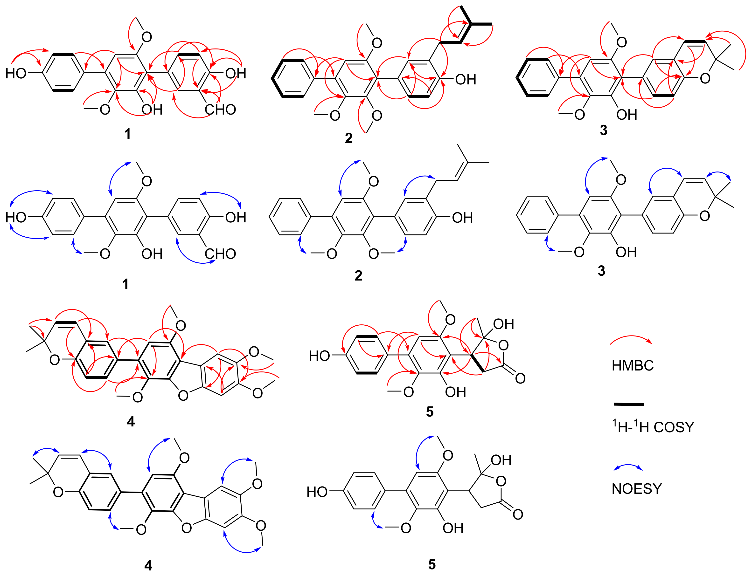
Sanshamycin A (1) was isolated as a yellow powder. The molecular formula was established as C21H18O6 (13 degrees of unsaturations) on the basis of its HRESIMS (Figure S8) and the 1H and 13C NMR data (Table 1 and Table 2). The initial analysis of the 1H (Figure S1), 13C (Figure S2), DEPT135 (Figure S3), and HSQC (Figure S4) spectra in acetone-d6 revealed that 1 was very similar to terphenyllin (6) [8], but with an aldehyde group (3-CHO) displaying characteristic proton and carbon signals at δH 10.06 (s) and δC 197.3. Further observation of the key HMBC correlations (Figure 2 and Figure S5) from δH 10.06 proton signal to C-2 (δC 136.2), C-3 (δC 120.6) and C-4 (δC 160.1), from H-2 (δH 7.83, d, J = 2.2 Hz) to δC 197.3 carbon signal, and the NOESY correlation (Figure 2 and Figure S7) from δH 10.06 proton signal to H-2 located the aldehyde group at C-3. Characteristic HMBC correlations from 4-OH (δH 11.02, s) to C-3, C-4, and C-5 (δC 116.1), 2′-OH (δH 7.90, s) to C-1′ (δC 114.8), C-2′ (δC 148.3), and C-3′ (δC 139.3), 4′′-OH (δH 8.55, s) to C-3′′ (δC 115.2), C-4′′ (δC 157.1), and C-5′′ (δC 115.2), 3′-OCH3 (δH 3.40, s) to C-3′, 6′-OCH3 (δH 3.78, s) to C-6′ (δC 153.5) were observed. Meanwhile, the ROESY spectrum showed correlations from 4-OH to H-5 (δH 7.02, d, J = 8.6 Hz), 3′-OCH3 to 2′-OH, H-2′′ (δH 7.55, d, J = 8.6 Hz) and H-6′′ (δH 7.55, d, J = 8.6 Hz), and 4′′-OH to H-3′′ (δH 6.96, d, J = 8.6 Hz) and H-5′′ (δH 6.96, d, J = 8.6 Hz). Further comprehensive interpretation of the HSQC, HMBC, 1H-1H COSY (Figure S6), and ROESY data allowed for the full assignment of the structure as shown in Figure 1.
Sanshamycin B (2) was isolated as a yellowish powder, and had the molecular formula C26H28O4 on the basis of the HRESIMS (Figure S18) and 1D NMR data (Table 1 and Table 2). Analysis of the 1H (Figure S11), 13C (Figure S12), DEPT135 (Figure S13) and HSQC (Figure S14) NMR data of 2 showed the presence of three methoxyl groups, two methyl groups, one methylene, and 20 sp2 carbons (ten of which were protonated). Analysis of the 13C NMR data, the coupling constants of the aromatic protons in the 1H NMR spectrum and also the correlations observed in the COSY spectrum revealed the presence of a pentasubstituted, a 1,3,4-trisubstituted, and a monosubstituted benzene rings, indicating a p-terphenyl framework structure similar to 1. The presence of an isopentyl group could be deduced from HMBC correlations (Figure S15) from H2-1′′′ (δH 3.40, d, J = 7.2 Hz), H3-4′′′ (δH 1.78, d, J = 1.4 Hz) and H3-5′′′ (δH 1.79, br s) to C-3′′′ (δC 134.7), H3-4′′′ and H3-5′′′ to C-2′′′ (δC 122.0), 1H-1H COSY correlations (Figure S16) from H2-1′′′ to H-2′′′ (δH 5.40, dq, J = 7.2, 1.4 Hz), and NOESY correlations (Figure S17) from H2-1′′′ to H3-5′′′ and H-2′′′ to H3-4′′′. That the isopentyl moiety was located at C-3 was evidenced from HMBC correlations from H2-1′′′ to C-2 (δC 132.1), C-3 (δC 125.9), and C-4 (δC 153.5), and NOESY correlation from H2-1′′′ to H-2 (δH 7.17, d, J = 2.1 Hz). Characteristic HMBC correlations from the methoxyl group at δH 3.66 to C-2′ (δC 152.0), the methoxyl group at δH 3.61 to C-3′ (δC 144.8) and the methoxyl group at δH 3.74 to C-6′ (δC 153.2) and NOESY correlations between 2′-OCH3 and H-6 (δH 7.17, overlap), between 3′-OCH3 and H-2′′(H-6′′) (δH 7.59, d, J = 7.5 Hz), and between 6′-OCH3 and H-5′ (δH 6.69, s), confirmed the positions of these methoxyl groups. The key HSQC, HMBC, 1H-1H COSY and ROESY of 2 was shown in Figure 2.
Sanshamycin C (3), obtained as a yellow powder, gave a molecular formula of C25H24O4, as determined by HRESIMS (Figure S28), and its 1H and 13C NMR data (Table 1 and Table 2), resembled those for sanshamycin B (2). The major differences observed in the 1H NMR spectrum (Figure S21) for 3 relative to those of 2 indicated the presence of an cis-disubstituted double bond due to the coupling constants of protons at δH 6.35 (d, J = 9.7 Hz, H-1′′′) and 5.60 (d, J = 9.7 Hz, H-2′′′) instead of a methylene signal at δH 3.40 and an olefinic methine signal at δH 5.40, and the absence of a methoxyl subtituent in 3. Comprehensive consideration of the HRESIMS and NMR data suggested 3 could be derived from the cyclization between the hydroxyl group 4-OH and C-3′′′ of the isopentyl group of 2. This suggestion was confirmed by HMBC correlations (Figure S25) from H-1′′′ to C-4 (δC 152.3) and C-3′′′ (δC 76.4), H-2′′′ to C-3 (δC 120.8), H-4′′′ (δH 1.47, s) and H-5′′′ (δH 1.47, s) to C-2′′′ (δC 130.3) and C-3′′′, H-2 (δH 7.09, d, J = 2.1 Hz) to C-1′′′ (δC 122.4), and NOESY correlation (Figure S27) from H-1′′′ to H-2. Correlations from the methoxyl group at δH 3.44 to C-3′ (δC 138.9) and the methoxyl group at δH 3.75 to C-6′ (δC 153.5) in the HMBC spectrum and NOESY correlations from 3′-OCH3 to H-2′′/H-6′′ (δH 7.64), and from 6′-OCH3 to H-5′ (δH 6.48, s) were observed. Overall analysis of the 1D and 2D NMR data permitted the structural assignment for 3 as showing in Figure 1.
Sanshamycin D (4) was isolated as a white powder whose molecular formula of C27H26O6 was obtained by analysis of HRESIMS (Figure S38) and NMR data (Figures S31–S37). The 1H and 13C NMR data of 4 were very similar to those of 4,5-dimethoxycandidusin A (9) [11], except for the presence of a prenyl and the absence of OH-4′′. The prenyl moiety was elucidated by HMBC correlations from H-4′′′/H-5′′′ (δH 1.49, s) to C-2′′′ (δC 130.8) and C-3′′′ (δC 76.5), and 1H-1H COSY correlations from H-1′′′ (δH 6.40, d, J = 9.7 Hz) to H-2′′′ (δH 5.66, d, J = 9.7 Hz). That the prenyl group was at C-3′′ was evidenced from the HMBC correlations from H-1′′′ to C-2′′ (δC 127.4) and C-4′′ (δC 152.3), H-2′′′ to C-3′′ (δC 121.0), and NOESY correlation from H-1′′′ to H-2′′ (δH 7.25, s). The HMBC spectrum displayed correlations from the methoxyl signals at δH 3.98, 4.02, 3.71, and 4.03 to C-4, C-5, C-3′, and C-6′, respectively. Significant NOESY correlations from 4-OCH3 to H-3 (δH 7.17, s), 5-OCH3 to H-6 (δH 7.56, s), 3′-OCH3 to H-6′′ (δH 7.38, d, J = 8.3 Hz), and 6′-OCH3 to H-5′ (δH 6.68, s), secured the positions of these methoxyl groups. Complete NMR analysis supported the full elucidation of the structure for compound 4 as shown in Figure 2.
Sanshamycin E (5) was isolated as a white powder. Its molecular formula was determined as C19H20O7 (10 degrees of unsaturations) by HRESIMS (Figure S48) in combination with 1H and 13C NMR data. The 1H (Figure S41), 13C (Figure S42), DEPT135 (Figure S43), and HSQC (Figure S44) NMR spectra (Table 1 and Table 2) showed resonances for 19 carbons, including 12 sp2 carbons (five of which are protonated), one ester carbonyl carbon, one quaternary sp3 carbon, two methoxyl carbons, one methylene sp2 carbon, one methine sp3 carbon, and one methyl carbon. Detailed analysis of the coupling constants of the protons in the 1H NMR spectrum and COSY correlations indicated the presence of a pentasubstituted and a 1,4-disubstituted benzene rings. Characteristic HMBC correlations (Figure S45) from H-2′′/H-6′′ (δH 7.41, d, J = 8.5 Hz) to C-4′′ (δC 155.1) and C-4′ (δC 136.4) and from H-3′′/H-5′′ (δH 6.90, d, J = 8.5 Hz) and H-5′ (δH 6.43, s) to C-1′′ (δC 130.5) secured the connectivity of C-1′′ to C-4′. HMBC correlations from the methoxyl proton signals at δH 3.66 to C-3′ (δC 136.0), at δH 3.84 to C-6′ (δC 151.5), from H-5′ to C-3′ and C-6′, and NOESY correlations (Figure S47) from 6′-OCH3 to H-5′ and from 3′-OCH3 to H-2′′/H-6′′ confirmed the locations of the methoxyl groups at C-3′ and C-6′. So far, nine degrees of unsaturation have been assigned, while the last degree of unsaturation could be completed by the presence of a γ-butyrolactone moiety, which was deduced from the HMBC correlations from H2-2 (δH 3.08, dd, J = 6.4, 4.5 Hz) to C-1 (δC 173.9) and C-4 (δC 118.3), H-3 (δH 3.96, dd, J = 6.4, 4.5 Hz) to C-1 and C-5 (δC 23.9), H3-5 (δH 1.94, s) to C-3 (δC 45.7) and C-4, and 1H-1H COSY correlation (Figure S46) from H2-2 to H-3. The high chemical shift value (δC 118.3) of C-4 is a characteristic of ketal carbon, which could suggest the connectivity of C-1 and C-4 through a ester bond. The γ-butyrolactone moiety was connected with the biphenyl moiety, which could be deduced from the HMBC correlations from H2-2 to C-1′ (δC 114.6) and from H-3 to C-2′ (δC 149.9) and C-6′. We have tried to determine the absolute configurations of C-3 and C-4 by comparing the experimental and calculated ECD data, but unable to reach a conclusion. Finally, the structure for 5 was determined as shown in Figure 1. A plausible biosynthetic pathway for 5 was tentatively proposed in the Supplementary Materials (Figure S51).
The known compounds terphenyllin (6) [8], 3-hydroxyterphenyllin (7) [9], candidusin A (8) [10], 4,5-dimethoxycandidusin A (9) [11], and candidusin C (10) [12] were identified by comparison of their NMR data with those reported in the literature.

2.2. In Vitro Evaluation of Type 4 Phosphodiesterase PDE4D Inhibitory Activity

p-Terphenyl compounds have been identified as phosphodiesterase (PDE) inhibitors. Terferol, derived from Streptomyces showdoensis SANK 65080, showed inhibitory activity towards cyclic adenosine 3′,5′-monophosphate phosphodiesterase (cAMP-PDE) and cyclic guanosine 3′,5′-monophosphate phosphodiesterase (cGMP-PDE) from various rat tissues [15]. Moreover, one metabolite with a similar structure to terferol from two different microbes was also found to possess inhibitory activities towards eukaryotic PDE11 and four PDE4s [14,16]. Given the close structural relationship of 110 with the known natural phosphodiesterase inhibitor terferol, the inhibitory activities of 16 and 9 were evaluated against the PDE4D, with rolipram as the positive control. As shown in Table 3, 3 was the most potent compound and displayed the best inhibition at 5 µM with inhibitory percentage of 49.4 %, while the other compounds showed weaker activities with inhibitory percentage of 4.8–23.2% (Table 3). Compound 3 was selected to test the concentration required for 50% inhibition of PDE4D (IC50), which gave an IC50 value of 5.543 ± 0.24 µM, while the positive drug rolipram exhibited an IC50 value of 0.588 ± 0.057 µM. Comparing with the other tested compounds, the two fused six-membered rings and the hydroxyl group at C-2′ in 3 may be responsible for the strong PDE4D inhibitory activity.

3. Materials and Methods

3.1. General Experimental Procedures

One- and two-dimensional NMR spectra were measured on Bruker AVIII-500 NMR spectrometer (Bruker Corporation, Karlsruhe, Germany). The chemical shifts of 1H (500 MHz) and 13C (125 MHz) NMR data were given in δ (ppm) and referenced to the solvent signal (CDCl3, δH 7.26 and δC 77.16; acetone-d6, δH 2.05 and δC 29.84). HRESIMS data were collected on an Agilent 6210 TOF LC-MS instrument (Agilent Technologies Inc., Palo Alto, CA, USA). Optical rotation value was recorded by a JASCO P-1020 digital polarimeter (JASCO, Tokyo, Japan). UV and IR data were measured on a UV-2550 spectrometer (Shimadzu, Kyoto, Japan) and Nicolet 380 Infrared Spectrometer (Thermo Electron Corporation, Madison, WI, USA), respectively. The ECD data were collected on JASCO J-715 spectropolarimeter (JASCO, Tokyo, Japan). The semi-preparative HPLC was conducted on a Waters 1525 HPLC equipped with a XBridge C18 column (5 μm, 250.0 mm × 10.0 mm; Waters Corporation, Milford, MA, USA). Thin-layer chromatography (TLC) was performed on pre-coated glass plates (silica gel GF254, Qingdao Marine Chemical Inc., Qingdao, China). Column chromatography (CC) was performed on silica gel (45–75 µm; Qingdao Marine Chemical Inc., Qingdao, China), ODS (40–60 µm; Osaka Soda Co., Ltd., Hyogo, Japan) and Sephadex LH-20 (Cytiva, Uppsala, Sweden).

3.2. Fungal Material and Fermentation

The fungal strain ITBBc1 was isolated from a coral reef in the South China Sea in Sansha, Hainan, China. This fungal strain was identified as Aspergillus sp. by internal transcribed spacer (ITS) sequence (GenBank accession number OP614945). The voucher specimen of this strain was deposited in the Hainan Key Laboratory of Tropical Microbe Resources, Institute of Tropical Bioscience and Biotechnology, Haikou, P.R. China. The strain was cultivated on a PDA agar plate (consisting of potato extract 200 g/L, glucose 20 g/L, agar 15 g/L, chloramphenicol 0.1 g/L and 1 L sterilized deionized water) at 28 °C for 5 days. Then, the agar plugs with mycelia were added into 1 L Erlenmeyer flasks, each containing 200 mL ME liquid medium (consisting of malt extract 10.0 g/L, sucrose 10.0 g/L, peptone 1.0 g/L and 1 L sterilized deionized water), which was cultivated on a rotary shaker at 160 rpm/min at 28 °C. After 4 days of fermentation, 15 mL of the seed cultures were inoculated into the rice solid media (consisting of rice 30 g and seawater 45 mL) in 1L-Erlenmeyer flasks and fermented at 28 °C for 45 days under static conditions.

3.3. Extraction and Isolation

The whole fermentation materials of strain ITBBc1 were collected and extracted with ethyl acetate (EtOAc) at room temperature for four times to yield a crude extract (120.0 g). Then, the crude extract was fractionated by silica gel CC using gradient elution of petroleum ether/EtOAc mixtures (v/v, 100:1, 50:1, 25:1, 10:1, 5:1, 2:1, 1:1, 0:1) to give 8 fractions (Fr.A–Fr.H). The fraction Fr.E (petroleum ether/EtOAc, v/v, 5:1) was separated by ODS reversed-phase CC eluted with aqueous methanol (40% to 100%) to yield the Fr.E7 (85% aqueous methanol), which was further purified by Sephadex LH-20 (eluted with 100% methanol) and then silica gel CC [using gradient elution of petroleum ether/EtOAc mixtures (v/v, 53:1, 25:1, 10:2)] to give compounds 2 (6.6 mg), 3 (5.0 mg) and 4 (2.2 mg). The fraction Fr.F (petroleum ether/EtOAc, v/v, 2:1) was subjected to ODS reversed-phase CC eluted with aqueous methanol (45% to 100%) to produce nine subfractions Fr.F1–F9. The Fr.F1 (45% aqueous methanol) was further subjected to Sephadex LH-20 (eluted with 100% methanol) and then silica gel CC [using gradient elution of petroleum ether/EtOAc mixtures (v/v, 5:3, 1:1)] to afford compound 5 (2.5 mg). The Fr.F3 (55% aqueous methanol) was purified by Sephadex LH-20 (eluted with 100% methanol) to give compound 1 (3.5 mg). The Fr.F5 (65% aqueous methanol) was subjected to Sephadex LH-20 (eluted with 100% methanol) and then purified by semi-preparative reverse-phase HPLC (eluted with 70% aqueous methanol; 3 mL/min, UV λmax 254 nm) to give compound 10 (42.1 mg, tR 9.1 min). The Fr.F6 (75% aqueous methanol) was purified by Sephadex LH-20 (eluted with 100% methanol) to give compound 9 (0.9 mg). Compound 6 was recrystallized from the fraction Fr.G (petroleum ether/EtOAc, v/v, 1:1) and obtained as white crystals (90 mg). Then, the rest of the Fr.G was subjected to ODS reversed-phase CC eluted with aqueous methanol (40% to 100%) to yield seven subfractions Fr.G1–G7. The Fr.G3 (50% aqueous methanol) was separated by Sephadex LH-20 (eluted with 100% methanol) and then purified by semi-preparative reverse-phase HPLC (3 mL/min, UV λmax 254 nm) to give compounds 7 (59.1 mg, tR 5.5 min; eluted with 60% aqueous methanol) and 8 (13.2 mg, tR 8.5 min; eluted with 65% aqueous methanol).
  • Sanshamycin A (1): yellow powder; UV (MeOH) λmax (log ε): 304 (2.81), 332 (2.52), 342 (2.54) nm; IR(KBr) νmax: 3415, 2918, 1652, 1403, 1118, 1075, 835, 759 cm−1; 1H and 13C NMR data, see Table 1 and Table 2; HRESIMS m/z 389.1001 [M + Na]+ (calculated for C21H18NaO6, 389.0996).
  • Sanshamycin B (2): yellowish powder; UV (MeOH) λmax (log ε): 258 (2.60), 298 (2.81) nm; IR(KBr) νmax: 3404, 2931, 1599, 1465, 1389, 1111, 1079, 1025, 616, 547 cm−1; 1H and 13C NMR data, see Table 1 and Table 2; HRESIMS m/z 427.1928 [M + Na]+ (calculated for C26H28NaO4, 427.1880).
  • Sanshamycin C (3): yellow powder; UV (MeOH) λmax (log ε): 273 (2.68), 302 (2.83) nm; IR(KBr) νmax: 3416, 2934, 2434, 1629, 1468, 1404, 1123, 1072, 795 cm−1; 1H and 13C NMR data, see Table 1 and Table 2; HRESIMS m/z 389.1752 [M + H]+ (calculated for C25H25O4, 389.1747).
  • Sanshamycin D (4): white powder; UV (MeOH) λmax (log ε): 258 (2.64), 310 (2.92), 337 (2.96) nm; IR(KBr) νmax: 3411, 2919, 1617, 1480, 1384, 1129, 1023, 617 cm−1; 1H and 13C NMR data, see Table 1 and Table 2; HRESIMS m/z 447.1801 [M + H]+ (calculated for C27H27O6, 447.1802).
  • Sanshamycin E (5): white powder; α D 25 68 (c 0.10, MeOH); UV (MeOH) λmax (log ε): 231 (2.50), 266 (2.67) nm; IR(KBr) νmax: 3430, 2922, 1628, 1382, 1097, 638, 534 cm−1; ECD (MeOH) λmax (∆ε): 190 (−19.29), 199 (+21.27), 208 (+17.37), 234 (−0.33) nm; 1H and 13C NMR data, see Table 1 and Table 2; HRESIMS m/z 383.1080 [M + Na]+ (calculated for C19H20NaO7, 383.1101).

3.4. Type 4 Phosphodiesterase PDE4D Inhibitiory Screening Assay

The type 4 phosphodiesterase PDE4D inhibitor assays were performed as described previously [30,31,32]. The inhibition of PDE4D by compounds 16 and 9 were assayed by a PerkinElmer 2910 (PerkinElmer, Inc., Waltham, MA, USA) liquid scintillation counter. Rolipram was used as a positive drug. Three independent experiments were conducted for the measurement of the inhibitory effect of compound 3 against PDE4D. The experimental data were analyzed via GraphPad Prism 5.1 (GraphPad Software, San Diego, CA, USA), and the IC50 values were calculated by nonlinear regression.

4. Conclusions

In conclusion, five new p-terphenyl derivatives, sanshamycins A–E (15), and five known analogues (610) were isolated and characterized from the coral-associated fungus Aspergillus sp. ITBBc1. The structures of the unreported compounds (15) were elucidated by interpretation of their 1D and 2D NMR and HRESIMS data whiles the structures of the previously reported compounds (610) were identified by comparison of their NMR data with those reported in literature. Sanshamycin A (1) represents the first example of p-terphenyls with an aldehyde substitution on the benzene ring. Sanshamycins B–D (24) feature varying methoxyl and isopentenyl substitutions, while sanshamycin E (5) features a five-membered lactone linked to a biphenyl. The inhibitory effects of 16 and 9 on PDE4D were assessed in vitro and 3 exhibited potent PDE4D inhibitory activity with an IC50 value of 5.543 µM. The current study revealed a new natural type 4 phosphodiesterase PDE4 inhibitor from the marine-derived fungus, Aspergillus sp. strain ITBBc1, could serve as a new structural motif for the future PDE4 inhibitor design. In general, the results of this study expand the knowledge of the chemical and biological diversity of the family of p-terphenyl natural products, and may provide a promising lead structure for the future development of PDE4 inhibitors. The inhibitory activity against other PDE members and the PDE4-inhibitory mechanism of sanshamycin C (3) deserves further investigation.

Supplementary Materials

The following supporting information can be downloaded at: https://www.mdpi.com/article/10.3390/md20110679/s1, Figures S1–S51: 1D, 2D NMR, MS, UV, and IR spectra of compounds 15.

Author Contributions

Conceptualization, Z.G., T.L. and R.W.; methodology, Z.G., A.A., S.Z. and L.H.; software, Z.G., L.H. and Z.X.; validation, Z.G. and L.H.; formal analysis, Z.G. and L.H.; investigation, A.A., Z.X. and S.Z.; resources, Z.G., T.L. and R.W.; data curation, Z.G. and L.H.; writing—original draft preparation, Z.G.; writing—review and editing, Z.G., T.L. and R.W.; visualization, Z.G. and R.W.; supervision, Z.X. and S.Z.; project administration, Z.G., T.L. and R.W.; funding acquisition, Z.G., Z.X., S.Z. and R.W. All authors have read and agreed to the published version of the manuscript.

Funding

This research was financially supported by the Hainan Provincial Basic and Applied Basic Research Fund for High-Level Talents in Natural Science (Nos. 2019RC352 and 2019RC306), Central Public-Interest Scientific Institution Basal Research Fund for CATAS-ITBB (Nos. 1630052022016, 1630052019011, and 1630052022028), the Natural Science Foundation of Hainan (No. 322QN387), and the Financial Fund of the Ministry of Agriculture and Rural Affairs, P. R. of China (No. NFZX2021).

Institutional Review Board Statement

Not applicable.

Informed Consent Statement

Not applicable.

Data Availability Statement

The authors declare that all relevant data supporting the results of this study are available within the article and its Supplementary Materials file, or from the corresponding authors upon request.

Conflicts of Interest

The authors declare no conflict of interest.

References

  1. Baillie, G.S.; Tejeda, G.S.; Kelly, M.P. Therapeutic targeting of 3′,5′-cyclic nucleotide phosphodiesterases: Inhibition and beyond. Nat. Rev. Drug Discov. 2019, 18, 770–796. [Google Scholar] [CrossRef] [PubMed]
  2. Crocetti, L.; Floresta, G.; Cilibrizzi, A.; Giovannoni, M.P. An overview of PDE4 inhibitors in clinical trials: 2010 to early 2022. Molecules 2022, 27, 4964. [Google Scholar] [CrossRef] [PubMed]
  3. Spina, D. PDE4 inhibitors: Current status. Brit. J. Pharmacol. 2008, 155, 308–315. [Google Scholar] [CrossRef] [PubMed] [Green Version]
  4. Lin, T.-T.; Huang, Y.-Y.; Tang, G.-H.; Cheng, Z.-B.; Liu, X.; Luo, H.-B.; Yin, S. Prenylated coumarins: Natural phosphodiesterase-4 inhibitors from Toddalia asiatica. J. Nat. Prod. 2014, 77, 955–962. [Google Scholar] [CrossRef] [PubMed]
  5. Cheng, Z.-B.; Deng, Y.-L.; Fan, C.-Q.; Han, Q.-H.; Lin, S.-L.; Tang, G.-H.; Luo, H.-B.; Yin, S. Prostaglandin derivatives: Nonaromatic phosphodiesterase-4 inhibitors from the soft coral Sarcophyton ehrenbergi. J. Nat. Prod. 2014, 77, 1928–1936. [Google Scholar] [CrossRef]
  6. Woo, S.; Kang, K.B.; Kim, J.; Sung, S.H. Molecular networking reveals the chemical diversity of selaginellin derivatives, natural phosphodiesterase-4 inhibitors from Selaginella tamariscina. J. Nat. Prod. 2019, 82, 1820–1830. [Google Scholar] [CrossRef]
  7. Fu, Y.; Ding, X.; Zhang, X.; Shao, X.; Zhao, J.; Xu, Y.; Luo, X.; Zhao, W. Diterpenoids from the root bark of Pinus massoniana and evaluation of their phosphodiesterase type 4D inhibitory activity. J. Nat. Prod. 2020, 83, 1229–1237. [Google Scholar] [CrossRef]
  8. Marchelli, R.; Vining, L.C. Terphenyllin, a novel p-terphenyl metabolite from Aspergillus candidus. J. Antibiot. 1975, 28, 328–331. [Google Scholar] [CrossRef]
  9. Kurobane, I.; Vining, L.C.; Mcinnes, A.G.; Smith, D.G. 3-Hydroxyterphenyllin, a new metabolite of Aspergillus candidus. Structure elucidation by H and C nuclear magnetic resonance spectroscopy. J. Antibiot. 1979, 32, 559–564. [Google Scholar] [CrossRef] [Green Version]
  10. Kobayashi, A.; Takemura, A.; Koshimizu, K.; Nagano, H.; Kawazu, K. Candidusin A and B: New p-terphenyls with cytotoxic effects on sea urchin embryos. Agric. Biol. Chem. 1982, 46, 585–589. [Google Scholar] [CrossRef]
  11. Guo, Z.K.; Yan, T.; Guo, Y.; Song, Y.C.; Tan, R.X.; Ge, H.M. p-Terphenyl and diterpenoid metabolites from endophytic Aspergillus sp. YXf3. J. Nat. Prod. 2012, 75, 15–21. [Google Scholar] [CrossRef]
  12. Rahbaek, L.; Frisvad, J.C.; Christophersen, C. An amendment of Aspergillus section Candidi based on chemotaxonomical evidence. Phytochemistry 2000, 53, 581–586. [Google Scholar] [CrossRef]
  13. Zhu, J.; Chen, W.; Li, Y.-Y.; Deng, J.-J.; Zhu, D.-Y.; Duan, J.; Liu, Y.; Shi, G.-Y.; Xie, C.; Wang, H.-X.; et al. Identification and catalytic characterization of a nonribosomal peptide synthetase-like (NRPS-like) enzyme involved in the biosynthesis of echosides from Streptomyces sp. LZ35. Gene 2014, 546, 352–358. [Google Scholar] [CrossRef] [PubMed]
  14. Biggins, J.B.; Liu, X.; Feng, Z.; Brady, S.F. Metabolites from the induced expression of cryptic single operons found in the genome of Burkholderia pseudomallei. J. Am. Chem. Soc. 2011, 133, 1638–1641. [Google Scholar] [CrossRef] [PubMed] [Green Version]
  15. Nakagawa, F.; Enokita, R.; Naito, A.; Iijima, Y.; Yamazaki, M. Terferol, an inhibitor of cyclic adenosine 3′,5′-monophosphate phosphodiesterase. I. Isolation and characterization. J. Antibiot. 1984, 37, 6–9. [Google Scholar] [CrossRef] [PubMed] [Green Version]
  16. El-Elimat, T.; Figueroa, M.; Raja, H.A.; Graf, T.N.; Adcock, A.F.; Kroll, D.J.; Day, C.S.; Wani, M.C.; Pearce, C.J.; Oberlies, N.H. Benzoquinones and terphenyl compounds as phosphodiesterase-4B inhibitors from a fungus of the order Chaetothyriales (MSX47445). J. Nat. Prod. 2013, 76, 382–387. [Google Scholar] [CrossRef] [PubMed] [Green Version]
  17. Guo, Z.-K.; Zhu, W.-Y.; Zhao, L.-X.; Chen, Y.-C.; Li, S.-J.; Cheng, P.; Ge, H.-M.; Tan, R.-X.; Jiao, R.-H. New antibacterial depsidones from an ant-derived fungus Spiromastix sp. MY-1. Chin. J. Nat. Med. 2022, 20, 627–632. [Google Scholar] [CrossRef]
  18. Xu, Y.; Xie, J.; Wu, W.C.; Chen, B.T.; Zhang, S.Q.; Wang, R.; Huang, J.; Guo, Z.K. Discovery of an unprecedented benz[α]anthraquinone-type heterodimer from a rare actinomycete Amycolatopsis sp. HCa1. Fitoterapia 2021, 155, 105039. [Google Scholar] [CrossRef]
  19. Tan, Y.-Z.; Guo, Z.-K.; Zhu, M.-Y.; Shi, J.; Li, W.; Jiao, R.-H.; Tan, R.-X.; Ge, H.-M. Anti-inflammatory spirobisnaphthalene natural products from a plant-derived endophytic fungus Edenia gomezpompae. Chin. Chem. Lett. 2020, 31, 1406–1409. [Google Scholar] [CrossRef]
  20. Han, H.; Guo, Z.-K.; Zhang, B.; Zhang, M.; Shi, J.; Li, W.; Jiao, R.-H.; Tan, R.-X.; Ge, H.-M. Bioactive phenazines from an earwig-associated Streptomyces sp. Chin. J. Nat. Med. 2019, 17, 475–480. [Google Scholar] [CrossRef]
  21. Ma, S.Y.; Xiao, Y.S.; Zhang, B.; Shao, F.L.; Guo, Z.K.; Zhang, J.J.; Jiao, R.H.; Sun, Y.; Xu, Q.; Tan, R.X.; et al. Amycolamycins A and B, two enediyne-derived compounds from a locust-associated actinomycete. Org. Lett. 2017, 19, 6208–6211. [Google Scholar] [CrossRef]
  22. Chen, B.; Wu, W.; Zhou, D.; Deng, X.; Zhang, S.; Yuan, J.; Xu, J.; Guo, Z. Bioactive components of two endophytic fungi from Hainan mangrove plants. J. Shenzhen Univ. Sci. Eng. 2022, 39, 245–252. [Google Scholar]
  23. Guo, Z.; Wang, R.; Wu, W.; Li, M.; He, W.; Zhou, S. Bioactive secondary metabolites from two sponge-derived actinomycetes. J. Shenzhen Univ. Sci. Eng. 2022, 39, 550–558. [Google Scholar]
  24. Guo, Z.; Ma, S.; Khan, S.; Zhu, H.; Zhang, B.; Zhang, S.; Jiao, R. Zhaoshumycins A and B, two unprecedented antimycin-type depsipeptides produced by a marine-derived Streptomyces sp. ITBB-ZKa6. Mar. Drugs 2021, 19, 624. [Google Scholar] [CrossRef]
  25. Guo, Z.-K.; Wang, R.; Chen, S.-Q.; Chen, F.-X.; Liu, T.-M.; Yang, M.-Q. Anthocidins A-D, new 5-hydroxyanthranilic acid related metabolites from the sea urchin-associated actinobacterium, Streptomyces sp. HDa1. Molecules 2018, 23, 1032. [Google Scholar] [CrossRef] [Green Version]
  26. Guo, Z.K.; Wang, R.; Chen, F.X.; Liu, T.M.; Yang, M.Q. Bioactive aromatic metabolites from the sea urchin-derived actinomycete Streptomyces spectabilis strain HDa1. Phytochem. Lett. 2018, 25, 132–135. [Google Scholar] [CrossRef]
  27. Guo, Z.-K.; Zhou, Y.-Q.; Han, H.; Wang, W.; Xiang, L.; Deng, X.-Z.; Ge, H.-M.; Jiao, R.-H. New antibacterial phenone derivatives asperphenone A-C from mangrove-derived fungus Aspergillus sp. YHZ-1. Mar. Drugs 2018, 16, 45. [Google Scholar] [CrossRef] [Green Version]
  28. Guo, Z.; Gai, C.; Cai, C.; Chen, L.; Liu, S.; Zeng, Y.; Yuan, J.; Mei, W.; Dai, H. Metabolites with insecticidal activity from Aspergillus fumigatus JRJ111048 isolated from mangrove plant Acrostichum specioum endemic to Hainan island. Mar. Drugs 2017, 15, 381. [Google Scholar] [CrossRef] [Green Version]
  29. Wang, R.; Guo, Z.K.; Li, X.M.; Chen, F.X.; Zhan, X.F.; Shen, M.H. Spiculisporic acid analogues of the marine-derived fungus, Aspergillus candidus strain HDf2, and their antibacterial activity. Antonie Leeuwenhoek 2015, 108, 215–219. [Google Scholar] [CrossRef]
  30. Pan, T.; Xie, S.; Zhou, Y.; Hu, J.; Luo, H.; Li, X.; Huang, L. Dual functional cholinesterase and PDE4D inhibitors for the treatment of Alzheimer’s disease: Design, synthesis and evaluation of tacrine-pyrazolo[3,4-b]pyridine hybrids. Bioorg. Med. Chem. Lett. 2019, 29, 2150–2152. [Google Scholar] [CrossRef]
  31. Liu, X.; Luo, H.-B.; Huang, Y.-Y.; Bao, J.-M.; Tang, G.-H.; Chen, Y.-Y.; Wang, J.; Yin, S. Selaginpulvilins A-D, new phosphodiesterase-4 inhibitors with an unprecedented skeleton from Selaginella pulvinata. Org. Lett. 2014, 16, 282–285. [Google Scholar] [CrossRef] [PubMed]
  32. Sun, Z.-H.; Cai, Y.-H.; Fan, C.-Q.; Tang, G.-H.; Huo, H.-B.; Yin, S. Six new tetraprenylated alkaloids from the South China Sea gorgonian Echinogorgia pseudossapo. Mar. Drugs 2014, 12, 672–681. [Google Scholar] [CrossRef] [PubMed]
Figure 1. The structures of 110.
Marinedrugs 20 00679 g001
Figure 2. Key HMBC, 1H-1H COSY and ROESY correlations of 15.
Marinedrugs 20 00679 g002
Table 1. 1H NMR (500 MHz) spectroscopic data for 15.
Position1 a2 b3 b4 b5 b
27.83, d (2.2)7.17, d (2.1)7.09, d (2.1) 3.08, dd (6.4, 4.5)
3 7.17, s3.96, dd (6.4, 4.5)
3-CHO10.06, s
4-OH11.02, s
4-OCH3 3.98, s
57.02, d (8.6)6.87, d (8.7)6.85, d (8.2) 1.94, s
5-OCH3 4.02, s
67.68, dd (8.6, 2.2)7.17, overlapping7.22, dd (8.2, 2.1)7.56, s
2′-OH7.90, s 5.92, (br s)
2′-OCH3 3.66, s
3′-OCH33.40, s3.61, s3.44, s3.71, s3.66, s
5′6.55, s6.69, s6.48, s6.68, s6.43, s
6′-OCH33.78, s3.74, s3.75, s4.03, s3.84, s
2′′7.55, d (8.6)7.59, d (7.5)7.64, m7.25, s7.41, d (8.5)
3′′6.96, d (8.6)7.45, t (7.5)7.46, t (7.5) 6.90, d (8.5)
4′′ 7.37, t (7.5)7.37, t (7.5)
4′′-OH8.55, s
5′′6.96, d (8.6)7.45, t (7.5)7.46, t (7.5)6.86, d (8.3)6.90, d (8.5)
6′′7.55, d (8.6)7.59, d (7.5)7.64, m7.38, d (8.3)7.41, d (8.5)
1′′′ 3.40, d (7.2)6.35, d (9.7)6.40, d (9.7)
2′′′ 5.40, dq (7.2, 1.4)5.60, d (9.7)5.66, d (9.7)
4′′′ 1.78, d (1.4)1.47, s1.49, s
5′′′ 1.79, br s1.47, s1.49, s
a Spectra were recorded in acetone-d6; b Spectra were recorded in CDCl3. δ in ppm.
Table 2. 13C NMR (125 MHz) spectroscopic data for 15.
Position1 a2 b3 b4 b5 b
1126.1, C126.1, C125.0, C115.3, C173.9, C
2136.2, CH132.1, CH128.7, CH150.6, C34.0, CH2
3120.6, C125.9, C120.8, C95.4, CH45.7, CH
3-CHO197.3, CH
4160.1, C153.5, C152.3, C149.1, C118.3, C
4-OCH3 56.3, CH3
5116.1, CH115.2, CH116.0, CH146.2, C23.9, CH3
5-OCH3 56.6, CH3
6140.0, CH130.0, CH131.5, CH104.3, CH
1′114.8, C124.7, C116.7, C114.8, C114.6, C
2′148.3, C 152.0, C 147.2, C 149.3, C 149.9, C
2′-OCH3 60.8, CH3
3′139.3, C144.8, C138.9, C136.7, C136.0, C
3′-OCH359.8, CH360.9, CH360.9, CH361.1, CH360.7, CH3
4′133.4, C134.7, C132.7, C131.1, C136.4, C
5′103.3, CH108.1, CH104.0, CH105.6, CH106.1, CH
6′153.5, C153.2, C153.5, C150.0, C151.5, C
6′-OCH355.3, CH356.1, CH356.0, CH355.9, CH356.0, CH3
1′′129.4, C138.5, C138.1, C131.0, C130.5, C
2′′129.9, CH129.2, CH128.8, CH127.4, CH130.5, CH
3′′115.2, CH128.2, CH128.5, CH121.0, C115.1, CH
4′′157.1, C127.3, CH127.5, CH152.3, C155.1, C
5′′115.2, CH128.2, CH128.5, CH116.1, CH115.1, CH
6′′129.9, CH129.2, CH128.8, CH130.2, CH130.5, CH
1′′′ 29.9, CH2122.4, CH122.4, CH
2′′′ 122.0, CH130.3, CH130.8, CH
3′′′ 134.7, C76.4, C76.5, C
4′′′ 25.8, CH328.4, CH328.2, CH3
5′′′ 17.9, CH328.4, CH328.2, CH3
a Spectra were recorded in acetone-d6; b Spectra were recorded in CDCl3. δ in ppm.
Table 3. Inhibitory activities of 16 and 9 towards PDE4D.
CompoundsPDE4D Inhibitory Percentage (100%)
Rolipram52.0
14.8
26.7
349.4
412.4
55.1
612.8
923.2
The test concentration of positive rolipram is 0.5 μM; The test concentrations of 16 and 9 are 5 μM.
Publisher’s Note: MDPI stays neutral with regard to jurisdictional claims in published maps and institutional affiliations.