Review

Marine Compounds, Mitochondria, and Malignancy: A Therapeutic Nexus

by
Sajad Fakhri
1,†,
Sadaf Abdian
2,†,
Seyed Zachariah Moradi
1,3,
Blake E. Delgadillo
4,
Carmela Fimognari
5 and
Anupam Bishayee
4,*
1
Pharmaceutical Sciences Research Center, Health Institute, Kermanshah University of Medical Sciences, Kermanshah 6734667149, Iran
2
Student Research Committee, Kermanshah University of Medical Sciences, Kermanshah 6714415153, Iran
3
Medical Biology Research Center, Health Technology Institute, Kermanshah University of Medical Sciences, Kermanshah 6734667149, Iran
4
College of Osteopathic Medicine, Lake Erie College of Osteopathic Medicine, Bradenton, FL 34211, USA
5
Department for Life Quality Studies, University of Bologna, 47921 Rimini, Italy
*
Author to whom correspondence should be addressed.
These authors contributed equally and are joint first authors.
Mar. Drugs 2022, 20(10), 625; https://doi.org/10.3390/md20100625
Submission received: 30 August 2022 / Revised: 27 September 2022 / Accepted: 27 September 2022 / Published: 30 September 2022
(This article belongs to the Section Marine Pharmacology)

Abstract

:
The marine environment is important yet generally underexplored. It contains new sources of functional constituents that can affect various pathways in food processing, storage, and fortification. Bioactive secondary metabolites produced by marine microorganisms may have significant potential applications for humans. Various components isolated from disparate marine microorganisms, including fungi, microalgae, bacteria, and myxomycetes, showed considerable biological effects, such as anticancer, antioxidant, antiviral, antibacterial, and neuroprotective activities. Growing studies are revealing that potential anticancer effects of marine agents could be achieved through the modulation of several organelles. Mitochondria are known organelles that influence growth, differentiation, and death of cells via influencing the biosynthetic, bioenergetic, and various signaling pathways related to oxidative stress and cellular metabolism. Consequently, mitochondria play an essential role in tumorigenesis and cancer treatments by adapting to alterations in environmental and cellular conditions. The growing interest in marine-derived anticancer agents, combined with the development and progression of novel technology in the extraction and cultures of marine life, led to revelations of new compounds with meaningful pharmacological applications. This is the first critical review on marine-derived anticancer agents that have the potential for targeting mitochondrial function during tumorigenesis. This study aims to provide promising strategies in cancer prevention and treatment.

1. Introduction

Cancer is the uncontrolled growth of cells and formation of tumors caused by genetic changes, and in recent decades, it has been considered to be one of the major causes of death in the world [1,2]. Genetic disorders, exposure to variant physical and chemical agents, tobacco, radiation, infections, environmental pollutants, obesity, and diet are some of the important risk factors for cancer [1,2,3,4,5,6,7]. Cancerous cells have a variety of basic origins and pathologies, such as aberrant cell cycles, proliferation, apoptosis, and angiogenesis [8,9]. Based on the grade, type, and location of cancers, several important treatments include chemotherapy, hormonal therapy, radiation therapy, and surgery [10]. Despite these different therapeutic methods, the majority of initially responsive tumors obtain a multidrug resistance phenotype; thus, combination therapy has been preferred and used to treat various cancers [11,12]. These treatments play significant roles in destroying cancerous cells, but they do not have the potency to suppress all types of cancer, which led researchers to investigate more efficacious therapeutic options [13]. Over the last several decades, many approved anticancer agents have either been obtained from natural sources or their synthetics analogs have been developed [14].
In the creation of new functional foods, the marine ecosystem remains a goldmine of bioactive components. Natural organic substances derived from marine bio-resources, such as fatty acids, peptides, enzymes, polyethers, polysaccharides, and proteins, are some of the most potent, specialized, and bioactive chemicals used [15]. Due to the various qualities of marine derived compounds, these chemicals exhibit significant biological activity, including antiviral, anticarcinoma, anticoagulant, antibacterial, antioxidant, antibacterial, antihypertensive, antiproliferative, and antidiabetic potential [16,17,18,19]. Several previous reviews emphasize that these products are considered the key structures in developing novel therapeutic strategies for the management of cancers. They exhibit promising results in suppressing the proliferation, growth, and metastases of several cancerous cells via targeting multiple signaling pathways [15,20,21,22,23,24,25]. Cytarabine (Cytosar), trabectedin (Yondelis), eribulin mesylate (Halaven), and brentuximab vedotin (Acentris) are some of the known marine-based products that are approved for the treatment of several types of cancer [23,24,26]. The mitochondrial pathway is one of the most important signaling pathways in modulating a cell’s function. Mitochondria are one of the vital components of eukaryotic cells, and they play a significant role in producing energy and balancing cell survival/death. Additionally, mitochondria control oncogenic signaling, redox homeostasis, innate apoptosis, and immunity. Given the important role that mitochondria play, malfunction of these organelles is one of the most important causes of cell disturbance, and by modulating mitochondrial signaling pathways, researchers may discover new ways to prevent and treat cancers [27,28,29,30,31,32,33]. Previously, several natural products, with no focus on marine compounds, have been highlighted in targeting mitochondria in cancer [34]. Several marine agents have been highlighted in combating cancer through different mechanisms with no focus on mitochondria-related pathways [20,21,35,36]. The current comprehensive and critical review discusses the antineoplastic potential and mechanism of natural marine drugs for diminishing the metastasis, proliferation, and angiogenesis of several cancerous cell lines via modulating mitochondrial signaling pathways.

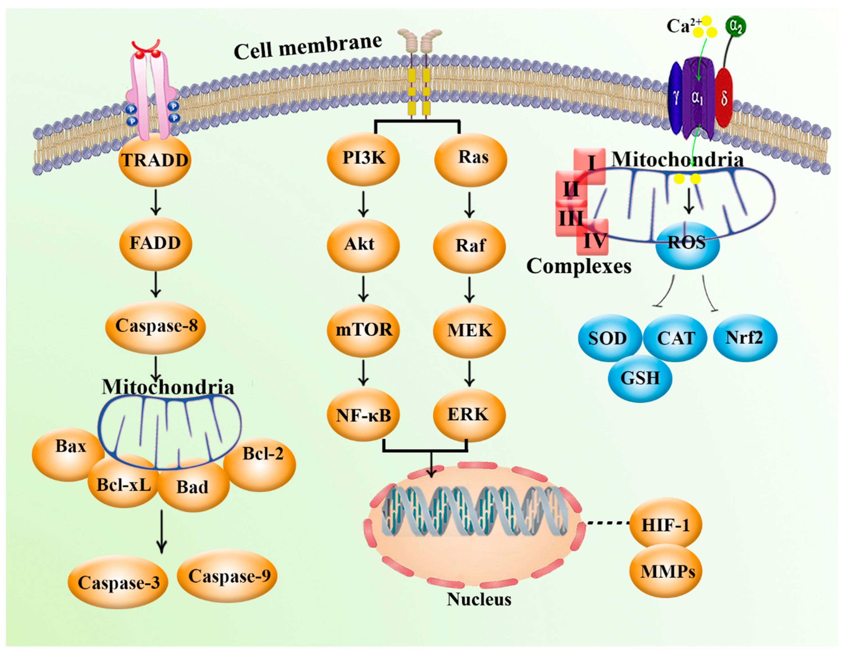
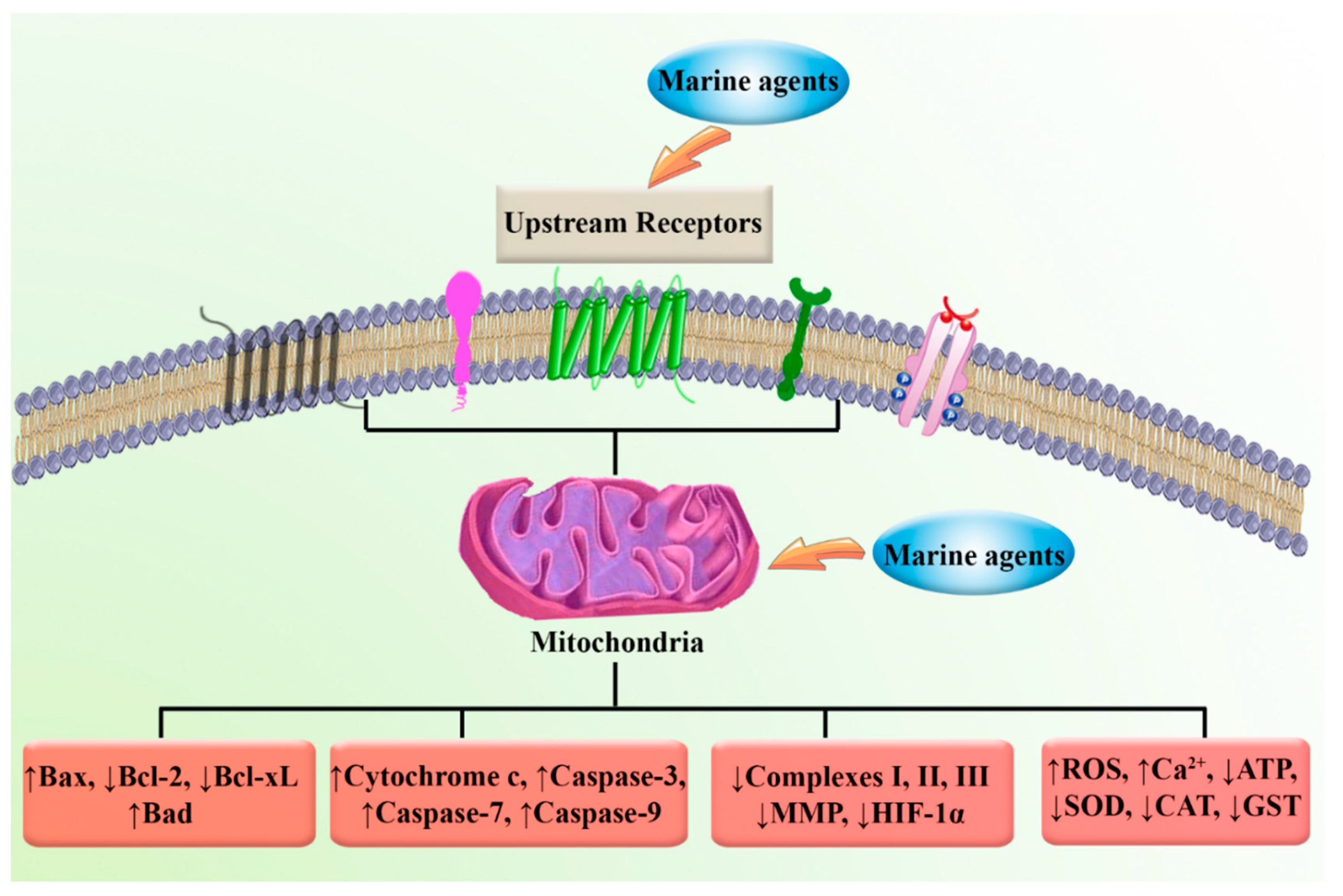
2. Mitochondria and Cancer: Biology and Cellular Signaling

Otto Warburg was the first to demonstrate that cancer cells, in the presence of oxygen, promote the fermentation of glucose, which may possibly be one of the underlying causes of cancer associated with mitochondrial respiration defects. In addition, researchers have concluded that genetic events that promote aberrant proliferation of cancer cells can also lead to alterations in biochemical metabolisms such as promoting aerobic glycolysis; although, this does not routinely impair mitochondrial function [37,38,39,40]. In most eukaryotic organisms, double-membrane-bound organelles, such as mitochondria, produce adenosine triphosphate (ATP) through aerobic respiration for use throughout the cell. Mitochondria control redox homeostasis, supply energy, influence oncogenic signaling, and regulate the cell cycle, apoptosis, and innate immunity. This mitochondrial biogenesis is often alternated in cancers. Mitochondria play an undeniable role in both lethal and vital functions in pathological and physiological conditions. In addition to the indispensable role of mitochondria in the production of energy and eukaryotic cell survival, mitochondria are important regulators of the intrinsic apoptotic pathway [37,38,39,40]. Numerous copies of the mitochondrial DNAs (mtDNAs) and nuclear DNA (nDNA)-encoded genes encompass the mitochondrial genome. In several cancers, mtDNA mutations have been found to affect mitochondrial metabolism, elevate tumorigenesis, and facilitate the adaptation of cancer cells to changing environmental conditions [40]. In addition, elimination of mtDNAs in cancer cells have been shown to diminish tumorigenicity via suppression of growth and progression of tumors. Thus, mitochondria have shown a meaningful multifunctional role in the progression of neoplasms and have made mitochondria an attractive target for therapeutic intervention [40].
Various mutations can affect mtDNA expression, and variant populations of mtDNA have been associated with cancer risk. African American women may have an increased risk of breast cancer risk if they have the macrohaplogroup N variant of the complex I subunit NADH dehydrogenase 3 gene (ND3) at nucleotide G10398A (resulting in an amino acid change of T114A). Endometrial cancer is associated with the 16519 T to C variant of the mtDNA control region [40,41,42]. In addition to mutations in several nDNA mitochondrial genes, the T6777C nucleotide variant of mtDNA’s cytochrome c (cyt. c) oxidase subunit 1 (MTCO1) has been associated with epithelial ovarian cancer [40]. Additionally, Chinese women with the variant C150 in the mtDNA control region have exhibited an elevated risk of cervical cancer and human papillomavirus infection [43]. Moreover, inhibition of mitochondrial oxidative phosphorylation (OXPHOS) is mentioned as another consequence of mtDNA mutations in many cancer cells [40]. In summary, two types of mutations in cancer cell mtDNA are important: mutations that promote the adaption of the cancer cells to changing bioenergetic environments and mutations that lead to stimulating neoplastic transformation via impairing OXPHOS [40].
Specific cancers have also been found to have mutations affecting mitochondrial enzymes encoded by nDNA and mutations directly affecting mtDNA. Succinate dehydrogenase (SDH) is one of these mitochondrial enzymes that may mutate [40,44]. Respiratory complex II (known as SDH) is an integral mitochondrial protein complex that transfers two electrons to coenzyme Q10 (CoQ) and oxidizes succinate to fumarate. SDHA-SDHD are the four main subunits that constitute this structure. SDH assembly factor 1 (SDHAF1) and SDHAF2 are two factors that are necessary for the formation and assembly of SDH. In preliminary research, SDHC, SDHB, and SDHD have been mentioned as mutated subunits of SDH in paragangliomas, and subsequent research confirmed the mutation in other parts, including SDHAF2 and SDHA [40,44,45]. Disruption of SDH elevated the cytosolic and mitochondrial levels of succinate, which suppressed the α-ketoglutarate dependent prolyl hydroxylase (PHDs). This resulted in the translocation of hypoxia-inducible factor 1α (HIF-1α) into the nucleus and its stabilization. Additionally, the inactivation of PHDs by reactive oxygen species (ROS) is another cause of HIF-1α stabilization; changing the energy metabolism from oxidative to glycolytic is the main result of these consecutive events. α-ketoglutarate-dependent dioxygenases, such as the TET family of 5-methylcytosine hydroxylase and Jumonji-C histone demethylases, have been shown to be suppressed by fumarate and succinate, which promotes the epigenetic dysregulation, DNA methylation, and alterations of histone [40,44,45].
Fumarate hydratase is another mitochondrial enzyme that converts fumarate to malate. Cells containing the mutant form of fumarate hydratase have markedly reduced levels of citrate and malate and elevated production of succinate and fumarate [37,40]. Several aggressive forms of renal carcinoma, uterine leiomyomas, and cutaneous leiomyomas have been associated with the fumarate hydratase gene mutations [46]. It has also been hypothesized that fumarate inhibits PHDs and stabilizes HIF-1α. Similarly, elevated levels of fumarate activate the stress response signaling that can be regulated by nuclear factor erythroid 2-related factor 2 (Nrf2). Degradation of Nrf2 by the ubiquitin E3 ligase cullin 3 and kelch-like ECH-associated protein 1 (KEAP1) keep its level low [37,40]. Additionally, excess fumarate leads to stabilized Nrf2 via inactivating KEAP1, which promotes turning on the nDNA stress-response genes, such as heme oxygenase 1 (HMOX1). This is the result of binding Nrf2 to antioxidant response elements (AREs). HMOX1 can increase the colony-forming capacity and activation of HMOX1 is an important marker of tumorigenesis [40,47].
Isocitrate dehydrogenase 1 (IDH1), a known enzyme encoded by the IDH1 gene, catalyzes the oxidative decarboxylation of isocitrate to 2-oxoglutarate (α-ketoglutarate) [48]. Several types of cancer, including gliomas, acute myeloid leukemia, astrocytoma, and chondromas, have been found to harbor heterozygous missense mutations in the nicotinamide adenine dinucleotide phosphate (NADP+)-dependent IDH enzymes, mitochondrial IDH2 enzymes, and cytosolic IDH1 enzymes [40,48,49]. IDH1-R132, IDH2-R140, and IDH2-R172 are common IDH mutations in gliomas. Mutant IDH2-R140 is the most common reported IDH mutation in acute myeloid leukemia [40,48,49,50]. IDH1 and IDH2, unlike IDH3, decrease the NADP+ instead of NAD+ in catalyzing the decarboxylation of isocitrate to α-ketoglutarate. The potential of NADPH to supply sufficient energy to perform the reaction led to reversibility of the NADP+-dependent reaction. Mutant IDH2-R172 and IDH1-R132 utilize NADPH to diminish α-ketoglutarate and convert it to R(−)-2-hydroxyglutarate, which is considered an “oncometabolite” [40]. Excess R(−)-2-hydroxyglutarate activates PHD1 and PHD2 resulting in reduced HIF-1α. R(−)-2-hydroxyglutarate is a potent inhibitor of JMJD2D, JMJD2C, and JMJD2A (α-ketoglutarate dependent Jumonji-C domain histone Nε-lysine demethylases) and is associated with interfering transcription patterns and methylating cellular genomes. Mutant IDH1 promotes enhancing trimethylation of histone H3 at lysine 9. Consequently, elevated methylation of DNA is found within the CpG island. In glioblastomas, extreme CpG island methylation can also be found [40,51].
Glioblastomas cells with mutant IDH1, IDH2, and CpG island methylator phenotype (CIMP) showed remarkable alteration in the expression of variant genes, including WNT, epidermal growth factor receptor (EGFR), transforming growth factor-β (TGFβ), RAS, and angiogenesis-related genes. Bioenergetic changes could accompany such alteration in chromatin structure, as the WNT pathway regulates mitochondrial energy metabolism [40,52,53]. In addition, Mn superoxide dismutase (MnSOD) dismutates mitochondrial matrix superoxide into H2O2, and cytosolic and intermembrane Cu/ZnSOD (SOD1) dismutate intermembrane-space superoxide [54,55]. Glutathione and glutathione peroxidases reduce H2O2 and other peroxides generated by mitochondria using NADPH. Mitochondrial ROS are potent mitogens and important signaling molecules, and the excess production of mitochondrial ROS can promote apoptosis or necrosis [40]. Furthermore, NADPH functions as a regulator of protein function through the reduction of thiols (-SH). It is possible for the bifunctional AP endonucleases 1 (APE1) protein to decrease the FOS–JUN sulfenic acids with NADPH via the thioredoxin 1. APE1 can regulate nuclear factor-κB (NF-κB), Nrf2, glucocorticoid receptor (GR), p53, NLR family pyrin domain containing 3 (NLRP3), and HIF-1α via decreasing cysteine residues in these proteins [40,56] (Figure 1).
Moreover, mutation of the promyelocytic leukemia gene or reduction of the potential of mitochondrial membrane diminishes the Ca2+ uptake capacity of mitochondria, which leads to the suppression of the activation of the intrinsic (mitochondrial) apoptosis pathway [40]. Moreover, decreased Ca2+ retention in mitochondria leads to elevated concentrations of cytosolic Ca2+. This condition promotes mitochondrial retrograde signaling via activation of IκBβ-dependent NF-κB, calcineurin, and enhanceosome-driven transcription, as well as elevation of the metastatic potential [40] (Figure 1).
Furthermore, one of the most general alterations found in cancer is the activation of the phosphoinositide 3-kinase (PI3K)–phosphatase and tensin homolog (PTEN)–protein kinase B (Akt) signaling pathway, which results after the shift from oxidative to glycolytic metabolism [57]. Similarly, p53, a tumor suppressor protein, can initiate apoptosis and promote suppression of cell growth. The limitation of energy can lead to induced phosphorylation of p53 by AMP-activated protein kinase (AMPK), which facilitates the activating checkpoints of the cell cycle. In addition, Akt and phosphoglycerate mutase can be negatively regulated by p53. This leads to the upregulation of OXPHOS complex IV and inhibition of glycolysis via the induction of the cyt. c oxidase (SCO2) [40,54,57]. Under low oxygen tension, the transcription factor HIF-1 promotes glycolysis via inhibition of mitochondrial function, which induces the upregulation of the expression of genes encoding glycolytic proteins, glucose transporters, as well as angiogenesis-related factors, including vascular endothelial growth factor (VEGF) and erythropoietin [58,59]. In addition, HIF-1 can affect mitochondria via targeting several mechanisms and parameters, such as inducing PDHK1 and cyt. c oxidase subunit 4 isoform 2 (COX4-2), inhibiting MYC signaling, and upregulating the transcription of miR-210 [40,58,59] (Figure 1).
In summary, the presence of mutations in mtDNA and nDNA, disturbances in the mitochondrial metabolism-related enzymes (such as IDH1, IDH2, SDH, and fumarate hydratase), activation of variant oncogenes and signaling pathways with main tumorigenesis potential (including PI3K–PTEN–Akt, HIF-1α, WNT, p53, and angiogenesis-related signaling), alterations of mitochondrial metabolism, cellular redox status, increased production of ROS, reduced mitochondrial Ca2+ retention, and mitochondrial membrane potential (MMP) are the main factors and signaling pathways that affect mitochondrial function and have been found in various cancers (Figure 1).

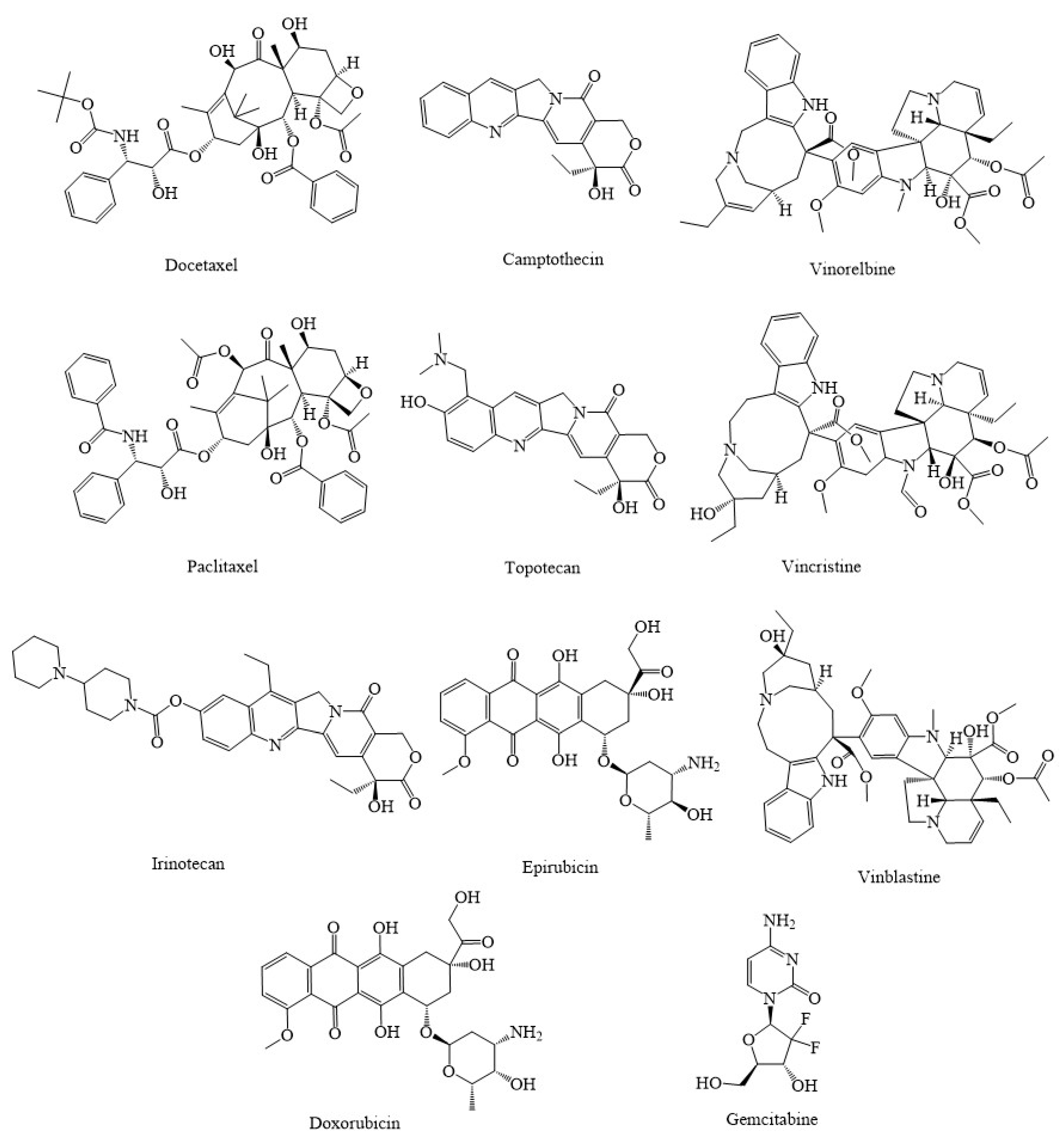
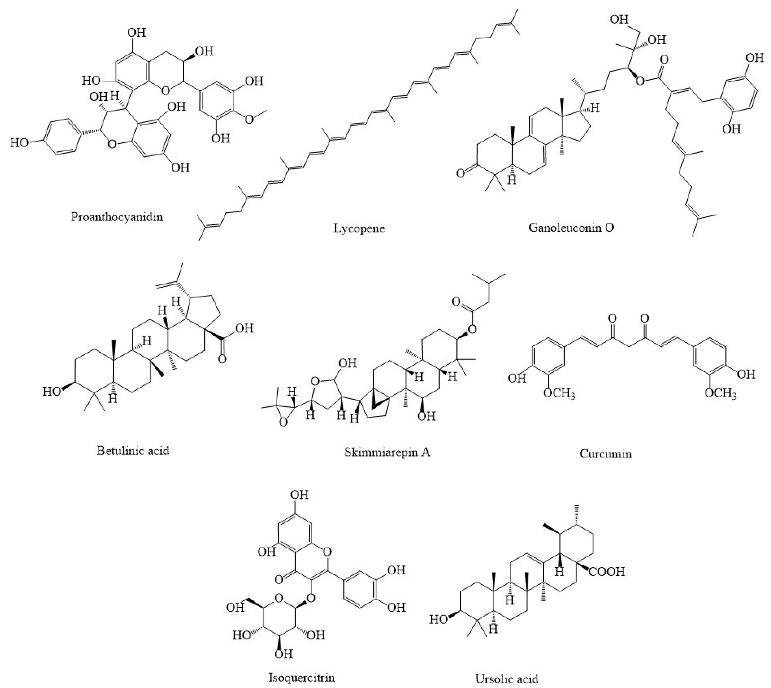
3. Synthetic Agents and Candidate Phytocompounds in Modulation of Cancer through the Regulation of Mitochondria Function

Nature is one of the main sources of active therapeutic agents, chemical compounds, and drugs with a wide variety of pharmacological effects, including antitumor, cardioprotective, antiviral, neuroprotective, anti-inflammatory, and antibacterial activities [60,61,62,63,64,65,66,67,68,69,70,71]. In recent decades, various mitochondrial-targeted components with significant anticarcinoma potential have been identified and obtained from natural compounds.
Paclitaxel (Taxol) and the analogs cabazitaxel (Jevtana) and docetaxel (Taxotere), camptothecin, vinorelbine (Navelbine), topotecan (Hycamtin), vincristine (Oncovin), irinotecan (Camptosar), epirubicin (Ellence), and vinblastine (Velban) (Figure 2) are some of the synthetic and semisynthetic analogs of antitumor drugs derived from plants that are routinely used in the treatment of various types of cancers through mitochondria [60]. Doxorubicin-loaded triphenylphosphine (TPP)-conjugated chitosan nanoparticles (NPs) (Dox-loaded TPP-NPs) played an essential role in controlling the apoptotic signaling pathway by lowering MMP, enhancing ROS, Bcl-2-associated X (Bax), and stimulating the release of cyt. c, which consequently led to activation of caspase-9 and caspase-3 [72]. The combination of paclitaxel and doxorubicin co-loaded liposomes (PD-LPs) has been demonstrated to be a useful strategy for clinical cancer medication with minimal toxicity and treatment failure. This was accomplished while boosting therapeutic potential in SMMC-7721 cells by modulating apoptotic pathways via increased production of Bax and caspase-3 and reduced levels of Bcl-2; thus, it can play an important role in the treatment of hepatocellular carcinoma [73].
Proanthocyanidin (Figure 3) combined with chitosan nanoparticles (CS-PAC-AgNPs) has been shown to be effective in the treatment of colorectal cancer by modulating mitochondria function. CS-PAC-AgNPs can activate the release of cyt. c, induce the activation of caspase-9 and caspase-3, inhibit cancerous cellular proliferation, and suppress the formation of antiapoptotic proteins in HT-29 cells [74]. Lycopene, a tetraterpene present in tomatoes and several foods, is the most abundant carotenoid accumulating in prostate tissues and has been found to have antineoplastic properties in prostate cancer. This antioxidant agent can regulate several cell signaling pathways that can lead to a decrease in the mitochondrial transmembrane potential, trigger the production of mitochondrial cyt. c, and induce apoptosis in LNCaP human prostate cancer cells [75,76]. Ganoleuconin O, a terpenoid compound obtained from Ganoderma leucocontextum, has shown anticancer effects in liver cancer cell line Huh-7.5 by inducing apoptosis pathway via increasing p53, Bax, caspase-9, and release of cyt. c while reducing the rate of ATP, Bcl-2; all of these lead to mitochondrial dysfunction [77]. Betulinic acid, another terpenoid compound isolated from Betula alba, has shown considerable anticancer activities in HeLa, PC12, and ACHN cancer cells by inducing mitochondrial dysfunction via enhancing the rate of ROS, caspase-3, caspase-9, Bax, and reducing Bcl-2 [77]. Skimmiarepin A and skimmiarepin C, two compounds derived from the Bael tree (Aegle marmelos), have shown antimalignancy impacts in human breast tumor T47D cells through blocking HIF-1 stimulation and silencing cellular respiration by selectively blocking the mitochondrial electron transport chain at complex I; thus, it may be a promising treatment for breast cancer [78]. Curcumin, a polyphenol effective factor isolated from the rhizome of Curcuma longa, has shown anticarcinogenic effect in HepG2 cells through stimulating apoptosis in mitochondria by increasing the production of proapoptotic protein Bax, decreasing the formation of antiapoptotic protein Bcl-2, promoting cyt. c release, and increasing the level of caspase-3 [79]. Isoquercitrin is a flavone compound that can diminish the growth of MDA-MB-231 by causing mitochondrial dysfunction via decreasing the levels and activities of MMP, Bcl-2, and increasing Bax in relation to mitochondria [77].
The role of natural and semisynthetic agents in the prevention, treatment, and decreasing the drug resistance in cancer is quite clear. Natural and synthetic agents apply these substantial pharmacological effects by affecting several signaling pathways. Natural products potentiate the interference with variant anticancer signals related to mitochondrial activities, including VEGF, p53, Bax/Bcl-2, caspases, cyt. c, HIF-1, MMP, and ROS-related pathways. Table 1 summarizes various anticancer phytochemicals and synthetic agents that interfere with the mitochondrial-related pathways.

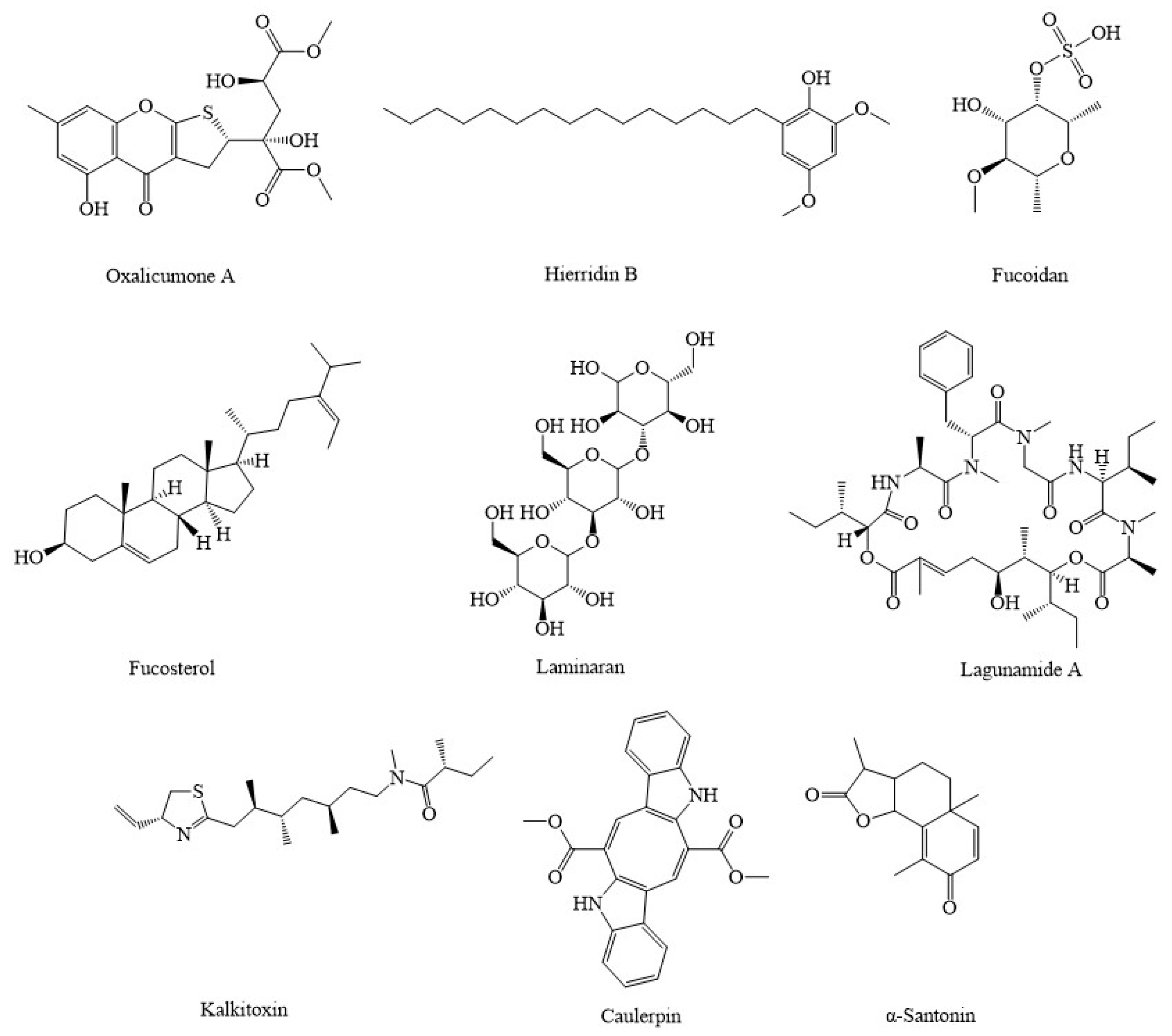
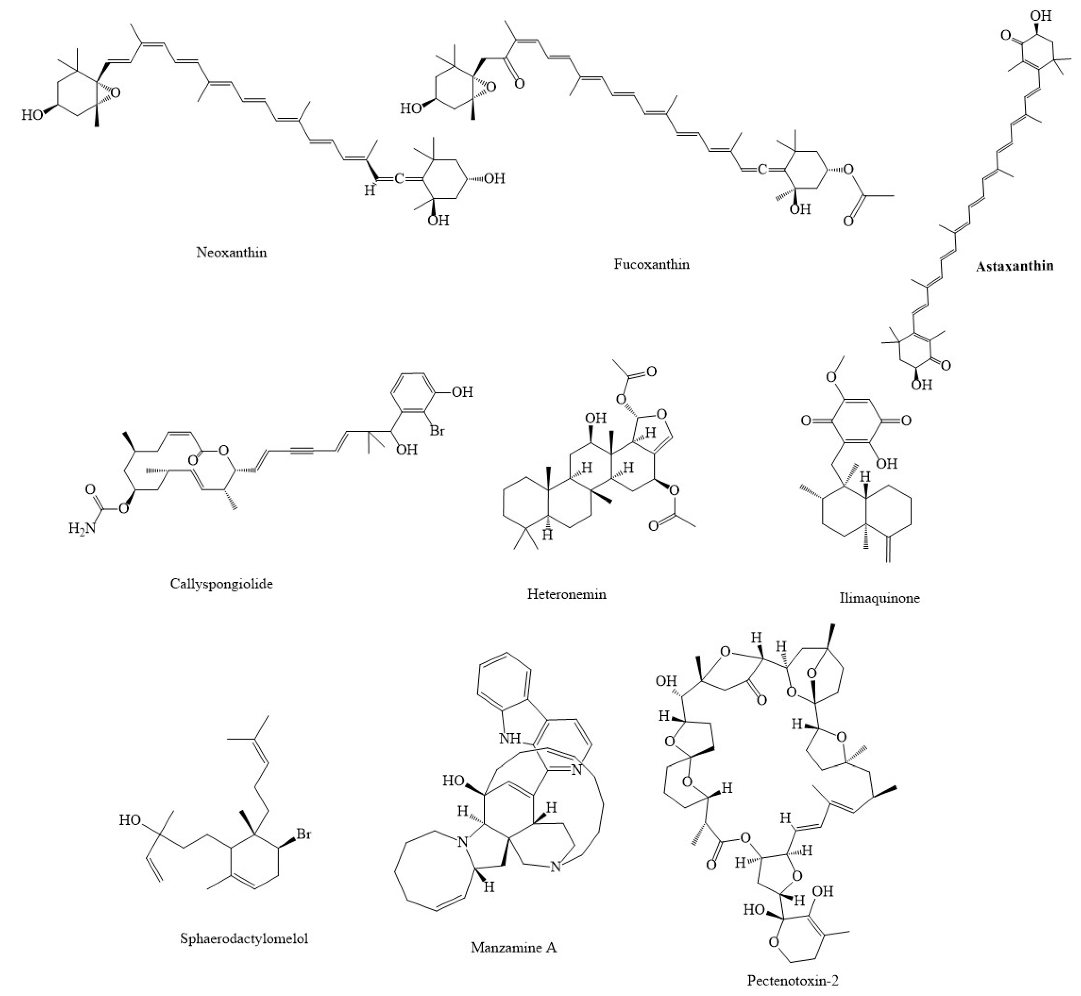
4. Marine Compounds Suppress Cancer through the Modulation of Mitochondria Function

The marine environment has long been a significant supplier of natural resources. With its vast expanse that covers 70% of the planet’s surface, it has the ability to hold a variety of different creatures [85,86]. Marine life are well-known makers of new small agents with distinct chemical structures and potentially beneficial biological effects [87]. These small compounds, known as secondary metabolites, and other various compounds are small molecules that are major sources of active ingredients and novel medications, which are abundant in nature [77]. Many of these marine-derived natural products have beneficial roles in the treatment of various types of cancer, so they can introduce a new way to pave the road against cancer [16,17,20,21,35,36].
Experiments with oxalicumone A (Figure 4), an organic chemical compound obtained from marine sources fungus Penicillium oxalicum, have proven that it is a powerful bioactive antitumor agent for a range of human carcinomas [88]. An immediate decrease in the rate of ATP production has been seen following attenuation of mitochondria function with oxalicumone A. This was followed by mitochondrial permeability transition pore (MPTP) activation and mitochondria enlargement, vacuolation, and reduced matrix density [88]. As another marine agent, hierridin B, is obtained from a marine cyanobacterium (Cyanobium sp.), which has recently been identified as a potential resource for the extraction and purification of chemicals having therapeutic properties, particularly as anticancer drugs [89]. Significant alterations in voltage-dependent anion channel-1 (VDAC1, a mitochondrial protein in the outer mitochondrial membrane that causes a central pore and is considered a major protein for mitochondria-mediated apoptosis) were caused by hierridin B on colon adenocarcinoma cell line HT-29. There was a drop in mRNA and protein expression of VDAC1, and the activity of mitochondrial was reduced [89]. As marine compounds isolated from algae, spatane diterpenoid, 5(R), 19-diacetoxy-15,18 (R and S), dihydro spata-13, 16 (E)-diene (DDSD), which were isolated from the brown marine algae Stoechospermum marginatum, were previously shown to have suppressive effects on various human cancer cell lines and murine melanoma cells [90]. DDSD triggered the production of ROS, which resulted in a change in the Bax/Bcl-2 ratio, which disturbed the inner mitochondrial transmembrane potential. This caused the redistribution of Cyt c to the cytoplasm and the stimulation of the caspase-mediated apoptotic pathway [90]. Another marine agent obtained from algae, fucoidan, which is isolated from brown algae, has been found to exhibit significant anticancer potential [91]. Cell death-inducing pathways, such as the c-Jun N-terminal kinase (JNK), p38, and extracellular signal-regulated kinases (ERK) 1/2, were activated in MCF-7 cells after exposure to fucoidan essence. These pathways are implicated in mechanisms that cause cell death [92]. The production of antiapoptotic proteins Bcl-2 and Bcl-xL was reduced in response to fucoidan essence therapy, although proapoptotic proteins Bax and Bad were slightly increased [92]. Fucoidan stimulated Cyt c production, and it significantly suppressed antiapoptotic proteins, such as Bcl-2 and Bcl-xL in MDA-MB-231 breast cancer cells [91,92]. Additionally, fucoidan led to decreasing Bax/Bcl-2 levels, which enhanced the production of ROS and Bax-to-Bcl-2 ratio in SMMC-7721 cells [93]. In this line, fucosterol (24-ethylidene cholesterol) is one of the phytosterols found in brown algae, and it exerts a variety of biological activities, notably anticancer capabilities. It can suppress the PI3K/Akt pathway and show apoptotic properties [94]. Fucosterol caused ROS production in HeLa cells and mitochondrial malfunction in ES2 and OV90 cells, activated caspase-3 and caspase-9, released cyt. c, and caused the impairment of MMP [17,94]. Brown algae are also a source of the β-glucan-like laminarin, which has several beneficial effects, including anti-inflammatory, antioxidant, and anticancer properties. Laminarin suppressed the PI3K/mitogen-activated protein kinase (MAPK), which modulated dysregulated pathways in ovarian cancer cells (ES2 and OV90). In addition, it had a continuous weakening effect on cell growth [95]. In ES2 and OV90 cells, laminarin caused MMP degradation, which led to an elevation in intracellular calcium concentrations, and it caused ROS production, which induced the production of Cyt c and enhanced the destruction of DNA [95]. Laminarin also increased ROS production, which stimulated MPTP, increased Ca2+, cyt. c, caspase-3, and Bax concentration, and decreased the MMP in LOVO cells [96]. Desmethylxestospongin B, also known as dmXe B, is a complex marine natural compound that displays unique capabilities as a calcium pathway modulator and has downstream impacts in the mitochondrial metabolism of cancer cells [97]. Through the suppression of inositol-1,4,5-triphosphate receptors, xestospongin B was able to exert an effect on mitochondrial metabolism. This was accomplished via regulating calcium communication between the endoplasmic reticulum (ER) and mitochondria that resulted in a decrease in mitochondrial respiration, which in turn caused the stimulation of AMPK and autophagy [97]. The sulfated polysaccharide (EI-SP), a compound that is present in marine algae and isolated from Enteromorpha intestinalis, has been demonstrated to have antioxidant, antineoplastic, and anti-inflammatory properties [98]. EI-SP elevated the amount of the proapoptotic Bax protein while it reduced the amount of the antiapoptotic protein Bcl-2 in human hepatoma hepG2 cells. It was concluded that a sulfated polysaccharide, EI-SP, exhibited anticancer properties. Additionally, it increased the level of cyt. c, which resulted in increased activity of caspase-9 [98]. Similar in source, thyrsiferol, extracted from the tropical sea red alga Laurencia thyrsifera J.Agardh, was discovered to have high anticancer potential against multiple tumor cells through different signaling pathways [99]. Thyrsiferol, which was also separated from the tropical marine red alga Laurencia thyrsifera J.Agardh, was found to suppress HIF-1 stimulation in T47D human breast tumor cells, as well as selectively block mitochondrial respiration at complex I and oxygen utilization in a concentration-dependent way [99]. Caulerpin, a secondary metabolite derived from the marine invasive green algae Caulerpa cylindracea, has been demonstrated to have antineoplastic impacts in many cancerous cell lines via multiple signaling pathways [100]. Caulerpin has been shown to block the function of complex II in the mitochondria respiratory system and can be beneficial in the treatment of human ovarian carcinoma [100]. Chlorella sorokiniana, a species of green algae commonly consumed as a food supplement in East-Asian countries, has shown a useful role on NSCLC by modulating apoptotic pathways [101]. Chlorella sorokiniana can enhance the rate of caspase-3, caspase-9, and Bax, reduce MMP, which induces the release of cyt. c, lower the rate of Bcl-2, and suppress the proliferation of non-small cell lung cancer [101].
Lagunamides A is a cyclic depsipeptide initially identified from the marine cyanobacterium Lyngbya majuscule. Lagunamides A has been shown to possess exceptional growth-suppressive properties against non-small-cell lung carcinoma [NSCLC] (A549), cervical carcinoma cells (HeLa), hepatocellular carcinoma cell (HepG2), BEL-7404, colorectal carcinoma cells (HCT116), and osteosarcoma (U2OS) cancer cells [102]. Additionally, lagunamide A enhanced the production of ROS, which caused cell death, and there was also a reduction in the MMP as a result of lagunamide A therapy [102]. As another peptide type marine compound, irciniastatin A (psymberin), a pederin-type natural compound isolated from a marine sponge Ircinia ramosa, showed particularly anticancer effects against human leukemia Jurkat cells [16]. Irciniastatin A is a strong inhibitor of protein production, and through a mitochondrial-mediated process, it caused sustained stimulation of the stress-activated protein kinases JNK and p38 by modulating production of ROS in mitochondria [16]. MSPs are marine antimicrobial peptides and natural compounds isolated from Nile tilapia (Oreochromis niloticus). MSPs have been shown to exert antitumor roles in the treatment of several cancers through different signaling pathways, especially by inducing apoptotic pathways [103]. MSP-4-peptide caused a decrease in the production of antiapoptotic proteins like Bcl-2 and increased proapoptotic proteins like Bax and Bid, releasing cyt. c, and increased caspase-9 and caspase-3 in mitochondria in MG63 cells in in vitro models of human osteosarcoma [103]
Conotoxins and conus textile, derived from marine cone snails in Iran’s southern seas, have been demonstrated to exhibit antitumor properties in U87MG human glioma cells in mitochondrial-related pathways [104]. Conus textile, a carnivorous species, has been shown to induce the activity of caspase-3, caspase-9, and Bax/Bcl-2 levels, and enhance the production of cyt. c and ROS in U87MG cells [104]. Complexes II and III were severely suppressed by 3,4,5-tribromo-2-(20,40-dibromophenoxy)-phenol, whereas complexes I and V were very mildly suppressed [105].
A solitary marine tunicate of the ascidian class in tropical seas, Phallusia nigra compounds generated considerable ROS production in skin mitochondria from the melanoma group, but they did not in the control group. Phallusia nigra, a unique marine tunicate with significant antitumor activities, exhibits high promise and practicality as a source of anticancer medications [106]. Additionally, Phallusia nigra compounds enhanced cyt. c released and reduced mitochondria membrane potential (MMP) in Albino/Wistar rats [106]. Aplidin (plitidepsin), a cyclic depsipeptide derived from the marine tunicate Aplidium albicans and now produced synthetically, has been demonstrated as having antineoplastic properties in many hematological and solid cancers through the apoptotic process and several signaling pathways, such as p38 and MAPK [107]. Aplidin has been demonstrated as having positive effects on leukemia lymphoma models in vitro and in vivo by modifying mitochondria through producing ROS and decreasing MMP and ATP concentrations [107]. Lipopeptide kalkitoxin, isolated from the Lyngbya majuscula marine cyanobacterium (often reported as Moorea producens), prevented hypoxic HIF-1 induction, reduced the quantity of hypoxia-induced secreted VEGF protein, and prevented tumor angiogenesis, as well as regulated mitochondrial respiration in tumor cells by blocking electron transport chain complex I [108].
Methyl 5-[(1E,5E)-2,6-Dimethyl octa-1,5,7-trienyl] furan-3-carboxylate (MDTFC), a furano-sesquiterpene bioactive component from a soft coral, has shown to induce apoptosis in THP-1 (human monocytoid) cells via reducing caspase-3, caspase-9, enhancing the rate of Bax/Bcl-2, releasing cyt. c, and decreasing MMP, which may make it an effective treatment in cancer [109]. 2-ethoxycarbonyl-2-β-hydroxy-A-nor-cholest-5-ene-4one (ECHC), a marine steroid compound derived from butanol extracts of the scleractinian coral Acropora formosa, may be useful in the treatment of lung cancer [110]. ECHC from scleractinian coral Acropora formosa can enhance the rate of ROS, cyt. c, Bax, tumor suppressor p53, and reduce the production of antiapoptotic factors, such as tumor necrosis factor-α (TNF-α), interleukin-8 (IL-8), Bcl-2, MMP-2, and MMP-9 in A549 human non-small cell lung cancer cell lines [110].
The Turbo coronatus, a Persian gulf marine mollusk that is a member of the Turbinidae family, was found to have antitumor and apoptotic impacts in epithelial ovarian cancer cells [111]. It has shown to inhibit SDH function, enhance the production of ROS and the release of cyt. c, reduce MMP, and enhance the activity of caspase-3 in mitochondria of human EOC cells [111]. Isolated from other marine sources, mansouramycin C, a cytotoxic isoquinolinequinone gained from a marine streptomycete, has been shown to have anticancer activity in tumoral cells via targeting different signaling pathways [112]. Mansouramycin C enhanced the production of ROS in cancerous cells, reduced the rate of ATP and MMP, and stimulated mitochondrial swelling via the opening of MPTP in A549 cells; thus, it may become a hopeful candidate for the treatment of cancer [112].
Highly polar xanthophylls of 9′-cis-neoxanthin and fucoxanthin (Figure 5), two carotenoids, have been found to diminish the survival of human prostate cancer cells by promoting apoptosis [113], reducing the rate of ATP synthesis, and enhancing the production of Bax and cyt. c in mitochondria [114]. Fucoxanthin has been shown to decrease cell cycle development in human neuroblastoma cells while causing cell death in HL-60 and human colon cancer cells [115]. Additionally, fucoxanthin enhanced ROS and reduced the levels and activity of MMP, SOD, catalase, and glutathione in oral squamous carcinoma (KB) cells [116]. Neoxanthin aggregation in mitochondria resulted in the reduction of MMP, which led to the production of cyt. c, apoptosis-inducing factor (AIF), and induced the activation of apoptotic processes [115]. Astaxanthin (3,3-dihydroxy-β,β′-carotene-4,4-dione), a carotenoid recognized for its powerful antioxidant capacity, has shown to have anticarcinoma properties in numerous cancer cell types via various signaling pathways. In this way, astaxanthin can enhance the level of proapoptotic proteins like Bax/Bad, reduce antiapoptotic proteins, such as Bcl-2, and can decrease the rate of ROS in glioblastoma multiforme cells [117]. Additionally, cellulose nanocrystals/nanofibrils loaded with astaxanthin nanoemulsion enhanced the production of ROS in L929 and NIH3T3 cells as in vitro models of skin cancer [118].
Arca inflata is a classical Chinese marine drug with a variety of therapeutic applications, including cancer therapy. It contains a potent peptide compound known as p6, which has been shown to have anticancer activities in human colorectal cancer cells via interfering with mitochondria-related pathways [119]. Acta inflata enhanced the rate of production of ROS, and Ca2+, released cyt. c, decreased MMP, and promoted the activity of caspase-3 in colorectal cancer cells, which causes apoptosis and led to mitochondrial malfunction. Additionally, p6 blocked the growth of colorectal cancer cells [119]. CS5931, a novel marine polypeptide extracted from sea squirt Ciona savignyi, has been shown to enhance the production of caspase-3, caspase-9, cyt. c, and Bax. It also interrupted MMP and suppressed the growth of HCT-8 colon cancer cells [120]. CS5931 has demonstrated strong cytotoxicity against several cancerous cells by intervening on caspase-3, caspase-9, cyt. c, Bax, and MMP [120]. Lamellarin D, a powerful marine alkaloid isolated from the marine mollusk Lamellaria, has been shown to block topoisomerase I and other signaling pathways, resulting in cytotoxic and antitumoral impacts in leukemia cells (p388) [121,122,123]. Lamellarin D increased the rate of Bax, stimulated the activity of caspase-3, caspase-9, and decreased Bcl-2, which led to mitochondrial malfunction and apoptosis in p388 leukemia cells [121,124].
GLP, a glycoprotein from Codium decorticatum, has been shown to stimulate the release of cyt. c and enhance the rate of ROS production, increase caspase-3, caspase-9, and decrease MMP in MDA-MB-231 cells [125]. It demonstrated cytotoxic impacts in human MDA-MB-231 cells via the apoptosis pathway. It reduced the growth of cancer cells and caused mitochondrial dysfunction [125]. In A549 lung cancer cells, the marine mangrove plant Avicennia marina combined with Ag NPs demonstrated an antineoplastic impact by enhancing the formation of ROS, destroying the mitochondrial membrane, and increasing the level of activated caspases, which resulted in apoptosis [126]. Pterocellin A, an alkaloid metabolite derived from the marine bryozoan Pterocella vesiculosa, has been shown to have antitumor effects against the human cervical cancer cell line HeLa. It has the potential to cause cytotoxic effects via inducing apoptosis and disrupting the morphology of cervical carcinoma cells (HeLa) [127]. Pterocellin A has been shown to induce the activation of caspase-3, and as a result of nucleus condensation, cause shrinkage, which led to the death of the HeLa cells [127]. Callyspongiolide decreased MMP and level of complex I and II, which decreased the activity of HIF-1A in mitochondria of various kinds of cancerous cell lines [128].
Heteronemin, a marine organic product obtained from Hippospongia sp., has been shown to defend against various cancerous cell lines by modulating the apoptosis pathway [129]. Heteronemin enhanced the rate of ROS, Bax, SOD2, and caspase-8 production, which decreased the level of Bcl-2 and SOD1 in human hepatocellular carcinoma cells. In addition, heteronemin led to mitochondrial dysfunction [129]. Brominated diterpene sphaerodactylomelol, derived from the red algae Sphaerococcus coronopifolius Stackhouse, has shown antitumor properties in breast cancer cells (MCF-7) via modulating the apoptosis signaling pathway [130]. Sphaerodactylomelol enhanced the rate of ROS, H2O2 production, caspase-3, caspase-9, Bax, and cyt. c, while decreasing antiapoptotic proteins, such as Bcl-2, and weakening MMP [130].
The marine compounds containing quinone moiety are known to target mitochondria. Ilimaquinone, a sesquiterpene quinone isolated from marine sponges (Halichondria sp.), has been shown to have antitumor effects on MCF-7 and MDA-MB-231 breast cancer cells by modulating various signaling pathways, particularly the mitochondrial pathway [131]. Ilimaquinone increased the rate of caspase-3, caspase-9, ROS, and MMP loss, which led to mitochondrial dysfunction in human breast cancer cells [131]. Another marine-derived polycyclic quinone-type metabolite, xestoquinone, decreased the growth of leukemia cancer cells, through multiple signaling mechanisms, including ROS-induced suppression of heat shock protein-90 [132]. A novel derivative of 1,4-naphthoquinone analogs isolated from the sea urchin pigments spinochromes, 6-S-(1,4-naphthoquinone-2-yl)-d-glucose chimera molecules, also showed anticancer potentials against human prostate cancer PC3 cells. The underlying mechanism of action included mitochondria membrane permeabilization, ROS upregulation, and release of AIF and cyt. c to the cytoplasm. This process led to the activation of caspase-9 and caspase-3, PARP cleavage, and apoptosis-like cell death [133]. Several other sesquiterpene quinones isolated from a marine sponge Hippospongia sp. Also suppressed the maturation of starfish oocytes and induced cell cycle arrest in HepG2 hepatocellular carcinoma cells [134].
Fraction 2.1, a polysaccharide gained from marine clam Donax variabilis, has shown anticarcinoma effects in A549 cells via inducing mitochondria malfunction by enhancing the level of Bax, activity of caspase-3, caspase-9, and stimulating the release of cyt. c while reducing Bcl-2 [135]. Manzamine A, an alkaloid obtained from sponges of the genera Haliclona sp., Xestospongia sp., and Pellina sp., has been shown to have the ability to destroy HCT116 cells by causing mitochondrial dysfunction by MMP loss, enhance the activity of caspase-3, caspase-7, induce the release of cyt. c, and decrease the level of Bcl-2 and Bcl-xL [136]. Aplysinopsins, a marine indole alkaloid isolated from genera of sponges and scleractinian corals, has exhibited antiproliferative effects on in K562 cells. EE-84, one of the analogs of aplysinopsins, reduced cell proliferation, intake of oxygen, level of antiapoptotic protein, Bcl-2, and caused MMP loss in K562 cells [137]. Another natural product from the marine sponge and fungus, Aspergillus sp., 18B-15-3, has exhibited anticarcinoma properties in pancreatic cancer. This substance exerted anticarcinoma effects by interfering with multiple pathways, especially the modulation of mitochondrial pathways [138]. 18B-15-3, marine-derived fungus Aspergillus sp., isolated from a marine sponge collected on Pramuka Island, suppressed complex III in the mitochondrial electron transport chain and reduced MMP, which blocked the normal function of mitochondria. Additionally, it blocked the intake of oxygen in pancreatic cancer cells [138]. 3,5-dibromo-2-(20,40-dibromophenoxy)-pheno and 3,4,5-tribromo-2-(20,40-dibromophenoxy)-pheno (20,40-dibromophenoxy)-phenol, other marine agents extracted from the marine sponge Dysidea sp., have been found to have anticancer effects in pancreatic cancer (PANC-1 cells) via increasing the levels of phosphorylated Akt [105].
Holothuroids (Holothuroidea class) are marine invertebrates which live in deep seas and are thought to be the origin of major drugs, such as anticancer medicines. Compounds obtained from sea cucumber (H. parva) and sponge (H. oculata) showed antitumor actions on liver mitochondria isolated from hepatocellular cancer in vivo [139]. H. parva and sponge H. oculata increased ROS concentration, decreased MMP, enhanced cyt. c release, and induced caspase-3 activity in the mitochondria, which had antineoplastic impacts in cancerous hepatocytes in male Sprague-Dawley rats [139]. Ciona savignyi, a marine agent containing a powerful polypeptide called Callyspongiolide, is an organic marine macrolide compound that exerted antimalignancy properties by inducing cell death in cancer [128]. Lipophilic 2,5-disubstituted pyrroles from the marine sponge Mycale sp. have been demonstrated to have antitumor actions in hypoxic conditions, which lead to the formation of solid tumors via mitochondria-related pathways [140]. Lipophilic 2,5-disubstituted pyrroles were shown to have antimalignancy effects in a human breast tumor T47D cell-based reporter assay by suppressing the function of HIF-1. This heterodimer protein was involved in the production of factors that are linked to angiogenesis, glucose metabolism, cell proliferation/survival, and invasion/metastasis. This was achieved via blocking the mitochondrial respiration through suppressing complex I and complex III, which generated ROS [140].
Urupocidin A, which is derived from marine sponges, is a bicyclic guanidine alkaloid that has been shown to have biological effects, particularly anticancer effects [87]. Urupocidin A was shown to increase the production of ROS, which was caused by a disruption in the integrity of mitochondria and MMP. It also reduced the expression of Bcl-2, which caused mitochondria to release various proteins which have antiapoptotic effects on prostate cancer cells [87]. The extraction of papuamine from Haliclona sp., a marine sponge, has been shown to exhibit anticancer effects in MCF-7 cells via regulating autophagy and mitochondrial dysfunction [141]. A furanoterpenoid isolated from irciformonin B found in a marine sponge, 10-acetylirciformonin B blocked Bcl-2 and Bcl-xL, enhanced Bax, stimulated the release of cyt. c, and increased the levels of ROS in mitochondria [142]. 10-acetylirciformonin B, has been shown to exhibit antitumor properties in leukemia HL 60 cells via promoting DNA destruction and apoptosis through several signaling pathways. 10-acetylirciformonin B dramatically reduced caspase-3 and caspase-9 activity and decreased topoisomerase II protein production [142]. The sponge Dysidea avara, which has a sesquiterpene lactone santonin, has been shown to have antitumor impacts in cancer cell lines; thus, marine drugs may be useful in treatment of cancer [143]. Santonin decreased MMP, enhanced ROS production, caspase-3, and released cyt. c in ALL B-lymphocytes, which caused apoptosis in all B-lymphocytes [143]. Mycothiazole, originally derived from the marine sponge Petrosaspongia mycofijiensis, has been shown to have anticarcinoma impacts in cancerous cells via inhibiting hypoxic HIF-1 signaling and causing mitochondrial dysfunction in human breast tumor T47D cells [144]. Mycothiazole has been shown to significantly block mitochondrial respiration at complex I [144]. A dipeptide Cyclo(-Pro-Tyr) (DP) isolated from marine sponge, increased the rate of Bax/Bcl-2 and caspase-3, had negative effects on MMP, stimulated the release of cyt. c, and enhanced the production of ROS in HepG2 cell lines [145]. DP, derived from the marine sponge Callyspongia fistularis, has demonstrated an apoptotic role in the destruction of liver cancer HepG2 cell lines and may be a promising medication for the treatment of hepatocellular carcinoma [145].
Table 2 summarizes various anticancer marine compounds that interfere with the mitochondrial-related pathways.
Briefly, biologically active secondary metabolites present in marine organisms show a wide range of antineoplastic activities. Marine-derived natural products act by diminishing cell viability, proliferation, promotion of ER stress, apoptosis, and ROS production by interfering with various cell signaling pathways in cancer.

5. Conclusion, Challenges, and Future Perspectives

Various dysregulated pathways can influence the growth, proliferation, invasion, and migration of cancer cells. In this line, mitochondria have received attention due to playing several important roles in growth, survival, and in cellular death. These roles and significant effects have made mitochondria and their related signaling pathways key points in preventing progression and aiding in the treatment of cancer. Targeting mitochondrial metabolism-related enzymes, including IDH1, IDH2, SDH, and fumarate hydratase, interfering with PI3K/PTEN/Akt, HIF-1α, WNT, p53, and angiogenesis-related signaling, interfering with mitochondrial metabolism, cellular redox status, increasing the production of ROS, reducing the mitochondrial Ca2+ retention, and MMP are some of the most important pathways related to mitochondria that can affect cancer. Several studies have shown the promising advantages of targeting pathways related to mitochondria to inhibit the growth and proliferation of different types of cancer cells. Marine-derived natural products are promising multi-targeted agents that can affect mitochondria signaling pathways. These products act as mediators to suppress cancer cell growth, proliferation, and angiogenesis with fewer side effects (Figure 6). Consequently, these compounds are suitable, promising options for the treatment of cancer through mitochondria.
Mitochondria were shown to be affected by several cytotoxic agents. However, it is often not the primary, but secondary and rather unspecific target that is affected due to the induction of other apoptosis-/cell death-related processes. Pan-Assay Interference Compounds (PAINS) are a recently discovered class of compounds that oppose the “target selectivity” point of view. Such compounds can affect intracellular signaling pathways in a non-specific way, and have been identified as a threat to the bioactivity of natural compounds [146]. PAINS-type behavior causes membrane disruption by decreasing the membrane dipole potential, which affects intracellular signaling pathways. The cell-based data of several natural products are likely to indicate such effects which may not be caused by specific binding to therapeutic targets [147]. This non-specific/specific targeting of mitochondria by marine compounds should be carefully considered in future studies.
Despite the effectiveness of marine agents, these compounds are often limited by poor bioavailability, low solubility, instability, and low selectivity, which restricts associated therapeutic uses in cancer. Growing clinical studies have demonstrated that marine natural products are encountered with chemical degradation and rapid metabolism and are absorbed with lower efficiency. The aforementioned pharmacokinetic limitations have been attenuated with nanotechnology to enhance bioavailability and anticancer properties of these compounds [4]. However, some clinical trials are in progression to evaluate the clinical potential and efficacy of marine compounds. For instance, the marine compound astaxanthin has shown potential cardioprotective [148] and immunomodulatory [149] effects. It also shows health benefits in overweight/obese adults [150,151], and cosmetic applications [152]. In a double-blind randomized controlled trial, the marine compound fucoidan improved disease control rate in patients with metastatic colorectal cancer [153]. Fucoidan also showed anticancer effects through regulating the expression of microRNAs [154]. In a randomized trial, sodium alginate was found to prevent radiation-induced esophagitis in advanced NSCLC [155]. Several new marine compounds are also being evaluated in patients with solid tumors [156,157,158,159,160].
This comprehensive and critical review underscores the importance of targeting malignancies by multi-targeted marine agents passing through the modulation of mitochondria function. A future area of research should focus more on developing well-controlled clinical trials to assess anticancer potentials of marine agents to overcome the toxicity and drug-resistance of present drugs in the prevention, treatment, and management of cancer.

Author Contributions

Conceptualization, S.F. and A.B.; software, S.F. and A.B., drafting the manuscript, S.F., S.A., S.Z.M. and A.B.; review and editing the manuscript: S.F., B.E.D., C.F. and A.B., supervision and project administration, A.B. All authors have read and agreed to the published version of the manuscript.

Funding

This research received no external funding.

Conflicts of Interest

The authors declared that there is no conflict of interest.

References

  1. Siegel, R.L.; Miller, K.D.; Jemal, A. Cancer statistics, 2019. CA A Cancer J. Clin. 2019, 69, 7–34. [Google Scholar] [CrossRef]
  2. Saini, A.; Kumar, M.; Bhatt, S.; Saini, V.; Malik, A. Cancer causes and treatments. Int. J. Pharm. Sci. Res. 2020, 11, 3109. [Google Scholar]
  3. Fakhri, S.; Moradi, S.Z.; Ash-Rafzadeh, A.; Bishayee, A. Targeting cellular senescence in cancer by plant secondary metabolites: A systematic review. Pharmacol. Res. 2022, 177, 105961. [Google Scholar] [CrossRef]
  4. Fakhri, S.; Moradi, S.Z.; Farzaei, M.H.; Bishayee, A. Modulation of dysregulated cancer metabolism by plant secondary metabolites: A mechanistic review. Semin. Cancer Biol. 2022, 80, 276–305. [Google Scholar] [CrossRef]
  5. Fakhri, S.; Moradi, S.Z.; Yarmohammadi, A.; Narimani, F.; Wallace, C.E.; Bishayee, A. Modulation of TLR/NF-κB/NLRP Signaling by Bioactive Phytocompounds: A Promising Strategy to Augment Cancer Chemotherapy and Immunotherapy. Front Oncol 2022, 12, 834072. [Google Scholar] [CrossRef]
  6. Fakhri, S.; Zachariah Moradi, S.; DeLiberto, L.K.; Bishayee, A. Cellular senescence signaling in cancer: A novel therapeutic target to combat human malignancies. Biochem. Pharmacol. 2022, 199, 114989. [Google Scholar] [CrossRef]
  7. Moradi, S.Z.; Nowroozi, A.; Sadrjavadi, K.; Moradi, S.; Mansouri, K.; Hosseinzadeh, L.; Shahlaei, M. Direct evidences for the groove binding of the Clomifene to double stranded DNA. Int. J. Biol. Macromol. 2018, 114, 40–53. [Google Scholar] [CrossRef]
  8. Adeyinka, A.; Bashir, K. Tumor lysis syndrome. JAMA Oncol. 2018, 4, 895. [Google Scholar]
  9. Tan, X.; Fu, J.; Yuan, Z.; Zhu, L.; Fu, L. ACNPD: The Database for Elucidating the Relationships Between Natural Products, Compounds, Molecular Mechanisms, and Cancer Types. Front. Pharmacol. 2021, 2214. [Google Scholar] [CrossRef]
  10. Al-Mahmood, S.; Sapiezynski, J.; Garbuzenko, O.B.; Minko, T. Metastatic and triple-negative breast cancer: Challenges and treatment options. Drug Deliv. Transl. Res. 2018, 8, 1483–1507. [Google Scholar] [CrossRef]
  11. Pomeroy, A.E.; Schmidt, E.V.; Sorger, P.K.; Palmer, A.C. Drug independence and the curability of cancer by combination chemotherapy. Trends Cancer 2022. [Google Scholar] [CrossRef]
  12. Rejhová, A.; Opattová, A.; Čumová, A.; Slíva, D.; Vodička, P. Natural compounds and combination therapy in colorectal cancer treatment. Eur. J. Med. Chem. 2018, 144, 582–594. [Google Scholar] [CrossRef]
  13. Meerson, A.; Khatib, S.; Mahajna, J. Flavonoids Targeting Cancer Stem Cells for Augmenting Cancer Therapeutics. Int. J. Mol. Sci. 2021, 22, 13044. [Google Scholar] [CrossRef]
  14. Schumacher, M.; Kelkel, M.; Dicato, M.; Diederich, M. A Survey of Marine Natural Compounds and Their Derivatives with Anti-Cancer Activity Reported in 2010. Molecules 2011, 16, 5629–5646. [Google Scholar] [CrossRef]
  15. Bhardwaj, N.; Goel, B.; Tripathi, N.; Sahu, B.; Jain, S.K. A comprehensive review on chemistry and pharmacology of marine bioactives as antimetastatic agents. Eur. J. Med. Chem. Rep. 2022, 4, 100023. [Google Scholar] [CrossRef]
  16. Chinen, T.; Nagumo, Y.; Watanabe, T.; Imaizumi, T.; Shibuya, M.; Kataoka, T.; Kanoh, N.; Iwabuchi, Y.; Usui, T. Irciniastatin A induces JNK activation that is involved in caspase-8-dependent apoptosis via the mitochondrial pathway. Toxicol. Lett. 2010, 199, 341–346. [Google Scholar] [CrossRef]
  17. Jiang, H.; Li, J.; Chen, A.; Li, Y.; Xia, M.; Guo, P.; Yao, S.; Chen, S. Fucosterol exhibits selective antitumor anticancer activity against HeLa human cervical cell line by inducing mitochondrial mediated apoptosis, cell cycle migration inhibition and downregulation of m-TOR/PI3K/Akt signalling pathway. Oncol. Lett. 2018, 15, 3458–3463. [Google Scholar] [CrossRef]
  18. Ngo, D.-H.; Vo, T.-S.; Ngo, D.-N.; Wijesekara, I.; Kim, S.-K. Biological activities and potential health benefits of bioactive peptides derived from marine organisms. Int. J. Biol. Macromol. 2012, 51, 378–383. [Google Scholar] [CrossRef]
  19. Ghosh, S.; Sarkar, T.; Pati, S.; Kari, Z.A.; Edinur, H.A.; Chakraborty, R. Novel Bioactive Compounds From Marine Sources as a Tool for Functional Food Development. Front. Mar. Sci. 2022, 9, 10–3389. [Google Scholar] [CrossRef]
  20. Sugumaran, A.; Pandiyan, R.; Kandasamy, P.; Antoniraj, M.G.; Navabshan, I.; Sakthivel, B.; Dharmaraj, S.; Chinnaiyan, S.K.; Ashokkumar, V.; Ngamcharussrivichai, C. Marine biome-derived secondary metabolites, a class of promising antineoplastic agents: A systematic review on their classification, mechanism of action and future perspectives. Sci. Total Environ. 2022, 836, 155445. [Google Scholar] [CrossRef]
  21. Florean, C.; Dicato, M.; Diederich, M. Immune-modulating and anti-inflammatory marine compounds against cancer. Semin. Cancer Biol. 2022, 80, 58–72. [Google Scholar] [CrossRef]
  22. El-Demerdash, A.; Atanasov, A.G.; Bishayee, A.; Abdel-Mogib, M.; Hooper, J.N.A.; Al-Mourabit, A. Batzella, Crambe and Monanchora: Highly Prolific Marine Sponge Genera Yielding Compounds with Potential Applications for Cancer and Other Therapeutic Areas. Nutrients 2018, 10, 33. [Google Scholar] [CrossRef] [Green Version]
  23. Catanzaro, E.; Calcabrini, C.; Bishayee, A.; Fimognari, C. Antitumor Potential of Marine and Freshwater Lectins. Mar. Drugs 2019, 18, 11. [Google Scholar] [CrossRef]
  24. Mondal, A.; Bose, S.; Banerjee, S.; Patra, J.K.; Malik, J.; Mandal, S.K.; Kilpatrick, K.L.; Das, G.; Kerry, R.G.; Fimognari, C.; et al. Marine Cyanobacteria and Microalgae Metabolites-A Rich Source of Potential Anticancer Drugs. Mar. Drugs 2020, 18, 476. [Google Scholar] [CrossRef]
  25. Catanzaro, E.; Bishayee, A.; Fimognari, C. On a Beam of Light: Photoprotective Activities of the Marine Carotenoids Astaxanthin and Fucoxanthin in Suppression of Inflammation and Cancer. Mar. Drugs 2020, 18, 544. [Google Scholar] [CrossRef]
  26. Newman, D.J.; Cragg, G.M. Current Status of Marine-Derived Compounds as Warheads in Anti-Tumor Drug Candidates. Mar Drugs 2017, 15, 99. [Google Scholar] [CrossRef]
  27. Fimognari, C.; Lenzi, M.; Ferruzzi, L.; Turrini, E.; Scartezzini, P.; Poli, F.; Gotti, R.; Guerrini, A.; Carulli, G.; Ottaviano, V. Mitochondrial pathway mediates the antileukemic effects of Hemidesmus indicus, a promising botanical drug. PLoS ONE 2011, 6, e21544. [Google Scholar] [CrossRef]
  28. Sainero-Alcolado, L.; Liaño-Pons, J.; Ruiz-Pérez, M.V.; Arsenian-Henriksson, M. Targeting mitochondrial metabolism for precision medicine in cancer. Cell Death Differ. 2022, 29, 1304–1317. [Google Scholar] [CrossRef]
  29. Passaniti, A.; Kim, M.S.; Polster, B.M.; Shapiro, P. Targeting mitochondrial metabolism for metastatic cancer therapy. Mol. Carcinog. 2022, 61, 827–838. [Google Scholar] [CrossRef]
  30. Yaqoob, M.D.; Xu, L.; Li, C.; Leong, M.M.L.; Xu, D.D. Targeting mitochondria for cancer photodynamic therapy. Photodiagnosis Photodyn. Ther. 2022, 38, 102830. [Google Scholar] [CrossRef]
  31. Xu, J.; Shamul, J.G.; Kwizera, E.A.; He, X. Recent Advancements in Mitochondria-Targeted Nanoparticle Drug Delivery for Cancer Therapy. Nanomaterials 2022, 12, 743. [Google Scholar] [CrossRef] [PubMed]
  32. Criscuolo, D.; Avolio, R.; Matassa, D.S.; Esposito, F. Targeting Mitochondrial Protein Expression as a Future Approach for Cancer Therapy. Front. Oncol. 2021, 11, 797265. [Google Scholar] [CrossRef] [PubMed]
  33. Yin, C.F.; Chang, Y.W.; Huang, H.C.; Juan, H.F. Targeting protein interaction networks in mitochondrial dynamics for cancer therapy. Drug Discov. Today 2022, 27, 1077–1087. [Google Scholar] [CrossRef]
  34. Mani, S.; Swargiary, G.; Singh, K.K. Natural Agents Targeting Mitochondria in Cancer. Int. J. Mol. Sci. 2020, 21, 6992. [Google Scholar] [CrossRef]
  35. Qi, W.J.; Sheng, W.S.; Peng, C.; Xiaodong, M.; Yao, T.Z. Investigating into anti-cancer potential of lycopene: Molecular targets. Biomed. Pharmacother. 2021, 138, 111546. [Google Scholar] [CrossRef]
  36. Ercolano, G.; De Cicco, P.; Ianaro, A. New drugs from the sea: Pro-apoptotic activity of sponges and algae derived compounds. Mar. Drugs 2019, 17, 31. [Google Scholar] [CrossRef]
  37. Zong, W.-X.; Rabinowitz, J.D.; White, E. Mitochondria and Cancer. Mol. Cell 2016, 61, 667–676. [Google Scholar] [CrossRef]
  38. Fulda, S.; Galluzzi, L.; Kroemer, G. Targeting mitochondria for cancer therapy. Nat. Rev. Drug Discov. 2010, 9, 447–464. [Google Scholar] [CrossRef]
  39. Grasso, D.; Zampieri, L.X.; Capelôa, T.; Van de Velde, J.A.; Sonveaux, P. Mitochondria in cancer. Cell Stress 2020, 4, 114–146. [Google Scholar] [CrossRef]
  40. Wallace, D.C. Mitochondria and cancer. Nat. Rev. Cancer 2012, 12, 685–698. [Google Scholar] [CrossRef]
  41. Canter, J.A.; Kallianpur, A.R.; Parl, F.F.; Millikan, R.C. Mitochondrial DNA G10398A polymorphism and invasive breast cancer in African-American women. Cancer Res. 2005, 65, 8028–8033. [Google Scholar] [CrossRef] [PubMed]
  42. Liu, V.W.; Wang, Y.; Hui-Juan, Y.; Tsang, P.C.; Ng, T.-Y.; Ling-Chui, W.; Nagley, P.; Ngan, H.Y. Mitochondrial DNA variant 16189T> C is associated with susceptibility to endometrial cancer. Hum. Mutat. 2003, 22, 173. [Google Scholar] [CrossRef] [PubMed]
  43. Zhai, K.; Chang, L.; Zhang, Q.; Liu, B.; Wu, Y. Mitochondrial C150T polymorphism increases the risk of cervical cancer and HPV infection. Mitochondrion 2011, 11, 559–563. [Google Scholar] [CrossRef] [PubMed]
  44. Bardella, C.; Pollard, P.J.; Tomlinson, I. SDH mutations in cancer. Biochim. Biophys. Acta (BBA)-Bioenerg. 2011, 1807, 1432–1443. [Google Scholar] [CrossRef] [PubMed]
  45. Aghamir, S.M.K.; Heshmat, R.; Ebrahimi, M.; Ketabchi, S.E.; Dizaji, S.P.; Khatami, F. The impact of succinate dehydrogenase gene (SDH) mutations in renal cell carcinoma (RCC): A systematic review. OncoTargets Ther. 2019, 12, 7929. [Google Scholar] [CrossRef] [PubMed]
  46. Toro, J.R.; Nickerson, M.L.; Wei, M.-H.; Warren, M.B.; Glenn, G.M.; Turner, M.L.; Stewart, L.; Duray, P.; Tourre, O.; Sharma, N. Mutations in the fumarate hydratase gene cause hereditary leiomyomatosis and renal cell cancer in families in North America. Am. J. Hum. Genet. 2003, 73, 95–106. [Google Scholar] [CrossRef]
  47. Yim, M.-S.; Ha, Y.-S.; Kim, I.Y.; Yun, S.-J.; Choi, Y.H.; Kim, W.-J. HMOX1 is an important prognostic indicator of nonmuscle invasive bladder cancer recurrence and progression. J. Urol. 2011, 185, 701–705. [Google Scholar] [CrossRef]
  48. Reitman, Z.J.; Yan, H. Isocitrate dehydrogenase 1 and 2 mutations in cancer: Alterations at a crossroads of cellular metabolism. J. Natl. Cancer Inst. 2010, 102, 932–941. [Google Scholar] [CrossRef]
  49. Thompson, C.B. Metabolic enzymes as oncogenes or tumor suppressors. New Engl. J. Med. 2009, 360, 813. [Google Scholar] [CrossRef]
  50. Ward, P.S.; Patel, J.; Wise, D.R.; Abdel-Wahab, O.; Bennett, B.D.; Coller, H.A.; Cross, J.R.; Fantin, V.R.; Hedvat, C.V.; Perl, A.E. The common feature of leukemia-associated IDH1 and IDH2 mutations is a neomorphic enzyme activity converting α-ketoglutarate to 2-hydroxyglutarate. Cancer Cell 2010, 17, 225–234. [Google Scholar] [CrossRef]
  51. Ooi, A.; Furge, K.A. Fumarate hydratase inactivation in renal tumors: HIF1α, NRF2, and “cryptic targets” of transcription factors. Chin. J. Cancer 2012, 31, 413. [Google Scholar] [CrossRef] [PubMed]
  52. Turcan, S.; Rohle, D.; Goenka, A.; Walsh, L.A.; Fang, F.; Yilmaz, E.; Campos, C.; Fabius, A.W.; Lu, C.; Ward, P.S. IDH1 mutation is sufficient to establish the glioma hypermethylator phenotype. Nature 2012, 483, 479–483. [Google Scholar] [CrossRef] [PubMed] [Green Version]
  53. Liu, H.; Colavitti, R.; Rovira, I.I.; Finkel, T. Redox-dependent transcriptional regulation. Circ. Res. 2005, 97, 967–974. [Google Scholar] [CrossRef] [PubMed]
  54. Wallace, D.C.; Fan, W.; Procaccio, V. Mitochondrial energetics and therapeutics. Annu. Rev. Pathol. 2010, 5, 297. [Google Scholar] [CrossRef] [PubMed]
  55. Guzy, R.D.; Hoyos, B.; Robin, E.; Chen, H.; Liu, L.; Mansfield, K.D.; Simon, M.C.; Hammerling, U.; Schumacker, P.T. Mitochondrial complex III is required for hypoxia-induced ROS production and cellular oxygen sensing. Cell Metab. 2005, 1, 401–408. [Google Scholar] [CrossRef]
  56. Tang, Z.; Wang, Y.; Wan, Y.; Xie, Y.; Li, S.; Tao, D.; Wang, C.; Wu, Y.-Z.; Sui, J.-D. Apurinic/apyrimidinic endonuclease 1/reduction-oxidation effector factor-1 (APE1) regulates the expression of NLR family pyrin domain containing 3 (NLRP3) inflammasome through modulating transcription factor NF-κB and promoting the secretion of inflammatory mediators in macrophages. Ann. Transl. Med. 2021, 9, 145. [Google Scholar]
  57. Jones, R.G.; Thompson, C.B. Tumor suppressors and cell metabolism: A recipe for cancer growth. Genes Dev. 2009, 23, 537–548. [Google Scholar] [CrossRef]
  58. Fukuda, R.; Zhang, H.; Kim, J.-w.; Shimoda, L.; Dang, C.V.; Semenza, G.L. HIF-1 regulates cytochrome oxidase subunits to optimize efficiency of respiration in hypoxic cells. Cell 2007, 129, 111–122. [Google Scholar] [CrossRef]
  59. Zhang, H.; Bosch-Marce, M.; Shimoda, L.A.; Tan, Y.S.; Baek, J.H.; Wesley, J.B.; Gonzalez, F.J.; Semenza, G.L. Mitochondrial autophagy is an HIF-1-dependent adaptive metabolic response to hypoxia. J. Biol. Chem. 2008, 283, 10892–10903. [Google Scholar] [CrossRef]
  60. Sharifi-Rad, J.; Ozleyen, A.; Boyunegmez Tumer, T.; Oluwaseun Adetunji, C.; El Omari, N.; Balahbib, A.; Taheri, Y.; Bouyahya, A.; Martorell, M.; Martins, N.; et al. Natural Products and Synthetic Analogs as a Source of Antitumor Drugs. Biomolecules 2019, 9, 679. [Google Scholar] [CrossRef]
  61. Moghaddam, R.H.; Samimi, Z.; Moradi, S.Z.; Little, P.J.; Xu, S.; Farzaei, M.H. Naringenin and naringin in cardiovascular disease prevention: A preclinical review. Eur. J. Pharmacol. 2020, 887, 173535. [Google Scholar] [CrossRef] [PubMed]
  62. Moradi, S.Z.; Momtaz, S.; Bayrami, Z.; Farzaei, M.H.; Abdollahi, M. Nanoformulations of herbal extracts in treatment of neurodegenerative disorders. Front. Bioeng. Biotechnol. 2020, 8, 238. [Google Scholar] [CrossRef] [PubMed]
  63. Fakhri, S.; Nouri, Z.; Moradi, S.Z.; Farzaei, M.H. Astaxanthin, COVID-19 and immune response: Focus on oxidative stress, apoptosis and autophagy. Phytother. Res. 2020, 34, 2790–2792. [Google Scholar] [CrossRef] [PubMed]
  64. Fakhri, S.; Pesce, M.; Patruno, A.; Moradi, S.Z.; Iranpanah, A.; Farzaei, M.H.; Sobarzo-Sánchez, E. Attenuation of Nrf2/Keap1/ARE in Alzheimer’s disease by plant secondary metabolites: A mechanistic review. Molecules 2020, 25, 4926. [Google Scholar] [CrossRef] [PubMed]
  65. Fakhri, S.; Iranpanah, A.; Gravandi, M.M.; Moradi, S.Z.; Ranjbari, M.; Majnooni, M.B.; Echeverría, J.; Qi, Y.; Wang, M.; Liao, P. Natural products attenuate PI3K/Akt/mTOR signaling pathway: A promising strategy in regulating neurodegeneration. Phytomedicine 2021, 91, 153664. [Google Scholar] [CrossRef] [PubMed]
  66. Fakhri, S.; Abbaszadeh, F.; Moradi, S.Z.; Cao, H.; Khan, H.; Xiao, J. Effects of polyphenols on oxidative stress, inflammation, and interconnected pathways during spinal cord injury. Oxidative Med. Cell. Longev. 2022, 2022, 1–34. [Google Scholar] [CrossRef]
  67. Fakhri, S.; Nouri, Z.; Moradi, S.Z.; Akkol, E.K.; Piri, S.; Sobarzo-Sanchez, E.; Farzaei, M.H.; Echeverría, J. Targeting multiple signal transduction pathways of SARS-CoV-2: Approaches to COVID-19 therapeutic candidates. Molecules 2021, 26, 2917. [Google Scholar] [CrossRef]
  68. Moradi, S.Z.; Jalili, F.; Farhadian, N.; Joshi, T.; Wang, M.; Zou, L.; Cao, H.; Farzaei, M.H.; Xiao, J. Polyphenols and neurodegenerative diseases: Focus on neuronal regeneration. Crit. Rev. Food Sci. Nutr. 2022, 62, 3421–3436. [Google Scholar] [CrossRef]
  69. Fakhri, S.; Piri, S.; Moradi, S.Z.; Khan, H. Phytochemicals Targeting Oxidative Stress, Interconnected Neuroinflammatory, and Neuroapoptotic Pathways Following Radiation. Curr. Neuropharmacol. 2022, 20, 836–856. [Google Scholar] [CrossRef]
  70. Sajadimajd, S.; Moradi, S.Z.; Akbari, V.; Aghaz, F.; Farzaei, M.H. Nanoformulated herbal bioactives for the treatment of neurodegenerative disorders. In Herbal Bioactive-Based Drug Delivery Systems; Elsevier: Amsterdam, The Netherlands, 2022; pp. 371–391. [Google Scholar]
  71. Fakhri, S.; Abdian, S.; Zarneshan, S.N.; Moradi, S.Z.; Farzaei, M.H.; Abdollahi, M. Nanoparticles in combating neuronal dysregulated signaling pathways: Recent approaches to the nanoformulations of phytochemicals and synthetic drugs against neurodegenerative diseases. Int. J. Nanomed. 2022, 17, 299. [Google Scholar] [CrossRef]
  72. Hou, J.; Yu, X.; Shen, Y.; Shi, Y.; Su, C.; Zhao, L. Triphenyl phosphine-functionalized chitosan nanoparticles enhanced antitumor efficiency through targeted delivery of doxorubicin to mitochondria. Nanoscale Res. Lett. 2017, 12, 1–9. [Google Scholar] [CrossRef] [PubMed]
  73. Fu, H.; Huang, L.; Xu, C.; Zhang, J.; Li, D.; Ding, L.; Liu, L.; Dong, Y.; Wang, W.; Duan, Y. Highly biocompatible thermosensitive nanocomposite gel for combined therapy of hepatocellular carcinoma via the enhancement of mitochondria related apoptosis. Nanomed. Nanotechnol. Biol. Med. 2019, 21, 102062. [Google Scholar] [CrossRef] [PubMed]
  74. Suganya, M.; Gnanamangai, B.M.; Govindasamy, C.; Elsadek, M.F.; Pugazhendhi, A.; Chinnadurai, V.; Selvaraj, A.; Ravindran, B.; Chang, S.W.; Ponmurugan, P. Mitochondrial dysfunction mediated apoptosis of HT-29 cells through CS-PAC-AgNPs and investigation of genotoxic effects in zebra (Danio rerio) fish model for drug delivery. Saudi J. Biol. Sci. 2019, 26, 767–776. [Google Scholar] [CrossRef] [PubMed]
  75. Gong, X.; Marisiddaiah, R.; Zaripheh, S.; Wiener, D.; Rubin, L.P. Mitochondrial β-carotene 9′, 10′ oxygenase modulates prostate cancer growth via NF-κB inhibition: A lycopene-independent function. Mol. Cancer Res. 2016, 14, 966–975. [Google Scholar] [CrossRef] [PubMed]
  76. Hantz, H.L.; Young, L.F.; Martin, K.R. Physiologically attainable concentrations of lycopene induce mitochondrial apoptosis in LNCaP human prostate cancer cells. Exp. Biol. Med. 2005, 230, 171–179. [Google Scholar] [CrossRef]
  77. Yang, Y.; He, P.-Y.; Zhang, Y.; Li, N. Natural products targeting the mitochondria in cancers. Molecules 2020, 26, 92. [Google Scholar] [CrossRef]
  78. Li, J.; Mahdi, F.; Du, L.; Datta, S.; Nagle, D.G.; Zhou, Y.-D. Mitochondrial respiration inhibitors suppress protein translation and hypoxic signaling via the hyperphosphorylation and inactivation of translation initiation factor eIF2α and elongation factor eEF2. J. Nat. Prod. 2011, 74, 1894–1901. [Google Scholar] [CrossRef]
  79. Li, W.; Chen, Y.; He, K.; Cao, T.; Song, D.; Yang, H.; Li, L.; Lin, J. The Apoptosis of Liver Cancer Cells Promoted by Curcumin/TPP-CZL Nanomicelles With Mitochondrial Targeting Function. Front. Bioeng. Biotechnol. 2022. [Google Scholar] [CrossRef]
  80. Wu, D.; Zhao, Y.; Fu, S.; Zhang, J.; Wang, W.; Yan, Z.; Guo, H.; Liu, A. Seleno-short-chain chitosan induces apoptosis in human breast cancer cells through mitochondrial apoptosis pathway in vitro. Cell Cycle 2018, 17, 1579–1590. [Google Scholar] [CrossRef]
  81. Yang, H.; Chen, W.; Ma, J.; Zhao, J.; Li, D.; Cao, Y.; Liu, P. Silver nanotriangles and chemotherapeutics synergistically induce apoptosis in glioma cells via a ROS-dependent mitochondrial pathway. Int. J. Nanomed. 2020, 15, 7791. [Google Scholar] [CrossRef]
  82. Piplani, H.; Vaish, V.; Rana, C.; Sanyal, S.N. Up-regulation of p53 and mitochondrial signaling pathway in apoptosis by a combination of cox-2 inhibitor, celecoxib and dolastatin 15, a marine mollusk linear peptide in experimental colon carcinogenesis. Mol. Carcinog. 2013, 52, 845–858. [Google Scholar] [CrossRef] [PubMed]
  83. Jin, H.; Pi, J.; Yang, F.; Jiang, J.; Wang, X.; Bai, H.; Shao, M.; Huang, L.; Zhu, H.; Yang, P. Folate-chitosan nanoparticles loaded with ursolic acid confer anti-breast cancer activities in vitro and in vivo. Sci. Rep. 2016, 6, 1–11. [Google Scholar] [CrossRef] [PubMed]
  84. Mariadoss, A.V.A.; Vinayagam, R.; Senthilkumar, V.; Paulpandi, M.; Murugan, K.; Xu, B.; Gothandam, K.; Kotakadi, V.S.; David, E. Phloretin loaded chitosan nanoparticles augments the pH-dependent mitochondrial-mediated intrinsic apoptosis in human oral cancer cells. Int. J. Biol. Macromol. 2019, 130, 997–1008. [Google Scholar] [CrossRef] [PubMed]
  85. Zinger, L.; Amaral-Zettler, L.A.; Fuhrman, J.A.; Horner-Devine, M.C.; Huse, S.M.; Welch, D.B.M.; Martiny, J.B.; Sogin, M.; Boetius, A.; Ramette, A. Global patterns of bacterial beta-diversity in seafloor and seawater ecosystems. PLoS ONE 2011, 6, e24570. [Google Scholar]
  86. Fakhri, S.; Yarmohammadi, A.; Yarmohammadi, M.; Farzaei, M.H.; Echeverria, J. Marine Natural Products: Promising Candidates in the Modulation of Gut-Brain Axis towards Neuroprotection. Mar. Drugs 2021, 19, 165. [Google Scholar] [CrossRef] [PubMed]
  87. Dyshlovoy, S.A.; Kudryashova, E.K.; Kaune, M.; Makarieva, T.N.; Shubina, L.K.; Busenbender, T.; Denisenko, V.A.; Popov, R.S.; Hauschild, J.; Fedorov, S.N. Urupocidin C: A new marine guanidine alkaloid which selectively kills prostate cancer cells via mitochondria targeting. Sci. Rep. 2020, 10, 1–12. [Google Scholar] [CrossRef]
  88. Shi, S.; Yao, L.; Guo, K.; Wang, X.; Wang, Q.; Li, W. Hepatocellular toxicity of oxalicumone A via oxidative stress injury and mitochondrial dysfunction in healthy human liver cells. Mol. Med. Rep. 2018, 17, 743–752. [Google Scholar] [CrossRef]
  89. Freitas, S.; Martins, R.; Costa, M.; Leão, P.N.; Vitorino, R.; Vasconcelos, V.; Urbatzka, R. Hierridin B isolated from a marine cyanobacterium alters VDAC1, mitochondrial activity, and cell cycle genes on HT-29 colon adenocarcinoma cells. Mar. Drugs 2016, 14, 158. [Google Scholar] [CrossRef]
  90. Velatooru, L.R.; Baggu, C.B.; Janapala, V.R. Spatane diterpinoid from the brown algae, Stoechospermum marginatum induces apoptosis via ROS induced mitochondrial mediated caspase dependent pathway in murine B16F10 melanoma cells. Mol. Carcinog. 2016, 55, 2222–2235. [Google Scholar] [CrossRef]
  91. Zhang, Z.; Teruya, K.; Eto, H.; Shirahata, S. Induction of apoptosis by low-molecular-weight fucoidan through calcium-and caspase-dependent mitochondrial pathways in MDA-MB-231 breast cancer cells. Biosci. Biotechnol. Biochem. 2013, 77, 235–242. [Google Scholar] [CrossRef]
  92. Zhang, Z.; Teruya, K.; Eto, H.; Shirahata, S. Fucoidan extract induces apoptosis in MCF-7 cells via a mechanism involving the ROS-dependent JNK activation and mitochondria-mediated pathways. PLoS ONE 2011, 6, e27441. [Google Scholar] [CrossRef]
  93. Yang, L.; Wang, P.; Wang, H.; Li, Q.; Teng, H.; Liu, Z.; Yang, W.; Hou, L.; Zou, X. Fucoidan derived from Undaria pinnatifida induces apoptosis in human hepatocellular carcinoma SMMC-7721 cells via the ROS-mediated mitochondrial pathway. Mar. Drugs 2013, 11, 1961–1976. [Google Scholar] [CrossRef] [PubMed]
  94. Bae, H.; Lee, J.-Y.; Song, G.; Lim, W. Fucosterol suppresses the progression of human ovarian cancer by inducing mitochondrial dysfunction and endoplasmic reticulum stress. Mar. Drugs 2020, 18, 261. [Google Scholar] [CrossRef] [PubMed]
  95. Bae, H.; Song, G.; Lee, J.-Y.; Hong, T.; Chang, M.-J.; Lim, W. Laminarin-derived from brown algae suppresses the growth of ovarian cancer cells via mitochondrial dysfunction and ER stress. Mar. Drugs 2020, 18, 152. [Google Scholar] [CrossRef] [PubMed] [Green Version]
  96. Ji, Y.B.; Ji, C.F.; Zhang, H. Laminarin induces apoptosis of human colon cancer LOVO cells through a mitochondrial pathway. Molecules 2012, 17, 9947–9960. [Google Scholar] [CrossRef]
  97. Podunavac, M.; Mailyan, A.K.; Jackson, J.J.; Lovy, A.; Farias, P.; Huerta, H.; Molgó, J.; Cardenas, C.; Zakarian, A. Scalable Total Synthesis, IP3R Inhibitory Activity of Desmethylxestospongin B, and Effect on Mitochondrial Function and Cancer Cell Survival. Angew. Chem. 2021, 133, 11378–11382. [Google Scholar] [CrossRef]
  98. Wang, X.; Chen, Y.; Wang, J.; Liu, Z.; Zhao, S. Antitumor activity of a sulfated polysaccharide from Enteromorpha intestinalis targeted against hepatoma through mitochondrial pathway. Tumor Biol. 2014, 35, 1641–1647. [Google Scholar] [CrossRef]
  99. Mahdi, F.; Falkenberg, M.; Ioannou, E.; Roussis, V.; Zhou, Y.-D.; Nagle, D.G. Thyrsiferol inhibits mitochondrial respiration and HIF-1 activation. Phytochem. Lett. 2011, 4, 75–78. [Google Scholar] [CrossRef]
  100. Ferramosca, A.; Conte, A.; Guerra, F.; Felline, S.; Rimoli, M.G.; Mollo, E.; Zara, V.; Terlizzi, A. Metabolites from invasive pests inhibit mitochondrial complex II: A potential strategy for the treatment of human ovarian carcinoma? Biochem. Biophys. Res. Commun. 2016, 473, 1133–1138. [Google Scholar] [CrossRef]
  101. Lin, P.-Y.; Tsai, C.-T.; Chuang, W.-L.; Chao, Y.-H.; Pan, I.; Chen, Y.-K.; Lin, C.-C.; Wang, B.-Y. Chlorella sorokiniana induces mitochondrial-mediated apoptosis in human non-small cell lung cancer cells and inhibits xenograft tumor growth in vivo. BMC Complement. Altern. Med. 2017, 17, 1–8. [Google Scholar] [CrossRef]
  102. Huang, X.; Huang, W.; Li, L.; Sun, X.; Song, S.; Xu, Q.; Zhang, L.; Wei, B.-G.; Deng, X. Structure determinants of lagunamide A for anticancer activity and its molecular mechanism of mitochondrial apoptosis. Mol. Pharm. 2016, 13, 3756–3763. [Google Scholar] [CrossRef] [PubMed]
  103. Kuo, H.-M.; Tseng, C.-C.; Chen, N.-F.; Tai, M.-H.; Hung, H.-C.; Feng, C.-W.; Cheng, S.-Y.; Huang, S.-Y.; Jean, Y.-H.; Wen, Z.-H. MSP-4, an antimicrobial peptide, induces apoptosis via activation of extrinsic Fas/FasL-and intrinsic mitochondria-mediated pathways in one osteosarcoma cell line. Mar. Drugs 2018, 16, 8. [Google Scholar] [CrossRef] [PubMed]
  104. Salimi, A.; Rahimitabar, N.; Vazirizadeh, A.; Adhami, V.; Pourahmad, J. Persian Gulf Snail Crude Venom (Conus textile): A potential source of anti-cancer therapeutic agents for glioblastoma through mitochondrial-mediated apoptosis. Asian Pac. J. Cancer Prev. 2021, 22, 49–57. [Google Scholar] [CrossRef] [PubMed]
  105. Arai, M.; Shin, D.; Kamiya, K.; Ishida, R.; Setiawan, A.; Kotoku, N.; Kobayashi, M. Marine spongean polybrominated diphenyl ethers, selective growth inhibitors against the cancer cells adapted to glucose starvation, inhibits mitochondrial complex II. J. Nat. Med. 2017, 71, 44–49. [Google Scholar] [CrossRef] [PubMed]
  106. Arast, Y.; Razi, N.S.; Seydi, E.; Naserzadeh, P.; Nazemi, M.; Pourahmad, J. Selective toxicity of non polar bioactive compounds of persian gulf sea squirt Phallusia nigra on skin mitochondria isolated from rat model of melanoma. Asian Pac. J. Cancer Prev. APJCP 2017, 18, 811. [Google Scholar] [PubMed]
  107. Humeniuk, R.; Menon, L.; Mishra, P.; Saydam, G.; Longo-Sorbello, G.; Elisseyeff, Y.; Lewis, L.; Aracil, M.; Jimeno, J.; Bertino, J. Aplidin synergizes with cytosine arabinoside: Functional relevance of mitochondria in Aplidin-induced cytotoxicity. Leukemia 2007, 21, 2399–2405. [Google Scholar] [CrossRef] [PubMed]
  108. Morgan, J.B.; Liu, Y.; Coothankandaswamy, V.; Mahdi, F.; Jekabsons, M.B.; Gerwick, W.H.; Valeriote, F.A.; Zhou, Y.-D.; Nagle, D.G. Kalkitoxin inhibits angiogenesis, disrupts cellular hypoxic signaling, and blocks mitochondrial electron transport in tumor cells. Mar. Drugs 2015, 13, 1552–1568. [Google Scholar] [CrossRef]
  109. Arepalli, S.; Sridhar, V.; Venkateswara Rao, J.; Kavin Kennady, P.; Venkateswarlu, Y. Furano-sesquiterpene from soft coral, Sinularia kavarittiensis: Induces apoptosis via the mitochondrial-mediated caspase-dependent pathway in THP-1, leukemia cell line. Apoptosis 2009, 14, 729–740. [Google Scholar] [CrossRef]
  110. Vaikundamoorthy, R.; Sundaramoorthy, R.; Krishnamoorthy, V.; Vilwanathan, R.; Rajendran, R. Marine steroid derived from Acropora formosa enhances mitochondrial-mediated apoptosis in non-small cell lung cancer cells. Tumor Biol. 2016, 37, 10517–10531. [Google Scholar] [CrossRef]
  111. Zangeneh, F.; Vazirizadeh, A.; reza Mirshamsi, M.; Fakhri, A.; Faizi, M.; Pourahmad, J. Induction of apoptosis by extract of Persian Gulf Marine Mollusk, Turbo Coronatus through the ROS-mediated mitochondrial targeting on human epithelial ovarian cancer cells. Iran. J. Pharm. Res. IJPR 2019, 18, 263. [Google Scholar]
  112. Kuang, S.; Liu, G.; Cao, R.; Zhang, L.; Yu, Q.; Sun, C. Mansouramycin C kills cancer cells through reactive oxygen species production mediated by opening of mitochondrial permeability transition pore. Oncotarget 2017, 8, 104057. [Google Scholar] [CrossRef] [PubMed]
  113. Miyashita, K.; Beppu, F.; Hosokawa, M.; Liu, X.; Wang, S. Nutraceutical characteristics of the brown seaweed carotenoid fucoxanthin. Arch. Biochem. Biophys. 2020, 686, 108364. [Google Scholar] [CrossRef] [PubMed]
  114. Li, G.; Chang, X.; Luo, X.; Zhao, Y.; Wang, W.; Kang, X. Fucoxanthin induces prostate cancer PC-3 cell apoptosis by causing mitochondria dysfunction and oxidative stress. Nan Fang Yi Ke Da Xue Xue Bao J. South. Med. Univ. 2021, 41, 953–959. [Google Scholar]
  115. Terasaki, M.; Asai, A.; Zhang, H.; Nagao, A. A highly polar xanthophyll of 9′-cis-neoxanthin induces apoptosis in HCT116 human colon cancer cells through mitochondrial dysfunction. Mol. Cell. Biochem. 2007, 300, 227–237. [Google Scholar] [CrossRef] [PubMed]
  116. Iyappan, P.; Bala, M.D.; Sureshkumar, M.; Veeraraghavan, V.P.; Palanisamy, A. Fucoxanthin induced apoptotic cell death in oral squamous carcinoma (KB) cells. Bioinformation 2021, 17, 181. [Google Scholar] [CrossRef]
  117. Shin, J.; Nile, A.; Saini, R.K.; Oh, J.-W. Astaxanthin Sensitizes Low SOD2-Expressing GBM Cell Lines to TRAIL Treatment via Pathway Involving Mitochondrial Membrane Depolarization. Antioxidants 2022, 11, 375. [Google Scholar] [CrossRef]
  118. Shanmugapriya, K.; Kim, H.; Lee, Y.W.; Kang, H.W. Cellulose nanocrystals/nanofibrils loaded astaxanthin nanoemulsion for the induction of apoptosis via ROS-dependent mitochondrial dysfunction in cancer cells under photobiomodulation. Int. J. Biol. Macromol. 2020, 149, 165–177. [Google Scholar] [CrossRef] [PubMed]
  119. Li, C.; Zhang, S.; Zhu, J.; Huang, W.; Luo, Y.; Shi, H.; Yu, D.; Chen, L.; Song, L.; Yu, R. A Novel Peptide Derived from Arca inflata Induces Apoptosis in Colorectal Cancer Cells through Mitochondria and the p38 MAPK Pathway. Mar. Drugs 2022, 20, 110. [Google Scholar] [CrossRef] [PubMed]
  120. Cheng, L.; Wang, C.; Liu, H.; Wang, F.; Zheng, L.; Zhao, J.; Chu, E.; Lin, X. A novel polypeptide extracted from Ciona savignyi induces apoptosis through a mitochondrial-mediated pathway in human colorectal carcinoma cells. Clin. Color. Cancer 2012, 11, 207–214. [Google Scholar] [CrossRef]
  121. Ballot, C.; Kluza, J.; Martoriati, A.; Nyman, U.; Formstecher, P.; Joseph, B.; Bailly, C.; Marchetti, P. Essential role of mitochondria in apoptosis of cancer cells induced by the marine alkaloid Lamellarin DMitochondria, DNA damage, and Lamellarin D. Mol. Cancer Ther. 2009, 8, 3307–3317. [Google Scholar] [CrossRef]
  122. Kluza, J.; Gallego, M.-A.; Loyens, A.; Beauvillain, J.-C.; Sousa-Faro, J.-M.F.; Cuevas, C.; Marchetti, P.; Bailly, C. Cancer cell mitochondria are direct proapoptotic targets for the marine antitumor drug lamellarin D. Cancer Res. 2006, 66, 3177–3187. [Google Scholar] [CrossRef] [PubMed]
  123. Sopha, P.; Phutubtim, N.; Chantrathonkul, B.; Ploypradith, P.; Ruchirawat, S.; Chittchang, M. Roles of autophagy in relation to mitochondrial stress responses of HeLa cells to lamellarin cytotoxicity. Toxicology 2021, 462, 152963. [Google Scholar] [CrossRef] [PubMed]
  124. Ballot, C.; Kluza, J.; Lancel, S.; Martoriati, A.; Hassoun, S.M.; Mortier, L.; Vienne, J.-C.; Briand, G.; Formstecher, P.; Bailly, C. Inhibition of mitochondrial respiration mediates apoptosis induced by the anti-tumoral alkaloid lamellarin D. Apoptosis 2010, 15, 769–781. [Google Scholar] [CrossRef] [PubMed]
  125. Thangam, R.; Senthilkumar, D.; Suresh, V.; Sathuvan, M.; Sivasubramanian, S.; Pazhanichamy, K.; Gorlagunta, P.K.; Kannan, S.; Gunasekaran, P.; Rengasamy, R. Induction of ROS-dependent mitochondria-mediated intrinsic apoptosis in MDA-MB-231 cells by glycoprotein from codium decorticatum. J. Agric. Food Chem. 2014, 62, 3410–3421. [Google Scholar] [CrossRef] [PubMed]
  126. Tian, S.; Saravanan, K.; Mothana, R.A.; Ramachandran, G.; Rajivgandhi, G.; Manoharan, N. Anti-cancer activity of biosynthesized silver nanoparticles using Avicennia marina against A549 lung cancer cells through ROS/mitochondrial damages. Saudi J. Biol. Sci. 2020, 27, 3018–3024. [Google Scholar] [CrossRef]
  127. Wang, A.T.; Prinsep, M.R.; Martinus, R.D. Pterocellin A isolated from marine bryozoan Pterocella vesiculosa is cytotoxic to human HeLa cells via mitochondrial apoptotic processes. Springerplus 2016, 5, 1–11. [Google Scholar] [CrossRef] [Green Version]
  128. Lee, S.; Jeong, Y.; Roe, J.-S.; Huh, H.; Paik, S.H.; Song, J. Mitochondrial dysfunction induced by callyspongiolide promotes autophagy-dependent cell death. BMB Rep. 2021, 54, 227. [Google Scholar] [CrossRef]
  129. Chang, W.-T.; Bow, Y.-D.; Fu, P.-J.; Li, C.-Y.; Wu, C.-Y.; Chang, Y.-H.; Teng, Y.-N.; Li, R.-N.; Lu, M.-C.; Liu, Y.-C. A marine terpenoid, heteronemin, induces both the apoptosis and ferroptosis of hepatocellular carcinoma cells and involves the ROS and MAPK pathways. Oxidative Med. Cell. Longev. 2021, 2021, 1–12. [Google Scholar] [CrossRef]
  130. Alves, C.; Silva, J.; Pinteus, S.; Alonso, E.; Alvariño, R.; Duarte, A.; Marmitt, D.; Goettert, M.I.; Gaspar, H.; Alfonso, A. Cytotoxic Mechanism of Sphaerodactylomelol, an Uncommon Bromoditerpene Isolated from Sphaerococcus coronopifolius. Molecules 2021, 26, 1374. [Google Scholar] [CrossRef]
  131. Bai, L.-Y.; Su, J.-H.; Chiu, C.-F.; Lin, W.-Y.; Hu, J.-L.; Feng, C.-H.; Shu, C.-W.; Weng, J.-R. Antitumor effects of a sesquiterpene derivative from marine sponge in human breast cancer cells. Mar. Drugs 2021, 19, 244. [Google Scholar] [CrossRef]
  132. Wang, K.-C.; Lu, M.-C.; Hsu, K.-C.; El-Shazly, M.; Shih, S.-P.; Lien, S.-T.; Kuo, F.-W.; Yang, S.-C.; Chen, C.-L.; Yang, Y.-C.S. The antileukemic effect of xestoquinone, a marine-derived polycyclic quinone-type metabolite, is mediated through ros-induced inhibition of hsp-90. Molecules 2021, 26, 7037. [Google Scholar] [CrossRef] [PubMed]
  133. Dyshlovoy, S.A.; Pelageev, D.N.; Hauschild, J.; Sabutskii, Y.E.; Khmelevskaya, E.A.; Krisp, C.; Kaune, M.; Venz, S.; Borisova, K.L.; Busenbender, T. Inspired by sea urchins: Warburg effect mediated selectivity of novel synthetic non-glycoside 1, 4-naphthoquinone-6S-glucose conjugates in prostate cancer. Mar. Drugs 2020, 18, 251. [Google Scholar] [CrossRef] [PubMed]
  134. Liu, H.; Wang, G.; Namikoshi, M.; Kobayashi, H.; Yao, X.; Cai, G. Sesquiterpene quinones from a marine sponge Hippospongia sp. that inhibit maturation of starfish oocytes and induce cell cycle arrest with HepG2 cells. Pharm. Biol. 2006, 44, 522–527. [Google Scholar] [CrossRef]
  135. Sahayanathan, G.J.; Padmanaban, D.; Raja, K.; Chinnasamy, A. Anticancer effect of purified polysaccharide from marine clam Donax variabilis on A549 cells. J. Food Biochem. 2020, 44, e13486. [Google Scholar] [CrossRef] [PubMed]
  136. Lin, L.-C.; Kuo, T.-T.; Chang, H.-Y.; Liu, W.-S.; Hsia, S.-M.; Huang, T.-C. Manzamine A exerts anticancer activity against human colorectal cancer cells. Mar. Drugs 2018, 16, 252. [Google Scholar] [CrossRef]
  137. Song, S.; Kim, S.; El-Sawy, E.R.; Cerella, C.; Orlikova-Boyer, B.; Kirsch, G.; Christov, C.; Dicato, M.; Diederich, M. Anti-leukemic properties of aplysinopsin derivative ee-84 alone and combined to bh3 mimetic a-1210477. Mar. Drugs 2021, 19, 285. [Google Scholar] [CrossRef]
  138. Abdel-Naime, W.A.; Kimishima, A.; Setiawan, A.; Fahim, J.R.; Fouad, M.A.; Kamel, M.S.; Arai, M. Mitochondrial Targeting in an Anti-Austerity Approach Involving Bioactive Metabolites Isolated from the Marine-Derived Fungus Aspergillus sp. Mar. Drugs 2020, 18, 555. [Google Scholar] [CrossRef]
  139. Seydi, E.; Motallebi, A.; Dastbaz, M.; Dehghan, S.; Salimi, A.; Nazemi, M.; Pourahmad, J. Selective toxicity of Persian Gulf sea cucumber (Holothuria parva) and sponge (Haliclona oculata) methanolic extracts on liver mitochondria isolated from an animal model of hepatocellular carcinoma. Hepat. Mon. 2015, 15, e33073. [Google Scholar] [CrossRef]
  140. Mao, S.-C.; Liu, Y.; Morgan, J.B.; Jekabsons, M.B.; Zhou, Y.-D.; Nagle, D.G. Lipophilic 2, 5-disubstituted pyrroles from the marine sponge Mycale sp. inhibit mitochondrial respiration and HIF-1 activation. J. Nat. Prod. 2009, 72, 1927–1936. [Google Scholar] [CrossRef]
  141. Kanno, S.-I.; Yomogida, S.; Tomizawa, A.; Yamazaki, H.; Ukai, K.; Mangindaan, R.E.; Namikoshi, M.; Ishikawa, M. Papuamine causes autophagy following the reduction of cell survival through mitochondrial damage and JNK activation in MCF-7 human breast cancer cells. Int. J. Oncol. 2013, 43, 1413–1419. [Google Scholar] [CrossRef]
  142. Shih, H.-C.; El-Shazly, M.; Juan, Y.-S.; Chang, C.-Y.; Su, J.-H.; Chen, Y.-C.; Shih, S.-P.; Chen, H.-M.; Wu, Y.-C.; Lu, M.-C. Cracking the cytotoxicity code: Apoptotic induction of 10-acetylirciformonin B is mediated through ROS generation and mitochondrial dysfunction. Mar. Drugs 2014, 12, 3072–3090. [Google Scholar] [CrossRef] [PubMed]
  143. Aghvami, M.; Keshavarz, A.; Nazemi, M.; Zarei, M.H.; Pourahmad, J. Selective Cytotoxicity of α-Santonin from the Persian Gulf Sponge Dysidea Avara on Pediatric ALL B-lymphocytes via Mitochondrial Targeting. Asian Pac. J. Cancer Prev. APJCP 2018, 19, 2149. [Google Scholar]
  144. Morgan, J.B.; Mahdi, F.; Liu, Y.; Coothankandaswamy, V.; Jekabsons, M.B.; Johnson, T.A.; Sashidhara, K.V.; Crews, P.; Nagle, D.G.; Zhou, Y.-D. The marine sponge metabolite mycothiazole: A novel prototype mitochondrial complex I inhibitor. Bioorganic Med. Chem. 2010, 18, 5988–5994. [Google Scholar] [CrossRef] [PubMed]
  145. Karanam, G.; Arumugam, M.K. Reactive oxygen species generation and mitochondrial dysfunction for the initiation of apoptotic cell death in human hepatocellular carcinoma HepG2 cells by a cyclic dipeptide Cyclo (-Pro-Tyr). Mol. Biol. Rep. 2020, 47, 3347–3359. [Google Scholar] [CrossRef]
  146. Magalhães, P.R.; Reis, P.B.; Vila-Viçosa, D.; Machuqueiro, M.; Victor, B.L. Identification of Pan-Assay INterference compoundS (PAINS) Using an MD-Based Protocol. In Computational Design of Membrane Proteins; Springer: Berlin/Heidelberg, Germany, 2021; pp. 263–271. [Google Scholar]
  147. De Matos, A.M.; Blázquez-Sánchez, M.T.; Sousa, C.; Oliveira, M.C.; de Almeida, R.F.; Rauter, A.P. C-Glucosylation as a tool for the prevention of PAINS-induced membrane dipole potential alterations. Sci. Rep. 2021, 11, 1–15. [Google Scholar] [CrossRef] [PubMed]
  148. Choi, H.D.; Youn, Y.K.; Shin, W.G. Positive effects of astaxanthin on lipid profiles and oxidative stress in overweight subjects. Plant Foods Hum. Nutr. 2011, 66, 363–369. [Google Scholar] [CrossRef]
  149. Park, J.S.; Chyun, J.H.; Kim, Y.K.; Line, L.L.; Chew, B.P. Astaxanthin decreased oxidative stress and inflammation and enhanced immune response in humans. Nutr. Metab. 2010, 7, 18. [Google Scholar] [CrossRef] [Green Version]
  150. Choi, H.D.; Kim, J.H.; Chang, M.J.; Kyu-Youn, Y.; Shin, W.G. Effects of astaxanthin on oxidative stress in overweight and obese adults. Phytother. Res. PTR 2011, 25, 1813–1818. [Google Scholar] [CrossRef]
  151. Yoshida, H.; Yanai, H.; Ito, K.; Tomono, Y.; Koikeda, T.; Tsukahara, H.; Tada, N. Administration of natural astaxanthin increases serum HDL-cholesterol and adiponectin in subjects with mild hyperlipidemia. Atherosclerosis 2010, 209, 520–523. [Google Scholar] [CrossRef]
  152. Yoon, H.-S.; Cho, H.H.; Cho, S.; Lee, S.-R.; Shin, M.-H.; Chung, J.H. Supplementing with dietary astaxanthin combined with collagen hydrolysate improves facial elasticity and decreases matrix metalloproteinase-1 and-12 expression: A comparative study with placebo. J. Med. Food 2014, 17, 810–816. [Google Scholar] [CrossRef]
  153. Tsai, H.L.; Tai, C.J.; Huang, C.W.; Chang, F.R.; Wang, J.Y. Efficacy of Low-Molecular-Weight Fucoidan as a Supplemental Therapy in Metastatic Colorectal Cancer Patients: A Double-Blind Randomized Controlled Trial. Mar. Drugs 2017, 15, 122. [Google Scholar] [CrossRef] [PubMed]
  154. Gueven, N.; Spring, K.J.; Holmes, S.; Ahuja, K.; Eri, R.; Park, A.Y.; Fitton, J.H. Micro RNA Expression after Ingestion of Fucoidan; A Clinical Study. Mar. Drugs 2020, 18, 143. [Google Scholar] [CrossRef] [PubMed]
  155. Ninomiya, K.; Ichihara, E.; Hotta, K.; Sone, N.; Murakami, T.; Harada, D.; Oze, I.; Kubo, T.; Tanaka, H.; Kuyama, S.; et al. Three-Arm Randomized Trial of Sodium Alginate for Preventing Radiation-Induced Esophagitis in Locally Advanced Non-Small Cell Lung Cancer Receiving Concurrent Chemoradiotherapy: The OLCSG1401 Study Protocol. Clin. Lung Cancer 2017, 18, 245–249. [Google Scholar] [CrossRef]
  156. Baird, R.D.; Kitzen, J.; Clarke, P.A.; Planting, A.; Reade, S.; Reid, A.; Welsh, L.; López Lázaro, L.; de las Heras, B.; Judson, I.R.; et al. Phase I safety, pharmacokinetic, and pharmacogenomic trial of ES-285, a novel marine cytotoxic agent, administered to adult patients with advanced solid tumors. Mol. Cancer Ther. 2009, 8, 1430–1437. [Google Scholar] [CrossRef] [PubMed]
  157. Sparidans, R.W.; Rosing, H.; Hillebrand, M.J.; López-Lázaro, L.; Jimeno, J.M.; Manzanares, I.; van Kesteren, C.; Cvitkovic, E.; van Oosterom, A.T.; Schellens, J.H.; et al. Search for metabolites of ecteinascidin 743, a novel, marine-derived, anti-cancer agent, in man. Anti-Cancer Drugs 2001, 12, 653–666. [Google Scholar] [CrossRef]
  158. Van Kesteren, C.; Twelves, C.; Bowman, A.; Hoekman, K.; López-Lázaro, L.; Jimeno, J.; Guzman, C.; Mathôt, R.A.; Simpson, A.; Vermorken, J.B.; et al. Clinical pharmacology of the novel marine-derived anticancer agent Ecteinascidin 743 administered as a 1- and 3-h infusion in a phase I study. Anti-Cancer Drugs 2002, 13, 381–393. [Google Scholar] [CrossRef]
  159. Faivre, S.; Chièze, S.; Delbaldo, C.; Ady-Vago, N.; Guzman, C.; Lopez-Lazaro, L.; Lozahic, S.; Jimeno, J.; Pico, F.; Armand, J.P.; et al. Phase I and pharmacokinetic study of aplidine, a new marine cyclodepsipeptide in patients with advanced malignancies. J. Clin. Oncol. Off. J. Am. Soc. Clin. Oncol. 2005, 23, 7871–7880. [Google Scholar] [CrossRef]
  160. Madden, T.; Tran, H.T.; Beck, D.; Huie, R.; Newman, R.A.; Pusztai, L.; Wright, J.J.; Abbruzzese, J.L. Novel marine-derived anticancer agents: A phase I clinical, pharmacological, and pharmacodynamic study of dolastatin 10 (NSC 376128) in patients with advanced solid tumors. Clin. Cancer Res. Off. J. Am. Assoc. Cancer Res. 2000, 6, 1293–1301. [Google Scholar]
Figure 1. The major mediatory role of mitochondria and related signaling mediators in cancer.
Marinedrugs 20 00625 g001
Figure 2. Chemical structures of several approved anticancer compounds combating cancers through different mitochondria-related signaling pathways.
Marinedrugs 20 00625 g002
Figure 3. Chemical structures of some candidate non-marine natural products combating cancers through different mitochondria-related signaling pathway.
Marinedrugs 20 00625 g003
Figure 4. Chemical structures of candidate marine compounds combating cancer through mitochondrial pathway.
Marinedrugs 20 00625 g004
Figure 5. Chemical structures of other marine compounds combating cancer through mitochondrial pathway.
Marinedrugs 20 00625 g005
Figure 6. Targeting mitochondria by marine compounds in cancer.
Marinedrugs 20 00625 g006
Table 1. Synthetic and plant-derived natural compounds combating cancers through different mitochondria-related signaling pathway.
Compound(s)Source(s)/ClassIn Vitro/In Vivo ModelsMechanisms & OutcomesReferences
SSCCSyntheticBreast cancer cells (MCF-7, BT-20)↓Cell size; ↑ROS; ↓MMP; ↑cyt. c[80]
CS-PAC-AgNPsSyntheticColorectal carcinoma cells (HT-29)↓Cell proliferation ↑caspase-3; ↑caspase-9; ↓Bcl-2; ↓Bcl-xL; ↑cyt. c[74]
PD-LPsSyntheticHepatocellular carcinoma cells (SMMC-7721)↑Caspase-3; ↑Bax; ↓Bcl-2[73]
Gemcitabine and AgNTsSyntheticGlioma cells (U87)↑ROS; ↓MMP; ↑cyt. c[81]
Celecoxib and dolastatin 15Synthetic + naturalSprague-Dawley rats↓Bcl-2; ↑caspase-3; ↑caspase-9; ↑cyt. c; ↑p53; ↓MMP [82]
Ganoleuconin OTerpenoidHepatocellular carcinoma (Huh7.5)↑Bax; ↑caspase-9; ↑p53; ↑cyt. c; ↓ATP; ↓Bcl-2[77]
Betulinic acidTerpenoidCervical carcinoma cells (HeLa);
Renal cell carcinoma (PC12, and ACHN)
↑Bax; ↑caspase-3; ↑caspase-9; ↑ROS; ↓Bcl-2[77]
Skimmiarepin A and CTerpenoidBreast cancer cells (T47D)↓HIF-1; ↓complex I[78]
Ursolic acidTerpenoidBreast cancer cells (MCF-7)↓Cell proliferation; ↓MMP; ↑ROS[83]
PhCsNPsPhenolic compoundOral squamous carcinoma cells (KB)↑ROS; ↓MMP; ↑cyt. c; ↑Bax; ↑caspase-3; ↑caspase-9; ↓Bcl-2[84]
CurcuminPhenolic compoundHepatocellular carcinoma cells (HepG2)↑Bax; ↑caspase-3; ↑cyt. c; ↓Bcl-2[79]
IsoquercitrinPhenolic compoundBreast cancer cells (MDA-MB-231)↓MMP; ↓Bcl-2; ↑Bax[77]
Abbreviations: AIF, apoptosis-inducing factor; ATP, adenosine triphosphate; Bax, Bcl-2-associated X protein; Bcl-2, B-cell lymphoma 2; Bcl-xL, B-cell lymphoma-extra-large; cyt. c, cytochrome c; HIF-1, hypoxia-inducible factor-1; MMP, mitochondrial membrane potential; MMPs, matrix metalloproteinases; NSCLC, non-small cell lung cancer; p53, tumor protein P53; ROS, reactive oxygen species.
Table 2. Marine compounds combating cancer through mitochondrial pathway.
Compound(s)Source(s)In Vitro/In Vivo ModelsMechanisms & OutcomesReferences
Oxalicumone A Fungus Penicillium oxalicumCervical adenocarcinoma cells
(L-02)
↓ATP; ↑MPTP; ↑Size of mitochondria; ↓matrix density[88]
Hierridin BMarine cyanobacterium Cyanobium sp.Colorectal carcinoma cells (HT-29)↓VDAC1; ↑Ca2+; ↑cyt. c[89]
DDSD Brown marine algae Stoechospermum marginatumMouse melanoma cells (B16F10)↑ROS; ↑cyt. c; ↓Bcl-2/Bax [90]
Fucoidan Brown algae Breast cancer cells (MCF-7
MCF-7, MDA-MB-231);
Hepatocellular carcinoma cells (SMMC-7721)
↓Bcl-2; ↑Bax; ↑cyt. c; ↑Bad; ↑ROS[91,92]
FucosterolBrown algaeCervical carcinoma cells (HeLa);
Ovarian cancer cells (ES2, OV90)
↑ROS; ↑caspase-3; ↑caspase-9; ↑cyt. c; ↓MMP[17,94]
LaminarinBrown algaeOvarian cancer cells (ES2, OV90);
Colon cancer cells (LOVO)
↓MMP; ↑ROS; ↑Ca2+; ↑cyt. c; ↑DNA destruction; ↑caspase-3; ↑Bax[95]
AstaxanthinMicroalgae Chlorella zofingiensis,
Chlorococcum sp., red yeast
Phaffia rhodozyma, and the
marine Agrobacterium aurantiacum
Glioblastoma multiforme cells↑Bax; ↑Bad; ↓Bcl-2[118]
Astaxanthin NanoemulsionMurine fibroblast cells (L929, NIH3T3)↑ROS[118]
9′-cis-neoxanthin and fucoxanthinGreen leafy vegetables, brown algaeColon cancer cells (HL-60)↑Bax; ↑cyt. c; ↓ATP; ↑AIF[113,114,115]
CaulerpinMarine green algae Caulerpa cylindraceaLeukemia cells (THP-1)↓Complex II[100]
Lagunamide AMarine cyanobacterium
Lyngbya majuscule
NSCLC (A549);
Cervical carcinoma cells (HeLa);
Hepatocellular carcinoma cell (HepG2);
Colorectal carcinoma cells (HCT116); Osteosarcoma cells (U2OS)
↓Cell proliferation; ↑ROS; ↓MMP; ↑caspase-3; ↓Bcl-2; ↓Bcl-xL; ↑Bax[102]
MSP-4-peptideFish Nile tilapia
(Oreochromis niloticus)
Osteosarcoma cells (MG63)↓Bcl-2; ↑Bax; ↑Bid; ↑cyt. c; ↑caspase-3; ↑caspase-9[103]
18B-15-3Marine fungus Aspergillus sp.Pancreatic cancer cells (PANC-1)↓MMP; ↓complex III; ↓intake of oxygen[138]
EI-SPMarine algae Enteromorpha intestinalisHepatocellular carcinoma cells (HepG2)↓Bcl-2; ↑Bax; ↑cyt. c; ↑caspase-9[98]
ThyrsiferolRed algae Laurencia thyrsifera J.AgardhBreast cancer cells (T47D)↓Complex I; ↓HIF-1; ↓oxygen utilization[99]
Conus textile Marine cone snailsGlioma cells (U87MG)↑Caspase-3; ↑caspase-9; ↑cyt. c; ↑ROS; ↑Bax/Bcl-2[104]
Phallusia nigra Marine tunicateAlbino\Wistar rats↑ROS; ↑cyt. c[106]
AplidinMarine tunicate Aplidium albicansleukemia lymphoma models↑ROS; ↓ATP; ↓MMP[107]
Holothuria parva and Haliclona oculataSea cucumberSprague-Dawley rats↑ROS; ↑cyt. c; ↑caspase-3[139]
KalkitoxinMarine cyanobacterium
Lyngbya majuscula
Breast cancer cells (T47D)↓Complex I; ↓HIF-1[108]
MDTFCSoft coralLeukemia cells (THP-1)↑Caspase-3; caspase-9; ↑Bax/Bcl-2; ↑cyt. c; ↓MMP[109]
ECHCCoral Acropora formosaNSCLC (A549)↑ROS; ↑cyt. c; ↑Bax; ↓Bcl-2; ↓TNF-α; ↓IL-8; ↓MMP2; ↓MMP9[110]
Turbo coronatus Marine molluskOvarian cancer cells (EOC)↑ROS; ↑cyt. c; ↑caspase-3; ↓MMP [111]
Mansouramycin CMarine streptomyceteNSCLC (A549)↑ROS; ↓ATP; ↓MMP; ↑mitochondrial swelling[112]
Arca inflataBivalve molluskColorectal cancer cells↑ROS; ↑Ca2+; ↑cyt. c; ↓MMP; ↑caspase-3; ↓cell growth[119]
CS5931Sea squirt Ciona savignyiColorectal carcinoma cells (HCT116)↑Caspase-3; ↑caspase-9; ↑cyt. c; ↑Bax; ↓MMP[120]
Lamellarin DMarine mollusk LamellariaLeukemia cells (p388)↑Bax; ↑caspase-3; ↑caspase-9; ↓Bcl-2[121,122,123]
Irciniastatin AMarine sponge Ircinia ramosaPancreatic cancer cells (PANC-1)↑JNK; ↑p38[16]
GLPGreen alga Codium decorticatumBreast cancer cells (MDA-MB-231)↑ROS; ↑cyt c; ↑caspase-3; ↑caspase-9; ↓MMP[125]
Avicennia marina combined with Ag NPsMarine mangrove plantNSCLC (A549)↑ROS; ↓MMP[126]
Pterocellin AMarine bryozoan Pterocella vesiculosaCervical carcinoma cells (HeLa)↑Caspase-3; ↑nucleus condensation[127]
Chlorella sorokinianaGreen algaeNSCLC↑Caspase-3; ↑caspase-9; ↑Bax; ↓MMP; ↑cyt. c; ↓Bcl-2[101]
HeteroneminMarine spong Hippospongia sp.Hepatocellular carcinoma cells↑ROS; ↑Bax; ↑SOD2; ↑caspase 8; ↓Bcl-2; ↓SOD1[129]
SphaerodactylomeloRed alga Sphaerococcus coronopifoliusBreast cancer cells (MCF-7)↑ROS; ↑caspase-3; caspase-9; ↑H2O2; ↓MMP; ↑cyt. c; ↓Bcl-2[130]
IlimaquinoneMarine sponges Halichondria sp.Breast cancer cells (MCF-7, MDA-MB-231)↑ROS; ↑caspase-3; ↑caspase-9; ↓MMP[131]
Manzamine ASponges Haliclona sp., Xestospongia sp., and Pellina sp.Colorectal carcinoma cells (HCT116)↑Caspase-3; ↑caspase-7; ↓MMP; ↓Bcl-2; ↓Bcl-xL[136]
AplysinopsinsGenera of sponges and scleractinian coralsLeukemia cells (K562)↓Cell proliferation; ↓Bcl-2; ↓MMP; ↓oxygen utilization; [137]
Lipophilic 2,5-disubstituted pyrrolesMarine sponge Mycale sp.Breast cancer cells (T47D)↓HIF-1; ↓complex I; ↑ROS[140]
Urupocidin A Marine spongesProstate cancer cells (22Rv1, LNCaP and MRC-9)↑ROS; ↓MMP; ↓Bcl-2[87]
PapuamineMarine sponge Neopetrosia chaliniformisBreast cancer cells (MCF-7)↓MMP; ↑cyt. c; ↑Bax[141]
10-acetylirciformonin BMarine spongeAcute myeloid leukemia cells (HL 60)↑ROS; ↑cyt. c; ↑Bax; ↓Bcl-2; ↓Bcl-xL[142]
α-SantoninSponge Dysidea avaraALL B-lymphocytes↑ROS; ↑caspase-3; ↓MMP; ↑cyt. c[143]
DPMarine sponge Callyspongia fistularisHepatocellular carcinoma cell (HepG2)↑Bax/Bcl-2; ↑caspase-3; ↓MMP; ↑ROS; ↑cyt. c[145]
Abbreviations: AIF, apoptosis-inducing factor; ATP, adenosine triphosphate; BAD, Bcl-2 associated agonist of cell death; Bax, Bcl-2-associated X protein; Bcl-2, B-cell lymphoma 2; Bcl-xL, B-cell lymphoma-extra-large; COX-2, cyclooxygenase-2; cyt. c, cytochrome c; DNA, deoxyribonucleic acid; DP: dipeptide Cyclo(-Pro-Tyr), GST, glutathione S-transferase; H2O2, hydrogen peroxide; HIF-1α, hypoxia-inducible factor 1α; IL-8, interleukin 8; JNK, c-Jun NH2-terminal kinase; MMP, mitochondrial membrane potential; MMPs, matrix metalloproteinases; MPTP, mitochondrial permeability transition pore; NSCLC, non-small cell lung cancer; ROS, reactive oxygen species; SOD1, superoxide dismutase; TNF-α, tumor necrosis factor-α; VDAC1, voltage-dependent anion channel-1.
Publisher’s Note: MDPI stays neutral with regard to jurisdictional claims in published maps and institutional affiliations.

