Article

Bioactive Chromodorolide Diterpenes from an Aplysillid Sponge

by
Wimolpun Rungprom
1,
Warinthorn Chavasiri
1,
Udom Kokpol
1,
Andrew Kotze
2 and
Mary J. Garson
3,*
1
Natural Products Research Unit, Department of Chemistry, Chulalongkorn University, Bangkok 10330, Thailand
2
CSIRO Livestock Industries, Queensland Bioscience Precinct, Brisbane 4067, Australia
3
Department of Chemistry, School of Molecular and Microbial Sciences, The University of Queensland, Brisbane 4072, Australia
*
Author to whom correspondence should be addressed.
Mar. Drugs 2004, 2(3), 101-107; https://doi.org/10.3390/md203101
Submission received: 7 May 2004 / Accepted: 1 August 2004 / Published: 25 August 2004

Abstract

:
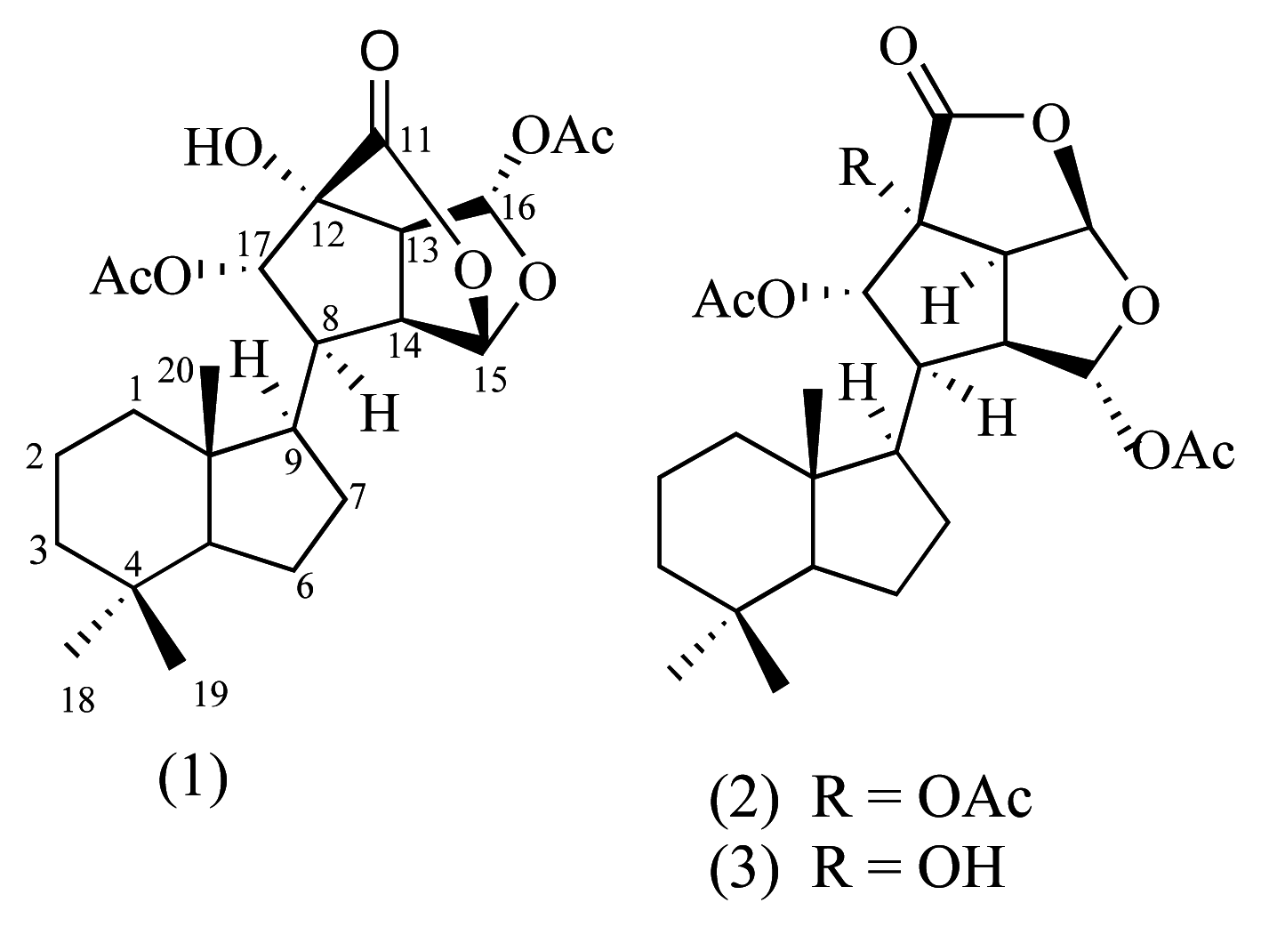
The known chromodorolides A (1) and B (2), and a new derivative chromodorolide C (3) have been isolated from an Australian sponge and characterized by 1D and 2D NMR experiments. The chromodorolides exhibited significant cytotoxicity against the P388 mouse leukaemia cell line, and also showed activity against the free-living larval stages of the parasitic nematodes Haemonchus contortus and Trichostrongylus colubriformis.

Introduction

Highly functionalized terpenoids with intact spongian carbon skeletons, or with rearranged carbon skeletons that can derive from a hypothetical spongian diterpenoid precursor, have been isolated from marine sponges and nudibranchs [14]. Nudibranchs belonging to the genus Chromodoris have proven to be an extremely rich source of diterpenoids related to compounds from Aplysillid sponges [2,49]. The nudibranchs usually form a specific chemical association with the sponge, on which they feed and from which they concentrate the diterpenes. The chromodorane diterpenes, chromodorolides A and B are diterpenoid examples with a highly rearranged carbon skeleton, and have previously been reported from the nudibranch Chromodoris cavae collected in Sri Lanka [8,9]. In this paper we wish to describe the new chromodorane diterpene chromodorolide C isolated together with chromodorolides A and B from an encrusting yellow sponge collected from Mooloolaba, Sunshine Coast, Queensland. We also report bioactivity testing of chromodorolides A – C.

Results and Discussion

The small scale extract of the sponge exhibited potent cytotoxicity (94 ± 3% inhibition at 100 μg/mL) against a P388 mouse leukemia cell line. The frozen sponge (5.87 g) was then extracted by dichloromethane/methanol (1:1) three times, and the resulting extract was further partitioned between EtOAc and water. The ethyl acetate layer was subjected to silica gel flash column chromatography using a step gradient from hexane to 100% EtOAc to yield two major fractions. The first fraction contained chromodorolide B (2) and the second fraction was purified by NPHPLC using hexane and EtOAc (7:2) to give chromodorolide A (1) and the new compound, chromodorolide C (3). The two known compounds 1 and 2 were identified by comparison of their spectroscopic data with those from the literature [8,9].
Marinedrugs 02 00101f1
The new chromodorane, compound 3 was isolated as a colorless oil and had a molecular peak at m/z 473.2148 corresponding to C24H34O8Na indicated by HRESIMS. The 1H NMR spectrum indicated the presence of three singlet methyls (δ 0.82, 0.83 and 0.76), and two additional singlet methyls (δ 2.05 and 2.13) that could be assigned to acetate moieties. Two methine protons (δ 6.51 and 6.00) supported the presence of acetal functionalities. Numerous complex signals in the region δ 0.93–1.48 were consistent with the compound having a terpene skeleton. In the 13C NMR spectrum, signals at δ 174.8, 168.8 and 169.2 were consistent with a lactone carbonyl and two acetate carbonyls respectively. The similarity of the 1H and 13C spectra of 3 to those of 2 identified the new compound as a chromodorane diterpene, possessing a 3,4 ring fused bisacetal-oxalone. HMBC correlations connected the acetal methine proton H-15 at δ 6.51 to C-14 at δ 45.4, and H-14 at δ 2.98 to C-15 at δ 98.1. DQFCOSY correlations then linked H-14 to H-13 at δ 3.52 which was further coupled to the second methine H-16 at δ 6.00. However, comparison of the spectral data showed that 3 only differed from 2 in its acetylation pattern. The HMBC spectrum showed the connectivity from H-15 at δ 6.51 to an acetate carbonyl at δ 169.2 and from H-17 at δ 5.19 to the acetate carbonyl at δ 168.8, and from two methyl acetate signals at δ 2.05 and δ 2.13 to the acetate carbonyls at δ 169.2 and δ 168.8 respectively, confirming the position of the acetate substituents. Additionally, in 3 a signal at δ 78.1, assigned to C-12, was evident and this suggested a quaternary carbon bearing a hydroxy group, whereas in compound 2, the corresponding signal resonated at δ 81.3. These data manifestly identified 3 as a new compound and allowed all resonances to be assigned (Table 1). In particular, the bicyclic terpene framework of 3 was carefully assigned by DQFCOSY and HMBC. The assignments for C-1 and C-3 were made on the basis of correlations to nearby methyl groups as shown in Table 1, while assignments for C-2 and C-6 were confirmed by HMBC correlations to H-3ax and to H-5 respectively. These data suggest that the NMR assignments published by Andersen et al. for chromodorolide B [9] may need revision. Small scale acetylation of 3 with acetic anhydride in pyridine provided an acetate product whose 1H NMR spectrum was identical to that of chromodorolide B 2.
This is the first report of these chromodorane diterpenes from a sponge source. It has been proposed by Andersen et al. [9] that the biosynthesis of the chromodorane skeleton starts from a spongian diterpene followed by opening and contraction of the six membered ring to the five membered ring [8,9]. Oxidative cleavage of a second six-membered ring, followed by lactonization of the C-11 carboxyl group with the hydroxy group at C-15 or C-16 then leads to two different bisacetal oxalone skeletons. The discovery of 3 indicates that acetylation could be a stepwise process in the sponge.
Chromodorolide A has been reported to exhibit cytotoxic and antimicrobial activity [9]. When tested individually against the P388 cell line, chromodorolides A, B and C displayed significant inhibition (66 (± 3), 70 (±2) and 42 (±4) %, respectively) at concentrations of 10 μg/mL, but did not show useful activity at concentrations of 1 μg/mL. Chromodorolide A 1 also showed nematocidal activity against the larval stages of Haemonchus contortus and Trichostrongylus colubriformis, two important pathogens of sheep and other ruminants. A concentration of 100 μg/mL caused 94 (± 3) % and 95 (± 4) % (Mean ± SE, n = 2 separate experiments) inhibition of the development of H. contortus and T. colubriformis larvae, respectively. A concentration of 10 μg/mL did not affect development for H. contortus, however T. colubriformis development was inhibited by 33 (± 1) %.

Conclusion

Chromodorolide diterpenes have been isolated for the first time from an Australian marine sponge and their cytotoxicity and also the anthelmintic activity of one of them against H. contortus and T. colubriformis has been demonstrated.

Experimental

General

1H and 13C NMR spectra were obtained in CDCl3 on a Bruker DRX spectrometer and were recorded at 500 and 125 MHz respectively. Electrospray mass spectra were recorded on a P E SCIEX API III triple quadropole mass spectrometer for solutions in methanol. Flash chromatography was carried out using Kieselgel 60 (230–400 mesh), while high performance liquid chromatography was carried out on a Waters μ-Porasil semi preparative column (300 mm × 7.8 mm) connected to a Waters 515 HPLC pump and a Gilson 132 refractive index detector. All solvents were freshly distilled or were of HPLC grade.

Animal Material

A single specimen of the sponge was collected from under a ledge at 10 m depth. The collection was made at the Fishhole at the Inner Gneerings Reef, Mooloolaba South-East Queensland in December 2002. The encrusting sponge was a bright lemon yellow colour underwater and developed the blue coloration on exposure to air that is typical of Aplysilla sulphurea. A voucher sample and photograph is held at the Department of Chemistry, The University of Queensland. A small sample of animal material has been submitted to The Queensland Museum for taxonomic identification.

Extraction and Purification

The frozen sponge (5.9 g) was cut into small pieces and extracted with 100 mL of DCM/MeOH (1:1) three times. The organic layer was filtered through a plug of cotton wool and the solvent was evaporated in vacuo to give the crude extract (624 mg) that was further partitioned with EtOAc and water (100 mL). The EtOAc layer was concentrated and subjected to silica flash column chromatography using a step gradient from hexane to 100% EtOAc to give two major fractions. The first fraction was identified as chromodorolide B (2) (5.7 mg) [9] and the second fraction was purified by NPHPLC using hexane and EtOAc (7:2) as eluent to furnish chromodorolide A (1) (5.7 mg) [8,9] and the new compound, chromodorolide C (3) (1.7 mg).

Spectral Data

Chromodorolide C (3): Colorless oil; [α]D -78° (c = 0.10, CH2Cl2); 1H-NMR (CDCl3; 500 MHz) 13C-NMR (CDCl3; 125 MHz) see Table 1, HRESIMS found 473.2148, C24H34O8Na (M+Na)+ requires 473.2151(+0.8 ppm).
Acetylation of Chromodorolide C (3): To a solution of chromodorolide C (3, 1.0 mg, 2.2×10⁻³ mmol) in dry pyridine (1.0 mL) was added acetic anhydride (0.1 mL). The mixture was stirred at room temperature overnight. The reaction was quenched by adding 1 mL of water, and extracted twice with 1 mL of EtOAc. The organic layer was blown down over N2 gas. The acetate product (0.9 mg) was obtained as a colorless oil by purifying on a silica-seppak using hexane:EtOAc (7:2) as eluent, and identified as (2) by comparison of 1H-NMR spectra.

Cytotoxicity assay

P388D1 mouse lymphoblast cells were grown at 37°C, 5% CO2 in DMEM media containing 10% horse sera with 2mM L-glutamine and 100 U/mL penicillin and 100 μg/mL streptomycin. Cells growing in log phase were diluted in this media and transferred (99 μL/well) to a 96-well tissue culture plate. Samples were tested in triplicate and included the crude DCM/MeOH extract from the sponge as well as purified samples of individual chromodorolide metabolites. Samples were dissolved in DMSO at an initial concentration of 10 mg/mL and diluted by serial dilution in DMSO. Samples were added to the wells (1 μL) so that final concentrations tested were 100, 10 and 1 μg/mL. Plates were incubated (37°C, 5% CO2) for 24 h. After this time, cytotoxicity was determined using an ATPLite assay which measures the amount of ATP present in the cells. The luminescence produced, proportional to the number of viable cells, was read on a Victor 2 multilabel plate reader (Wallac).

Anthelmintic assay

Nematocidal activity was determined using the method of Lacey et al [10]. Nematode eggs were placed into wells of a mictotitre plate containing chromodorolide A in 2% agar. The eggs hatched overnight, a nutrient medium was added, and the larvae were held at 27°C for 6 days. The number of larvae that developed to the L3 stage was counted and compared to control wells. For each nematode species, the effect of chromodorolide C was examined in two separate experiments at concentrations of 100 and 10 μg/mL (duplicate or triplicate assays wells at each concentration for experiments one and two, respectively) and the percentage development was calculated for each assay relative to the mean development in twelve control assay wells. Data is therefore presented as Mean ± SE, n = 2 separate experiments.
Table 1. 1H (500 MHz) and 13C (125 MHz) data for chromodorolide C (3)
Atom# δ13Caδ1H1H-1H COSYHMBCb
139.0ax 0.93, dt (J = 2.0, 12.0 Hz)H-1eqMe-20
eq 1.53, mH-1ax
219.9ax 1.38, mH-2eq, H-3ax, H-3eqH-3ax
eq 1.55, mH-2ax, H-3ax
340.9ax 1.03, dt (J = 3.0, 12.0 Hz)H-2ax, H-2eq, H-3eqMe-18, Me-19
eq 1.38, mH-2ax, H-3ax
433.0--H-5, Me-18, Me-19
557.01.09, dd (J = 6.5, 13.0 Hz)H-6ax, H-6eqMe-18, Me-19, Me-20
621.10ax 1.30, mH-5, H-6eq, H-7H-5
eq 1.55, mH-5, H-6ax, H-7
725.2ax 1.42, mH-6ax, H-6eq, H-9-
eq 1.55, mH-6ax, H-6eq, H-9
847.52.58, ddd (J = 8.2, 12.0, 12.0 Hz)H-9, H-14, H-17H-15, H-17
950.21.71, dd (J = 9.8, 12.0)H-7ax, H-7eq, H-8H-8, H-17. Me-20
1043.8--H-5, Me-20
11174.8--H-16, H-17
1278.1--H-13, H-14, H-16,
1352.13.52, dd (J = 6.0, 8.2 Hz)H-14, H-16H-15, H-16
1445.42.98, t (J = 8.2 Hz)H-8, H-13H-8, H-15
1598.16.51, s-H-8, H-13, H-14
16103.36.00, d (J = 6.0 Hz)H-13H-15
1775.35.19, d (J = 12.0 Hz)H-8H-8, H-13, H-14
Me-1833.40.82, s-Me-19
Me-1921.00.83, s-H-5, Me-18,
Me-2013.60.76, s-H-5, H-9
OAc169.2--H-15, -OAc
20.82.05-
OAc168.8--H-17, -OAc
20.92.13-
aInverse detection at 500 MHz; solution in CDCl3; 13C = 77.0 ppm and 1H = 7.25 ppm.
bInverse detection at 500 MHz; correlations observed when 1J13C-1H = 135 Hz and long range nJ13C-1H = 8 Hz.

Acknowledgments

We thank Dr David De Vries and Ms Linda Banbury, Centre for Phytochemistry, Southern Cross University for P388 cytotoxicity screening. This research was funded by the Australia Research Council. Wimolpun Rungprom thanks the Thailand Royal Golden Jubilee Fund for the award of a postgraduate scholarship.
  • Samples Availability: Samples and copies of 1H and 13C NMR spectra are available from the corresponding author.

References and Notes

  1. Tischler, M.; Andersen, R. J.; Choudhary, M. I.; Clardy, J. Terpenoids from the sponge Aplysilla glacialis and specimens of the nudibranch Cadlina luteomarginata found on the sponge. J. J. Org. Chem 1991, 56, 42–47. [Google Scholar]
  2. Hochlowski, J. E.; Faulkner, D. J.; Matsumoto, G. K.; Clardy, J. Norrisolide, a novel diterpene from the dorid nudibranch Chromodoris norrisi. J. J. Org. Chem 1983, 48, 1141–1142. [Google Scholar]
  3. Molinski, T. F.; Faulkner, D.J. A new diterpene lactone from an Australian Aplysilla species. J. Org. Chem 1986, 51, 1144–1146. [Google Scholar]
  4. Karuso, P.; Taylor, W. C.; White, A. H. The constituents of marine sponges. 11. Isolation of 8 diterpenes from Aplysilla rosea. Aust. J. Chem 1986, 39, 1629–1641. [Google Scholar]
  5. Hochlowski, J. E.; Faulkner, D. J. Chemical constituents of the nudibranch Chromodoris marislae. Tetrahedron Lett 1981, 22, 271–274. [Google Scholar]
  6. Bobzin, S. C.; Faulkner, D. J. Diterpenes from the marine sponge Aplysilla polyrhapsis and the dorid nudibranch Chromodoris norrisi. J. Org. Chem 1989, 54, 3902–3907. [Google Scholar]
  7. Cimino, G.; Fontana, A.; Gavagnin, M. Marine opistobranch mollusks: chemistry and ecology in sacoglossans and dorids. Curr. Org. Chem 1999, 3, 327–372. [Google Scholar]
  8. Dumdei, E. J.; de Silva, E. D.; Andersen, R. J. Chromodorolide A, a rearranged diterpene with a new carbon skeleton from the Indian ocean nudibranch Chromodoris cavae. J. Am. Chem Soc 1989, 111, 2712–2713. [Google Scholar]
  9. Morris, S. A.; Dumdei, E. J.; de Silva, E. D.; Andersen, R. J. Chromodorane diterpenes from the tropical dorid nudibranch Chromodoris cavae. Can. J. Chem 1991, 69, 768–771. [Google Scholar]
  10. Lacey, E; Redwin, J. M.; Gill, J.H.; Demargheriti, V.M; Waller, P.J. Resistance of Parasites to Antiparasitic Drugs; Boray, J.C., Martin, P.J., Roush, R.P., Eds.; MSD AGVET: Rahway, NJ, 1990; pp. 177–184. [Google Scholar]