Article

The Protect Effects of Chitosan Oligosaccharides on Intestinal Integrity by Regulating Oxidative Status and Inflammation under Oxidative Stress

College of Coastal Agriculture Sciences, Guangdong Ocean University, Zhanjiang 524088, China
*
Author to whom correspondence should be addressed.
Mar. Drugs 2021, 19(2), 57; https://doi.org/10.3390/md19020057
Submission received: 6 January 2021 / Revised: 20 January 2021 / Accepted: 21 January 2021 / Published: 25 January 2021
(This article belongs to the Special Issue Marine Carbohydrate-Based Compounds with Medicinal Properties)

Abstract

:
The aim of this study was to evaluate the effects of the dietary supplementation of chitosan oligosaccharides (COS) on intestinal integrity, oxidative status, and the inflammation response with hydrogen peroxide (H2O2) challenge. In total, 30 rats were randomly assigned to three groups with 10 replications: CON group, basal diet; AS group, basal diet + 0.1% H2O2 in drinking water; ASC group, basal diet + 200 mg/kg COS + 0.1% H2O2 in drinking water. The results indicated that COS upregulated (p < 0.05) villus height (VH) of the small intestine, duodenum, and ileum; mucosal glutathione peroxidase activity; jejunum and ileum mucosal total antioxidant capacity; duodenum and ileum mucosal interleukin (IL)-6 level; jejunum mucosal tumor necrosis factor (TNF)-α level; duodenum and ileum mucosal IL-10 level; the mRNA expression level of zonula occludens (ZO)-1 in the jejunum and ileum, claudin in the duodenum, nuclear factor-erythroid 2-like 2 in the jejunum, and heme oxygenase-1 in the duodenum and ileum; and the protein expression of ZO-1 and claudin in jejunum; however, it downregulated (p < 0.05) serum diamine oxidase activity and D-lactate level; small intestine mucosal malondialdehyde content; duodenum and ileum mucosal IL-6 level; jejunum mucosal TNF-α level; and the mRNA expression of IL-6 in the duodenum and jejunum, and TNF-α in the jejunum and ileum. These results suggested COS could maintain intestinal integrity under oxidative stress by modulating the intestinal oxidative status and release of inflammatory cytokines.

1. Introduction

The pernicious effects of oxidative stress on intestinal function have been widely studied [1,2]. Studies have demonstrated oxidative stress was one of the vital factors contributing to intestinal injury and disfunction [3,4]. Oxidative stress was frequently related to increases in interleukin (IL)-1β, IL-6, and tumor necrosis factor (TNF)-α [3,5], and undermined intestinal function [6]. Both oxidative stress and inflammation could disturb intestinal function by over-production of reactive oxygen species (ROS) and pro-inflammatory cytokines [2,7]. Impaired intestinal integrity, accompanied with intestinal permeability and histological changes [8,9,10], promote the transportation of toxic luminal substances, which may contribute to intestinal disease and even death [10,11,12]. The intestine predominantly responds to virous stressors, especially oxidative stress and inflammation [3,13,14,15]. Hence, alleviating intestinal oxidative stress and inflammation are important in maintaining intestinal function. Therefore, an ideal candidate, which has free-radical scavenging activity, and antioxidant and anti-inflammation capacities, is urgently needed to maintain intestinal function under oxidative stress.
Chitosan oligosaccharides (COS) are the degraded products of chitosan; compared to chitosan, COS are non-toxic, non-allergenic, less viscous, and entirely soluble in water [16]. COS also have multiple properties, including free-radical scavenging, and antioxidant, anti-inflammatory, antibacterial, and immune-enhancing activities, which capture much attention, and they are widely used in biomedical medicine and agriculture science [17]. Qiao et al. [18] reported COS could relieve sepsis by virtue of their antioxidation property and anti-inflammatory effect. Liu et al. [19] demonstrated COS could remit oxidative damage in umbilical vein endothelial cells. Lan et al. [20] demonstrated COS remitted H2O2-induced oxidative stress in the liver, kidney and spleen by alleviating oxidative and inflammation stress. Furthermore, COS also have positive effects on intestinal health [21,22]. It is believed that intestinal oxidative stress and the inflammation response are highly related to intestinal integrity and function. COS captured attention due to antioxidant and anti-inflammatory activities. Therefore, this study was done to evaluate the effects of dietary COS supplementation on intestinal integrity, oxidative status, and the inflammatory response with H2O2 challenge.

2. Results

2.1. Intestinal Mucosal Morphology

Figure 1 and Table 1 show intestinal morphology indices. Compared with the CON group, the villus height (VH) and VH to crypt depth (CD) ratio of the small intestine were decreased (p < 0.05) in the AS group (basal diet + 0.1% H2O2 in drinking water). Compared with the AS group, COS increased the VH of the small intestine, and the VH:CD of the duodenum and jejunum.

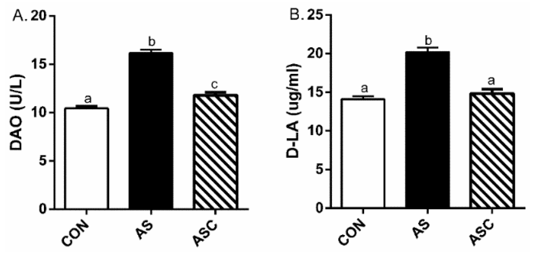
2.2. Intestinal Permeability

Compared with the CON group, serum diamine oxidase (DAO) activity and d-lactate acid (D-LA) content were upregulated (p < 0.05) in the AS group (Figure 2). Compared with the AS group, COS downregulated (p < 0.05) serum DAO activity and D-LA content.

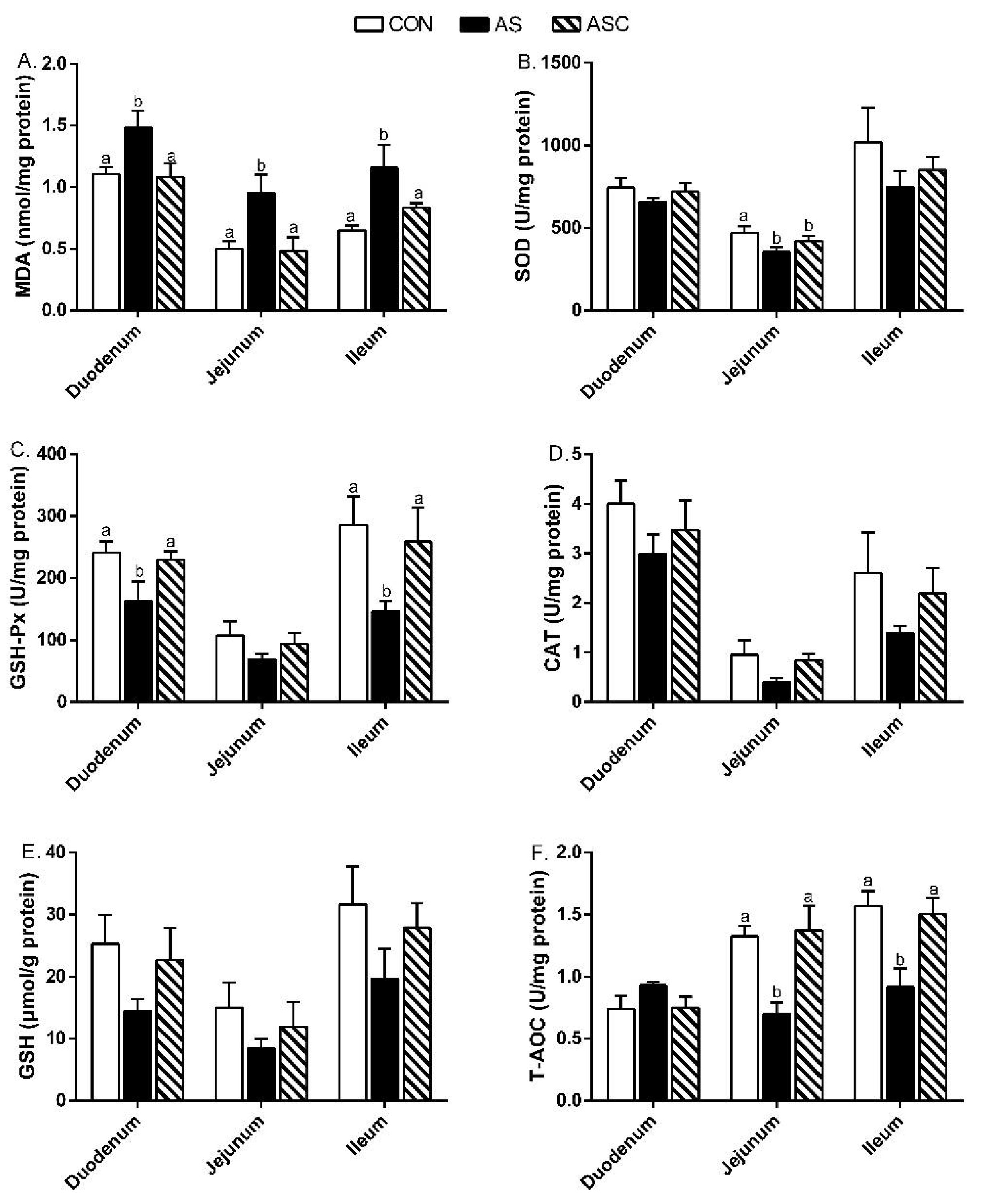
2.3. Antioxidant Capacity

Figure 3 presents the antioxidant indicators in the small intestinal mucosa. In duodenum, the malondialdehyde (MDA) content in the AS group was higher (p < 0.05) than the contents in the CON and ASC groups, but the glutathione peroxidase (GSH-Px) activity was lower (p < 0.05). In the jejunum, the MDA content in the AS group was higher (p < 0.05) than in the CON and ASC groups; the superoxide dismutase (SOD) activity in the CON group was higher (p < 0.05) than in the AS and ASC groups; the total antioxidant capacity (T-AOC) activity in the AS group was lower (p < 0.05) than in the CON and ASC groups. In the ileum, the MDA contents in the CON and ASC groups were lower (p < 0.05) than that in the AS group, but the GSH-Px and T-AOC activities were higher.

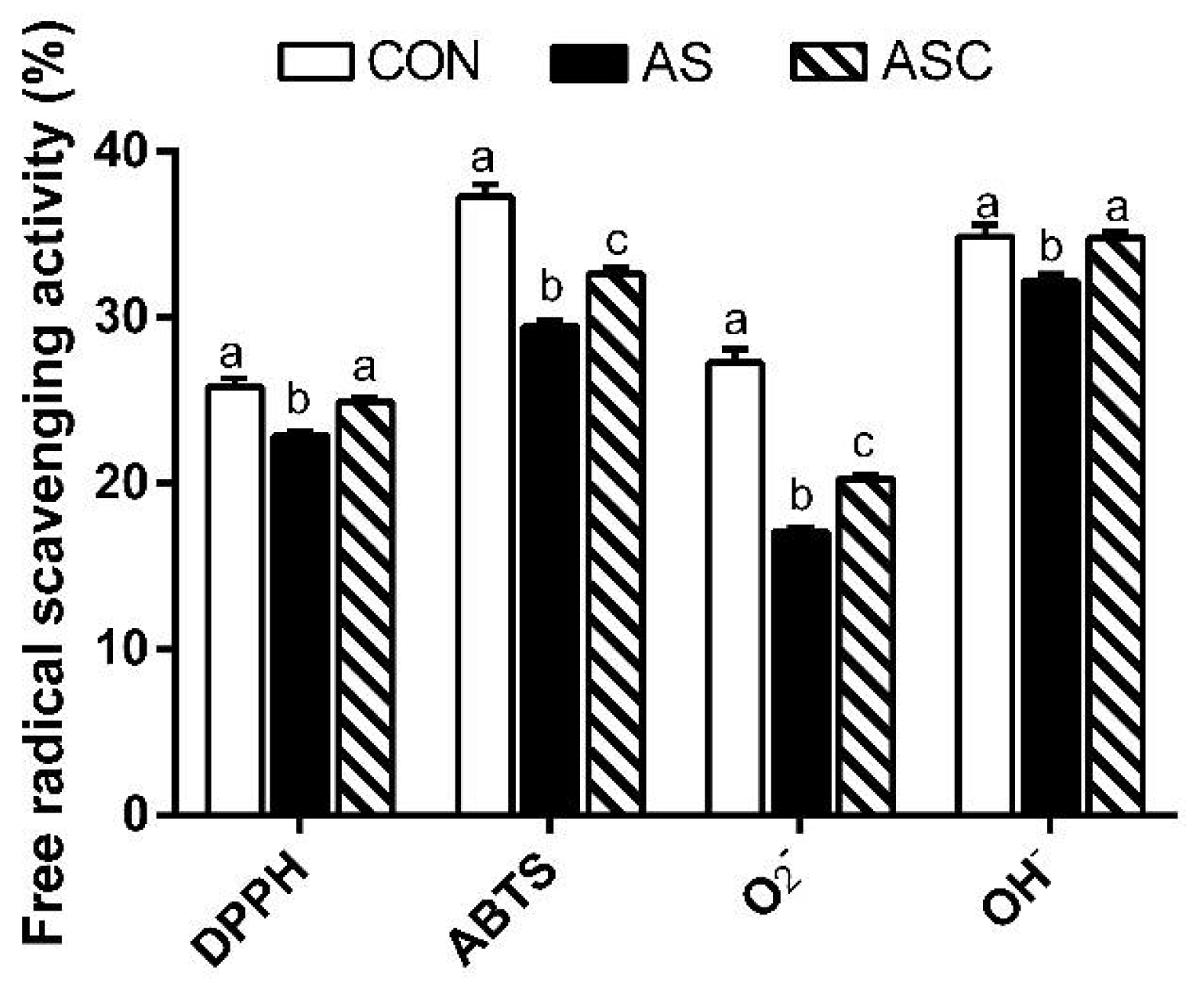
2.4. Free Radical Scavenging Activity

Figure 4 presents the free-radical scavenging capacities in jejunum mucosa. The 1,1-diphenyl-2-picrylhydrazyl (DPPH), 2,2′-azino-bis-3-ethylbenzothiazoline-6-sulfonic acid (ABTS), O2 (superoxide radical) and hydroxyl radical (OH) scavenging activities in the CON and ASC groups were higher (p < 0.05) than in the AS group.

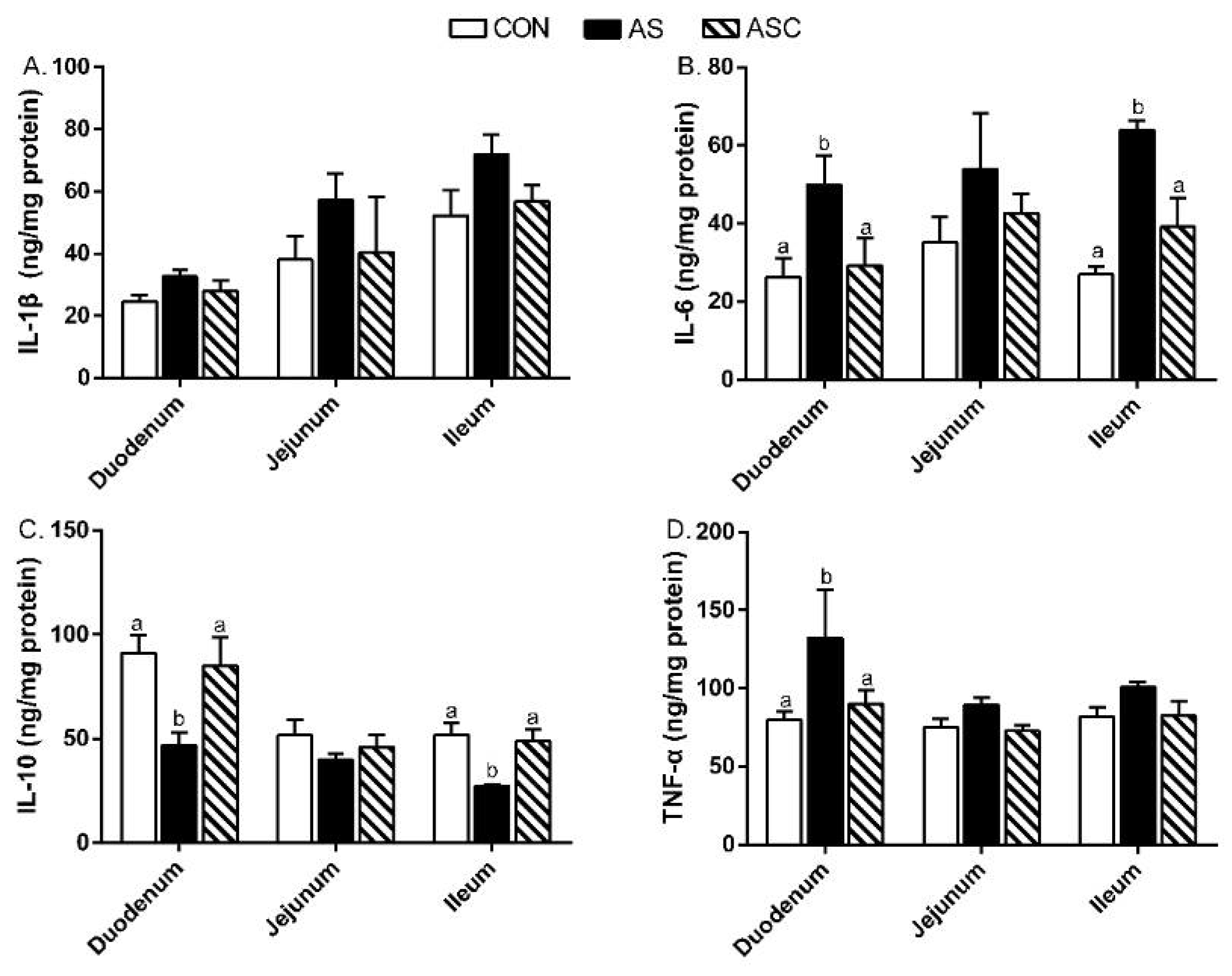
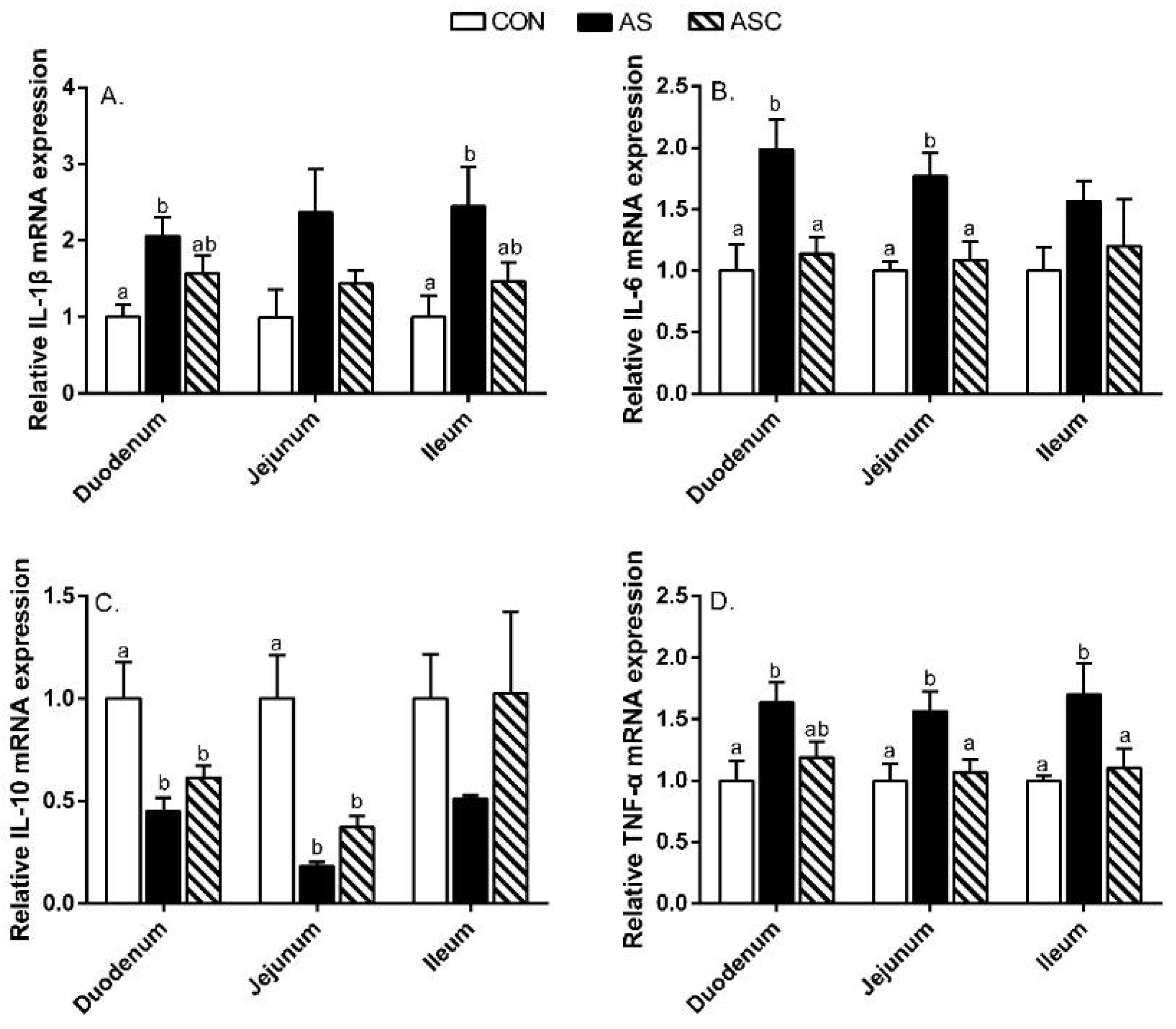
2.5. Intestinal Mucosal Immunity

In the duodenum, the IL-6 and TNF-α contents in the AS group were enhanced (p < 0.05) compared to CON and ASC groups (Figure 5), but the IL-10 content was decreased (p < 0.05). In the ileum, the IL-6 content in the AS group was increased (p < 0.05) over CON and ASC groups, but the IL-10 content was decreased (p < 0.05).
Furthermore, we also examined the related cytokines’ gene expression in the small intestine (Figure 6). In the duodenum, the relative gene expression levels of IL-1β and TNF-α in the AS group were higher (p < 0.05) than in the CON group; IL-6 in the AS group was enhanced (p < 0.05) over CON and ASC groups; IL-10 in the AS and ASC groups was less present (p < 0.05) compared to the CON group. In the jejunum, the relative gene expression levels of IL-6 and TNF-α in the AS group were increased (p < 0.05) compared to CON and ASC groups; IL-10 in the AS and ASC groups was decreased (p < 0.05) compared to the CON group. In the ileum, the relative gene expression of IL-1β in the AS group was enhanced (p < 0.05) compared to the CON group; TNF-α in the AS group was higher (p < 0.05) than in CON and ASC groups.

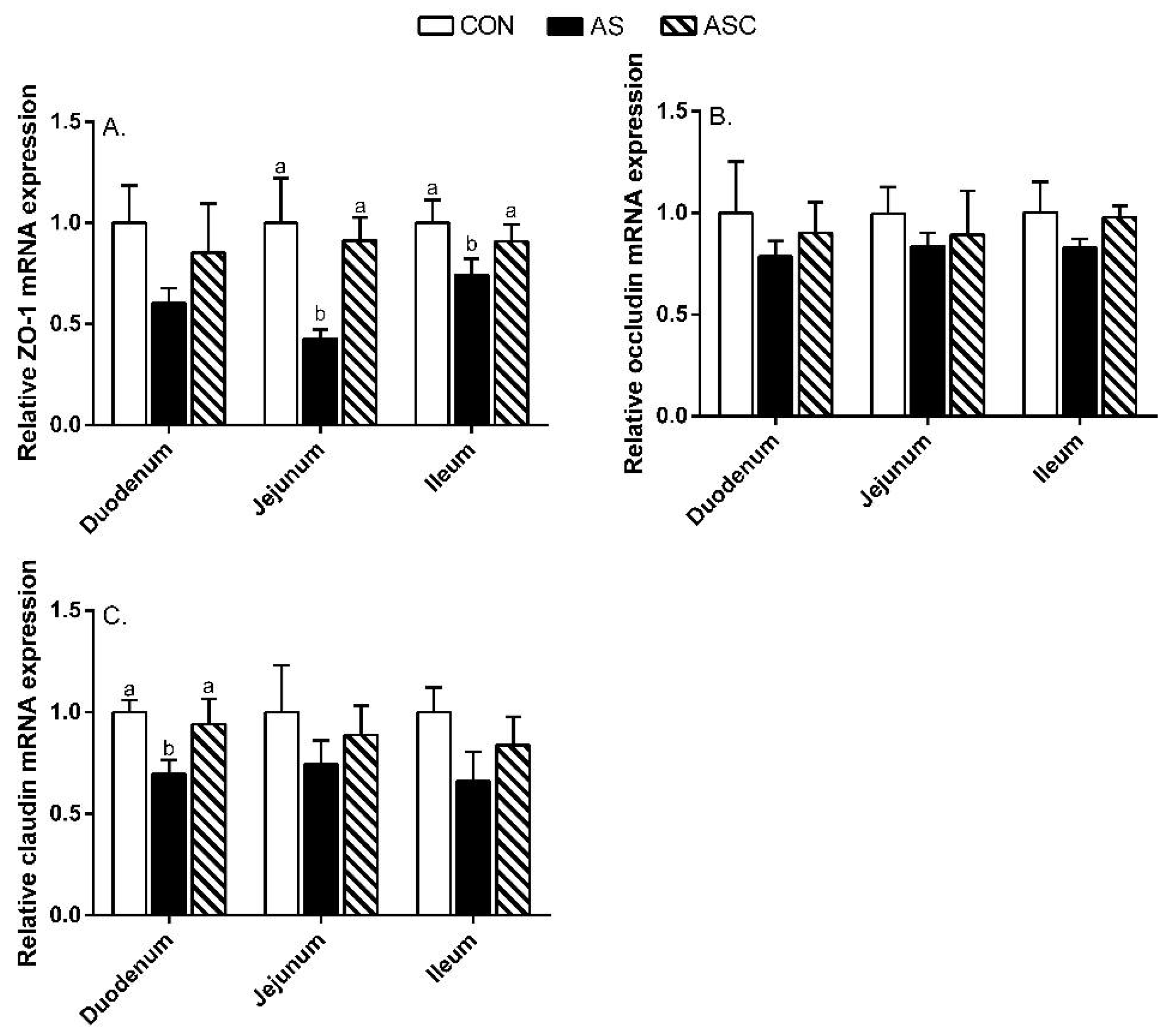
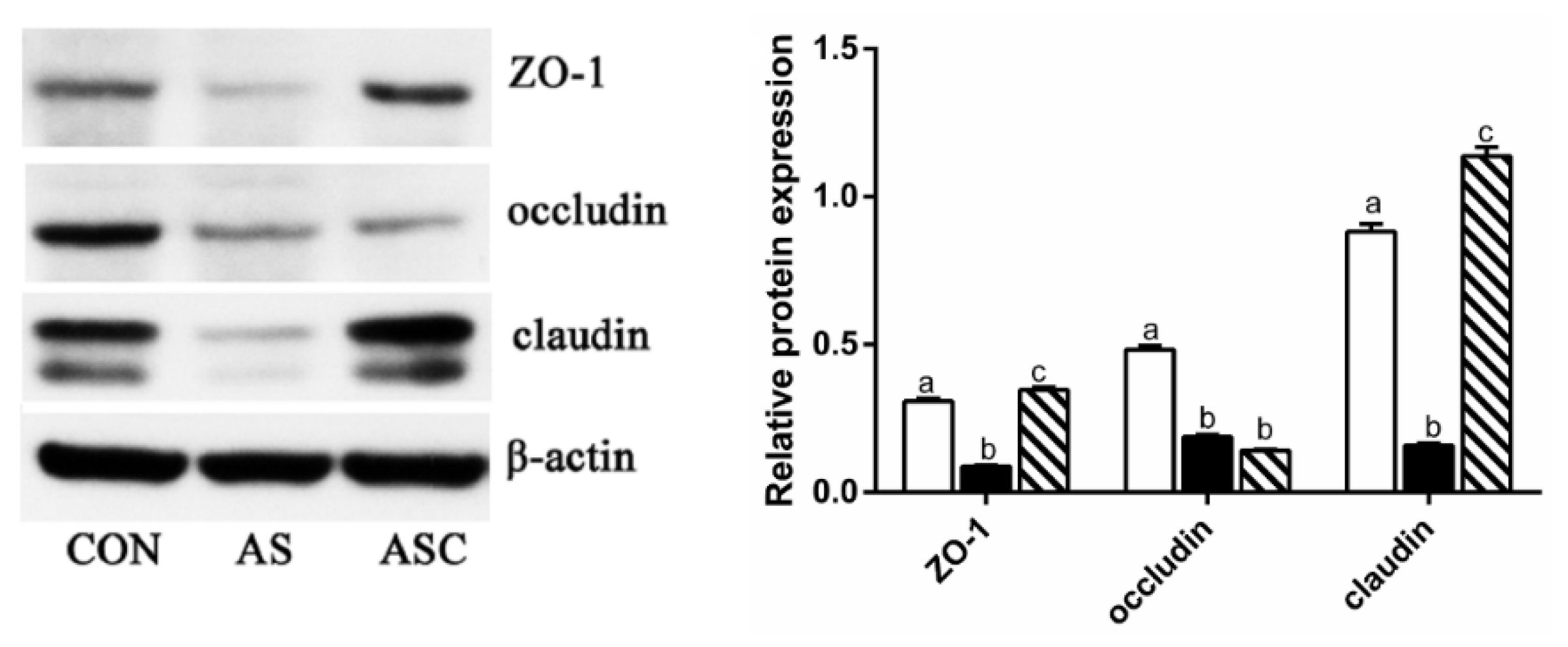
2.6. Intestinal Barrier Function-Related Gene Expression

In the duodenum, the relative gene expression of claudin in the AS group was decreased (p < 0.05) compared to CON and ASC groups (Figure 7). In the jejunum and ileum, the relative gene expression of zonula occludens (ZO)-1 in the AS group was decreased (p < 0.05) compared to CON and ASC groups. In addition, we found that the expression levels of ZO-1 and claudin protein in the AS group were decreased (p < 0.05) compared to CON and ASC groups (Figure 8); and the expression of occludin protein in the AS and ASC groups was decreased (p < 0.05) compared to the CON group in jejunum mucosa.
We also examined the critical gene expression in the antioxidant signaling pathway. As shown in Figure 9, in the duodenum, the relative gene expression of heme oxygenase (HO)-1 in the AS group was decreased (p < 0.05) compared to CON and ASC groups. In the jejunum, the relative gene expression of nuclear factor-erythroid 2-like 2 (Nrf2) in the AS group was lower (p < 0.05) than that in the CON group. In the ileum, the relative gene expression of Nrf2 in the AS group was decreased (p < 0.05) compared to the CON group; and the relative gene expression of HO-1 in the AS group was decreased (p < 0.05) compared to CON and ASC groups.

3. Discussion

Intestinal barrier integrity was commonly assessed by gut morphology, serum DAO activity, and D-LA activity. The gut morphology was a useful biomarker of the stress response of the intestinal tract [9,23,24]. The DAO activity and D-LA content were biomarkers of intestinal permeability [3,11,25]. In this study, oxidative stress decreased the VH of the small intestine, and enhanced DAO content and D-LA activity, demonstrating that oxidative stress resulted in intestinal injury by increasing intestinal permeability and decreasing VH [3,9]. As expected, COS decreased DAO content and D-LA activity, and increased the VH of the small intestine; hence, COS had positive effects on intestinal permeability. These results are also in line with former studies by Li et al. [21] and Zhao et al. [26], who indicated COS decreased serum DAO activity. The structure of the intestinal morphology reflected gut health status. Li et al. [21] illustrated COS increased the VH of the duodenum and ileum in broilers. Liu et al. [27] and Liu et al. [28] reported dietary COS supplementation increased the VH of the jejunum and ileum in weaning pigs. The positive effects of COS on intestinal permeability and morphology could explain the improving intestinal function with COS supplementation.
The tight junction proteins maintained and regulated the intestinal barrier function. The tight junction proteins mainly consisted of the transmembrane proteins claudin and occludin, and peripheral membrane protein ZO-1. Therefore, the decreased mRNA expression of ZO-1, claudin, and occludin reflected intestinal barrier dysfunction [4]. In this study, oxidative stress downregulated the protein expression of ZO-1, occludin, and claudin in the jejunum; and the mRNA expression of claudin in the duodenum and ZO-1 in the jejunum and ileum, which were all consistent with the results reported by Song et al. [29] and Cao et al. [1]. COS upregulated the protein expression of ZO-1 and claudin in the jejunum, and the mRNA expression of ZO-1 in the jejunum and ileum, and claudin in the duodenum—similarly to other studies on mice fed high-fat diets [30], dexamethasone-challenged broilers [31], and weaning pigs [32,33]. These results demonstrated that COS could alleviate oxidative-induced intestinal barrier function partly by maintaining the intestinal structure, intestinal permeability, and tight junction functionality.
Accumulating evidence indicates that oxidative status was an important factor in intestinal barrier function. An imbalance between oxidation and the antioxidant defense system leads to oxidative stress and inflammation, and finally, induces intestinal barrier dysfunction [12]. SOD, GSH-Px, and CAT were regarded as the main antioxidant enzymes for scavenging free radicals. GSH was regarded as the most important non-enzymatic antioxidant which scavenges single oxygen molecules and hydroxyl radicals. The T-AOC can reflect the total antioxidant capacity. MDA is an indicator of oxidative stress [34]. H2O2 can stimulate ROS over-production, disrupt the activity of antioxidant enzymes, and induce lipid peroxidation [35,36]. Consistently, in this study, oxidative stress induced higher small intestine MDA content, along with lower duodenum and ileum mucosal GSH-Px activity, jejunum mucosal SOD activity, and jejunum and ileum mucosal T-AOC activity. COS reduced duodenum, jejunum, and ileum mucosal MDA content; and increased duodenum and ileum mucosal GSH-Px activity, and jejunum and ileum mucosal T-AOC activity, suggesting COS could alleviate intestinal mucosal oxidative stress by improving the antioxidative enzyme activity and decreasing MDA content. These results are consistent with Lan et al. [20], who indicated COS could increase SOD, CAT, GSH-Px, and T-AOC activity, and decrease the MDA level with an H2O2 challenge. Li et al. [21] also indicated COS increased SOD activity in the duodenum’s mucosa and decrease the MDA level in the jejunum and ileum’s mucosa in broilers. Similarly, Li et al. [22] indicated that dietary COS supplementation increased the inhibition of hydroxy radical capacity, and GSH, T-AOC, GSH-Px, and SOD activity, whereas decreased MDA content in the ileum mucosa of broilers. Nrf2 is a nuclear transcription factor and plays a vital role in antagonizing oxidative stress [37]. Our results show decreased mRNA expression of Nrf2 in the jejunum and ileum, and HO-1 in the duodenum and ileum by H2O2 challenge. Other studies illustrated the increased Nrf2 mRNA expression level could increase the mRNA expression of SOD and GSH-Px [3]; the decreased Nrf2 and HO-1 mRNA expression levels may be related to a response to oxidative stress. As expected, COS enhanced the mRNA expression of Nrf2 in the jejunum and HO-1 in the duodenum and ileum—similarly to other studies on doxorubicin-challenged rats [38] and mice fed a high-fat diet [39]. Collectively, the combined results illustrate that the increased antioxidant enzyme activity may be mediated by Nrf2/HO-1 signaling pathway. Additionally, the efficiency of the free-radical scavenging capacities reflected the neutralization of free radical capacities, or hydrogen donor capacity [40]. In this study, COS improved the radical scavenging capacity of the jejunum mucosa; that may relate to antioxidant capacity and hydrogen donation ability [17].
Inflammation cytokines play vital roles in the inflammatory and immune responses. The accumulating literature illustrates inflammation is an important marker in intestinal disfunction [3,12]. Chen et al. [12] indicated that over-production of cytokines could change the intestinal permeability and tight junction structure by modulating tight junction-related genes expression in weaning piglets. The over-production of IL-1β, IL-6, and TNF-α directly resulted in intestinal mucosal injury [41,42]. Therefore, suppressing the over-production of intestinal mucosal IL-1β, IL-6, and TNF-α was a useful way to maintain the intestinal function. Previous studies indicated that stressors could disturb the balance between anti- and pro-inflammatory responses by increasing pro-inflammatory cytokines’ production [12,43,44]. In this study, the duodenum and ileum mucosal IL-6 content, and jejunum mucosal TNF-α level were higher, whereas the duodenum and ileum mucosal IL-10 levels were decreased, in the AS group compared to the CON group, indicating that oxidative stress resulted in inflammation in the intestine. Furthermore, the levels of mRNA expression of IL-1β in the duodenum and ileum, IL-6 in the duodenum and jejunum, and TNF-α in the small intestine in the AS group were increased compared to the CON group, but the expression levels of IL-10 in the duodenum and jejunum were decreased. Dietary COS supplementation decreased the duodenum and ileum mucosal IL-6 level and jejunum mucosal TNF-α level; inhibited the expression of IL-6 in the jejunum and ileum, and TNF-α in the jejunum and ileum; and increased the duodenum and ileum mucosal IL-10 levels, all of which was in consistent with the results of Hu et al. [33], who reported COS reduced IL-1β and TNF-α mRNA expression levels in jejunum mucosa in weaning pigs. Besides, COS decreased the IL-6 and TNF-α mRNA expression levels in the liver of mice fed a high-fat diet [39]. These results suggest that COS may alleviate intestinal inflammation by suppressing the levels of IL-1β, IL-6, and TNF-α [45,46]. However, in this study, we ignored the immune cells in the mucosal immune system, especially the mast cells, which play a vital role in the regulation of intestinal mucosal immune function and intestinal barrier function. Further study would focus on this point.
In conclusion, COS had beneficial effects on intestinal integrity by improving the antioxidant capacity and suppressing the release of inflammatory cytokines. Dietary COS supplementation may be an effective nutritional strategy to alleviate the detrimental effects of oxidative stress.

4. Materials and Methods

4.1. Animals, Diets, and Experimental Design

In total, 30 male Sprague–Dawley rats (8–10 weeks old, 178.39 ± 5.12 g) were purchased from Beijing Administration Office of Laboratory Animals and acclimatized for 7 days before the experiment. The rats were provided with a pelleted diet, had free access to diet and water, and were housed at constant temperature (24 ± 2 °C) and relative humidity (60% ± 5%) on a 12-h light–dark cycle. The basal diet composition is shown in Table 2. The experimental protocol and use of rats were approved by the Animal Care and Use Committee of Guangdong Ocean University, Zhanjiang, China (SYXK-2018-0147, 2018).
H2O2 induced oxidative stress by generation of potent ROS [23,24,25]. ROS caused lipid peroxidation, membrane disintegration, and endothelial cell damage [26]. The 30 rats were randomly divided into three groups: CON, basal diet; AS group, basal diet + 0.1% H2O2 in drinking water; ASC, basal diet + 200 mg/kg COS + 0.1% H2O2 in drinking water.

4.2. Sample Collection

At the end of the experiment, all rats were anesthetized and sacrificed to collected blood samples through the eyeballs. The blood samples were centrifuged at 4 °C, 3200× g, for 10 min to obtain serum for further analysis. The duodenum, jejunum, and ileum were divided into two parts, one part fixed in 10% buffered formalin for morphology analysis, the rest for collecting mucosa, and frozen in liquid nitrogen for further analysis.

4.3. Serum Diamine Oxidase (DAO) and d-Lactate Acid (D-LA)

Serum DAO activity and d-LA level were measured with commercial kits (Nanjing Jiancheng Institute of Bioengineering, Nanjing, China).

4.4. Intestinal Morphology

The intestinal morphology analysis was performed according our previously described methods [2].

4.5. Intestinal Antioxidant Parameters and Inflammatory Cytokines

About 1 g of each mucosal sample was homogenized at a ratio of 1:9 (weight/volume) with ice-cold PBS. Homogenate was centrifuged at 3200× g for 10 min at 4 °C to obtain supernatant; Bradford method was used to determine the supernatant protein concentration. The antioxidant parameters and inflammatory cytokines were measured with corresponding assay kits (Nanjing Jiancheng Bioengineering Institute, Nanjing, China).

4.6. Free Radical Scavenging Activities

DPPH, ABTS, O2, and OH scavenging activities were evaluated by following the methods described in another study [20].

4.7. Gene Expression Analysis

Total RNA extraction, cDNA reverse transcription, and real-time polymerase chain reaction analysis were done according to our previous described methods [47]. The primers are shown in Table 3. The relative mRNA expression was calculated by 2−∆∆Ct method.

4.8. Western Blot Analysis

The procedures of the Western blot assay were according to the description of Alhaithloul et al. [37].

4.9. Statistical Analysis

The individual rat was regarded as the experiment unit, SAS 2003 (version 9.1, SAS Institute Inc., Cary, NC, USA) was used to analyze the statistic. Duncan’s multiple range test was used to check the variance among the groups, and the differences were considered significant at p < 0.05.

Author Contributions

R.L. and Z.Z. designed the experiment. R.L. did the manuscript writing. Q.C. and L.W. were involved in experimental analysis and data collection. All authors have read and agreed to the published version of the manuscript.

Funding

This research was supported by the scientific research start-up funds of Guangdong Ocean University (R18005).

Institutional Review Board Statement

The study was approved by the Animal Care and Use Committee of Guangdong Ocean University (SYXK-2018-0147, 2018).

Data Availability Statement

The data presented in this study are available on request from the corresponding author.

Acknowledgments

Special thanks to Yiqi Lu, who provided assistance in rat management.

Conflicts of Interest

The authors declare no conflict of interest.

References

  1. Cao, S.; Wang, C.; Yan, J.; Li, X.; Hu, C. Curcumin ameliorates oxidative stress-induced intestinal barrier injury and mitochondrial damage by promoting Parkin dependent mitophagy through AMPK-TFEB signal pathway. Free Radic. Bio. Med. 2020, 147, 8–22. [Google Scholar] [CrossRef] [PubMed]
  2. Lan, R.; Li, Y.; Chang, Q.; Zhao, Z. Dietary chitosan oligosaccharides alleviate heat stress–induced intestinal oxidative stress and inflammatory response in yellow-feather broilers. Poult. Sci. 2020, 99, 6745–6752. [Google Scholar] [CrossRef] [PubMed]
  3. Cheng, K.; Song, Z.; Li, S.; Yan, E.; Zhang, H.; Zhang, L.; Wang, C.; Wang, T. Effects of resveratrol on intestinal oxidative status and inflammation in heat-stressed rats. J. Therm. Biol. 2019, 85, 102415. [Google Scholar] [CrossRef] [PubMed]
  4. Cheng, Y.; Chen, Y.; Chen, R.; Su, Y.; Zhang, R.; He, Q.; Wang, K.; Wen, C.; Zhou, Y. Dietary mannan oligosaccharide ameliorates cyclic heat stress-induced damages on intestinal oxidative status and barrier integrity of broilers. Poult. Sci. 2019, 98, 4767–4776. [Google Scholar] [CrossRef] [PubMed]
  5. Lan, R.; Li, S.; Chang, Q.; Zhao, Z. Chitosan Oligosaccharides Protect Sprague Dawley Rats from Cyclic Heat Stress by Attenuation of Oxidative and Inflammation Stress. Animals 2019, 9, 1074. [Google Scholar] [CrossRef] [Green Version]
  6. Wang, S.; Li, X.; Wang, W.; Zhang, H.; Xu, S. Application of transcriptome analysis: Oxidative stress, inflammation and microtubule activity disorder caused by ammonia exposure may be the primary factors of intestinal microvilli deficiency in chicken. Sci. Total. Environ. 2019, 696, 134035. [Google Scholar] [CrossRef]
  7. Liu, Y.; Chen, F.; Odle, J.; Lin, X.; Jacobi, S.K.; Zhu, H.; Wu, Z.; Hou, Y. Fish oil enhances intestinal integrity and inhibits TLR4 and NOD2 signaling pathways in weaned pigs after LPS challenge. J. Nutr. 2012, 142, 2017–2024. [Google Scholar] [CrossRef] [Green Version]
  8. Wu, S.; Pan, L.; Liao, H.; Yao, W.; Shen, N.; Chen, C.; Liu, D.; Ge, M. High-fat diet increased NADPH-oxidase-related oxidative stress and aggravated LPS-induced intestine injury. Life Sci. 2020, 253, 117539. [Google Scholar] [CrossRef]
  9. Liu, G.; Zhu, H.; Ma, T.; Yan, Z.; Zhang, Y.; Geng, Y.; Zhu, Y.; Shi, Y. Effect of chronic cyclic heat stress on the intestinal morphology, oxidative status and cecal bacterial communities in broilers. J. Therm. Biol. 2020, 91, 102619. [Google Scholar] [CrossRef]
  10. Wu, Q.; Liu, N.; Wu, X.; Wang, G.; Lin, L. Glutamine alleviates heat stress-induced impairment of intestinal morphology, intestinal inflammatory response, and barrier integrity in broilers. Poult. Sci. 2018, 97, 2675–2683. [Google Scholar] [CrossRef]
  11. Song, Z.; Cheng, K.; Zhang, L.; Wang, T. Dietary supplementation of enzymatically treated Artemisia annua could alleviate the intestinal inflammatory response in heat-stressed broilers. J. Therm. Biol. 2017, 69, 184–190. [Google Scholar] [CrossRef] [PubMed]
  12. Chen, J.; Yu, B.; Chen, D.; Huang, Z.; Mao, X.; Zheng, P.; Yu, J.; Luo, J.; He, J. Chlorogenic acid improves intestinal barrier functions by suppressing mucosa inflammation and improving antioxidant capacity in weaned pigs. J. Nutr. Biochem. 2018, 59, 84–92. [Google Scholar] [CrossRef] [PubMed]
  13. Yun, S.H.; Moon, Y.S.; SoHn, S.H.; Jang, I.S. Effects of cyclic heat stress or vitamin C supplementation during cyclic heat stress on HSP70, inflammatory cytokines, and the antioxidant defense system in Sprague Dawley rats. Exp. Anim. 2012, 61, 543–553. [Google Scholar] [CrossRef] [PubMed] [Green Version]
  14. Lee, S.I.; Kang, K.S. Function of capric acid in cyclophosphamide-induced intestinal inflammation, oxidative stress, and barrier function in pigs. Sci. Rep. 2017, 7, 1–12. [Google Scholar] [CrossRef] [PubMed]
  15. Qiao, R.; Sheng, C.; Lu, Y.; Zhang, Y.; Ren, H.; Lemos, B. Microplastics induce intestinal inflammation, oxidative stress, and disorders of metabolome and microbiome in zebrafish. Sci. Total Environ. 2019, 662, 246–253. [Google Scholar] [CrossRef] [PubMed]
  16. Zou, P.; Yang, X.; Wang, J.; Li, Y.; Yu, H.; Zhang, Y.; Liu, G. Advances in characterisation and biological activities of chitosan and chitosan oligosaccharides. Food Chem. 2016, 190, 1174–1181. [Google Scholar] [CrossRef] [PubMed]
  17. Naveed, M.; Phil, L.; Sohail, M.; Hasnat, M.; Baig, M.M.F.A.; Ihsan, A.U.; Shumzaid, M.; Kakar, M.U.; Husain, T.; Akabar, M. Chitosan oligosaccharide (COS): An overview. Int. J. Biol. Macromol. 2019, 129, 827–843. [Google Scholar] [CrossRef]
  18. Qiao, Y.; Bai, X.; Du, Y. Chitosan oligosaccharides protect mice from LPS challenge by attenuation of inflammation and oxidative stress. Int. Immunopharmacol. 2011, 11, 121–127. [Google Scholar] [CrossRef]
  19. Liu, H.; Li, W.; Xu, G.; Li, X.; Bai, X.; Wei, P.; Yu, C.; Du, Y. Chitosan oligosaccharides attenuate hydrogen peroxide-induced stress injury in human umbilical vein endothelial cells. Pharmacol. Res. 2009, 59, 167–175. [Google Scholar] [CrossRef]
  20. Lan, R.; Chang, Q.; An, L.; Zhao, Z. Dietary supplementation with chitosan oligosaccharides alleviates oxidative stress in rats challenged with hydrogen peroxide. Animals 2020, 10, 55. [Google Scholar] [CrossRef]
  21. Li, J.; Cheng, Y.; Chen, Y.; Qu, H.; Zhao, Y.; Wen, C.; Zhou, Y. Dietary chitooligosaccharide inclusion as an alternative to antibiotics improves intestinal morphology, barrier function, antioxidant capacity, and immunity of broilers at early age. Animals 2019, 9, 493. [Google Scholar] [CrossRef] [PubMed] [Green Version]
  22. Li, X.; Ding, X.; Peng, X.; Chi, X.; Cui, H.; Zuo, Z.; Fang, J. Effect of chitosan oligosaccharides on antioxidant function, lymphocyte cycle and apoptosis in ileum mucosa of broiler. Kafkas Univ. Vet. Fak. Derg. 2017, 23, 571–577. [Google Scholar]
  23. Zou, Y.; Wei, H.; Xiang, Q.; Wang, J.; Zhou, Y.; Peng, J. Protective effect of quercetin on pig intestinal integrity after transport stress is associated with regulation oxidative status and inflammation. J. Vet. Med. Sci. 2016, 78, 1487–1494. [Google Scholar] [CrossRef] [PubMed] [Green Version]
  24. Viveros, A.; Chamorro, S.; Pizarro, M.; Arija, I.; Centeno, C.; Brenes, A. Effects of dietary polyphenol-rich grape products on intestinal microflora and gut morphology in broiler chicks. Poult. Sci. 2011, 90, 566–578. [Google Scholar] [CrossRef] [PubMed]
  25. Wei, L.; Li, Y.; Chang, Q.; Guo, G.; Lan, R. Effects of chitosan oligosaccharides on intestinal oxidative stress and inflammation response in heat stressed rats. Exp. Anim. 2021, 70. [Google Scholar] [CrossRef]
  26. Zhao, P.; Piao, X.; Zeng, Z.; Li, P.; Xu, X.; Wang, H. Effect of Forsythia suspensa extract and chito-oligosaccharide alone or in combination on performance, intestinal barrier function, antioxidant capacity and immune characteristics of weaned piglets. Anim. Sci. J. 2017, 88, 854–862. [Google Scholar] [CrossRef]
  27. Liu, P.; Piao, X.; Kim, S.; Wang, L.; Shen, Y.; Lee, H.; Li, S. Effects of chito-oligosaccharide supplementation on the growth performance, nutrient digestibility, intestinal morphology, and fecal shedding of Escherichia coli and Lactobacillus in weaning pigs. J. Anim. Sci. 2008, 86, 2609–2618. [Google Scholar] [CrossRef] [Green Version]
  28. Liu, P.; Piao, X.; Thacker, P.; Zeng, Z.; Li, P.; Wang, D.; Kim, S. Chito-oligosaccharide reduces diarrhea incidence and attenuates the immune response of weaned pigs challenged with Escherichia coli K88. J. Anim. Sci. 2010, 88, 3871–3879. [Google Scholar] [CrossRef] [Green Version]
  29. Song, D.; Cheng, Y.; Li, X.; Wang, F.; Lu, Z.; Xiao, X.; Wang, Y. Biogenic nanoselenium particles effectively attenuate oxidative stress-induced intestinal epithelial barrier injury by activating the Nrf2 antioxidant pathway. ACS Appl. Mater. Interfaces 2017, 9, 14724–14740. [Google Scholar] [CrossRef]
  30. He, N.; Wang, S.; Lv, Z.; Zhao, W.; Li, S. Low molecular weight chitosan oligosaccharides (LMW-COSs) prevent obesity-related metabolic abnormalities in association with the modification of gut microbiota in high-fat diet (HFD)-fed mice. Food Funct. 2020, 11, 9947–9959. [Google Scholar] [CrossRef]
  31. Osho, S.; Adeola, O. Chitosan oligosaccharide supplementation alleviates stress stimulated by in-feed dexamethasone in broiler chickens. Poult. Sci. 2020, 99, 2061–2067. [Google Scholar] [CrossRef] [PubMed]
  32. Wan, J.; Jiang, F.; Xu, Q.; Chen, D.; Yu, B.; Huang, Z.; Mao, X.; Yu, J.; He, J. New insights into the role of chitosan oligosaccharide in enhancing growth performance, antioxidant capacity, immunity and intestinal development of weaned pigs. Rsc. Adv. 2017, 7, 9669–9679. [Google Scholar] [CrossRef] [Green Version]
  33. Hu, S.; Wang, Y.; Wen, X.; Wang, L.; Jiang, Z.; Zheng, C. Effects of low-molecular-weight chitosan on the growth performance, intestinal morphology, barrier function, cytokine expression and antioxidant system of weaned piglets. BMC Vet. Res. 2018, 14, 215. [Google Scholar] [CrossRef] [PubMed] [Green Version]
  34. Wu, X.; Cao, W.; Jia, G.; Zhao, H.; Chen, X.; Wu, C.; Tang, J.; Wang, J.; Liu, G. New insights into the role of spermine in enhancing the antioxidant capacity of rat spleen and liver under oxidative stress. Anim. Nutr. 2017, 3, 85–90. [Google Scholar] [CrossRef] [PubMed]
  35. Ganie, S.; Haq, E.; Hamid, A.; Masood, A.; Zargar, M. Long dose exposure of hydrogen peroxide (H2O2) in albino rats and effect of Podophyllum hexandrum on oxidative stress. Eur. Rev. Med. Pharmacol. Sci. 2011, 15, 906–915. [Google Scholar] [CrossRef]
  36. Duan, J.; Yin, J.; Ren, W.; Liu, T.; Cui, Z.; Huang, X.; Wu, L.; Kim, S.W.; Liu, G.; Wu, X. Dietary supplementation with L-glutamate and L-aspartate alleviates oxidative stress in weaned piglets challenged with hydrogen peroxide. Amino Acids 2016, 48, 53–64. [Google Scholar] [CrossRef]
  37. Alhaithloul, H.A.; Alotaibi, M.F.; Bin-Jumah, M.; Elgebaly, H.; Mahmoud, A.M. Olea europaea leaf extract up-regulates Nrf2/ARE/HO-1 signaling and attenuates cyclophosphamide-induced oxidative stress, inflammation and apoptosis in rat kidney. Biomed. Pharmacother. 2019, 111, 676–685. [Google Scholar] [CrossRef]
  38. Zhang, Y.; Ahmad, K.A.; Khan, F.U.; Yan, S.; Ihsan, A.U.; Ding, Q. Chitosan oligosaccharides prevent doxorubicin-induced oxidative stress and cardiac apoptosis through activating p38 and JNK MAPK mediated Nrf2/ARE pathway. Chem. Biol. Interact. 2019, 305, 54–65. [Google Scholar] [CrossRef]
  39. Tao, W.; Sun, W.; Liu, L.; Wang, G.; Xiao, Z.; Pei, X.; Wang, M. Chitosan oligosaccharide attenuates nonalcoholic fatty liver disease induced by high fat diet through reducing lipid accumulation, inflammation and oxidative stress in C57BL/6 mice. Mar. Drugs 2019, 17, 645. [Google Scholar] [CrossRef] [Green Version]
  40. Zhang, J.; Hu, Z.; Lu, C.; Bai, K.; Zhang, L.; Wang, T. Effect of various levels of dietary curcumin on meat quality and antioxidant profile of breast muscle in broilers. J. Agr. Food Chem. 2015, 63, 3880–3886. [Google Scholar] [CrossRef]
  41. Al-Sadi, R.; Boivin, M.; Ma, T. Mechanism of cytokine modulation of epithelial tight junction barrier. Front. Biosci. 2009, 14, 2765. [Google Scholar] [CrossRef] [PubMed] [Green Version]
  42. Hu, C.; Xiao, K.; Luan, Z.; Song, J. Early weaning increases intestinal permeability, alters expression of cytokine and tight junction proteins, and activates mitogen-activated protein kinases in pigs. J. Anim. Sci. 2013, 91, 1094–1101. [Google Scholar] [CrossRef] [PubMed] [Green Version]
  43. Pié, S.; Lallès, J.P.; Blazy, F.; Laffitte, J.; Sève, B.; Oswald, I. Weaning is associated with an upregulation of expression of inflammatory cytokines in the intestine of piglets. J. Nutr. 2004, 134, 641–647. [Google Scholar] [CrossRef] [PubMed] [Green Version]
  44. Ruan, Z.; Liu, S.; Zhou, Y.; Mi, S.; Liu, G.; Wu, X.; Yao, K.; Assaad, H.; Deng, Z.; Hou, Y. Chlorogenic acid decreases intestinal permeability and increases expression of intestinal tight junction proteins in weaned rats challenged with LPS. PLoS ONE 2014, 9, e97815. [Google Scholar] [CrossRef]
  45. Hyung, J.H.; Ahn, C.B.; Kim, B.I.; Kim, K.; Je, J.Y. Involvement of Nrf2-mediated heme oxygenase-1 expression in anti-inflammatory action of chitosan oligosaccharides through MAPK activation in murine macrophages. Eur. J. Pharmacol. 2016, 793, 43–48. [Google Scholar] [CrossRef]
  46. Liu, H.T.; Huang, P.; Ma, P.; Liu, Q.S.; Yu, C.; Du, Y.G. Chitosan oligosaccharides suppress LPS-induced IL-8 expression in human umbilical vein endothelial cells through blockade of p38 and Akt protein kinases. Acta Pharmacol. Sin. 2011, 32, 478–486. [Google Scholar] [CrossRef]
  47. Lan, R.; Liu, F.; He, Z.; Chen, C.; Liu, S.; Shi, Y.; Liu, Y.; Yoshimura, Y.; Zhang, M. Immunolocalization of GnRHRI, gonadotropin receptors, PGR, and PGRMCI during follicular development in the rabbit ovary. Theriogenology 2014, 81, 1139–1147. [Google Scholar] [CrossRef]
Figure 1. Effects of chitosan oligosaccharides on intestinal morphology when challenged with hydrogen peroxide. CON group, basal diet; AS group, basal diet + 0.1% hydrogen peroxide (H2O2) in drinking water; ASC group, basal diet + 200 mg/kg COS + 0.1% H2O2 in drinking water. Scale label: 50 μm.
Marinedrugs 19 00057 g001
Figure 2. Effects of chitosan oligosaccharides on serum DAO and D-LA after hydrogen peroxide challenge. CON group, basal diet; AS group, basal diet + 0.1% H2O2 in drinking water; ASC, basal diet + 200 mg/kg COS + 0.1% H2O2 in drinking water. (A) DAO content; (B) D-LA activity. Values are means ± standard errors. Columns that have different numbers above them statistically differ (p < 0.05). a,b,c Columns that have different letter statistically differ (p < 0.05).
Marinedrugs 19 00057 g002
Figure 3. Effects of chitosan oligosaccharides on small intestine antioxidant indicators when challenged with hydrogen peroxide. CON group, basal diet; AS group, basal diet + 0.1% H2O2 in drinking water; ASC, basal diet + 200 mg/kg COS + 0.1% H2O2 in drinking water. (A) MDA content; (B) SOD activity; (C) GSH-Px activity; (D) CAT activity; (E) GSH content; (F) T-AOC activity. Values indicate means ± standard errors. a,b Columns that have different letter statistically differ (p < 0.05).
Marinedrugs 19 00057 g003
Figure 4. Effects of chitosan oligosaccharides on the free-radical scavenging activity of the jejunum when challenged with hydrogen peroxide. CON group, basal diet; AS group, basal diet + 0.1% H2O2 in drinking water; ASC group, basal diet + 200 mg/kg COS + 0.1% H2O2 in drinking water. The free-radical scavenging activity was calculated on the basis of the protein content (mg/mL) of the jejunum mucosa. Values indicate means ± standard errors. a,b,c Columns that have different letter statistically differ (p < 0.05).
Marinedrugs 19 00057 g004
Figure 5. Effects of chitosan oligosaccharides on inflammatory cytokines in the small intestine when challenged with hydrogen peroxide. CON group, basal diet; AS group, basal diet + 0.1% H2O2 in drinking water; ASC group, basal diet + 200 mg/kg COS + 0.1% H2O2 in drinking water. (A) IL-1β content; (B) IL-6 content; (C) IL-10 content; (D) TNF-α content. Values are indicated as means ± standard errors. a,b Columns that have different letter statistically differ (p < 0.05).
Marinedrugs 19 00057 g005
Figure 6. Effects of chitosan oligosaccharides on inflammatory cytokine-related gene mRNA expression in the small intestine when challenged with hydrogen peroxide. CON group, basal diet; AS group, basal diet + 0.1% H2O2 in drinking water; ASC group, basal diet + 200 mg/kg COS + 0.1% H2O2 in drinking water. (A) relative IL-1β mRNA level; (B) relative IL-6 mRNA level; (C) relative IL-10 mRNA level; (D) relative TNF-α mRNA level. Values indicate means ± standard errors. a,b Columns that have different letter statistically differ (p < 0.05).
Marinedrugs 19 00057 g006
Figure 7. Effects of chitosan oligosaccharides on intestinal barrier-related gene mRNA expression of the small intestine when challenged with hydrogen peroxide. CON group, basal diet; AS group, basal diet + 0.1% H2O2 in drinking water; ASC group, basal diet + 200 mg/kg COS + 0.1% H2O2 in drinking water. (A) relative ZO-1 mRNA expression level; (B) relative occludin mRNA expression level; (C) relative claudin mRNA expression level. Values indicated as means ± standard errors. a,b Columns that have different letter statistically differ (p < 0.05).
Marinedrugs 19 00057 g007
Figure 8. Effects of chitosan oligosaccharides on tight junction protein expression in the jejunum when challenged with hydrogen peroxide. CON group, basal diet; AS group, basal diet + 0.1% H2O2 in drinking water; ASC group, basal diet + 200 mg/kg COS + 0.1% H2O2 in drinking water. a,b,c Columns that have different letter statistically differ (p < 0.05).
Marinedrugs 19 00057 g008
Figure 9. Effects of chitosan oligosaccharides on Nrf2 and HO-1 gene mRNA expression in the small intestine when challenged with hydrogen peroxide. CON group, basal diet; AS group, basal diet + 0.1% H2O2 in drinking water; ASC group, basal diet + 200 mg/kg COS + 0.1% H2O2 in drinking water. (A) relative Nrf2 mRNA expression level; (B) relative HO-1 mRNA expression level. Values indicated as means ± standard errors. a,b Columns that have different letter statistically differ (p < 0.05).
Marinedrugs 19 00057 g009
Table 1. Effects of chitosan oligosaccharides on intestinal mucosal morphology when challenged with hydrogen peroxide.
Items 1CONASASCSEM 2p-Value
Duodenum
VH (μm)277.71 a219.07 b258.72 c5.70<0.0001
CD (μm)104.3297.34104.082.480.1265
VH:CD2.67 a2.26 b2.50 a0.060.0021
Jejunum
VH (μm)209.73 a157.59 b190.96 a6.860.0010
CD (μm)84.6879.4781.853.410.5751
VH:CD2.48 a2.00 b2.34 a0.050.0002
Ileum
VH (μm)170.16 a116.56 b150.19 a7.260.0013
CD (μm)67.6961.1965.812.410.1969
VH:CD2.52 a1.91 b2.29 ab0.130.0221
1 COS, chitosan oligosaccharide; CON group, basal diet; AS group, basal diet + 0.1% hydrogen peroxide (H2O2) in drinking water; ASC group, basal diet + 200 mg/kg COS + 0.1% H2O2 in drinking water; VH, villus height; CD, crypt depth. 2 SEM, standard error of mean. a,b,c Two different superscripts indicate a significant difference in the same row (p < 0.05).
Table 2. The composition of basal diet.
Ingredients (g/kg)Basal DietBasal Diet with 200 mg/kg COS
Cornstarch464.00463.80
Chitosan oligosaccharides00.20
Casein140.00140.00
Dextrinized cornstarch155.00155.00
Sucrose100.00100.00
Soybean oil40.0040.00
Cellulose acetate50.0050.00
Mineral premix 135.0035.00
Vitamin premix 210.0010.00
l-Methionine1.801.80
l-Cystine1.801.80
Choline bitartrate2.302.30
Tert-butylhydroquinone0.100.10
Gross energy (MJ/kg)16.2216.20
1 Mineral premix (mg/kg of premix): CaCO3, 3.70 × 10⁵; KH2PO4, 1.96 × 10⁵; K3C6H5O7·H2O, 7.08 × 10⁴; NaCl, 7.4 × 10⁴; K2SO4, 4.66 × 10⁴; MgO, 2.4 × 10⁴; FeC6H5O7H2O, 6.06 × 10³; ZnCO3, 1.65 × 10³; MnCO3, 630; CuCO3, 324; NaSiO3·9H2O, 1.45 × 10³; CrK(SO4)·12H2O, 275; LiCl, 17.4; H3BO3, 81.5; NaF, 63.5; NiCO3·2Ni(OH)2·4H2O, 30.6; NH4VO3, 6.6; sucrose was added to make a total of 1 kg; 2 vitamin premix (mg/kg of premix): nicotinic, 3.0 × 10³; calcium pantothenate, 1.6 × 10³; pyridoxine hydrochloride, 700; thiamine hydrochloride, 600; riboflavin, 600; folic acid, 200; d-biotin, 20; cyanocobalamin, 2.5 × 10³; a-tocopherol, 1.5 × 10⁴; cholecalciferol, 250; phylloquinone, 75; sucrose was added to make a total of 1 kg.
Table 3. Primers for real-time PCR.
GeneAccession NO.Primer Sequence (5′ to 3′)Product Size (bp)
GAPDHNM_017008.4F: GGCAAGTTCAACGGCACAG
R: GACGCCAGTAGACTCCACGAC
144
IL-1βNC_005102.4F: CCACCTCCAGGGACAGGATA
R: TGGGATCTACACTCTCCAGC
132
IL-6NM_012589.2F: CAAGTCCGGAGAGGAGACT
R: TTCTGACAGTGCATCATCGC
172
IL-10NM_012854.2F: TGCGACGCTGTCATCGATTT
R: GTAGATGCCGGGTGGTTCAA
186
TNF-αNM_012675.3F: ACACACGAGACGCTGAAGT
R: TCCAGTGAGTTCCGAAAGCC
93
ZO-1NM_001106266.1F: GCCAGCTTTAAGCCTCCAGA
R: TGGCTTCGCTTGAGGTTTCT
144
OccludinNM_031329.2F: GATCTAGAGCCTGGAGCAACG
R: ATTGGGTTTGAATTCATCCGGC
166
Claudin-1NM_031699.2F: GCTGTCATCGGGGGCATAAT
R: CCTGGCCAAATTCATACCTGG
136
Nrf2NM_031789.2F: TTTGTAGATGACCATGAGTCG
R: TGTCCTGCTGTATGCTGCTT
142
HO-1NM_012580.2F: TTAAGCTGGTGATGGCCTCC
R: GTGGGGCATAGACTGGGTTC
90
Publisher’s Note: MDPI stays neutral with regard to jurisdictional claims in published maps and institutional affiliations.

