
Metabolites with Anti-Inflammatory Activity from the Mangrove Endophytic Fungus Diaporthe sp. QYM12

 ,
,
Abstract
1. Introduction
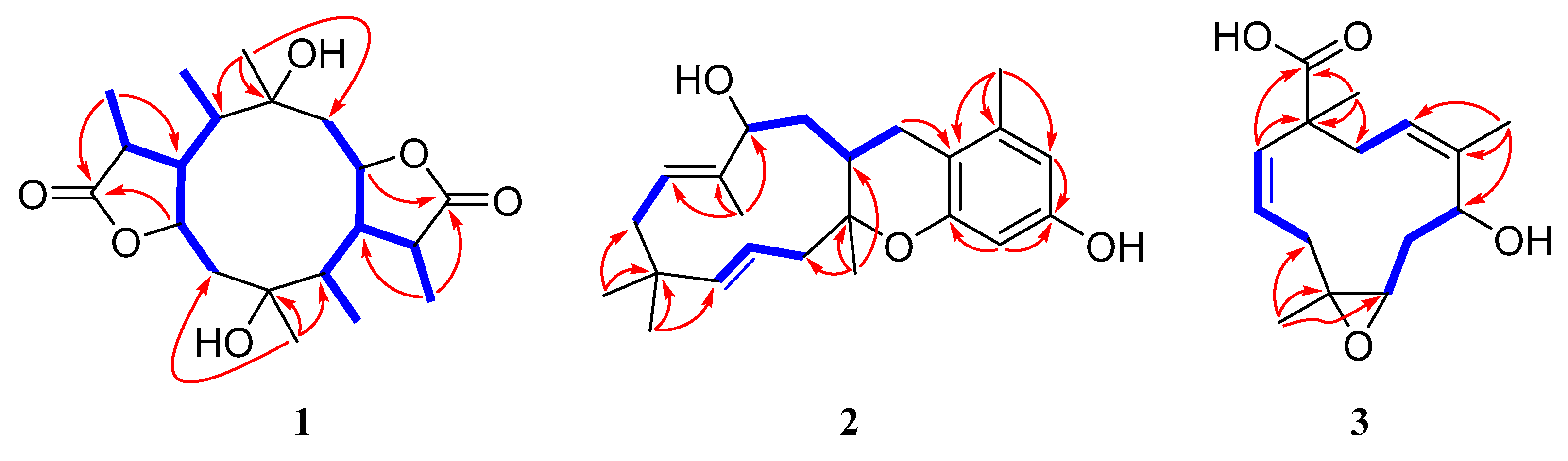
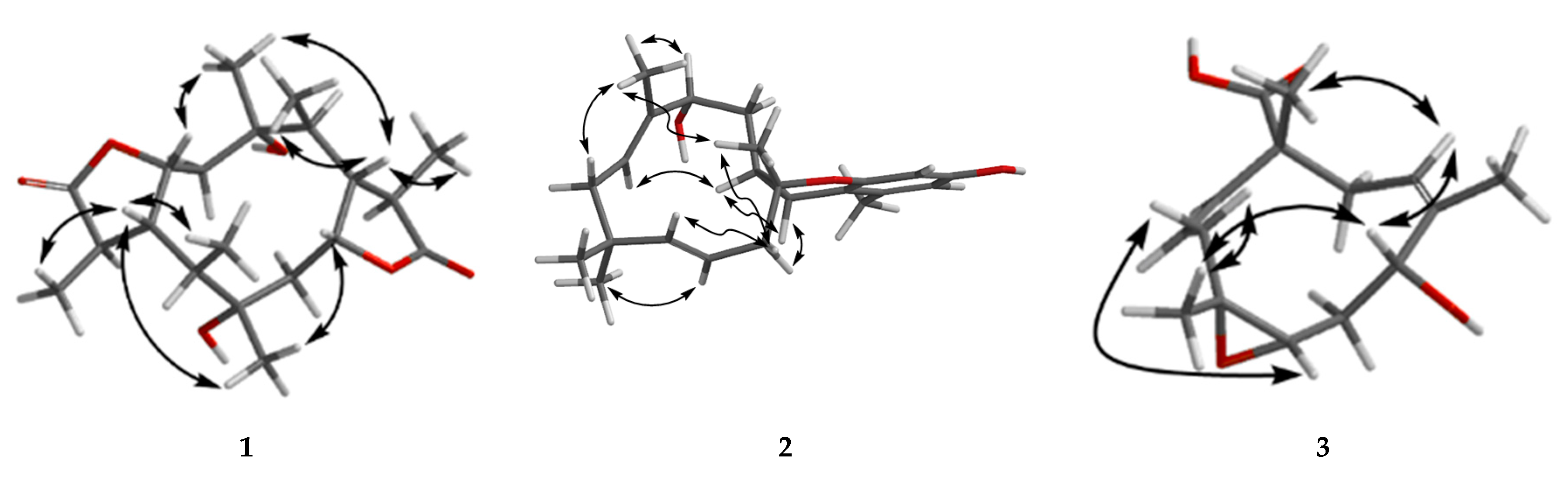
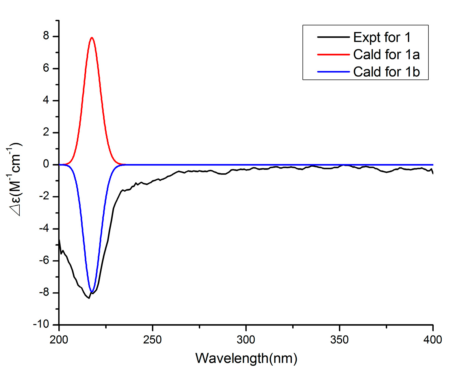
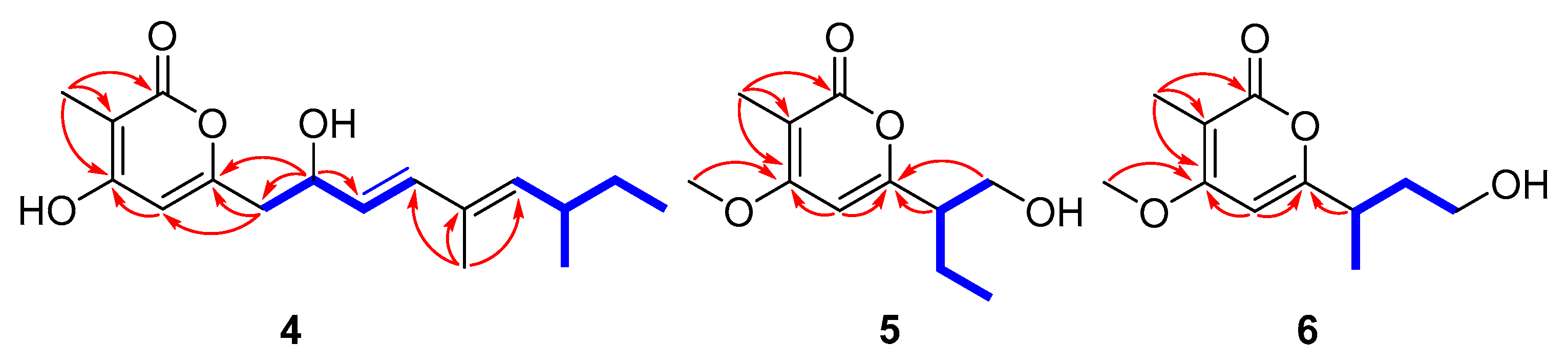
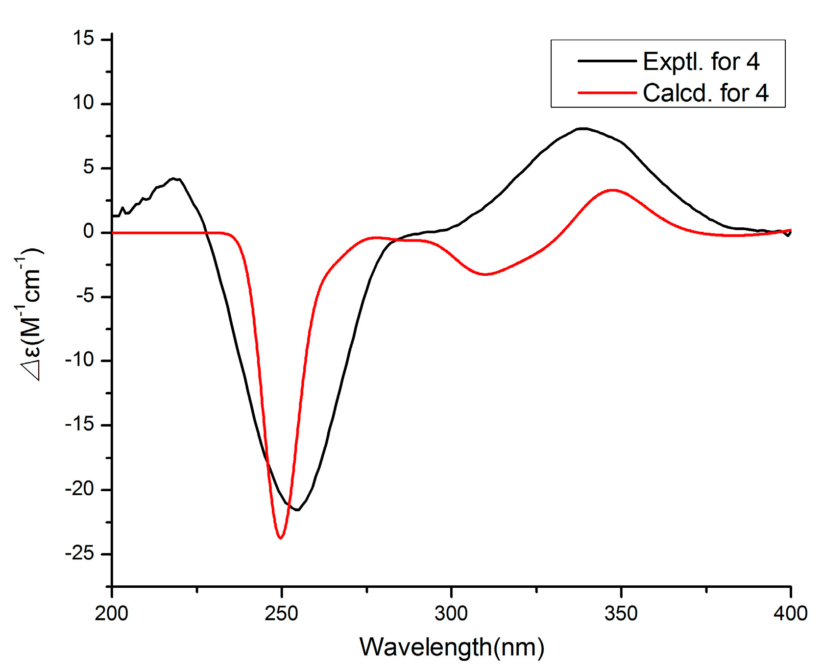
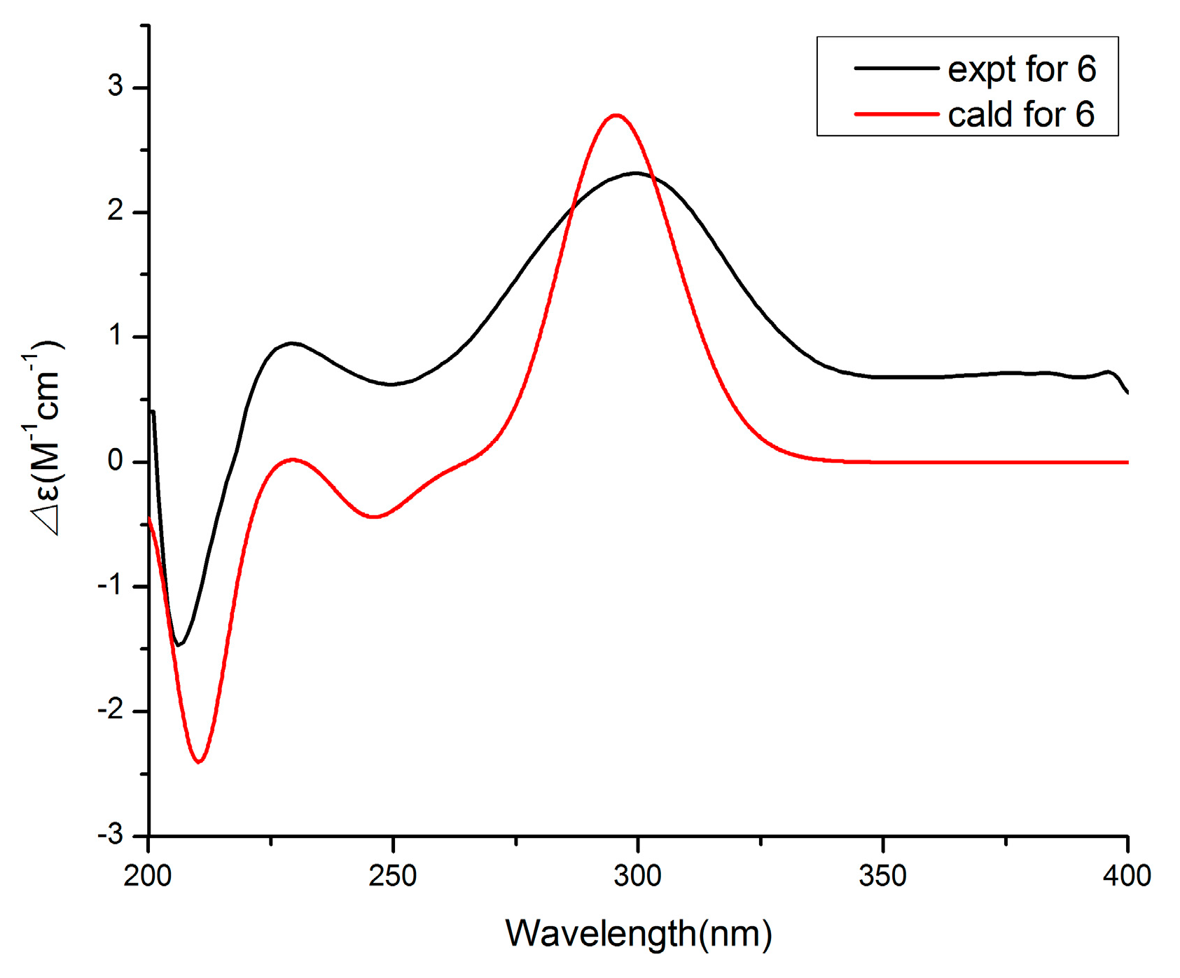
2. Results
3. Experimental Section
3.1. General Experimental Procedures
3.2. Fungal Material, Fermentation and Isolation
3.3. ECD Calculation Methods
3.4. Anti-Inflammatory Assay
4. Conclusions
Supplementary Materials
Author Contributions
Funding
Institutional Review Board Statement
Data Availability Statement
Conflicts of Interest
References
- Cheng, Z.S.; Pan, J.H.; Tang, W.C.; Chen, Q.J.; Lin, Y.C. Biodiversity and biotechnological potential of mangrove-associated fungi. J. For. Res. 2009, 20, 63–72. [Google Scholar] [CrossRef]
- Sebastianes, F.L.S.; Cabedo, N.; Aouad, N.E. 3-Hydroxypropionic Acid as an Antibacterial Agent from Endophytic Fungi Diaporthe phaseolorum. Curr. Microbiol. 2012, 65, 622–632. [Google Scholar] [CrossRef] [PubMed]
- Zhu, F.; Lin, Y.C. Marinamide, a novel alkaloid and its methyl ester produced by the application of mixed fermentation technique to two mangrove endophytic fungi from the South China Sea. Chin. Sci. Bull. 2006, 51, 1426–1430. [Google Scholar] [CrossRef]
- Rosario, N.; Maria, S.; Anna, A. Secondary Metabolites of Mangrove-Associated Strains of Talaromyces. Mar. Drugs 2018, 16, 12. [Google Scholar]
- Deshmukh, S.K.; Gupta, M.K.; Prakash, V. Mangrove-Associated Fungi: A Novel Source of Potential Anticancer Compounds. J. Fungi 2018, 4, 101. [Google Scholar] [CrossRef]
- Carvalho, C.R.D.; Ferreira-D’Silva, A.; Wedge, D.E. Antifungal activities of cytochalasins produced by Diaporthe miriciae, an endophytic fungus associated with tropical medicinal plants. Can. J. Microbiol. 2018, 64, 835–843. [Google Scholar] [CrossRef]
- Sousa, J.P.B.; Aguilar-Pérez, M.M.; Arnold, A.E. Chemical constituents and their antibacterial activity from the tropical endophytic fungus Diaporthe sp. F2934. J. Appl. Microbiol. 2016, 120, 1501–1508. [Google Scholar] [CrossRef][Green Version]
- Yang, X.; Wu, P.; Xue, J. Cytochalasans from endophytic fungus Diaporthe sp. SC-J0138. Fitoterapia 2020, 145, 104611. [Google Scholar] [CrossRef]
- Dettrakul, S.; Kittakoop, P.; Isaka, M. Antimycobacterial pimarane diterpenes from the Fungus Diaporthe sp. Bioorg. Med. Chem. Lett. 2003, 13, 1253–1255. [Google Scholar] [CrossRef]
- Chepkirui, C.; Stadler, M. The genus Diaporthe: A rich source of diverse and bioactive metabolites. Mycol. Prog. 2017, 16, 477–494. [Google Scholar] [CrossRef]
- Kumaran, R.S.; Hur, B.K. Screening of species of the endophytic fungus Phomopsis for the production of the anticancer drug taxol. Biotechnol. Appl. Bioc. 2011, 54, 21–30. [Google Scholar] [CrossRef] [PubMed]
- Du, Y.; Sun, J.; Gong, Q. New α-Pyridones with Quorum Sensing Inhibitory Activity from Diversity-Enhanced Extracts of a Marine Algae-Derived Streptomyces sp. J. Agric. Food Chem. 2018, 66, 1807–1812. [Google Scholar] [CrossRef] [PubMed]
- Pittayakhajonwut, P.; Theerasilp, M.; Kongsaeree, P.; Pughiinin, A. A Sesquiterpene from the Fungus Kionochaeta pughii BCC 3878. Planta Med. 2002, 68, 1017–1019. [Google Scholar] [CrossRef] [PubMed]
- Cai, P.; Smith, D.; Cunningham, B. Epolones: Novel Sesquiterpene-Tropolones from Fungus OS-F69284 That Induce Erythropoietin in Human Cells. J. Nat. Prod. 1998, 61, 791–795. [Google Scholar] [CrossRef] [PubMed]
- Chiang, Y.M.; Oakley, C.E.; Ahuia, M. An efficient system for heterologous expression of secondary metabolite genes in Aspergillus nidulans. J. Am. Chem. Soc. 2013, 135, 7720–7731. [Google Scholar] [CrossRef] [PubMed]
- Zhang, H.; Saurav, K.; Yu, Z. alpha-Pyrones with Diverse Hydroxy Substitutions from Three Marine-Derived Nocardiopsis Strains. J. Nat. Prod. 2016, 79, 1610–1618. [Google Scholar] [CrossRef] [PubMed]
- Aoki, Y.; Matsumoto, D.; Kawaide, H. Physiological role of germicidins in spore germination and hyphal elongation in Streptomyces coelicolor A3(2). J. Antibiot. 2011, 64, 607–611. [Google Scholar] [CrossRef]
- Weber, D.; Gorzalczany, S.; Martino, V. Metabolites from Endophytes of the Medicinal Plant Erythrina crista-galli. Z. Naturforsch. C. Biosci. 2005, 60, 5–6. [Google Scholar] [CrossRef]
- Iadecola, C.; Pelligrino, D.A.; Moskowitz, M.A. Nitric oxide synthase inhibition and cerebrovascular regulation. J. Cereb. Blood Flow Metab. 1994, 14, 175–192. [Google Scholar] [CrossRef]
- Chen, Y.; Liu, Z.M.; Liu, H.J. Dichloroisocoumarins with Potential Anti-Inflammatory Activity from the Mangrove Endophytic Fungus Ascomycota sp. CYSK-4. Mar. Drugs 2018, 16, 54. [Google Scholar] [CrossRef]
- Chen, Y.; Liu, Z.M.; She, Z.G. Ascomylactams A-C, Cytotoxic 12- or 13-Membered-Ring Macrocyclic Alkaloids Isolated from the Mangrove Endophytic Fungus Didymella sp. CYSK-4, and Structure Revisions of Phomapyrrolidones A and C. J. Nat. Prod. 2019, 82, 1752–1758. [Google Scholar] [CrossRef] [PubMed]
- Thomas, B.; Robert, K.; Bernhard, L. Production of Macrocyclic Sesqui- and Diterpenes in Heterologous Microbial Hosts: A Systems Approach to Harness Nature’s Molecular Diversity. Chemcatchem 2014, 6, 1142–1165. [Google Scholar]
- Wu, J.R.; Li, X.D.; Lin, W.H. Briarane-type diterpenoids from a gorgonian coral Ellisella sp. with anti-HBV activities. Bioorg. Chem. 2020, 105, 104423. [Google Scholar] [CrossRef] [PubMed]
- Tseng, W.-R.; Ahmed, A.F.; Huang, C.-Y.; Tsai, Y.-Y.; Tai, C.-J.; Orfali, R.S.; Hwang, T.-L.; Wang, Y.-H.; Dai, C.-F.; Sheu, J.-H. Bioactive Capnosanes and Cembranes from the Soft Coral Klyxum flaccidum. Mar. Drugs 2019, 17, 461. [Google Scholar] [CrossRef] [PubMed]
- Chang, Y.C.; Chiang, C.C.; Chang, Y.S.; Chen, J.J. Novel Caryophyllane-Related Sesquiterpenoids with Anti-Inflammatory Activity from Rumphella antipathes (Linnaeus, 1758). Mar. Drugs 2020, 18, 554. [Google Scholar] [CrossRef]
- Zhou, B.; Wu, Y.; Yue, J.M. Euphorbesulins A–P, Structurally Diverse Diterpenoids from Euphorbia esula. J. Nat. Prod. 2016, 79, 1952–1961. [Google Scholar] [CrossRef]
- Lee, S.J.; Lee, I.S.; Mar, W. Inhibition of inducible nitric oxide synthase and cyclooxygenase-2 activity by 1,2,3,4,6-penta-O-galloyl-beta-D-glucose in murine macrophage cells. Arch. Pharm. Res. 2003, 26, 832–839. [Google Scholar] [CrossRef]


| No. | 1 | 2 | ||
|---|---|---|---|---|
| δC, Type | δH (J in Hz) | δC, Type | δH (J in Hz) | |
| 1 | 80.9, C | 137.8, C | ||
| 2 | 46.4, CH2 | 2.16, m | 108.9, CH | 6.26, d (2.2) |
| 3 | 81.5, CH | 4.95, td (2.5, 7.3) | 154.4, C | |
| 4 | 54.1, CH | 2.16, m | 101.4, CH | 6.18, d (2.3) |
| 4a | 154.5, C | |||
| 5 | 50.6, CH | 1.96, dt (6.8, 13.2) | 79.4, C | |
| 6α | 81.2, C | 42.8, CH2 | 2.51, d (14.5) | |
| 6β | 2.22, m | |||
| 7 | 46.0, CH2 | 2.21, m | 121.3, CH | 5.14, m |
| 2.05, m | ||||
| 8 | 81.0, CH | 4.86, td (2.5, 6.8) | 141.4, CH | 5.15, d (2.2) |
| 9 | 49.6, CH | 2.56, dt (7.2, 10.0) | 38.5, C | |
| 10 | 44.0, CH | 2.05, m | 40.6, CH2 | 2.23, m |
| 1.77, m | ||||
| 11 | 23.7, CH3 | 1.20, s | 123.5, CH | 5.17, m |
| 12 | 181.1, C | 138.6, C | ||
| 13 | 42.6, CH | 2.72, qd (3.1, 7.6) | 78.2, CH | 3.99, d (9.6) |
| 14α | 18.3, CH3 | 1.33, d (7.6) | 39.7, CH2 | 1.76, m |
| 14β | 1.11, dd (9.3, 13.5) | |||
| 14a | 34.2, CH | 1.69, m | ||
| 15α | 15.9, CH3 | 0.97, d (7.3) | 27.3, CH2 | 2.88, dd (5.6, 16.4) |
| 15β | 2.24, m | |||
| 15a | 112.7, C | |||
| 16 | 23.8, CH3 | 1.22, s | 19.3, CH3 | 2.19, s |
| 17 | 180.5, C | 19.8, CH3 | 1.06, s | |
| 18 | 38.3, CH | 2.90, dq (7.3, 9.9) | 24.1, CH3 | 1.01, s |
| 19 | 11.6, CH3 | 1.31, d (7.3) | 30.4, CH3 | 1.06, s |
| 20 | 15.8, CH3 | 0.99, d (7.2) | 10.6, CH3 | 1.65, s |
| No. | 3 | 4 | |||
|---|---|---|---|---|---|
| δC, Type | δH (J in Hz) | No. | δC, Type | δH (J in Hz) | |
| 1 | 138.8, CH | 5.50, d (15.8) | 2 | 167.7, C | |
| 2 | 124.6, CH | 5.45, ddd (4.7, 10.6, 15.8) | 3 | 97.8, C | |
| 3α | 44.1, CH2 | 2.60, dd (4.7, 11.9) | 4 | 166.4, C | |
| 3β | 1.57, dd (10.6, 11.9) | 5 | 101.7, CH | 6.06, s | |
| 4 | 64.6, C | 6 | 160.0, C | ||
| 5 | 60.7, CH | 2.45, dd (5.2, 9.7) | 7 | 41.4, CH2 | 2.65, m |
| 6α | 34.4, CH2 | 2.19, ddd (5.1, 10.0, 13.3) | 8 | 69.8, CH | 4.47, d (6.5) |
| 6β | 1.61, m | 9 | 127.7, CH | 5.58, dd (6.8, 15.6) | |
| 7 | 76.4, CH | 4.10, dd (6.6, 10.1) | 10 | 135.8, CH | 6.25, d (15.6) |
| 8 | 137.3, C | 11 | 131.6, C | ||
| 9 | 126.5, CH | 5.16, brd (11.4) | 12 | 139.6, CH | 5.23, d (10.0) |
| 10α | 36.4, CH2 | 2.71, dd (12.2, 13.3) | 13 | 34.2, CH | 2.40, m |
| 10β | 2.08, brd (12.2) | 14α | 30.1, CH2 | 1.38, m | |
| 11 | 49.1, C | 14β | 1.24, m | ||
| 12 | 17.0, CH3 | 1.34, s | 15 | 10.9, CH3 | 0.83, t (7.4) |
| 13 | 10.8, CH3 | 1.64, s | 16 | 19.6, CH3 | 0.94, d (6.6) |
| 14 | 19.7, CH3 | 1.39, s | 17 | 11.5, CH3 | 1.74, s |
| 15 | 181.5, C | 18 | 6.8, CH3 | 1.85, s | |
| No. | 5 | 6 | ||
|---|---|---|---|---|
| δC, Type | δH (J in Hz) | δC, Type | δH (J in Hz) | |
| 2 | 165.6, C | 166.2, C | ||
| 3 | 101.3, C | 101.2, C | ||
| 4 | 163.9, C | 166.2, C | ||
| 5 | 96.2, CH | 6.10, s | 93.8, CH | 6.25, s |
| 6 | 165.5, C | 167.6, C | ||
| 7 | 49.4, CH | 2.56, m | 35.5, CH | 2.82, dq (6.8, 13.7) |
| 8 | 63.6, CH2 | 3.88, m | 37.4, CH2 | 1.93, m |
| 1.75, dt (6.1, 13.6) | ||||
| 9 | 22.1, CH2 | 1.65, m | 60.3, CH2 | 3.63, m |
| 10 | 11.7, CH3 | 0.92, t (7.4) | 18.7, CH3 | 1.25, d (6.9) |
| 11 | 8.5, CH3 | 1.91, s | 8.5, CH3 | 1.87, s |
| 12 | 56.2, CH3 | 3.90, s | 56.4, CH3 | 3.86, s |
| Compound | 1 | 2 | 3 | 4 | 5 | 6 | 7 | L-NMMA a |
|---|---|---|---|---|---|---|---|---|
| IC50 (μM) | 21.5 | 36.8 | 50.0 | 12.5 | - | - | 50.0 | 15.0 |
Publisher’s Note: MDPI stays neutral with regard to jurisdictional claims in published maps and institutional affiliations. |
© 2021 by the authors. Licensee MDPI, Basel, Switzerland. This article is an open access article distributed under the terms and conditions of the Creative Commons Attribution (CC BY) license (http://creativecommons.org/licenses/by/4.0/).
Share and Cite
Chen, Y.; Zou, G.; Yang, W.; Zhao, Y.; Tan, Q.; Chen, L.; Wang, J.; Ma, C.; Kang, W.; She, Z. Metabolites with Anti-Inflammatory Activity from the Mangrove Endophytic Fungus Diaporthe sp. QYM12. Mar. Drugs 2021, 19, 56. https://doi.org/10.3390/md19020056
Chen Y, Zou G, Yang W, Zhao Y, Tan Q, Chen L, Wang J, Ma C, Kang W, She Z. Metabolites with Anti-Inflammatory Activity from the Mangrove Endophytic Fungus Diaporthe sp. QYM12. Marine Drugs. 2021; 19(2):56. https://doi.org/10.3390/md19020056
Chicago/Turabian StyleChen, Yan, Ge Zou, Wencong Yang, Yingying Zhao, Qi Tan, Lin Chen, Jinmei Wang, Changyang Ma, Wenyi Kang, and Zhigang She. 2021. "Metabolites with Anti-Inflammatory Activity from the Mangrove Endophytic Fungus Diaporthe sp. QYM12" Marine Drugs 19, no. 2: 56. https://doi.org/10.3390/md19020056
APA StyleChen, Y., Zou, G., Yang, W., Zhao, Y., Tan, Q., Chen, L., Wang, J., Ma, C., Kang, W., & She, Z. (2021). Metabolites with Anti-Inflammatory Activity from the Mangrove Endophytic Fungus Diaporthe sp. QYM12. Marine Drugs, 19(2), 56. https://doi.org/10.3390/md19020056