Article

Isolation of Scalarane-Type Sesterterpenoids from the Marine Sponge Dysidea sp. and Stereochemical Reassignment of 12-epi-Phyllactone D/E

1
Korea Institute of Ocean Science & Technology (KIOST), Busan 49111, Korea
2
Department of Marine Biotechnology, University of Science & Technology, Daejeon 34113, Korea
3
Department of Radiation Oncology, Samsung Medical Center, Seoul 06351, Korea
*
Author to whom correspondence should be addressed.
Mar. Drugs 2021, 19(11), 627; https://doi.org/10.3390/md19110627
Submission received: 12 October 2021 / Revised: 4 November 2021 / Accepted: 8 November 2021 / Published: 9 November 2021

Abstract

:
The chemical investigation of the marine sponge Dysidea sp., which was collected from Bohol province in the Philippines, resulted in the identification of 15 new scalarane-type sesterterpenoids (114, 16), together with 15 known compounds. The chemical structures of the new compounds were elucidated based on NMR spectroscopy and HRMS. The structure of 12-epi-phyllactone D/E (15) isolated during this study was originally identified in 2007. However, careful inspection of our experimental 13C NMR spectrum revealed considerable discrepancies with the reported data at C-9, C-12, C-14, and C-23, leading to the correction of the reported compound to the C-12 epimer of 15, phyllactone D/E. The biological properties of compounds 116 were evaluated using the MDA-MB-231 cancer cell line. Compound 7, which bears a pentenone E-ring, exhibits significant cytotoxicity with a GI50 value of 4.21 μM.

Graphical Abstract

1. Introduction

Sesterterpenoids, such as the ophiobolins, sterigmatocystin, and hippolide A, have attracted a lot of attentions as potent pharmaceutical compounds because of their unique anti-inflammatory activity and cytotoxicity in various cancer cell lines [1]. These compounds are ubiquitous in a broad range of natural sources, from easily accessible terrestrial plants and insects to hard-to-access marine organisms. In marine nature, the scalarane-type scaffolds have emerged as one of the most prevalent structural features of the sesterterpenoids [2]. Since scalarin, a pentacyclic scalarane, was first isolated from the marine sponge Cacospongia scalaris in 1972 [3], a number of scalarane-type sesterterpenoids has been isolated from Dysidea sp. [4,5], Phyllospongia sp. [6,7,8,9,10,11], Strepsichordaia sp. [12], Cateriospongia sp. [13,14], Smenospongia sp. [15], and Hyrtios sp. [16], belonging to the order Dictyoceratida [17].
This family of scalarane derivatives is featured with a trans-fused 6/6/6/6 ring system and can be further categorized into three structural subgroups, namely scalarane, homoscalarane, and bishomoscalarane, based on the presence of single carbon substituents at C-20 and/or C-24 (Figure 1). Among them, bishomoscalarane exhibits an exceptionally broad range of diversity in the carbon framework, arising from two distinctive sites: C-20 and C-24/C-25 (Figure 2). Therefore, cyclopropane or alcohol/esters are frequently found at C-20 adjacent to the A ring [12]. The oxidation of C-24 and C-25 results in the formation of an extra E ring in the form of a lactone or cyclopentenone [18]; 24-oxo-25-nor-bishomoscalarane has also been identified as another feature of the D ring [8]. In addition, oxidation of the backbone usually occurs at C-3 [6], C-12 [19], and C-16 [12] to produce hydroxyl or ester substituents. A large group of bishomoscalarane derivatives found in nature is considered to be the outcome of these variations occurring in combinations.
The marine sponge Dysidea sp. is known to be a rich source of scalaranes, which exhibits useful pharmacological properties, such as anticancer and antimicrobial activities [17,20,21,22,23,24]. In the course of our studies on bioactive natural products from marine organisms, we inspected the chemical components of Dysidea sp. collected from the Bohol province in the Philippines. As a result, we identified 15 new scalarane derivatives, including one scalarane, four 20,24-bishomo-25-norscalaranes, and 10 bishomoscalaranes (Figure 3), along with 14 known compounds (Figure S1, Supplementary Materials). In this report, we disclose the structural assignment of the new scalarane sesterterpenoids and their pharmacological properties as anti-cancer agents. In addition, the C-12 configuration of compound 15, which was assigned by Li in 2007 [11], was reinvestigated because of the significant differences observed between the reported and experimental 13C chemical shifts at C-9, C-12, C-14, and C-23.

2. Results and Discussion

2.1. Structure Elucidation

Compound 1 was isolated as a colorless oil, and its molecular formula was determined to be C31H48O5 using HRESIMS (m/z [M + Na]+ 523.3382, calcd 523.3394), corresponding to eight degrees of unsaturation (DOU). The 1H NMR spectrum of 1 exhibited three singlet methyl groups at δH 0.77, 0.82, and 1.04; three doublet methyl groups at δH 1.07, 1.23, and 1.37; and three oxymethines at δH 4.03, 4.19, and 5.41. Furthermore, unique upfield signals at δH 0.57 and −0.49 indicated the presence of a cyclopropane. Analysis of the 13C NMR and HSQC spectra revealed the presence of two ester carbons (δC 174.8 and 171.7), three oxymethine carbons (δC 80.4, 74.8, 64.5), 10 methylene carbons, six methine carbons, and six methyl groups. The HMBC data showed notable correlations between the singlet methyl groups and methines from δH 0.77 to δC 51.4/50.3, δH 0.82 to δC 54.3/51.4, and δH 1.04 to 
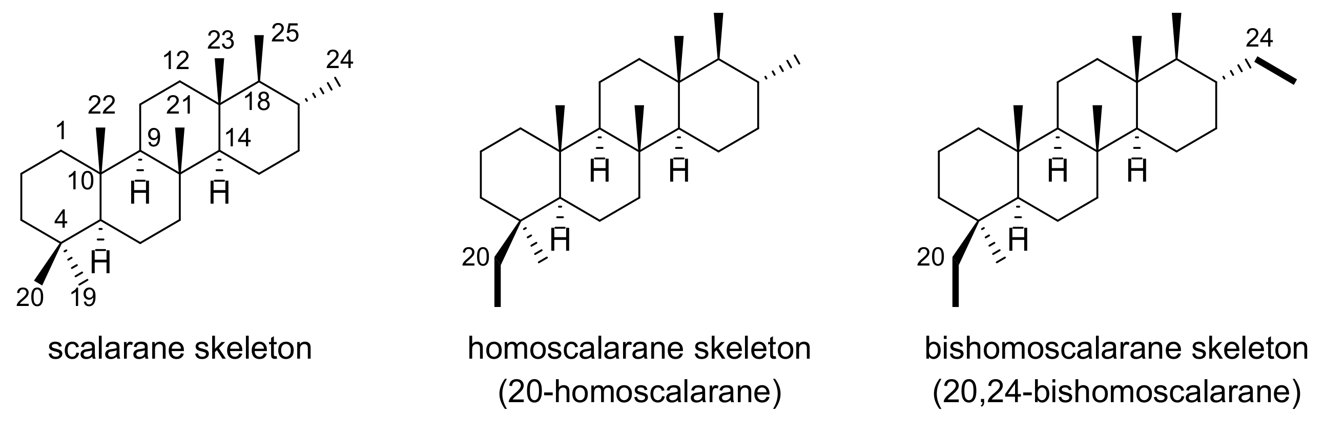
δC 54.3, which are known as characteristic correlations occurring from the ring junctions of scalarane-type 6/6/6/6 fused-cyclic systems (Figure 4). Additional HMBC correlations from the doublet methyl group at δH 1.37 to δC 80.4/44.9 and from the methine at δH 2.34 to δC 174.8/44.9 suggested the existence of a γ-valerolactone moiety. Therefore, our preliminary findings led to the hypothesis that compound 
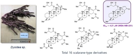
1 possessed a honulactone A-like scaffold (B+D type shown in Figure 2) [12].
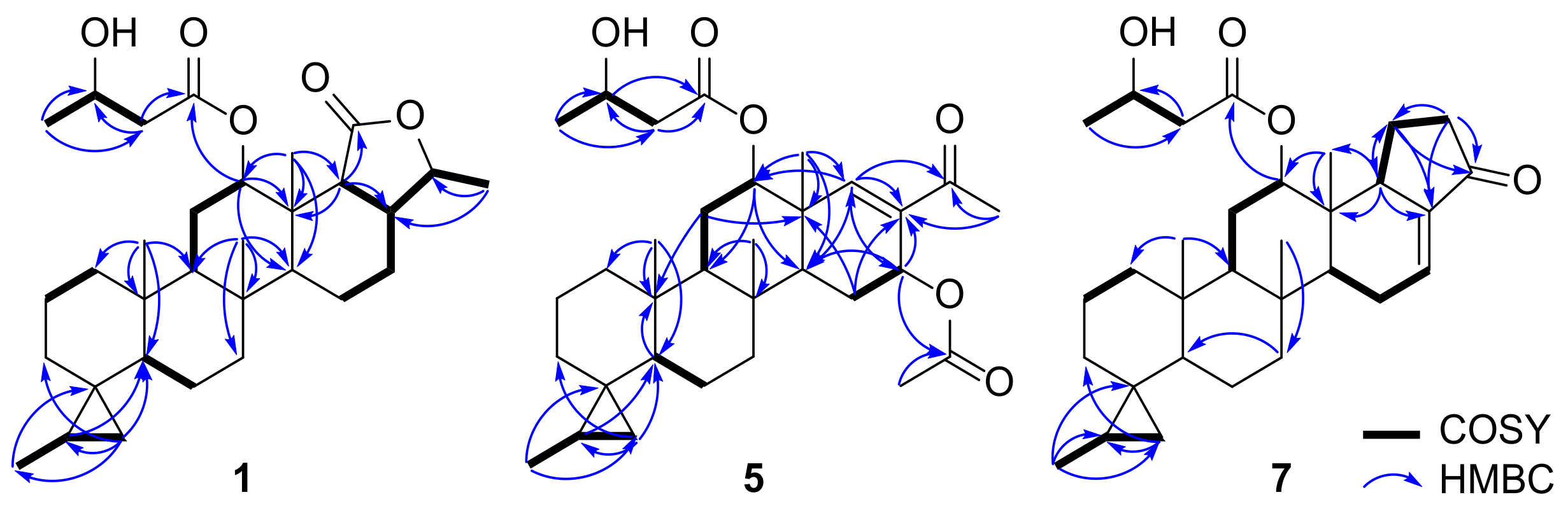
While the △17,18-olefin in honulactones is considered one of the structural features that forms the unsaturated lactone E-ring, the initially identified γ-valerolactone and DOU suggest the possibility of a saturated terminal lactone in compound 1. This speculation was confirmed by the 1H-1H COSY cross peak observed for H2-15–H2-16–H-17–H-18, as well as HMBC correlations from CH3-23 (δH 1.04) to C-18 (δC 52.5) and from H-18 (δH 2.34) to C-13 (δC 38.7). In addition, the cyclopropane moiety inferred from the 1H NMR data was positioned at C-4 based on the HMBC correlations from H2-19 (δH 0.57, and –0.49) to C-3 (δC 33.2)/C-5 (δC 50.3) and from CH3-27 (δH 1.07) to C-4 (δC 22.7), and the spin system for CH3-27–H-20 (δH 0.72)–H2-19 (δH 0.57, −0.49) in the 1H-1H COSY spectrum. Interpretation of the remaining HMBC correlations from CH3-4′ (δH 1.23) to C-2′ (δC 43.3)/C-3′ (δC 64.5), H2-2′ (δH 2.49/2.42) to C-1′ (δC 171.7)/C-3′, and H-12 (δH 5.41) to C-1′ elucidated the 3-hydroxyl butanoate group at C-12.
The trans-fused cyclic scaffold in 1 was determined from the NOESY cross peaks observed between H-11β (δH 1.71) and CH3-21 (δH 0.82)/CH3-22 (δH 0.77), and CH3-23 and CH3-21/H-17 (δH 1.86) (Figure 5). The NOESY correlations between H-12 and CH3-23, and H-18 and H-14 (δH 1.23)/H-24 (δH 4.03) suggested the β-orientations of H-12 and CH3-26, respectively. Moreover, the 20S* configuration of CH3-27 was determined based on the NOESY signals observed between H-19cisH –0.49) and H-3β (δH 1.24)/CH3-27, and H-19trans (δH 0.57) and H2-6.
Compound 2 was isolated as a colorless oil, and its molecular formula was determined to be C31H46O6 by HRESIMS (m/z [M + Na]+ 537.3167, calcd 537.3187), corresponding to nine degrees of unsaturation. Analysis of the 1D and 2D NMR spectra obtained for 2 indicated a similar carbon framework to 1, but the higher oxidation state of the lactone in E ring appeared as a major difference. HMBC correlations from CH3-23 (δH 1.22) to C-18 (δC 133.7) and from CH3-26 (δH 1.56) to C-17 (δC 162.9) revealed an α,β-unsaturated lactone in the E ring, which was responsible for the one degree higher DOU than that of 1. In addition, the 13C chemical shift of C-24 (δC 104.4) was characteristic of a ketal carbon atom, of which the position was confirmed by HMBC correlations from CH3-26 to C-24. The β-configuration of OH-24 was determined by the NOESY correlation observed between H-16α (δH 2.28) and CH3-26 (Figure S3, Supplementary Materials).
Compound 3 was isolated as a mixture of two inseparable epimers. The molecular formula of 3 was deduced to be C31H44O6 by HRESIMS (m/z [M + Na]+ 535.3011, calcd 535.3030), corresponding to 10 degrees of unsaturation. An initial inspection of the 13C NMR spectrum revealed that most of the peaks were split into a doublet-like shape, indicating a 1:1 mixture of diastereomers. The 1D and 2D NMR spectra obtained for compound 3 exhibited most of the structural features of 2, except for one more disubstituted olefin observed at δH (6.38/6.37)/δC (138.84/138.80) and δH (6.29/6.25)/δC (118.6/118.4). The location of the double bond was determined to be △15,16 using the consecutive 1H-1H COSY correlations observed for H-14 (δH 2.69/2.62)–H-15 (δH 6.38/6.37)–H-16 (δH 6.29/6.25). The splittings observed in the 13C NMR spectrum were most prominent at CH3-26 (ΔδC 1.13 ppm), informing a mixture of C-24 epimers. This phenomenon has often been observed in the case of 24-homoscalaranes, which possess both an △15,16-olefin and 24-hydroxy pentenolide E-ring [25,26]. Since the △15,16-olefin increases the planarity of the D-ring and renders the C-24 stereocenter more isolated, the 24R* and 24S* diastereomers exhibit almost identical spectroscopic and chromatographic behaviors to give an inseparable mixture.
Compound 4 was isolated as an inseparable mixture and its molecular formula was determined to be C32H46O6 by HRESIMS (m/z [M + Na]+ 549.3163, calcd 549.3187), indicating 10 degrees of unsaturation. The NMR spectra of 4 were only discriminated from those of 3 by the extra methylene group observed at δH 1.50 and δC 29.5/29.4, which was also supported by the mass difference of +14. The extra methylene group was observed in the ester side chain located at C-12, which formed a 3-hydroxypentanoate moiety, as supported by the spin system for H2-2′ (δH 2.35)–H-3′ (δH 3.90/3.86)–H2-4′ (δH 1.50)–CH3-5′ (δH 0.95) in the 1H-1H COSY spectrum.
Compound 5 was isolated as a colorless oil. Its molecular formula was determined to be C32H48O6 by HRESIMS (m/z [M + Na]+ 551.3310, calcd 551.3343), corresponding to nine degrees of unsaturation. Our initial analysis of the 1H NMR spectrum obtained for compound 5 indicated the presence of the scalarane-type scaffold: five singlet methyl groups at δH 0.80, 0.84, 1.06, 2.02, and 2.22; two doublet methyl groups at δH 1.08 and 1.25; three oxymethines at δH 4.19, 5.11, and 5.76; unique cyclopropane signals at δH 0.59 and –0.49; and one singlet olefin at δH 6.72. The 13C NMR and HSQC spectra showed one ketone (δC 197.7), two ester carbons (δC 172.0, 170.2), one trisubstituted olefin carbon (δC 153.2, 135.1), three oxymethine carbons (δC 76.8, 65.3, 64.6), eight methylenes, four methines, and seven methyl groups. In addition, the HMBC correlation from the singlet methyl at δH 2.22 to δC 197.7 suggested the presence of a methyl ketone moiety, instead of the lactone E-ring observed in compounds 14, leading to the conclusion that 5 had a B+F type skeleton, as shown in Figure 2.
Detailed interpretation of the combined spectral data of 5 revealed that the features related to the A-B-C ring system were identical to those of 14. As anticipated, the methyl ketone was positioned at C-17 to form an unsaturated ketone in the D ring on the basis of HMBC correlations between CH3-26 (δH 2.22) and C-17 (δC 135.1), and H-18 (δH 6.72) and C-17/C-24 (δC 197.7) (Figure 4). Moreover, the 1H-1H COSY cross peak for H-14 (δH 1.76)–H2-15 (δH 1.89, 1.61)–H-16 (δH 5.76) and HMBC correlations from δH 2.02 (CH3CO2–) to δC 170.2 (CH3CO2–) and from H-16 to δC 170.2 positioned an acetate substituent at C-16, of which the relative configuration was assigned to be α-orientation based on the small coupling constants between H2-15 and H-16 (dd, JH-15–H-16 = 4.3, 1.6 Hz).
Compound 6 was isolated as an amorphous solid. Its molecular formula was determined to be C30H46O5 by HRESIMS (m/z [M + Na]+ 509.3215, calcd 509.3237), corresponding to eight degrees of unsaturation. The 1H and 13C NMR spectra obtained for compound 6 were almost identical to those of 5. However, the absence of one ester carbon and the singlet methyl group at δH 2.02 suggested deacetylation from 5, which was further supported by an upfield shift of H-16 (δH 4.62). The relative configuration of OH-16 was assigned as β-orientation based on the large coupling constant observed between H-16 and H-15β (dd, JH-15–H-16 = 9.6, 5.1 Hz).
Compound 7 was isolated as a yellow oil. Its molecular formula was determined to be C31H44O4 by HRESIMS (m/z [M + Na]+ 503.3113, calcd 503.3132), corresponding to 10 degrees of unsaturation. Preliminary analysis of the 1H and 13C NMR data revealed that the scalarane-type scaffold had a cyclopropane substituent on the A ring. Interpretation of the 13C NMR and HSQC spectra exhibited the sp2 carbons in the enone systems: three sp2 methines at δH 7.38/δC 157.5, δH 6.34/δC 137.4, and δH 6.64/δC 130.4; and one trisubstituted sp2 carbon atom at δC 136.4. Therefore, HMBC correlations observed from H-25 (δH 7.38) to C-17 (δC 136.4)/C-18 (δC 49.3)/C-24 (δC 195.9), H-26 (δH 6.34) to C-17/C-24/C-25 (δC 157.5), and H-18 (δH 3.35) to C-17/C-23 (δC 14.5), as well as the 1H-1H COSY cross peak for H-18–H-25–H-26, confirmed the presence of a △25,26-cyclopenten-24-one subunit for the E-ring and the trisubstituted double bond at △16,17 (Figure 4).
Compound 8 was isolated as a yellow oil, and its molecular formula was determined to be C34H52O8 by HRESIMS (m/z [M + Na]+ 611.3541, calcd 611.3554), corresponding to nine degrees of unsaturation. The 1H NMR spectrum obtained for compound 8 showed similar patterns to that of 5. However, the upfield peaks observed for the cyclopropane moiety in 5 were substituted by an oxymethine at δH 5.35, a methyl singlet at δH 1.09, and an acetate at δH 2.03, suggesting the C+F type scaffold shown in Figure 2. Therefore, the connectivity of C-27–C-20–C-4–C-19 was determined using the HMBC correlations observed from CH3-19 (δH 0.99) to C-20 (δC 73.2) and from CH3-27 (δH 1.09) to C-4 (δC 39.4)/C-20 (Figure 6). In addition, the acetate at 
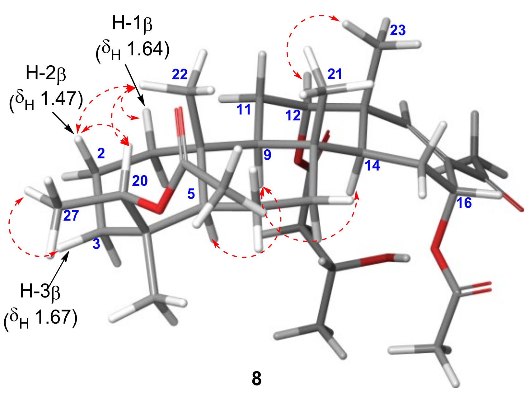
δH 2.03 exhibited a HMBC correlation with C-20 to be located at C-20. The relative configuration at C-20 was assigned as 20R* from the NOESY correlations observed between H-20 (δH 5.35) and H-2β (δH 1.47)/CH3-22 (δH 0.87), and H-3β (δH 1.67) and CH3-27 (Figure 7). Similarly, the configuration of the acetate group at C-16 was assigned as α-orientation based on the small coupling constant observed for H-16 (dd, JH-15–H-16 = 4.3, 1.6 Hz).
Compound 9 was isolated as a colorless oil, and its molecular formula was determined to be C30H48O6 by HRESIMS (m/z [M + NH4]+ 522.3810, calcd 522.3789) corresponding to seven degrees of unsaturation. Analysis of the 1D and 2D NMR data provided almost identical features to those of 8 to determine the carbon skeleton of compound 9. In this case, only one ester carbon atom (δC 172.2) was observed in the 13C NMR spectrum, and the acetate groups shown in the 1H NMR spectrum of 8 disappeared. This information indicated that compound 9 was the deacetylation product of 8. Accordingly, the upfield shifts of H-20 (δH 4.32) and H-16 (δH 4.55) were the major differences, compared to compound 8.
Compound 10 was isolated as a yellow oil, and its molecular formula was determined to be C33H50O8 by HRESIMS (m/z [M + Na]+ 597.3404, calcd 597.3398), corresponding to nine degrees of unsaturation. Preliminary inspection of the 13C NMR and HSQC data of 10 identified four singlet methyl groups (δH 0.87/δC 16.6, δH 0.87/δC 16.8, δH 0.96/δC 23.3, δH 1.13/δC 19.8), three doublet methyl groups (δH 1.07/δC 16.0, δH 1.18/δC 22.5, δH 1.39/δC 18.2), and one acetate group (δH 2.03/δC 22.0), indicating a honulactone C-like scaffold (C+D type shown in Figure 2) [12]. A detailed analysis of the 1H NMR spectrum identified an oxymethine group at δH 4.44 as a major difference from honulactone C. The location of the oxymethine was determined to be C-16, as indicated by the HMBC correlations from H-16 (δH 4.44) to C-17 (δC 162.1)/C-18 (δC 135.6) and 1H-1H COSY cross peak for H2-15 (δH 1.91, 1.84)–H-16 (Figure 6). The configuration of the OH-16 group was assigned as α-orientation based on the small coupling constant observed for H-16 (dd, JH-15–H-16 = 4.7, 1.4 Hz), and compound 10 was named as 16α-hydroxyhonulactone C [12].
Compound 11 was isolated as a yellow oil. Its molecular formula was determined as C33H50O8 by HRESIMS (m/z [M + Na]+ 597.3396, calcd 597.3398), corresponding to nine degrees of unsaturation. The 1H and 13C NMR data of 11 were almost identical to those of 10, but a ketal moiety (δC 104.4) was observed instead of one doublet methyl group and two oxymethines in compound 10. As shown in compounds 24, the hemiketal functionality in the scalarane-type scaffold usually occurs at C-24 in the E-ring, which was also applicable in this case, as indicated by the HMBC correlations from CH3-26 (δH 1.56) to C-17 (δC 162.9)/C-24 (δC 104.4). The α-orientation of the hydroxyl group at C-24 was determined by the NOESY correlation between H-16β (δH 2.33) and CH3-26. Thus, compound 11 was named 24α-hydroxyhonulactone C [12].
Compound 12 was isolated as an inseparable mixture. Its molecular formula was determined as C33H48O8 by HRESIMS (m/z [M + Na]+ 595.3241, calcd 595.3241), corresponding to 10 degrees of unsaturation. Compared to 11, two more sp2 methines at δC 138.9/138.7 and δH 6.38, and δC 118.44/118.35 and δH 6.28/6.26 were observed in the 13C NMR and HSQC spectra, indicating the presence of a disubstituted double bond. These sp2 protons were involved in a spin system for H-14 (δH 2.66/2.62)–H-15 (δH 6.38)–H-16 (δH 6.28/6.26) in the 1H-1H COSY spectrum and used to confirm the presence of the △15,16-olefin, which was further supported by HMBC correlations from H-15 to C-13 (δC 40.1/40.0)/C-14 (δC 53.96/53.90)/C-17 (δC 157.3) and from H-16 to C-14/C-18 (δC 130.9). As discussed in the cases of 3 and 4, the presence of the olefin at △15,16 and the hemiketal at C-24 rendered compound 12 an inseparable mixture of C-24 epimers.
Compound 13 was isolated as an amorphous solid. Its molecular formula was determined as C31H48O6 by HRESIMS (m/z [M + Na]+ 539.3325, calcd 539.3343), corresponding to eight degrees of unsaturation. Inspection of the 1H NMR spectrum of 13 revealed most of the structural features of the bishomoscalarane-type skeletons. Precise analysis of the 13C NMR and HSQC data revealed the presence of a triplet methyl group (δH 0.67/δC 8.80) and ketal carbon (δC 104.4), suggesting the A+D type skeleton shown in Figure 2. While most of the spectral data of 13 were identical to phyllofolactone H, the ketal carbon indicated the oxidation of C-24 to give a 24-hydroxy pentenolide E ring. This insight can be confirmed by the HMBC correlation from CH3-26 (δH 1.48) to C-17 (δC 163.0)/C-24 (δC 104.4). The configuration of OH-24 was determined to be α-orientation by the NOESY correlation between H-16β (δH 2.33) and CH3-26. Thus, compound 13 was named 24α-hydroxyphyllofolactone H [19].
Compound 14 was isolated as an inseparable mixture. Its molecular formula was determined as C31H46O6 by HRESIMS (m/z [M + Na]+ 537.3175, calcd 537.3187), corresponding to nine degrees of unsaturation. Similar to compound 3, the 13C NMR spectrum of 14 showed a 1:1 splitting pattern corresponding to a mixture of two diastereomers. The distinctive spectral features of 14, differentiated from 13, were observed as the two sp2 methines at δH 6.40/6.38 and 6.28/6.27, suggesting an unsaturated derivative of 13. The methines belonged in the 1H-1H COSY correlation for H-14 (δH 2.68/2.62)–H-15 (δH 6.40/6.38)–H-16 (δH 6.28/6.27) to identify the olefin at C-15 (Figure 6). In addition, 14 was determined to be a mixture of C-24 epimers, considering the largest splitting observed at CH3-26 (ΔδC 1.13 ppm).
Compound 15 was isolated as an inseparable mixture. Its molecular formula was determined to be C32H48O6 by HRESIMS (m/z [M + Na]+ 551.3366, calcd 551.3343), corresponding to nine degrees of unsaturation. The MS data indicated an additional methylene relative to 14, which was further supported by the change observed in the coupling pattern of the terminal methyl group of the side chain at C-12 from a doublet to triplet. The 13C NMR and HSQC data identified the methylene group at δH 1.51/1.25 and δC 29.5/29.4, which were involved in the spin system for H2-2′–H-3′–H2-4′–CH3-5′ in the 1H-1H COSY spectrum to confirm the presence of the 3-hydroxypentanoate side chain. The orientation of the ester at C-12 was assigned as α by the NOESY signal between H-12 (δH 5.55/5.49) and CH3-23 (δH 1.06/1.05), as well as the small coupling constant observed for H-12 (dd, J = 2.3, 1.8 Hz), to identify 12-epi-phyllactone D/E.
Interestingly, the identified structure was previously isolated as a mixture of C-24 epimers by Li et al. in 2007 [11], but our experimental 13C NMR data showed some discrepancies with the previously reported data at C-9 (Δ 4.28 ppm), C-11 (Δ 2.7 ppm), C-12 (Δ 2.08 ppm), C-14 (Δ 4.46 ppm), and C-23 (Δ 4.35 ppm) (Figure 8a). In addition, another identification of 12-epi-phyllactone D/E was reported by Andersen et al. in 2009 [13]. Although they acquired almost identical experimental NMR data with ours rather than those reported by Li, the isolated compound was estimated to be same as Li’s without consideration of the differences in NMR data (Tables S17 and S18, Supplementary Materials). Therefore, we investigated the variations in 13C chemical shifts depending on the orientation of the substituents at C-12.
Phyllactone D (17) and E (18), the reported 12β-epimers of 15, were selected for comparison [25]. While C-12 in phyllactones D and E was observed at δC 75.1 and 75.8, respectively, the corresponding chemical shifts of the reported and isolated 15 were observed at δC 75.3 and 73.2/73.1, respectively. The deviations observed for isolated 15 from phyllactone D/E became more obvious at C-9, C-14, and C-23 (Figure 8b). However, the reported chemical shifts for 15 were better aligned with those of phyllactone D/E. Furthermore, the differences in the 13C NMR chemical shifts observed between isolated 15 and compounds 3, 4, 12, and 14, which share an identical substructure for the B-E ring system, showed negligible values (< 0.5 ppm) around the C-ring (Table S19, Supplementary Materials). Accordingly, isolated 15 is more likely to be the 12α-epimer. Even though Li determined the 12a-configuration observing the NOESY signal between H-12 and CH3-23 and JH-12–H-13 calculation (3.0, 2.5 Hz), the NMR database suggests that the compound previously reported by Li is presumed to be a mixture of phyllactone D (17) and E (18).
Compound 16 was isolated as a yellowish oil. Its molecular formula was determined as C27H40O5 by HRESIMS (m/z [M + Na]+ 467.2762, calcd 467.2768), corresponding to eight degrees of unsaturation. The 1H NMR spectrum of 16 revealed five singlet methyl groups at δH 0.73, 0.74, 0.79, 0.85, and 0.86; one acetate group at δH 1.95; one oxymethine at δH 4.80; one olefin at δH 7.30; and one aldehyde at δH 9.41. The 13C and HSQC NMR spectra showed characteristic peaks for the aldehyde carbon atom at δC 196.4, two carbonyl carbons at δC 169.6 and 169.6, one trisubstituted olefin at δC 145.8 and 124.2, and one oxymethine at δC 76.9. The HMBC correlation between the two methyl groups at δC 33.3 and 21.4 was identified as a characteristic feature of the 4-dimethyl-sesterterpenoid scaffold (Figure 9). The aldehyde at δH 9.41 exhibited a HMBC correlation with C-18 (δC 58.7) to be located at C-25. Additional HMBC correlations from H-18 (δH 3.07) to C-16 (δC 145.8)/C-17 (δC 124.2)/C-24 (δC 169.6), along with the 1H-1H COSY cross peak for H-14–H2-15–H-16, indicated the presence of the acid at C-24 and trisubstituted olefin at C-16. The acetate group (δC 21.3/δH 1.95) was positioned at C-12, as indicated by the HMBC correlation from H-12 (δH 4.80) to C-1′ (δC 169.6) and 1H-1H COSY cross peak for H2-11–H-12. Thus, the planar structure of 16 was found to be the deacetalization product of scalarin (19) [3]. The NOESY correlations between CH3-23 (δH 0.86) and H-12/H-18 determined the configuration of the C-12 acetate and C-18 formyl groups as α.
Whereas scalarin (19) exists only in its hemiacetal form, the formation of 18-epi-19 or 19 via the acetalization of 16 was not observed. To rationalize the observed difference in reactivity, 18-epi-16 was proposed as a plausible precursor of scalarin, and geometrical optimization of 16 and 18-epi-16 was performed at the B3LYP/6-31G** level of theory. The atomic distance between O-24 to C-25 was calculated to be 3.37 Å for 16 and 2.68 Å for 18-epi-16 (Figure 10). This result suggests that 18-epi-16 can undergo acetalization to form scalarin because the β-orientation of C-25 increases its proximity to the acid at C-24. However, the acetalization of the 25α-formyl group in 16 will be restricted due to its remoteness to OH-24 to exist as its aldehyde form.

2.2. Biological Activity

The cytotoxicity of compounds 116 against MDA-MB-231 (a human breast cancer cell line) was evaluated to elucidate their potential as anticancer agents. Compounds 16, 8, 11, and 1315 exhibited moderate cytotoxicity with GI50 values ranging from 40 to 72 μM. Compounds 9, 10, 12, and 16 were inactive toward the cancer cell line (Table 1). Among the bishomoscalaranes, the highest anticancer activity was exhibited by compound 7, which has a relatively rare cyclopentenone E-ring (B+E type scaffold in Figure 2), with a GI50 value of 4.2 μM.
The highly diversified structures of the isolated scalaranes provided some information on their structure–activity relationship (SAR). The presence of the △15,16-olefin generally had a detrimental effect that reduced the cytotoxicity in the range of 12–30 μM, as shown by the sets of 2 and 3 (B+D type), 13 and 14 (C+D type), and 13 and 14 (A+D type). Comparing 3 with 4 and 14 with 15, the homologation of one methylene group at C-4′ was beneficial toward increasing the activity to ~20 μM. A series of compounds 2, 11, and 13, which only differ at the C-4 substituent, indicated the disadvantageous effect of oxidation at C-20 on the anticancer activity. The negative effect of oxidation at C-20 was also observed in the inactive series of compounds 9, 10, and 12.

3. Materials and Methods

3.1. General Experimental Procedures

Specific optical rotations were collected on a Rudolph Research Analytical (Autopol III) polarimeter (Rudolph Research Analytical, Hackettstown, NJ, USA). IR spectra were measured on a JASCO FT/IR-4100 spectrophotometer (JASCO Corporation, Tokyo, Japan). The 1D and 2D NMR spectra were taken in CDCl3 using a Bruker 600 MHz spectrometer (Bruker BioSpin GmbH, Rheinstetten, Germany) at 297.1 K. 1H NMR spectra were collected after 64–128 scans, and 13C NMR spectra were collected at a range of 10,000–15,000 scans depending on the sample concentrations. The mixing time for NOESY experiments was set as 0.3 s. Chemical shifts were reported in parts per million relative to CHCl3 residue (δH 7.26, δC 77.1) in CDCl3. High resolution mass-spectra were obtained on a Sciex X500R Q-TOF spectrometer (Framingham, MA, USA) equipped with an ESI source. MPLC was performed using the TELEDYNE ISCO CombiFlash Companion with the TELEDYNE ISCO RediSep Normal-phase Silica Flash Column (Teledyne ISCO, Lincoln, NE, USA). HPLC was performed on a PrimeLine Binary pump (Analytical Scientific Instruments, Inc., El Sobrante, CA, USA) utilizing silica columns (YMC-Pack Silica, 250 × 10 mm I.D., or 250 × 4.6 mm I.D., 5 µm; YMC Co. Ltd., Kyoto, Japan), the Shodex RI-101 (Shoko Scientific Co. Ltd., Yokohama, Japan), or the UV-M201.

3.2. Biological Material

The marine sponge used in this study was collected in March 2016 from the Bohol province in the Philippines (N 9°43′31.39″ E 124°32′19.86″) at a depth of 15 m using scuba diving. The sponge was directly kept frozen at −20 °C until identified as Dysidea sp. and chemically analyzed. A voucher sample (163PIL-267) has been stored at the Marine Biotechnology Research Center, Korea Institute of Ocean Science & Technology (KIOST).

3.3. Extraction and Isolation

The lyophilized specimen (wet wt. 1.5 kg) was extracted with MeOH (2.0 L × 3) and CH2Cl2 (4.0 L × 2) at room temperature. The combined extracts were concentrated under reduced pressure. The dried residue (89.5 g) was partitioned with n-butanol (5.0 L) and water (5.0 L). The n-butanol layer was concentrated and further partitioned between n-hexane (3.0 L) and 15% aqueous methanol (3.0 L). A portion (12.2 g) of the concentrated 15% aqueous methanol fraction (31.7 g) was subjected to flash column chromatography over C18 (YMC Gel ODS-A, 60 Å, 230 mesh (YMC Co, Ltd., Kyoto, Japan)) with a stepwise gradient solvent system (50%, 60%, 70%, 80%, 90%, and 100% MeOH, acetone, and EtOAc).
The 80% MeOH fraction (612.7 mg) was further separated using MPLC on C18 with a gradient solvent system from 70% MeOH to 100% MeOH over 40 minutes to yield 4 fractions. The third subfraction (250.1 mg) was separated using HPLC (eluent 65% MeOH) to yield 8 (3.9 mg, tR = 38 min), 9 (2.5 mg, tR = 42 min), 10 (2.3 mg, tR = 58 min), honulactone C (9.8 mg), and honulactone D (9.0 mg). The fourth subtractions (175.3 mg) was separated using HPLC (eluent 70% MeOH) to yield 11 (1.8 mg, tR = 28 min), 12 (1.4 mg, tR = 28 min), and honulactone I+J mixture (1.6 mg).
The 100% MeOH fraction (4.22 g) was further separated using MPLC on C18 with a gradient solvent system from 30% MeOH to 100% MeOH over 40 minutes to yield 4 fractions. The second fraction (2.49 g) was directly separated using MPLC on SiO2 with a gradient solvent system from 70% HX to 100% EtOAc over 80 minutes to yield 8 subfractions (based on TLC analysis). Scalarin (19, 213.0 mg) was recrystallized from the second subfraction (572.8 mg) under the HX-EtOAc solvent conditions. The residue (250.0 mg) of the second subfraction was separated using HPLC (HX/acetone = 7/1) to yield 4 (5.2 mg, tR = 54 min), 15 (5.5 mg, tR = 48 min), phyllofolactone H (5.7 mg), and phyllofolactone I (11.5 mg). The third subfraction (295.5 mg) was separated using HPLC (HX/acetone = 7/1) to yield 1 (3.4 mg, tR = 34 min), 3 (7.0 mg, tR = 76 min), 14 (6.0 mg, tR = 66 min), 13 (2.3 mg, tR = 60 min), 16 (4.5 mg, tR = 45 min, honulactone A (21.6 mg), honulactone B (26.2 mg), honulactone E+F mixture (21.4 mg), and phyllofolactone J+K (2.7 mg). The fourth subfraction (380.0 mg) was separated using HPLC (HX/acetone = 5/1) to yield 2 (3.1 mg, tR = 36 min), 5 (1.6 mg, tR = 31 min), 6 (5.6 mg, tR = 32 min), 7 (5.0 mg, tR = 30 min), and phyllofenone C (2.3 mg).

3.4. Assay

Human breast cancer MDA-MB-231 cells were purchased from the American Type Culture Collection (ATCC, Manassas, VA, USA) and were cultured in Dulbecco’s modified Eagle medium (DMEM) supplemented with 10% fetal bovine serum (FBS, Gibco, Carlsbad, CA, USA), 1 × antibiotic-antimycotic solution (Thermo Fisher Scientific, Waltham, MA, USA), and 25 mM HEPES (Gibco). Cultures were maintained in a humidified atmosphere of 95% air/5% CO2 at 37 °C.
Cell viability was determined using a CCK-8 (Cell Counting Kit-8, Dojindo Laboratory, Kumamoto, Japan) assay according to the manufacturer’s instructions. MDA-MB-231 cells were seeded at 5 × 10³ cells/well into a 96-well plate and then were treated with various concentrations of compounds 116. Following treatment for 48 h, the cells were incubated with the CCK-9 solution, and the absorbance was measured at 450 nm using a SpectraMax i3 microplate reader (Molecular Devices, Sunnyvale, CA, USA). GI50 values were calculated from a non-linear regression fit using GraphPad Prism version 9.2.0 (GraphPad Software, La Jolla, CA, USA).
1: colorless oil; [ α ] D 20 + 20.0 (c 0.2, CHCl3); IR (ATR) νmax 3131, 2954, 2929, 2581, 1770, 1734, 1452, 1381, 1261, 1176, 1027 cm−1; 1H NMR and 13C NMR, see Tables S1 and S2, Supplementary Materials; HRESIMS m/z 523.3382 [M + Na]+ (calcd for C31H48O5Na, 523.3394).
2: colorless oil; [ α ] D 20 + 40.0 (c 0.2, CHCl3); IR (ATR) νmax 3735, 2954, 2925, 2851, 1731, 1689, 1452, 1374, 1278, 1176, 1014 cm−1; 1H NMR and 13C NMR, see Tables S1 and S2, Supplementary Materials; HRESIMS m/z 537.3167 [M + Na]+ (calcd for C31H46O6Na, 537.3187).
3: colorless oil; [ α ] D 20 + 45.0 (c 0.2, CHCl3); IR (ATR) νmax 3727, 2957, 2922, 2865, 2848, 1738, 1657, 1458, 1371, 1286, 1621, 1173, 1031 cm−1; 1H NMR and 13C NMR, see Tables S1 and S2, Supplementary Materials; HRESIMS m/z 535.3011 [M + Na]+ (calcd for C31H44O6Na, 535.3030).
4: colorless oil; [ α ] D 20 + 48.3 (c 0.2, CHCl3); IR (ATR) νmax 3735, 2954, 2922, 2869, 2855, 1731, 1685, 1452, 1374, 1286, 1173, 1021 cm−1; 1H NMR and 13C NMR, see Tables S1 and S2, Supplementary Materials; HRESIMS m/z 549.3163 [M + Na]+ (calcd for C32H46O6Na, 549.3187).
5: colorless oil; [ α ] D 20 – 20.0 (c 0.1, CHCl3); IR (ATR) νmax 3727, 2961, 2929, 2851, 1734, 1678, 1452, 1367, 1254, 1173, 1027 cm−1; 1H NMR and 13C NMR, see Tables S1 and S2, Supplementary Materials; HRESIMS m/z 551.3310 [M + Na]+ (calcd for C32H48O6Na, 551.3343).
6: amorphous powder; [ α ] D 20 + 45.0 (c 0.2, CHCl3); IR (ATR) νmax 3735, 2971, 2929, 2865, 1724, 1678, 1657, 1452, 1371, 1296, 1173, 1080, 1027 cm−1; 1H NMR and 13C NMR, see Tables S1 and S2, Supplementary Materials; HRESIMS m/z 509.3215 [M + Na]+ (calcd for C30H46O5Na, 509.3237).
7: colorless oil; [ α ] D 20 + 33.3 (c 0.1, CHCl3); IR (ATR) νmax 3735, 2957, 2918, 2848, 1727, 1702, 1657, 1458, 1371, 1254, 1176, 1038 cm−1; 1H NMR and 13C NMR, see Tables S1 and S2, Supplementary Materials; HRESIMS m/z 503.3113 [M + Na]+ (calcd for C31H44O4Na, 503.3132).
8: colorless oil; [ α ] D 20 + 8.3 (c 0.2, CHCl3); IR (ATR) νmax 3727, 2961, 2929, 2851, 1738, 1721, 1671, 1505, 1452, 1374, 1246, 1031 cm−1; 1H NMR and 13C NMR, see Tables S1 and S2, Supplementary Materials; HRESIMS m/z 611.3541 [M + Na]+ (calcd for C34H52O8Na, 611.3554).
9: colorless oil; [ α ] D 20 – 6.7 (c 0.1, CHCl3); IR (ATR) νmax 2961, 2925, 2851, 1745, 1727, 1505, 1265, 1031 cm−1; 1H NMR and 13C NMR, see Tables S1 and S2, Supplementary Materials; HRESIMS m/z 522.3810 [M + NH4]+ (calcd for C30H52NO6, 522.3789).
10: colorless oil; [ α ] D 20 + 73.3 (c, 0.1, CHCl3); IR (ATR) νmax 3477, 3388, 2966, 2923, 2866, 1729, 1457, 1368, 1250 cm−1; 1H NMR and 13C NMR, see Tables S1 and S2, Supplementary Materials; HRESIMS m/z 597.3404 [M + Na]+ (calcd for C33H50O8Na, 597.3398).
11: colorless oil; [ α ] D 20 + 40.0 (c 0.1, CHCl3); IR (ATR) νmax 2965, 2918, 2855, 1731, 1649, 1458, 1374, 1250, 1169, 1035 cm−1; 1H NMR and 13C NMR, see Tables S1 and S2, Supplementary Materials; HRESIMS m/z 597.3396 [M + Na]+ (calcd for C33H50O8Na, 597.3398).
12: colorless oil; [ α ] D 20 + 71.7 (c 0.2, CHCl3); IR (ATR) νmax 3392, 2946, 2925, 2858, 1734, 1455, 1367, 1243, 1180 cm−1; 1H NMR and 13C NMR, see Tables S1 and S2, Supplementary Materials; HRESIMS m/z 595.3241 [M + Na]+ (calcd for C33H48O8Na, 595.3241).
13: amorphous powder; [ α ] D 20 + 6.7 (c 0.2, CHCl3); IR (ATR) νmax 3727, 2957, 2929, 2848, 1727, 1657, 1455, 1374, 1278, 1176 cm−1; 1H NMR and 13C NMR, see Tables S1 and S2, Supplementary Materials; HRESIMS m/z 539.3325 [M + Na]+ (calcd for C31H48O6Na, 539.3343).
14: colorless oil; [ α ] D 20 + 31.7 (c 0.2, CHCl3); IR (ATR) νmax 3717, 2961, 2925, 2872, 1727, 1649, 1458, 1374, 1275, 1257, 1176 cm−1; 1H NMR and 13C NMR, see Tables S1 and S2, Supplementary Materials; HRESIMS m/z 537.3175 [M + Na]+ (calcd for C31H46O6Na, 537.3187).
15: colorless oil; [ α ] D 20 + 30.0 (c 0.2, CHCl3); IR (ATR) νmax 3735, 2957, 2925, 2869, 1731, 1448, 1363, 1278, 1176, 1014 cm-1; 1H NMR and 13C NMR, see Tables S1 and S2, Supplementary Materials; HRESIMS m/z 551.3366 [M + Na]+ (calcd for C32H48O6Na, 551.3343).
16: colorless oil; [ α ] D 20 – 70.0 (c 0.2, CHCl3); IR (ATR) νmax 3727, 2961, 2922, 2851, 1738, 1646, 1452, 1381, 1225, 1021 cm−1; 1H NMR and 13C NMR, see Tables S1 and S2, Supplementary Materials; HRESIMS m/z 467.2762 [M + Na]+ (calcd for C27H40O5Na, 467.2768).

4. Conclusions

A total of 15 novel scalaranes 114 and 16, including 14 bishomoscalaranes and one scalarin derivative, has been isolated from the marine sponge, Dysidea sp., and characterized using a combination of 1D and 2D NMR spectroscopy. The isolation and structural identification of compound 15 resulted in the reassignment of the previously characterized 12-epi-phyllactone D/E. The actual structure of the reported 12-epi-phyllactone D/E was determined to be a mixture of known phyllactones D and E through the precise analysis of the experimental and reported 13C chemical shifts. In addition, the effect of the C-18 configuration in 16 on the formation of the hemiacetal E-ring was rationalized by measuring the atomic distances between C-25 and O-24 in 16 and 18-epi-16. Finally, the evaluation of the anticancer activities of compounds 116 against MDA-MB-231 revealed that compound 7 exhibited significant cytotoxicity with a GI50 value of 4.2 μM. Detailed studies to elucidate the biological mechanism of 7 are currently underway in our laboratory.

Supplementary Materials

The following are available online at https://www.mdpi.com/article/10.3390/md19110627/s1, I. Experimental procedure; Figure S1: Structures of known compounds isolated from Dysidea sp., Tables S1–S16: 13C/1H chemical shifts for 116, Figure S2: Key 1H-1H COSY, HMBC correlations of 116, Figure S3: Key NOESY correlations of 116, Tables S17–S19, Figures S4 and S5: Comparison of 13C/1H chemical shifts for 12β epimer (17, 18) and 12α epimer (15) reported by Li et al., Andersen et al., and our experiment, II. Computational methods; Supporting Information II; Figures S-1–S-112: 1H NMR, 13C NMR, COSY, HSQC, HMBC, NOESY, and HRMS spectra of 116.

Author Contributions

A.-Y.S. worked on isolation and structure elucidation. A.S. and C.C. performed the biological evaluation. J.L. collected the marine sponge and supervised the whole research work. All authors have read and agreed to the published version of the manuscript.

Funding

This work was supported by grants from the Ministry of Ocean and Fisheries (PM62520) and the Korea Institute of Ocean Science and Technology (PE99921).

Institutional Review Board Statement

Not applicable.

Informed Consent Statement

Not applicable.

Data Availability Statement

All data presented in this study are available from the corresponding author on reasonable request.

Acknowledgments

We are grateful to Young-A Kim at Hannam University for the helpful discussions on the identification of the marine sponge and Byungho Park at EMNI for the technical support on the quantum mechanical calculations performed in this study. We thank Wilfredo Campos at University of the Philippines Visayas (UPV) for requesting and receiving permission to collect the marine sponge.

Conflicts of Interest

The authors declare no conflict of interest.

References

  1. Evidente, A.; Kornienko, A.; Lefranc, F.; Cimmino, A.; Dasari, R.; Evidente, M.; Mathieu, V.; Kiss, R. Sesterterpenoids with anticancer activity. Curr. Med. Chem. 2015, 22, 3502–3522. [Google Scholar] [CrossRef] [Green Version]
  2. Pour, P.M.; Behzad, S.; Asgari, S.; Khankandi, H.P.; Farzaei, M.H. Sesterterpenoids. In Recent Advances in Natural Products Analysis; Elsevier: Amsterdam, The Netherlands, 2020; pp. 347–391. [Google Scholar]
  3. Fattorusso, E.; Magno, S.; Santacroce, C.; Sica, D. Scalarin, a new pentacyclic C-25 terpenoid from the sponge Cacospongia scalaris. Tetrahedron 1972, 28, 5993–5997. [Google Scholar] [CrossRef]
  4. Yang, X.; Shao, Z.; Zhang, X. Sesterterpenes from the Sponge Dysidea sp. Z. Naturforsch. B 2010, 65, 625–627. [Google Scholar] [CrossRef] [Green Version]
  5. Wang, Q.; Sun, Y.; Yang, L.; Luo, X.; de Voogd, N.J.; Tang, X.; Li, P.; Li, G. Bishomoscalarane sesterterpenoids from the sponge Dysidea granulosa collected in the South China Sea. J. Nat. Prod. 2020, 83, 516–523. [Google Scholar] [CrossRef]
  6. Chee, C.L.; Otero-Quintero, S.; Nicholas, G.M.; Bewley, C.A. Phyllolactones AE: New bishomoscalarane sesterterpenes from the marine sponge Phyllospongia lamellosa. Tetrahedron 2001, 57, 5731–5738. [Google Scholar] [CrossRef]
  7. Zeng, L.; Fu, X.; Su, J.; Pordesimo, E.O.; Traeger, S.C.; Schmitz, F.J. Novel bishomoscalarane sesterterpenes from the sponge Phyllospongia foliascens. J. Nat. Prod. 1991, 54, 421–427. [Google Scholar] [CrossRef]
  8. Zhang, H.J.; Tang, H.F.; Yi, Y.H.; Lin, H.W. Scalarane sesterterpenes from the Chinese sponge Phyllospongia foliascens. Helv. Chim. Acta 2009, 92, 762–767. [Google Scholar] [CrossRef]
  9. Fu, X.; Zeng, L.; Su, J.; Schmitz, F.J. Phyllofolactones C and D, two new minor Homoscalarane Sesterterpenes from the Chinese sponge Phyllospongia foliascens. J. Nat. Prod. 1999, 62, 644–646. [Google Scholar] [CrossRef]
  10. Ponomarenko, L.P.; Kalinovsky, A.I.; Stonik, V.A. New Scalarane-based Sesterterpenes from the Sponge Phyllospongia m adagascarensis. J. Nat. Prod. 2004, 67, 1507–1510. [Google Scholar] [CrossRef]
  11. Lan, W.J.; Li, H.J. New sesterterpenoids from the marine sponge Phyllospongia papyracea. Helv. Chim. Acta 2007, 90, 1218–1222. [Google Scholar] [CrossRef]
  12. Jiménez, J.I.; Yoshida, W.Y.; Scheuer, P.J.; Lobkovsky, E.; Clardy, J.; Kelly, M. Honulactones: New bishomoscalarane sesterterpenes from the Indonesian sponge Strepsichordaia aliena. J. Org. Chem. 2000, 65, 6837–6840. [Google Scholar] [CrossRef]
  13. Williams, D.E.; Hollander, I.; Feldberg, L.; Frommer, E.; Mallon, R.; Tahir, A.; van Soest, R.; Andersen, R.J. Scalarane-based sesterterpenoid RCE-protease inhibitors isolated from the Indonesian marine sponge Carteriospongia foliascens. J. Nat. Prod. 2009, 72, 1106–1109. [Google Scholar] [CrossRef]
  14. Cao, F.; Wu, Z.-H.; Shao, C.-L.; Pang, S.; Liang, X.-Y.; de Voogd, N.J.; Wang, C.-Y. Cytotoxic scalarane sesterterpenoids from the South China Sea sponge Carteriospongia foliascens. Org. Biomol. Chem. 2015, 13, 4016–4024. [Google Scholar] [CrossRef]
  15. Song, J.; Jeong, W.; Wang, N.; Lee, H.-S.; Sim, C.J.; Oh, K.-B.; Shin, J. Scalarane sesterterpenes from the sponge Smenospongia sp. J. Nat. Prod. 2008, 71, 1866–1871. [Google Scholar] [CrossRef]
  16. Kwon, O.-S.; Kim, D.; Kim, C.-K.; Sun, J.; Sim, C.J.; Oh, D.-C.; Lee, S.K.; Oh, K.-B.; Shin, J. Cytotoxic scalarane sesterterpenes from the sponge Hyrtios erectus. Mar. Drugs 2020, 18, 253. [Google Scholar] [CrossRef]
  17. Abdelaleem, E.R.; Samy, M.N.; Desoukey, S.Y.; Liu, M.; Quinn, R.J.; Abdelmohsen, U.R. Marine natural products from sponges (Porifera) of the order Dictyoceratida (2013 to 2019): A promising source for drug discovery. RSC Adv. 2020, 10, 34959–34976. [Google Scholar] [CrossRef]
  18. Gonzalez, A.M. Scalarane sesterterpenoids. Curr. Bioact. 2010, 6, 178–206. [Google Scholar] [CrossRef] [Green Version]
  19. Jiménez, J.I.; Yoshida, W.Y.; Scheuer, P.J.; Kelly, M. Scalarane-based sesterterpenes from an Indonesian sponge Strepsichordaia aliena. J. Nat. Prod. 2000, 63, 1388–1392. [Google Scholar] [CrossRef] [PubMed]
  20. Zhang, X.; Xu, H.-Y.; Huang, A.-M.; Wang, L.; Wang, Q.; Cao, P.-Y.; Yang, P.-M. Antibacterial meroterpenoids from the South China Sea sponge Dysidea sp. Chem. Pharm. Bull. 2016, 64, 1036–1042. [Google Scholar] [CrossRef] [PubMed] [Green Version]
  21. Lu, Y.; Zhao, M. Two highly acetylated sterols from the marine sponge Dysidea sp. Z. Naturforsch B 2017, 72, 49–52. [Google Scholar] [CrossRef]
  22. Liu, L.; Wu, W.; Li, J.; Jiao, W.-H.; Liu, L.-Y.; Tang, J.; Liu, L.; Sun, F.; Han, B.-N.; Lin, H.-W. Two sesquiterpene aminoquinones protect against oxidative injury in HaCaT keratinocytes via activation of AMPKα/ERK-Nrf2/ARE/HO-1 signaling. Biomed. Pharm. 2018, 100, 417–425. [Google Scholar] [CrossRef] [PubMed]
  23. Diaz-Marrero, A.R.; Austin, P.; Van Soest, R.; Matainaho, T.; Roskelley, C.D.; Roberge, M.; Andersen, R.J. Avinosol, a meroterpenoid-nucleoside conjugate with antiinvasion activity isolated from the marine sponge Dysidea sp. Org. Lett. 2006, 8, 3749–3752. [Google Scholar] [CrossRef] [PubMed]
  24. Zhang, H.; Skildum, A.; Stromquist, E.; Rose-Hellekant, T.; Chang, L.C. Bioactive polybrominated diphenyl ethers from the marine sponge Dysidea sp. J. Nat. Prod. 2008, 71, 262–264. [Google Scholar] [CrossRef] [PubMed]
  25. Fu, X.; Zeng, L.M.; Su, J.Y.; Pais, M.; Potier, P. Scalarane-type bishomosesterterpenes from the sponge Phyllospongia foliascens. J. Nat. Prod. 1992, 55, 1607–1613. [Google Scholar] [CrossRef] [PubMed]
  26. Fu, X.; Zeng, L.; Su, J.; Pais, M.; Potier, P. Two new sesterterpenes from a South China sea sponge. J. Nat. Prod. 1993, 56, 1985–1988. [Google Scholar] [CrossRef]
Figure 1. Subtypes of the scalarane skeleton found in marine nature.
Marinedrugs 19 00627 g001
Figure 2. Diversity of bishomoscalarane skeletons frequently found in nature.
Marinedrugs 19 00627 g002
Figure 3. The structures of compounds 116 isolated from Dysidea sp.
Marinedrugs 19 00627 g003
Figure 4. COSY and HMBC correlations observed for compounds 1, 5, and 7.
Marinedrugs 19 00627 g004
Figure 5. NOESY correlations observed for compound 1.
Marinedrugs 19 00627 g005
Figure 6. COSY and HMBC correlations observed for compounds 8, 10, and 14.
Marinedrugs 19 00627 g006
Figure 7. NOESY correlations observed for compound 8.
Marinedrugs 19 00627 g007
Figure 8. (a) The differences observed between the experimental and reported 13C chemical shifts (Li et al.) of 15 in CDCl3. (b) The deviations in the 13C NMR chemical shifts observed for reported (Li et al.) and isolated 15 relative to phyllactone D and phyllactone E. (c) The structures of phyllactone D and E.
Marinedrugs 19 00627 g008
Figure 9. COSY, HMBC, and NOESY correlations observed for compound 16.
Marinedrugs 19 00627 g009
Figure 10. Atomic distances between O-24 and C-25 in compounds 16 and 18-epi-16.
Marinedrugs 19 00627 g010
Table 1. The results of the cytotoxicity tests against MDA-MB-231 (human breast cancer cell line) obtained for compounds 116.
Cytotoxicity 1 (MDA-MB-231)
Compound12345678
GI50 (μM)69.9443.3872.4954.0253.5850.84.2153.55
Compound910111213141516
GI50 (μM)>100>10071.14>10050.7163.5440.82>100
1 Cisplatin (Sigma-Aldrich, St. Louis, MO) was used as a positive control (GI50 = 1.31 μM).
Publisher’s Note: MDPI stays neutral with regard to jurisdictional claims in published maps and institutional affiliations.

