Article

Novel Fredericamycin Variant Overproduced by a Streptomycin-Resistant Streptomyces albus subsp. chlorinus Strain

by
Marta Rodríguez Estévez
1,
Maksym Myronovskyi
1,
Birgit Rosenkränzer
1,
Thomas Paululat
2,
Lutz Petzke
3,
Jeanette Ristau
3 and
Andriy Luzhetskyy
1,4,*
1
Pharmazeutische Biotechnologie, Universität des Saarlandes, 66123 Saarbrücken, Germany
2
Organische Chemie II, Universität Siegen, 57068 Siegen, Germany
3
BASF SE, 67056 Ludwigshafen, Germany
4
Helmholtz-Institut für Pharmazeutische Forschung Saarland, 66123 Saarbrücken, Germany
*
Author to whom correspondence should be addressed.
Mar. Drugs 2020, 18(6), 284; https://doi.org/10.3390/md18060284
Submission received: 30 April 2020 / Revised: 22 May 2020 / Accepted: 27 May 2020 / Published: 28 May 2020
(This article belongs to the Special Issue Marine Anti-infective Agents 2020)

Abstract

:
Streptomycetes are an important source of natural products potentially applicable in the pharmaceutical industry. Many of these drugs are secondary metabolites whose biosynthetic genes are very often poorly expressed under laboratory cultivation conditions. In many cases, antibiotic-resistant mutants exhibit increased production of natural drugs, which facilitates the identification and isolation of new substances. In this study, we report the induction of a type II polyketide synthase gene cluster in the marine strain Streptomyces albus subsp. chlorinus through the selection of streptomycin-resistant mutants, resulting in overproduction of the novel compound fredericamycin C2 (1). Fredericamycin C2 (1) is structurally related to the potent antitumor drug lead fredericamycin A.

1. Introduction

The bacterial genus Streptomyces is well-known for producing a huge variety of bioactive secondary metabolites with potential pharmaceutical applications [1,2]. The genes responsible for their biosynthesis are generally clustered together. However, many of these biosynthetic genes are poorly or not expressed (silent) under laboratory cultivation conditions. Thus, the activation of silent secondary metabolite gene clusters is an essential step for the discovery of new natural drugs. A simple strategy to activate or upregulate the expression of these genes consists in screening for antibiotic-resistant mutants [3,4]. This approach is based on the work of Ochi and his team. They discovered that certain mutations in the rpsL and rpoB genes, which code for the ribosomal protein S12 and the β-subunit of RNA polymerase, respectively, lead to an altered gene product that confers resistance to streptomycin (str mutants) or to rifampicin (rif mutants), respectively. The mutations in these genes also increase the production of secondary metabolites in several Streptomyces strains [3,4,5,6,7]. Presumably, some of the str and rpoB mutations give rise to diverse metabolic changes which typically occur during the stringent response. The stringent response is triggered in E. coli [8] and other prokaryotic microorganisms [9,10] under amino acid starvation conditions by the signaling molecule, guanosine tetraphosphate (ppGpp). This molecule generates a series of physiological changes, including a significant decrease of protein synthesis [11], downregulation of rRNA transcription [12], and activation of stationary-phase metabolic processes, such as the expression of secondary metabolite biosynthetic genes [9]. Thus, it is suggested that the mutant gene products of rpsL and rpoB may be responsible for antibiotic overproduction, mimicking the role of ppGpp in the stringent response [3,4,5,6].
In previous studies, the marine strain Streptomyces albus subsp. chlorinus NRRL B-24108 has been shown to harbor genes for the production of several bioactive secondary metabolites, such as the herbicide albucidin or the antibiotic nybomycin [13,14]. Here, we report the induction of a type II polyketide synthase (PKS) gene cluster in S. albus subsp. chlorinus, leading to overproduction of the novel compound fredericamycin C2 (1). This substance is structurally related to fredericamycin C (2), a secondary metabolite first isolated in 1981, together with the compounds fredericamycin A and fredericamycin B, from the culture broth of Streptomyces griseus ATCC 49344 [15]. All fredericamycin variants known to date (A, B, C, C1, and E) share a similar structure that involves two aromatic moieties, which in the case of fredericamycins A and E are linked by a rare stereogenic spiro carbon center [16,17,18]. Fredericamycin A displays strong in vivo anticancer activity against several mammal tumor cell lines [19], and it has been patented as an effective treatment for certain types of cancer in humans [20]. Additionally, fredericamycins A, B, C, and C1 exhibit moderate antibacterial and antifungal activities [19]. In this study, we present the generation of the high-level streptomycin-resistant strain, Streptomyces albus subsp. chlorinus JR1, which overproduces the novel compound fredericamycin C2 (1). We also describe the mutation likely causing this phenotype and propose the initial biosynthetic steps of fredericamycin C2 (1), based on the gene cluster homology with that of fredericamycin A.

2. Results

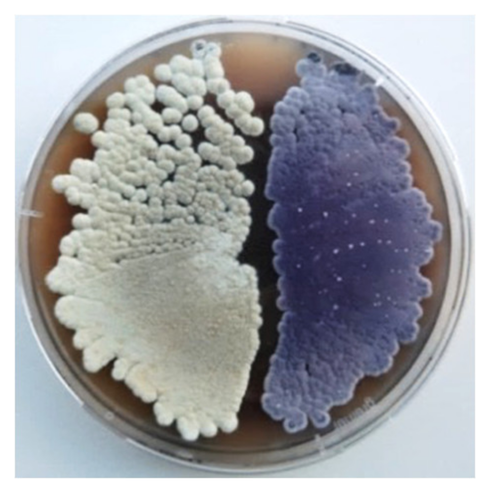
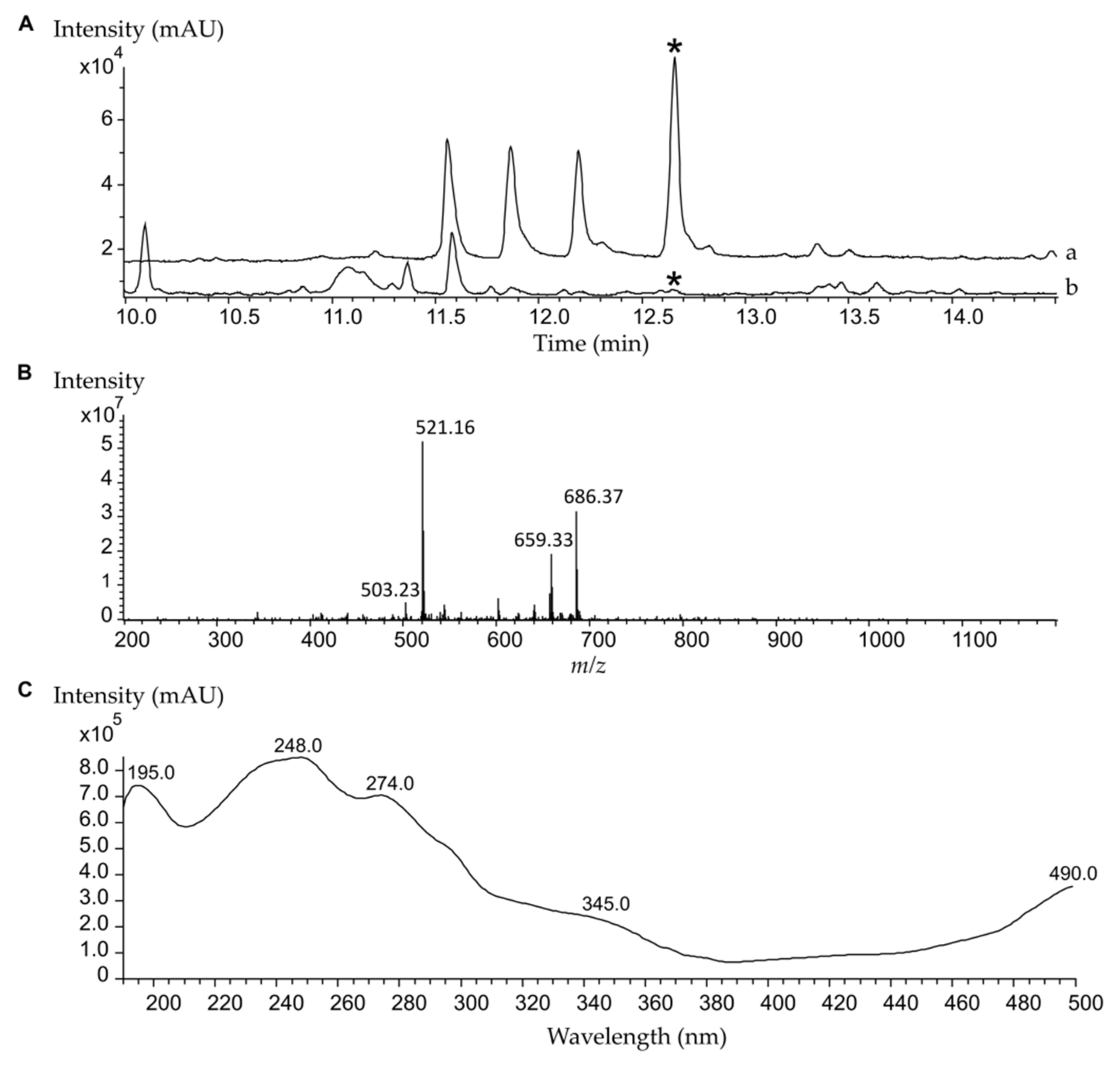
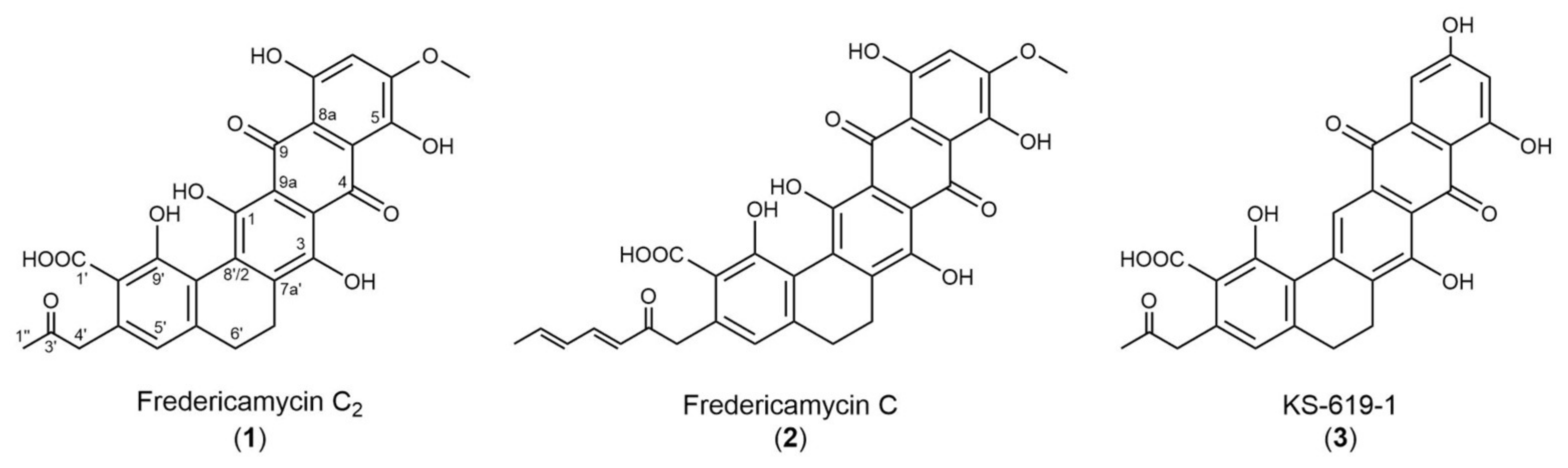
2.1. High-Level Streptomycin-Resistant Mutant S. albus subsp. chlorinus Overproduces the Novel Compound Fredericamycin C2

The strain S. albus subsp. chlorinus NRRL B-24108 was cultured in MS agar medium containing increasing concentrations of streptomycin. After several rounds of selection, we isolated a streptomycin-resistant colony that displayed a distinctive violet color when cultured on a solid medium, in contrast to the characteristic white color of the parental strain (Figure 1). We named the mutant strain S. albus subsp. chlorinus JR1. While the growth of the wild type of S. albus subsp. chlorinus was inhibited by a streptomycin concentration of 50 μg/mL, the mutant strain was able to grow in the presence of up to 200 μg/mL of antibiotic (Table S1). The metabolic profiles of both strains were analyzed using liquid chromatography and mass spectrometry, revealing the presence of a peak at tR = 12.7 min that corresponds to an [M + H]+ ion of m/z 521.107 and displays UV absorption signals at λmax 195, 248, 274, 345, and 490 nm (Figure 2). The peak area revealed an over 50-fold greater production of the corresponding compound by S. albus subsp. chlorinus JR1 compared to the parental strain’s yield (Table S2, Figure S1). The search of this mass in a natural product database yielded no coincidences, suggesting a potentially undescribed metabolite overproduced by S. albus subsp. chlorinus JR1. The compound was extracted from a 2 l solid culture of S. albus subsp. chlorinus JR1 and purified by normal phase chromatography through a silica column followed by reverse phase chromatography.
The molecular formula of the substance was determined to be C27H20O11 based on high-resolution MS (ESI) showing the quasi molecular ion m/z 521.107 ([M + H]+, calculated for C27H21O11). In the proton NMR spectrum, two broad multiplets are visible at δH 2.63 (t, 6’-H2) and 2.79 (t, 7’-H2) ppm, which are two neighbored methylene groups. All other signals are singlets: One methoxy group at δH 3.94 (6-OCH3), one additional methylene group at δH 4.06 (4’-H2), one methyl group at δH 2.17 (1’’-H3), and two methines at δH 6.52 (5’-H) and 6.88 (7-H). Moreover, broad singlets at δH 5.5, 12.8, and 13.2 ppm indicate hydroxyl groups to be present in the molecule (Table 1). Typical quinone carbonyl signals at δC 186.9 (C-4) and 187.6 (C-9) are visible in the 13C-NMR spectrum. A signal at δC 169.3 ppm (C-1’) shows an acid functionality which is attached at C-8’ proven from an HMBC (Heteronuclear Multiple Bond Coherence) correlation C-1’/ 5’-H. A very weak signal at δH 204.1 ppm (C-3’) shows a ketone supported by an HMBC cross-peak C-3’/1’’-H3 (Table 1). This ketone is part of a propan-2-on sidechain which is attached at C-6’ based on HMBC signals C-6’/4’-H2, C-6’/1’’-H3, C-5’/4’-H2, and C-7’/4’-H2. The HMBC signal C-6/6-OCH3 indicates the methoxy group to be attached at C-6 next to the aromatic proton 7-H. One of the rings contains a CH2-CH2 moiety (C-6’-C7’) which is in ring D proven from HMBC signals C-3/7’-H2, C-7a’/7’-H2, C-8’/7’-H2, C-7a’/6’-H2, and C-5a’/6’-H2 (Figure S2). Comparison to literature shows high similarity of the new compound named fredericamycin C2 (1) to fredericamycin C (2) [17], which differs only in the sidechain attached at C-6’ (fredericamycin C) and to KS-619-1 (3) [21] with a different substitution pattern of the pentacyclic ring system (KS-619-1) (Figure 3).

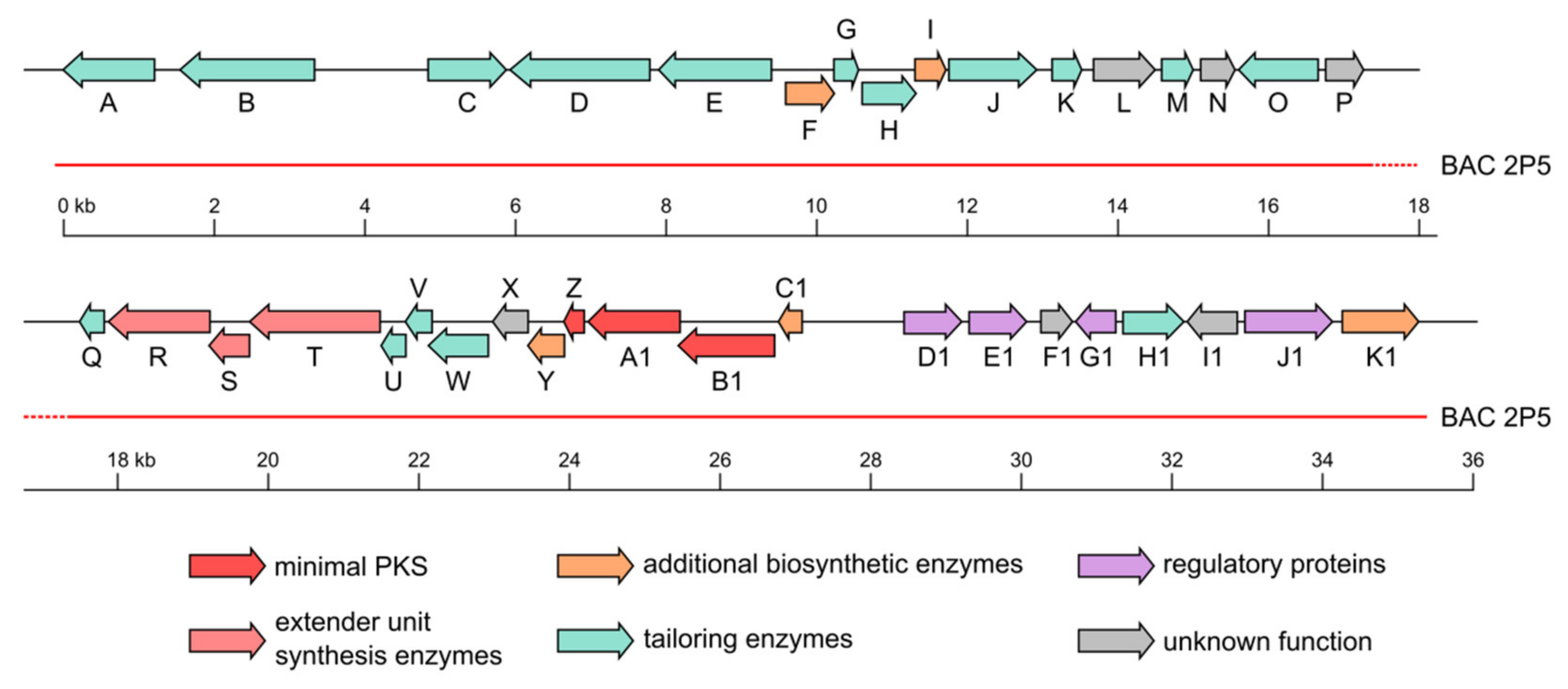
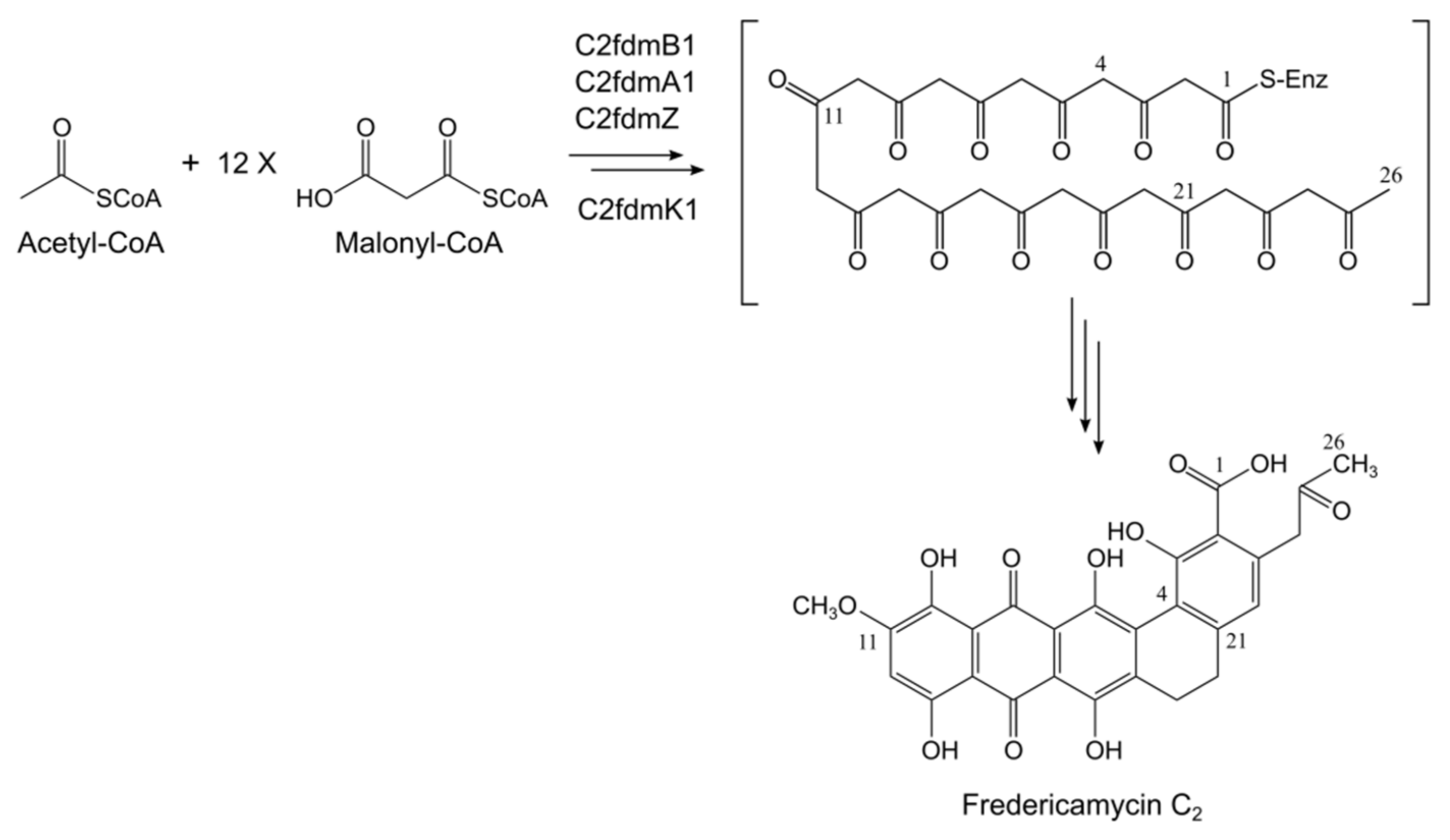
2.2. Fredericamycin C2 Is Biosynthesized by a Type II PKS Gene Cluster

Fredericamycin C2 (1) is structurally related to other fredericamycin variants, such as C and A, which are biosynthesized by a type II PKS gene cluster [22]. This suggests the involvement of a type II PKS system in the production of fredericamycin C2 (1). Although a subclass of type I PKSs (iterative type I PKSs) have also been reported to synthesize aromatic compounds [23,24,25,26], their products are structurally smaller and simpler than the complex multicyclic metabolites produced by type II PKSs [27]. The pentacyclic aromatic polyketide structure of fredericamycin C2 (1) (Figure 3) further supports the assumption of type II PKS genes involved in its biosynthesis. The genome of S. albus subsp. chlorinus was screened for secondary metabolite genes, revealing the presence of one type II PKS gene cluster. Based on protein BLAST analysis, we assigned the genes putative functions, which are summarized in Table 2. To test whether the expression of these genes leads to fredericamycin C2 (1) production, BAC 2P5 containing the identified type II PKS cluster was isolated from a genomic library of S. albus subsp. chlorinus NRRL B-24108 and transferred via intergeneric conjugation into the heterologous host Streptomyces albus Del14. HPLC-MS analysis of the extract from the resulting ex-conjugant S. albus 2P5 revealed the presence of a peak with identical retention time and m/z to those of fredericamycin C2 (1), demonstrating that the type II PKS cluster from S. albus subsp chlorinus is responsible for fredericamycin C2 (1) biosynthesis (Figure S3, Figure 2). BAC 2P5 comprises a 35 kb genomic region containing a total of 37 open reading frames (ORFs), 10 of which share homology at protein level with the fredericamycin A gene cluster from S. griseus (Table 2, Figure 4), which is also responsible for fredericamycin C (2) biosynthesis [22]. The homologue genes include those coding for the minimal PKS ketosynthase subunits (KSα and KSβ), two polyketide cyclases, four tailoring enzymes, a transcriptional regulator, and a protein of unknown function. The gene similarity with fredericamycin A cluster, together with the heterologous expression results (Figure S3), indicates the relevance of these genes in fredericamycin C2 (1) biosynthesis.

2.3. Screening for the Mutation Causing Fredericamycin C2 Overproduction and Streptomycin Resistance in S. albus subsp. chlorinus JR1

Frequently, streptomycin resistance results from a point mutation in the rpsL gene, which codes for the ribosomal protein S12 [3,4,28,29,30]. Following genome sequencing of S. albus subsp. chlorinus JR1, the resulting reads were mapped to the genome of the wild type strain and single nucleotide mutations were searched in the sequence corresponding to the rpsL gene. No point mutations were found in this sequence, indicating that the mutation responsible for streptomycin resistance in S. albus subsp. chlorinus JR1 is located elsewhere in the genome. A total of fifteen point mutations were detected in the genome of S. albus subsp. chlorinus JR1 (Table S3). A nucleotide insertion was detected within the coding sequence of the jag gene (SACHL2_00217; position 3714884) that codes for a single-stranded DNA binding protein. This gene partially overlaps with the adjacent downstream gene rsmG (SACHL2_00216), indicating their co-transcription in a bicistronic operon. The insertion of a cytosine nucleotide in the sequence of jag creates a premature stop codon that truncates the protein translation, also affecting the expression of the co-translating rsmG gene. rsmG codes for a 16S rRNA methyltransferase, and it has been reported that point-nonsense mutations in its sequence, as well as the deletion of this gene, lead to increased resistance to streptomycin and enhanced production of secondary metabolites in different bacterial strains [31,32]. Therefore, we believe that a point mutation in the jag gene is responsible for high-level streptomycin resistance and upregulation of fredericamycin C2 (1) biosynthetic gene expression in S. albus subsp. chlorinus JR1 by preventing the translation of the rsmG gene.

2.4. Biological Activity of Fredericamycin C2

Fredericamycin C2 (1) was tested for antibacterial activity against a Gram-positive (Bacillus subtilis) and two Gram-negative strains (Escherichia coli and Pseudomonas putida) through disk diffusion test. The new fredericamycin variant displays a growth inhibition zone against P. putida at a minimal concentration of 2.5 mg/mL (Figure S4). Fredericamycin C2 (1) shows no inhibitory activity against the growth of B. subtilis and E. coli.

3. Discussion

Fredericamycin variants constitute a family of aromatic polyketides with significant toxicity against tumor cells as well as moderate antibiotic and antifungal activity. Here, we present the novel variant fredericamycin C2 (1), which is overproduced by the strain S. albus subsp. chlorinus JR1, a spontaneous streptomycin-resistant mutant derived from S. albus subsp. chlorinus NRRL B-24108. We suggest that a mutation in the jag gene affecting the translation of the adjacent gene rsmG is responsible for the phenotype of the mutant strain. The frame shift originated by the point insertion putatively generates a truncated non-functional Jag protein. In previous studies, deletion of a jag homologue in Streptococcus pneumoniae led to retarded growth and smaller cell size compared to the wild type strain, indicating that Jag is likely involved in cell division [33]. However, no association of jag deletion with increased antibiotic resistance or induction of secondary metabolite production has been reported before. The point mutation in the jag gene has a polar effect on the overlapping gene rsmG, preventing its transcription. The enhanced production of fredericamycin C2 (1), as well as the increased streptomycin resistance observed in S. albus subsp. chlorinus JR1, is most likely derived from the lack of RsmG function. This is consistent with previous studies where rsmG deletion mutants showed higher resistance to streptomycin and improved yields of secondary metabolites at a late-growth phase [34,35]. rsmG encodes a methyltransferase that catalyzes the methylation of 16S rRNA at the residue G527 (E. coli numbering). This residue, together with C526 and the S12 protein, interacts with the antibiotic streptomycin [36]. These interactions tend to stabilize the tRNA-mRNA tandem, which affects the proof-reading process and results in misreading of the genetic code [37]. The absence of RsmG would generate 16S rRNA molecules non-methylated at residue G527, causing a weaker binding to streptomycin and making the strain resistant to the antibiotic. Although the mechanism by which the lack of 16S rRNA methyltransferase may induce the expression of fredericamycin C2 (1) biosynthetic genes in S. albus subsp. chlorinus JR1 remains unknown, we hypothesize that the mutant experiences an increased protein synthesis rate at a stationary phase, as it has been previously observed [34,35]. The increased protein synthetic activity leads to expression of both pleiotropic and pathway-specific regulatory proteins, which eventually enhance the transcription of poorly expressed secondary metabolite gene clusters. Several attempts to complement the jag and rsmG gene functions in S. albus subsp. chlorinus JR1 resulted in no recombinant colonies, suggesting the genetic intractability of the strain (data not shown).
The structure of fredericamycin C2 (1) presented in this paper only differs from that of fredericamycin C (2) in the polyketide chain length. While fredericamycin C (2) backbone consists of 30 carbon atoms, fredericamycin C2 (1) contains a C26 polyketide chain (Figure 3). Fredericamycin C (2) biosynthesis begins with the generation of a C6 primer unit (hexadienyl-ACP) by the PKS initiation module. This starter unit is then transferred to the elongation module, which presumably catalyzes the sequential decarboxylative condensation of 12 malonyl-CoA molecules, delivering a C30 polyketide chain [22,38]. In the case of fredericamycin C2 (1), we propose that acetyl-CoA functions as the starter unit, which is elongated by the polyketide synthase through successive incorporation of malonyl-CoA extender units (Figure 5). This process is most likely catalyzed by the minimal PKS enzymes encoded by the genes c2fdmB1, c2fdmA1, and c2fdmZ, and the ACP S-malonyltransferase encoded by c2fdmK1 (Table 2). The resulting C26 polyketide chain is subsequently modified by tailoring enzymes to eventually yield the product fredericamycin C2 (1) (Figure 5).

4. Materials and Methods

4.1. General Experimental Procedures

All strains and BACs (bacterial artificial chromosomes) used in this work are listed in Table S4. Escherichia coli strains were cultured in LB medium [39]. Streptomyces strains were grown on soy flour mannitol agar (MS agar) [40] for sporulation and conjugation and in liquid tryptic soy broth (TSB; Sigma-Aldrich, St. Louis, MO, USA). For metabolite expression, liquid DNPM medium (40 g/L dextrin, 7.5 g/L soytone, 5 g/L baking yeast, and 21 g/L MOPS, pH 6.8) or MS agar were used. The antibiotics kanamycin, apramycin, and nalidixic acid were supplemented when required.

4.2. Isolation and Manipulation of DNA

BAC extraction from a Streptomyces albus subsp. chlorinus-constructed genomic library (Intact Genomics, St. Louis, MO, USA), DNA manipulation, E. coli transformation, and E. coli/Streptomyces intergeneric conjugation were performed according to standard protocols [39,40,41]. Plasmid DNA was purified with the BACMAX™ DNA purification kit (Lucigen, Middleton, WI, USA). Restriction endonucleases were used according to manufacturer’s recommendations (New England Biolabs, Ipswich, MA, USA).

4.3. Strain Generation, Metabolite Extraction, and Analysis

The spontaneous streptomycin-resistant mutant Streptomyces albus subsp. chlorinus JR1 was obtained after several rounds of selection of streptomycin-resistant colonies growing on MS agar medium containing increasing concentrations of the antibiotic. MICs of streptomycin were determined by spreading spores on MS agar plates containing 50, 100, and 200 μg/mL of streptomycin. Metabolites were extracted from the agar with ethyl acetate acidified with 100% acetic acid up to pH = 2.0, evaporated and dissolved in methanol. One μL of extract was separated using a Dionex Ultimate 3000 UPLC (Thermo Fisher Scientific, Waltham, MA, USA), a 10-cm ACQUITY UPLC® BEH C18 column, 1.7 μm (Waters, Milford, MA, USA), and a linear gradient of 0.1% formic acid solution in acetonitrile against 0.1% formic acid solution in water from 5% to 95% in 18 min at a flow rate of 0.6 mL/min. Samples were analyzed using an Orbitrap speed mass spectrometer (Thermo Scientific, Waltham, MA, USA). Data were collected and analyzed with the Thermo Xcalibur software, version 3.0 (Thermo Scientific, Waltham, MA, USA). The monoisotopic mass was searched in a natural product database.

4.4. Fredericamycin C2 Purification and Quantification

S. albus subsp. chlorinus JR1 was grown for 8 days at 28 °C on 50 Petri dishes, each containing 40 mL of DNPM agar. Fredericamycin C2 (1) was extracted from the solid agar using ethyl acetate acidified with 100% acetic acid up to pH = 2.0. The crude extract was first separated by normal phase chromatography on a prepacked silica cartridge (Biotage, Uppsala, Sweden) using hexane (solvent A), chloroform (solvent B), ethyl acetate (solvent C), and methanol (solvent D) (1:1:1:1) as the mobile phase, in a linear gradient from 0% to 100% of each pair of solvents (A-B, B-C, and C-D). Fractions containing fredericamycin C2 (1) were detected by LC-MS analysis, pooled together, and further fractionated by semi-preparative HPLC (Dionex UltiMate 3000, Thermo Fisher Scientific, Waltham, MA, USA) using a C18 column (Synergi 10 μm, 250 × 10 mm; Phenomenex, Aschaffenburg, Germany) and a 0.1% formic acid solution in acetonitrile as the mobile phase in a linear gradient. UV spectra were recorded with a DAD detector at 274 nm and 525 nm. Finally, 0.8 g of fredericamycin C2 (1) was collected in a single fraction. For quantification, a calibration curve with different fredericamycin C2 (1) concentrations was constructed (Figure S1).
Fredericamycin C2 (1): Violet, amorphous solid; m/z 521.1077 [M + H]+ (calculated for C27H21O11, 521.1084); UV λmax (MeOH) 195, 248, 274, 345, 490 nm; 1H and 13C-NMR data, Table 1 and Supplementary Information Figures S2, S5–S10.

4.5. H-NMR Spectroscopy

NMR data were measured using a Varian VNMR-S600 spectrometer equipped with 3 mm triple resonance inverse and 3 mm dual broadband probes. Fredericamycin C2 (1) samples were dissolved in 150 µL DMSO-d6/Pyridine-d5 95:5 and measured at 35 °C. The residual solvent signal of DMSO was used as an internal reference.

4.6. Antimicrobial Susceptibility Test

Disk diffusion tests were performed according to [42]. Ten mL of LB soft agar (10 g/L tryptone, 10 g/L NaCl, 5 g/L yeast extract, 7 g/L agar) was inoculated with the strains Escherichia coli GB2005, Bacillus subtilis ATCC 6633, or Pseudomonas putida KT2440, and poured on LB agar plates. Five paper disks (Macherey and Nagel, Düren, Germany) were coated with 10, 5, 2.5, 0.5, or 0.25 mg/mL of fredericamycin C2 (1) solved in methanol and placed onto the solidified soft agar. An additional disk loaded with methanol was used as a negative control and the antibiotics nalidixic acid, ampicillin, and chloramphenicol (50 μg/mL, respectively) were used as positive controls. The plates were incubated at 28 °C overnight.

4.7. Genome Sequencing, Genome Assembly, and Analysis

S. albus subsp. chlorinus JR1 was sequenced using an Illumina MiSEQ library with 301-bp inserts (Illumina, San Diego, CA, USA). S. albus subsp. chlorinus JR1 strain genome assembly has a total of 57 contigs, and 5 final scaffolds—7,539,766 bp; 63,833 bp; 4,962 bp; 2,694 bp; and 2,548 bp (assembled with Newbler version 2, Roche, Basel, Switzerland). Sequencing reads coverage against the S. albus subsp. chlorinus B-24108 genome (Genbank accession number VJOK00000000) was examined with Geneious, version 11.0.3 (Biomatters Ltd., Auckland, New Zealand).

4.8. Genome Mining and Bioinformatics Analysis

The genome of S. albus subsp. chlorinus was screened for secondary metabolite biosynthetic gene clusters using the antiSMASH [43] online tool (https://antismash.secondarymetabolites.org/#!/start). Analysis of genetic data was performed using the Geneious software, version 11.0.3 (Biomatters Ltd., Auckland, New Zealand) [44].

5. Conclusions

Here, we demonstrate the significance of inducing poorly expressed secondary metabolite gene clusters for the identification of new microbial natural products. A mutation in the genome of the strain S. albus subsp. chlorinus JR1 obtained by selection of streptomycin-resistant colonies has led to overproduction and the discovery of the so far undescribed compound fredericamycin C2 (1). This secondary metabolite expands the structural variability of the fredericamycin family, whose most prominent member, fredericamycin A, is a potent antitumor drug. Screening for antibiotic-resistant strains represents a simple and inexpensive approach that has enabled the improvement of secondary metabolite production in the genetically intractable bacterial strain S. albus subsp. chlorinus.

Supplementary Materials

The following are available online at https://www.mdpi.com/1660-3397/18/6/284/s1; Table S1: Streptomycin MICs for S. albus subsp. chlorinus and S. albus subsp. chlorinus JR1; Table S2: Quantification of fredericamycin C2; Table S3: Type and location of point mutations in the genome of S. albus subsp. chlorinus JR1; Table S4: Bacterial strains and BACs used in this work; Figure S1: Calibration curve for fredericamycin C2 quantification; Figure S2: Important 2D NMR correlations in fredericamycin C2; Figure S3: HPLC-MS chromatograms of crude extracts from S. albus subsp. chlorinus JR1, S. albus 2P5, and its parental strain S. albus Del14; Figure S4: Antibacterial evaluation of fredericamycin C2. Figure S5: 1H NMR; Figure S6: 13C NMR; Figure S7: COSY NMR; Figure S8: HSQC NMR; Figure S9: HMBC NMR; Figure S10: ROESY NMR.

Author Contributions

M.R.E., M.M., and A.L. designed experiments; M.R.E., M.M., and B.R. performed experiments; T.P. performed and evaluated the NMR analysis; L.P. and J.R. developed and provided the mutant strain; M.R.E., M.M., and A.L. analyzed the data and wrote the manuscript; all authors reviewed the manuscript. Special thanks to M. Stierhof for chemical advice. All authors have read and agreed to the published version of the manuscript.

Funding

This research has received funding from BMBF grant “EXPLOMARE“ 031B0868A.

Conflicts of Interest

The authors declare no conflict of interest.

References

  1. Protasov, E.S.; Axenov-Gribanov, D.V.; Rebets, Y.V.; Voytsekhovskaya, I.V.; Tokovenko, B.T.; Shatilina, Z.M.; Luzhetskyy, A.N.; Timofeyev, M.A. The diversity and antibiotic properties of actinobacteria associated with endemic deepwater amphipods of Lake Baikal. Antonie Van Leeuwenhoek 2017, 110, 1593–1611. [Google Scholar] [CrossRef]
  2. Axenov-Gribanov, D.V.; Voytsekhovskaya, I.V.; Tokovenko, B.T.; Protasov, E.S.; Gamaiunov, S.V.; Rebets, Y.V.; Luzhetskyy, A.N.; Timofeyev, M.A. Actinobacteria isolated from an underground lake and moonmilk speleothem from the biggest conglomeratic karstic cave in Siberia as sources of novel biologically active compounds. PLoS ONE 2016, 11, e0152957. [Google Scholar] [CrossRef] [PubMed] [Green Version]
  3. Shima, J.; Hesketh, A.; Okamoto, S.; Kawamoto, S.; Ochi, K. Induction of actinorhodin production by rpsL (encoding ribosomal protein S12) mutations that confer streptomycin resistance in Streptomyces lividans and Streptomyces coelicolor A3(2). J. Bacteriol. 1996, 178, 7276–7284. [Google Scholar] [CrossRef] [PubMed] [Green Version]
  4. Hosoya, Y.; Okamoto, S.; Muramatsu, H.; Ochi, K. Acquisition of certain streptomycin-resistant (str) mutations enhances antibiotic production in bacteria. Antimicrob. Agents Chemother. 1998, 42, 2041–2047. [Google Scholar] [CrossRef] [PubMed] [Green Version]
  5. Hesketh, A.; Ochi, K. A novel method for improving Streptomyces coelicolor A3(2) for production of actinorhodin by introduction of rpsL (encoding ribosomal protein S12) mutations conferring resistance to streptomycin. J. Antibiot. 1997, 50, 532–535. [Google Scholar] [CrossRef] [PubMed]
  6. Hu, H.; Ochi, K. Novel approach for improving the productivity of antibiotic-producing strains by inducing combined resistant mutations. Appl. Environ. Microbiol. 2001, 67, 1885–1892. [Google Scholar] [CrossRef] [PubMed] [Green Version]
  7. Wang, L.Y.Z.; Liu, Q.; Huang, Y.; Hu, C.; Liao, G. Improvement of A21978C production in Streptomyces roseosporus by reporter-guided rpsL mutation selection. J. Appl. Microbiol. 2012, 112, 1095–1101. [Google Scholar] [CrossRef]
  8. Cashel, M.; Rudd, K.E. The stringent response. In Escherichia Coli and Salmonella Typhimurium: Cellular and Molecular Biology; Neidhart, F.C., Ed.; American Society for Microbiology: Washington, DC, USA, 1987. [Google Scholar]
  9. Ochi, K. Occurrence of the stringent response in Streptomyces sp. and its significance for the initiation of morphological and physiological differentiation. J. Gen. Microbiol. 1986, 132, 2621–2631. [Google Scholar] [CrossRef] [Green Version]
  10. Strauch, E.; Takano, E.; Baylis, H.A.; Bibb, M.J. The stringent response in Streptomyces coelicolor A3(2). Mol. Microbiol. 1991, 5, 289–298. [Google Scholar] [CrossRef] [PubMed] [Green Version]
  11. Sands, M.K.; Roberts, R.B. The effects of a tryptophan-histidine deficiency in a mutant of Escherichia coli. J. Bacteriol. 1952, 63, 505–511. [Google Scholar] [CrossRef] [Green Version]
  12. Gralla, J.D. Escherichia coli ribosomal RNA transcription: Regulatory roles for ppGpp, NTPs, architectural proteins and a polymerase-binding protein. Mol. Microbiol. 2005, 55, 973–977. [Google Scholar] [CrossRef] [PubMed]
  13. Myronovskyi, M.; Rosenkranzer, B.; Stierhof, M.; Petzke, L.; Seiser, T.; Luzhetskyy, A. Identification and Heterologous Expression of the Albucidin Gene Cluster from the Marine Strain Streptomyces Albus Subsp. Chlorinus NRRL B-24108. Microorganisms 2020, 8, 237. [Google Scholar] [CrossRef] [PubMed] [Green Version]
  14. Rodriguez Estevez, M.; Myronovskyi, M.; Gummerlich, N.; Nadmid, S.; Luzhetskyy, A. Heterologous expression of the nybomycin gene cluster from the marine strain Streptomyces albus subsp. chlorinus NRRL B-24108. Mar. Drugs 2018, 16, 435. [Google Scholar] [CrossRef] [PubMed] [Green Version]
  15. Pandey, R.C.; Toussaint, M.W.; Stroshane, R.M.; Kalita, C.C.; Aszalos, A.A.; Garretson, A.L.; Wei, T.T.; Byrne, K.M.; Geoghegan, R.F., Jr.; White, R.J. Fredericamycin A, a new antitumor antibiotic. I. Production, isolation and physicochemical properties. J. Antibiot. 1981, 34, 1389–1401. [Google Scholar] [CrossRef]
  16. Misra, R.; Pandey, R.C.; Hilton, B.D.; Roller, P.P.; Silverton, J.V. Structure of fredericamycin A, an antitumor antibiotic of a novel skeletal type; spectroscopic and mass spectral characterization. J. Antibiot. 1987, 40, 786–802. [Google Scholar] [CrossRef] [Green Version]
  17. Sontag, B.; Muller, J.G.; Hansske, F.G. Fredericamycin B and C from Streptomyces griseus: Structure elucidation after 23 years. J. Antibiot. 2004, 57, 823–828. [Google Scholar] [CrossRef] [Green Version]
  18. Chen, Y.; Luo, Y.; Ju, J.; Wendt-Pienkowski, E.; Rajski, S.R.; Shen, B. Identification of fredericamycin E from Streptomyces griseus: Insights into fredericamycin A biosynthesis highlighting carbaspirocycle formation. J. Nat. Prod. 2008, 71, 431–437. [Google Scholar] [CrossRef]
  19. Warnick-Pickle, D.J.; Byrne, K.M.; Pandey, R.C.; White, R.J. Fredericamycin A, a new antitumor antibiotic. II. Biological properties. J. Antibiot. 1981, 34, 1402–1407. [Google Scholar] [CrossRef] [Green Version]
  20. Lu, K.P.; Fischer, G. Methods of Inhibiting Pin1-Associated States Using a Fredericamycin A Compound. WO/2004/002429. 2004. Available online: https://patentscope2.wipo.int/search/en/detail.jsf?docId=WO2004002429 (accessed on 2 March 2020).
  21. Yasuzawa, T.; Yoshida, M.; Shirahata, K.; Sano, H. Structure of a novel Ca2+ and calmodulin-dependent cyclic nucleotide phosphodiesterase inhibitor KS-619-1. J. Antibiot. 1987, 40, 1111–1114. [Google Scholar] [CrossRef] [Green Version]
  22. Wendt-Pienkowski, E.; Huang, Y.; Zhang, J.; Li, B.; Jiang, H.; Kwon, H.; Hutchinson, C.R.; Shen, B. Cloning, sequencing, analysis, and heterologous expression of the fredericamycin biosynthetic gene cluster from Streptomyces griseus. J. Am. Chem. Soc. 2005, 127, 16442–16452. [Google Scholar] [CrossRef]
  23. Zhang, Q.; Pang, B.; Ding, W.; Liu, W. Aromatic Polyketides Produced by Bacterial Iterative Type I Polyketide Synthases. ACS Catal. 2013, 3, 1439–1447. [Google Scholar] [CrossRef]
  24. Kudo, F.; Kasama, Y.; Hirayama, T.; Eguchi, T. Cloning of the pactamycin biosynthetic gene cluster and characterization of a crucial glycosyltransferase prior to a unique cyclopentane ring formation. J. Antibiot. 2007, 60, 492–503. [Google Scholar] [CrossRef] [PubMed] [Green Version]
  25. Zhao, Q.; He, Q.; Ding, W.; Tang, M.; Kang, Q.; Yu, Y.; Deng, W.; Zhang, Q.; Fang, J.; Tang, G.; et al. Characterization of the azinomycin B biosynthetic gene cluster revealing a different iterative type I polyketide synthase for naphthoate biosynthesis. Chem. Biol. 2008, 15, 693–705. [Google Scholar] [CrossRef] [PubMed] [Green Version]
  26. Xiao, Y.; Li, S.; Niu, S.; Ma, L.; Zhang, G.; Zhang, H.; Zhang, G.; Ju, J.; Zhang, C. Characterization of tiacumicin B biosynthetic gene cluster affording diversified tiacumicin analogues and revealing a tailoring dihalogenase. J. Am. Chem. Soc. 2011, 133, 1092–1105. [Google Scholar] [CrossRef]
  27. Hertweck, C.; Luzhetskyy, A.; Rebets, Y.; Bechthold, A. Type II polyketide synthases: Gaining a deeper insight into enzymatic teamwork. Nat. Prod. Rep. 2007, 24, 162–190. [Google Scholar] [CrossRef]
  28. Funatsu, G.; Wittmann, H.G. Ribosomal proteins. 33. Location of amino-acid replacements in protein S12 isolated from Escherichia coli mutants resistant to streptomycin. J. Mol. Biol. 1972, 68, 547–550. [Google Scholar] [CrossRef]
  29. Finken, M.; Kirschner, P.; Meier, A.; Wrede, A.; Bottger, E.C. Molecular basis of streptomycin resistance in Mycobacterium tuberculosis: Alterations of the ribosomal protein S12 gene and point mutations within a functional 16S ribosomal RNA pseudoknot. Mol. Microbiol. 1993, 9, 1239–1246. [Google Scholar] [CrossRef]
  30. Meier, A.; Kirschner, P.; Bange, F.C.; Vogel, U.; Bottger, E.C. Genetic alterations in streptomycin-resistant Mycobacterium tuberculosis: Mapping of mutations conferring resistance. Antimicrob. Agents Chemother. 1994, 38, 228–233. [Google Scholar] [CrossRef] [Green Version]
  31. Tanaka, Y.; Tokuyama, S.; Ochi, K. Activation of secondary metabolite-biosynthetic gene clusters by generating rsmG mutations in Streptomyces griseus. J. Antibiot. 2009, 62, 669–673. [Google Scholar] [CrossRef]
  32. Mikheil, D.M.; Shippy, D.C.; Eakley, N.M.; Okwumabua, O.E.; Fadl, A.A. Deletion of gene encoding methyltransferase (gidB) confers high-level antimicrobial resistance in Salmonella. J. Antibiot. 2012, 65, 185–192. [Google Scholar] [CrossRef]
  33. Ulrych, A.; Holeckova, N.; Goldova, J.; Doubravova, L.; Benada, O.; Kofroňová, O.; Halada, P.; Branny, P. Characterization of pneumococcal Ser/Thr protein phosphatase phpP mutant and identification of a novel PhpP substrate, putative RNA binding protein Jag. BMC Microbiol. 2016, 16, 247. [Google Scholar] [CrossRef] [PubMed] [Green Version]
  34. Nishimura, K.; Hosaka, T.; Tokuyama, S.; Okamoto, S.; Ochi, K. Mutations in rsmG, encoding a 16S rRNA methyltransferase, result in low-level streptomycin resistance and antibiotic overproduction in Streptomyces coelicolor A3(2). J. Bacteriol. 2007, 189, 3876–3883. [Google Scholar] [CrossRef] [PubMed] [Green Version]
  35. Lopatniuk, M.; Myronovskyi, M.; Nottebrock, A.; Busche, T.; Kalinowski, J.; Ostash, B.; Fedorenko, V.; Luzhetskyy, A. Effect of “ribosome engineering” on the transcription level and production of S. albus indigenous secondary metabolites. Appl. Microbiol. Biotechnol. 2019, 103, 7097–7110. [Google Scholar] [CrossRef] [PubMed]
  36. Carter, A.P.; Clemons, W.M.; Brodersen, D.E.; Morgan-Warren, R.J.; Wimberly, B.T.; Ramakrishnan, V. Functional insights from the structure of the 30S ribosomal subunit and its interactions with antibiotics. Nature 2000, 407, 340–348. [Google Scholar] [CrossRef]
  37. Davies, J.; Gorini, L.; Davis, B.D. Misreading of RNA codewords induced by aminoglycoside antibiotics. Mol. Pharmacol. 1965, 1, 93–106. [Google Scholar]
  38. Das, A.; Szu, P.H.; Fitzgerald, J.T.; Khosla, C. Mechanism and engineering of polyketide chain initiation in fredericamycin biosynthesis. J. Am. Chem. Soc. 2010, 132, 8831–8833. [Google Scholar] [CrossRef] [Green Version]
  39. Green, M.R.; Sambrook, J. Molecular Cloning: A Laboratory Manual, 4th ed.; Cold Spring Harbor Laboratory Press: New York, NY, USA, 2012. [Google Scholar]
  40. Kieser, T.B.M.J.; Buttner, M.J.; Charter, K.F.; Hopwood, D.A. Practical Streptomyces Genetics; John Innes Foundation: Norwich, UK, 2000. [Google Scholar]
  41. Rebets, Y.; Kormanec, J.; Lutzhetskyy, A.; Bernaerts, K.; Anne, J. Cloning and expression of metagenomic DNA in Streptomyces lividans and subsequent fermentation for optimized production. In Metagenomics. Methods in Molecular Biology; Streit, W., Daniel, R., Eds.; Humana Press: New York, NY, USA, 2017. [Google Scholar]
  42. Bauer, A.W.; Kirby, W.M.; Sherris, J.C.; Turck, M. Antibiotic susceptibility testing by a standardized single disk method. Am. J. Clin. Pathol. 1966, 45, 493–496. [Google Scholar] [CrossRef]
  43. Weber, T.; Blin, K.; Duddela, S.; Krug, D.; Kim, H.U.; Bruccoleri, R.; Lee, S.Y.; Fischbach, M.A.; Muller, R.; Wohlleben, W.; et al. antiSMASH 3.0-a comprehensive resource for the genome mining of biosynthetic gene clusters. Nucleic Acids Res. 2015, 43, W237–W243. [Google Scholar] [CrossRef] [Green Version]
  44. Kearse, M.; Moir, R.; Wilson, A.; Stones-Havas, S.; Cheung, M.; Sturrock, S.; Buxton, S.; Cooper, A.; Markowitz, S.; Duran, C.; et al. Geneious Basic: An integrated and extendable desktop software platform for the organization and analysis of sequence data. Bioinformatics 2012, 28, 1647–1649. [Google Scholar] [CrossRef]
Figure 1. Streptomyces albus subsp. chlorinus NRRL B-24108 (left) and Streptomyces albus subsp. chlorinus JR1 (right) spores on MS agar medium.
Marinedrugs 18 00284 g001
Figure 2. HPLC-MS analysis of crude extract from solid cultures of S. albus subsp. chlorinus JR1 (a) and its parental strain S. albus subsp. chlorinus NRRL B-24108 (b). (A) UV chromatogram. The asterisk (*) indicates the peak corresponding to fredericamycin C2 (1) at tR = 12.7 min. (B) Mass spectrum associated with tR = 12.7 min from the UV chromatogram displayed in (A). (C) UV spectrum of purified fredericamycin C2 (1).
Marinedrugs 18 00284 g002
Figure 3. Structures of fredericamycin C2 (1), fredericamycin C (2) and KS-619-1 (3).
Marinedrugs 18 00284 g003
Figure 4. Map of the genes encoded in BAC 2P5 isolated from a genomic library of S. albus subsp. chlorinus. Characters from A to K1 indicate the corresponding c2fdm gene described in Table 2.
Marinedrugs 18 00284 g004
Figure 5. Proposed early biosynthesis steps of fredericamycin C2 (1) in S. albus subsp. chlorinus.
Marinedrugs 18 00284 g005
Table 1. NMR data of fredericamycin C2 (1) (600/150 MHz, DMSO-d6/Pyridine-d5 95:5, 35 °C, solvent (DMSO) as internal reference).
Pos. aδCδH (J Hz)COSY bHMBC bROESY b
9’164.3 5’-H
9a’115.3 4’-H2, 5’-H
3’140.7 4’-H2, (5’-H)
5’120.46.52 s(4‘-H2)1J, 4’-H2, (5’-H), 6’-H24’-H2, 6’-H2, (7’-H2)
5a’144.3 5‘-H, 6‘-H2, 7‘-H2
6’28.82.63 br t (7Hz)7’-H25’-H, 7’-H25’-H, 7’-H2
7’21.22.79 br t (7Hz)6’-H26’-H2
7a’137.3 6’-H2, 7’-H2
3152.5 7’-H2
3a112.2
4186.9
4a113.2 (7-H)
5148.8 c 7-H
6157.6 6-OCH3, 7-H
6-OCH356.43.94 s6-H1J7-H
7105.86.88 s6-OCH36-OCH36-OCH3
8158.6 7-H
8a105.8 7-H
9187.6 7-H
9a114.4
1156.3
2 = 8’136.3 (5’-H), 7’-H2
8a’121.0 5’-H
1’169.3 5’-H
4’49.54.06 s(5’-H, 1’’-H3)5’-H, (1’’-H3)5’-H, 1’’-H3
3’(204.1) d
1’’29.72.17 s 4’-H2
OH 13.22 br s
12.80 br s
5.5 br s
a Numbering according to fredericamycin C (2); b weak signals in brackets; c from HMBC; d very weak signal in carbon NMR spectrum.
Table 2. Proposed functions of genes present in the type II polyketide synthase (PKS) cluster of S. albus subsp. chlorinus and homology with fredericamycin A gene cluster.
GeneSize (aa)Proposed FunctionGenBank Homologue 1Identity/Similarity (%)Fredericamycin A Gene Cluster HomologueIdentity/Similarity (%)
c2fdmA406Cytochrome P450 oxygenaseWP_017596471.166/73--
c2fdmB594MonooxygenaseWP_017596470.167/75--
c2fdmC337O-methyltransferaseWP_043504920.142/55--
c2fdmD620Asparagine synthaseWP_017596467.179/88--
c2fdmE497MonooxygenaseWP_081620749.159/70--
c2fdmF216Polyketide cyclaseREH43750.143/54--
c2fdmG107MonooxygenaseWP_027732672.144/64--
c2fdmH2373-ketoacyl-ACP reductaseWP_017596481.172/83--
c2fdmI134Polyketide cyclaseWP_109361109.172/83--
c2fdmJ389O-methyltransferaseWP_061257536.167/79fdmN56/70
c2fdmK138OxidoreductaseWP_020573863.156/68--
c2fdmL263UnknownWP_017596478.156/71--
c2fdmM138OxidoreductaseWP_017596477.170/85--
c2fdmN150UnknownWP_017596476.176/89--
c2fdmO358O-methyltransferaseWP_043504920.140/54--
c2fdmP169UnknownWP_017596474.162/75--
c2fdmQ113MonooxygenaseWP_017596454.165/76--
c2fdmR454Biotin carboxylaseWP_017596455.180/87--
c2fdmS175Biotin carboxyl carrier proteinWP_026120848.160/71--
c2fdmT585Carboxyl transferaseWP_017596457.175/80--
c2fdmU111MonooxygenaseWP_017596458.170/78fdmQ50/66
c2fdmV113MonooxygenaseWP_017596459.176/83fdmP51/70
c2fdmW2483-ketoacyl-ACP reductaseWP_017596460.183/92fdmO55/71
c2fdmX153UnknownWP_015621174.161/74--
c2fdmY156Polyketide cyclaseWP_075740187.167/82fdmI56/74
c2fdmZ87ACPWP_017596463.146/71--
c2fdmA1409KSβWP_017596464.177/86fdmG61/72
c2fdmB1422KSαWP_017596465.183/90fdmF63/77
c2fdmC1112Polyketide cyclaseWP_017596466.183/90fdmD64/76
c2fdmD1254Transcriptional regulatorWP_116247593.158/78fdmR146/64
c2fdmE1256Transcriptional regulatorWP_081620746.167/80--
c2fdmF1144UnknownWP_017596472.178/89fdmE59/72
c2fdmG1179Transcriptional regulatorKPC87453.180/84--
c2fdmH1271Serine hydrolaseWP_099880484.190/93--
c2fdmI1225UnknownWP_099880487.181/86--
c2fdmJ1394Transcriptional regulatorWP_055497612.198/98--
c2fdmK1315ACP S-malonyltransferaseWP_099880491.187/92--
1 NCBI accession numbers are given.

