11β,20β-Epoxybriaranes from the Gorgonian Coral Junceella fragilis (Ellisellidae)


 , , and
Abstract
1. Introduction
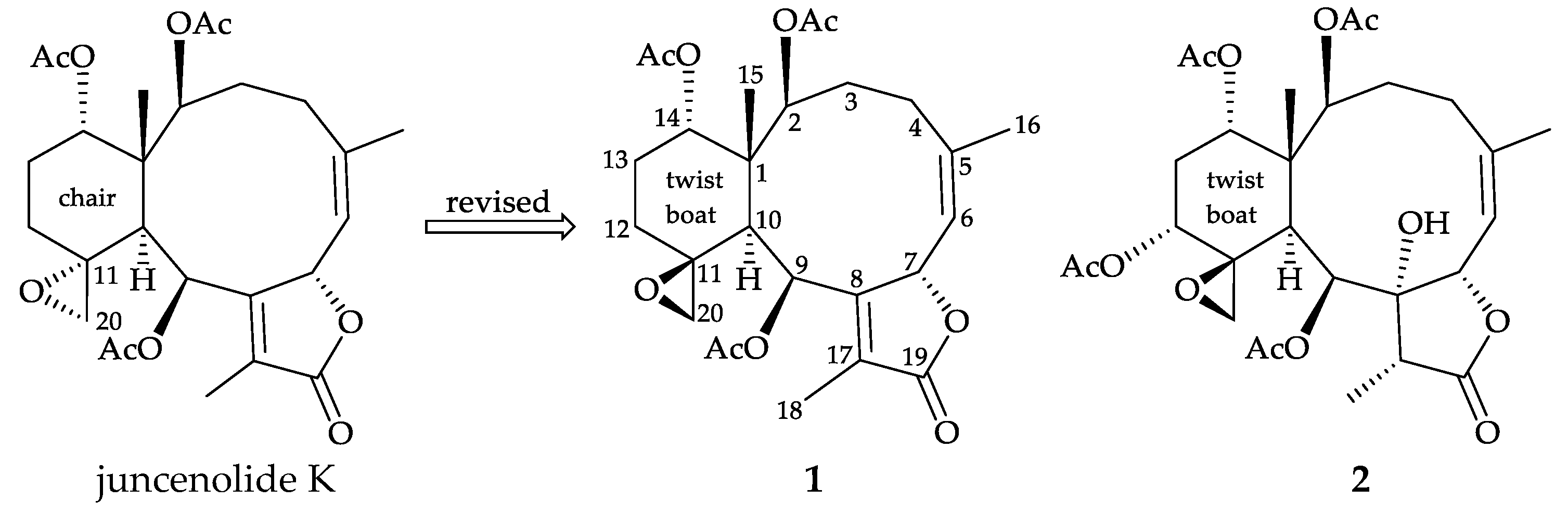
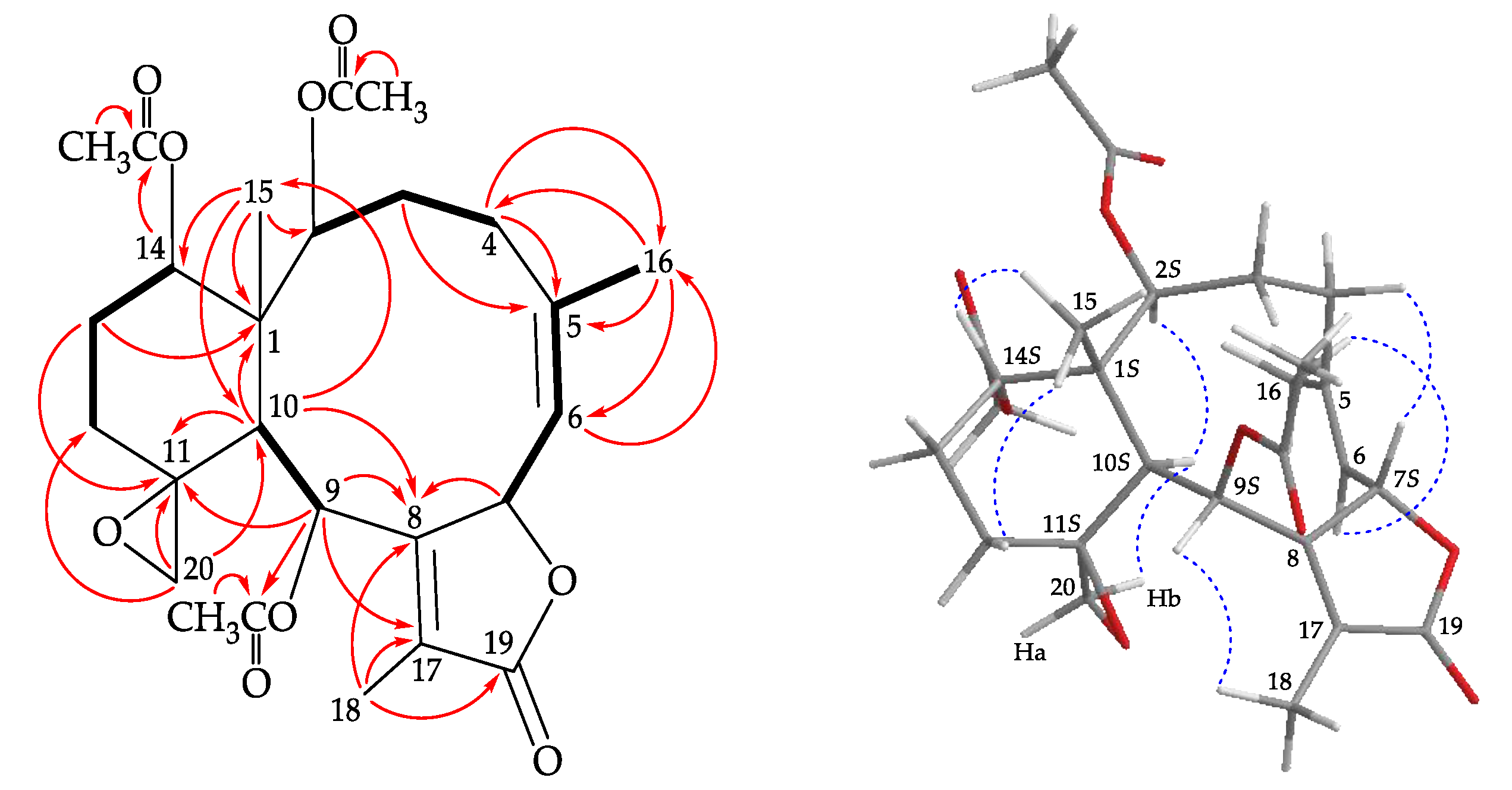
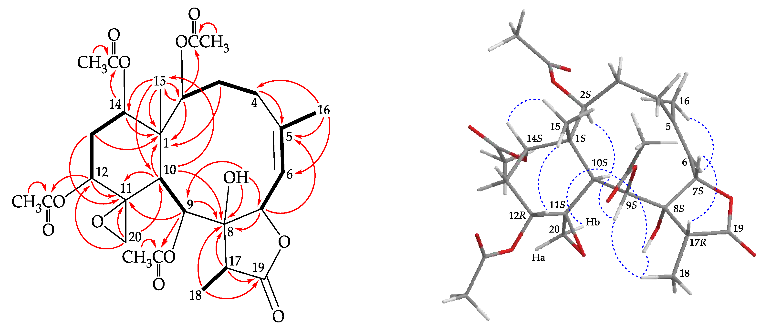
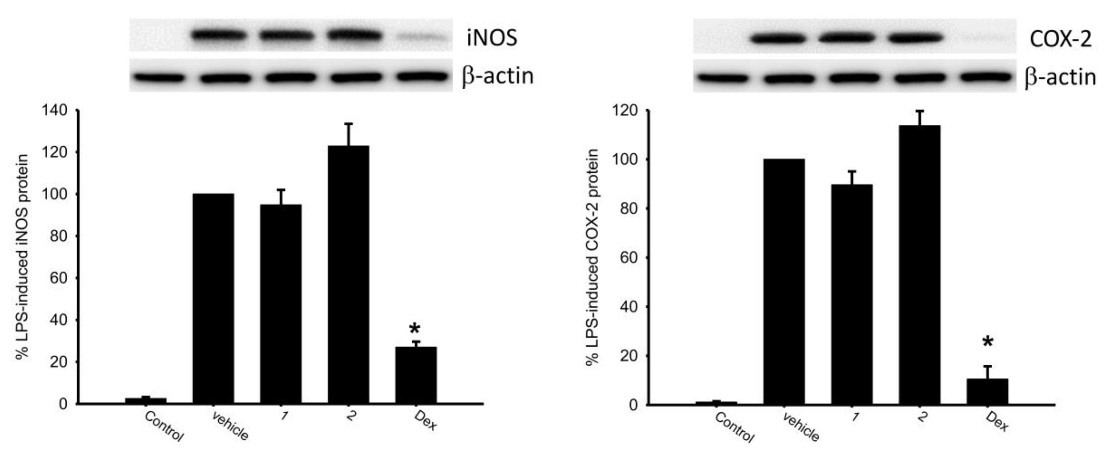
2. Results and Discussion
3. Materials and Methods
3.1. General Experimental Procedures
3.2. Animal Material
3.3. Extraction and Isolation
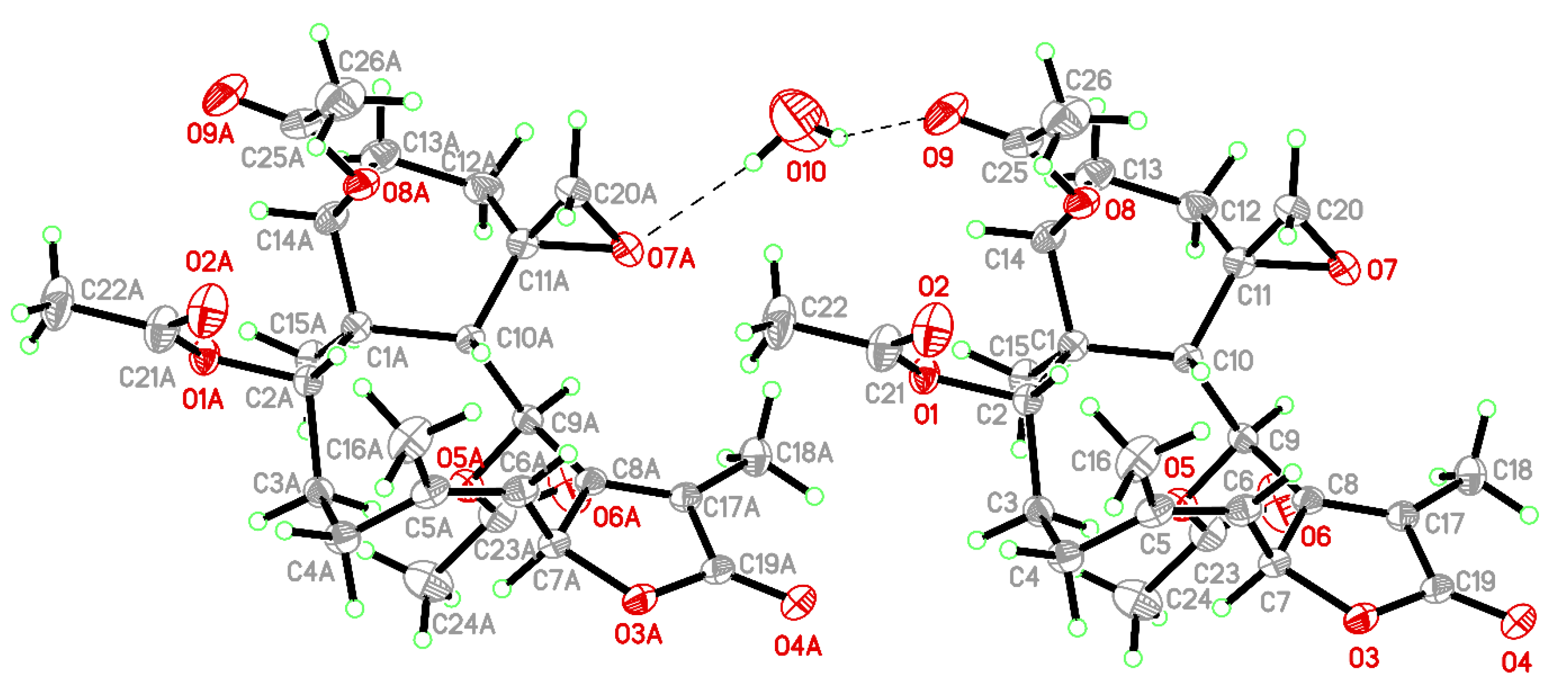
3.4. Single-Crystal X-ray Crystallography of Juncenolide K (1)
3.5. In Vitro Inflammatory Assay
4. Conclusions
Supplementary Materials
Author Contributions
Funding
Conflicts of Interest
References
- Bayer, F.M. Key to the genera of octocorallia exclusive of Pennatulacea (Coelenterata: Anthozoa), with diagnoses of new taxa. Proc. Biol. Soc. Wash. 1981, 94, 902–947. [Google Scholar]
- Bayer, F.M.; Grasshoff, M. The genus group taxa of the family Ellisellidae, with clarification of the genera established by J.E. Gray (Cnidaria: Octocorallia). Senckenb. Biol. 1994, 74, 21–45. [Google Scholar]
- Chen, C.-C.; Chang, K.-H. Gorgonacea (Coelenterata: Anthozoa: Octocorallia) of Southern Taiwan. Bull. Inst. Zool. Acad. Sin. 1991, 30, 149–181. [Google Scholar]
- Chung, H.-M.; Wang, Y.-C.; Tseng, C.-C.; Chen, N.-F.; Wen, Z.-H.; Fang, L.-S.; Hwang, T.-L.; Wu, Y.-C.; Sung, P.-J. Natural product chemistry of gorgonian corals of genus Junceella–Part III. Mar. Drugs 2018, 16, 339, and review articles in this series. [Google Scholar] [CrossRef] [PubMed]
- Su, Y.-M.; Fan, T.-Y.; Sung, P.-J. 11,20-Epoxybriaranes from the gorgonian coral Ellisella robusta (Ellisellidae). Nat. Prod. Res. 2007, 21, 1085–1090. [Google Scholar] [CrossRef] [PubMed]
- Shin, J.; Park, M.; Fenical, W. The junceellolides, new anti-inflammatory diterpenoids of the briarane class from the Chinese gorgonian Junceella fragilis. Tetrahedron 1989, 45, 1633–1638. [Google Scholar] [CrossRef]
- Sheu, J.-H.; Chen, Y.-P.; Hwang, T.-L.; Chiang, M.Y.; Fang, L.-S.; Sung, P.-J. Junceellolides J–L, 11,20-epoxybriaranes from the gorgonian coral Junceella fragilis. J. Nat. Prod. 2006, 69, 269–273. [Google Scholar] [CrossRef] [PubMed]
- Shen, Y.-C.; Chen, Y.-H.; Hwang, T.-L.; Guh, J.-H.; Khalil, A.T. Four new briarane diterpenoids from the gorgonian coral Junceella fragilis. Helv. Chim. Acta 2007, 90, 1391–1398. [Google Scholar] [CrossRef]
- Sung, P.-J.; Chen, Y.-P.; Su, Y.-M.; Hwang, T.-L.; Hu, W.-P.; Fan, T.-Y.; Wang, W.-H. Fragilide B: A novel briarane-type diterpenoid with a S-cis diene moiety. Bull. Chem. Soc. Jpn. 2007, 80, 1205–1207. [Google Scholar] [CrossRef]
- Sung, P.-J.; Lin, M.-R.; Su, Y.-D.; Chiang, M.Y.; Hu, W.-P.; Su, J.-H.; Cheng, M.-C.; Hwang, T.-L.; Sheu, J.-H. New briaranes from the octocorals Briareum excavatum (Briareidae) and Junceella fragilis (Ellisellidae). Tetrahedron 2008, 64, 2596–2604. [Google Scholar] [CrossRef]
- Sung, P.-J.; Pai, C.-H.; Su, Y.-D.; Hwang, T.-L.; Kuo, F.-W.; Fan, T.-Y.; Li, J.-J. New 8-hydroxybriarane diterpenoids from the gorgonians Junceella juncea and Junceella fragilis (Ellisellidae). Tetrahedron 2008, 64, 4224–4232. [Google Scholar] [CrossRef]
- Hwang, T.-L.; Lin, M.-R.; Tsai, W.-T.; Yeh, H.-C.; Hu, W.-P.; Sheu, J.-H.; Sung, P.-J. New polyoxygenated briaranes from octocorals Briareum excavatum and Ellisella robusta. Bull. Chem. Soc. Jpn. 2008, 81, 1638–1646. [Google Scholar] [CrossRef]
- Wang, S.-S.; Chen, Y.-H.; Chang, J.-Y.; Hwang, T.-L.; Chen, C.-H.; Khalil, A.T.; Shen, Y.-C. Juncenolides H–K, new briarane diterpenoids from Junceella juncea. Helv. Chim. Acta 2009, 92, 2092–2100. [Google Scholar] [CrossRef]
- Chang, Y.-C.; Hwang, T.-L.; Huang, S.-K.; Huang, L.-W.; Lin, M.-R.; Sung, P.-J. 12-epi-Fragilide G, a new briarane-type diterpenoid from the gorgonian coral Ellisella robusta. Heterocycles 2010, 81, 991–996. [Google Scholar] [CrossRef]
- Wang, S.-H.; Chang, Y.-C.; Chiang, M.Y.; Chen, Y.-H.; Hwang, T.-L.; Weng, C.-F.; Sung, P.-J. Chlorinated briarane diterpenoids from the sea whip gorgonian corals Junceella fragilis and Ellisella robusta (Ellisellidae). Chem. Pharm. Bull. 2010, 58, 928–933. [Google Scholar] [CrossRef]
- Chang, J.-Y.; Liaw, C.-C.; Fazary, A.E.; Hwang, T.-L.; Shen, Y.-C. New briarane diterpenoids from the gorgonian coral Junceella juncea. Mar. Drugs 2012, 10, 1321–1330. [Google Scholar] [CrossRef]
- Cheng, W.; Li, X.; Yin, F.; van Ofwegen, L.; Lin, W. Halogenated briarane diterpenes with acetyl migration from the gorgonian coral Junceella fragilis. Chem. Biodivers. 2017, 14, e1700053. [Google Scholar] [CrossRef]
- Zheng, L.-G.; Chang, Y.-C.; Hu, C.-C.; Wen, Z.-H.; Wu, Y.-C.; Sung, P.-J. Fragilides K and L, new briaranes from the gorgonian coral Junceella fragilis. Molecules 2018, 23, 1510. [Google Scholar] [CrossRef]
- Su, T.-P.; Yuan, C.-H.; Jhu, Y.-M.; Peng, B.-R.; Wen, Z.-H.; Wu, Y.-J.; Wu, T.-Y.; Liu, H.-W.; Sung, P.-J. Fragilides U–W: New 11,20-epoxybriaranes from the sea whip gorgonian coral Junceella fragilis. Mar. Drugs 2019, 17, 706. [Google Scholar] [CrossRef]
- Zheng, L.-G.; Chang, Y.-C.; Chen, J.-J.; Wen, Z.-H.; Hwang, T.-L.; Sung, P.-J. (+)-12-epi-Fragilide G, a new chlorinated briarane from the sea whip gorgonian coral Junceella fragilis. Heterocycles 2019, 96, 1601–1609. [Google Scholar]
- Hamann, M.T.; Harrison, K.N.; Carroll, A.R.; Scheuer, P.J. Briarane diterpenes from Micronesian gorgonians. Heterocycles 1999, 42, 325–331. [Google Scholar]
- El Sayed, K.A.; Dunbar, D.C.; Perry, T.L.; Wilkins, S.P.; Hamann, M.T.; Greenplate, J.T.; Wideman, M.A. Marine natural products as prototype insecticidal agents. J. Agric. Food Chem. 1997, 45, 2735–2739. [Google Scholar] [CrossRef]
- Shen, Y.-C.; Lin, Y.-C.; Chiang, M.Y. Juncenolide A, a new briarane from the Taiwanese gorgonian Junceella juncea. J. Nat. Prod. 2002, 65, 54–56. [Google Scholar] [CrossRef] [PubMed]
- Shen, Y.-C.; Lin, Y.-C.; Ko, C.-L.; Wang, L.-T. New briaranes from the Taiwanese gorgonian Junceella juncea. J. Nat. Prod. 2003, 66, 302–305. [Google Scholar] [CrossRef] [PubMed]
- Tanaka, C.; Yamamoto, Y.; Otsuka, M.; Tanaka, J.; Ichiba, T.; Marriott, G.; Rachmat, R.; Higa, T. Briarane diterpenes from two species of octocorals, Ellisella sp. and Pteroeides sp. J. Nat. Prod. 2004, 67, 1368–1373. [Google Scholar] [CrossRef] [PubMed]
- Qi, S.-H.; Zhang, S.; Wen, Y.-M.; Xiao, Z.-H.; Li, Q.-X. New briaranes from the South China Sea gorgonian Junceella fragilis. Helv. Chim. Acta 2005, 88, 2349–2354. [Google Scholar] [CrossRef]
- Sun, J.-F.; Huang, H.; Chai, X.-Y.; Yang, X.-W.; Meng, L.; Huang, C.-G.; Zhou, X.-F.; Yang, B.; Hu, J.; Chen, X.-Q.; et al. Dichotellides A–E, five new iodine-containing briarane type diterpenoids from Dichotella gemmacea. Tetrahedron 2011, 67, 1245–1250. [Google Scholar] [CrossRef]
- Li, C.; La, M.-P.; Sun, P.; Kurtan, T.; Mandi, A.; Tang, H.; Liu, B.-S.; Yi, Y.-H.; Li, L.; Zhang, W. Bioactive (3Z,5E)-11,20-epoxybriara-3,5-dien-7,18-olide diterpenoids from the South China Sea gorgonian Dichotella gemmacea. Mar. Drugs 2011, 9, 1403–1418. [Google Scholar] [CrossRef]
- Li, C.; La, M.-P.; Tang, H.; Pan, W.-H.; Sun, P.; Krohn, K.; Yi, Y.-H.; Li, L.; Zhang, W. Bioactive briarane diterpenoids from the South China Sea gorgonian Dichotella gemmacea. Bioorg. Med. Chem. Lett. 2012, 22, 4368–4372. [Google Scholar] [CrossRef]
- Li, C.; Jiang, M.; La, M.-P.; Li, T.-J.; Tang, H.; Sun, P.; Liu, B.-S.; Yi, Y.-H.; Liu, Z.; Zhang, W. Chemistry and tumor cell growth inhibitory activity of 11,20-epoxy-3Z,5(6)E-diene briaranes from the South China Sea gorgonian Dichotella gemmacea. Mar. Drugs 2013, 11, 1565–1582. [Google Scholar] [CrossRef]
- La, M.-P.; Li, J.; Li, C.; Tang, H.; Liu, B.-S.; Sun, P.; Zhuang, C.-L.; Li, T.-J.; Zhang, W. Briarane diterpenoids from the gorgonian Dichotella gemmacea. Mar. Drugs 2014, 12, 6178–6189. [Google Scholar] [CrossRef] [PubMed]
- Li, C.; La, M.-P.; Tang, H.; Sun, P.; Liu, B.-S.; Zhuang, C.-L.; Yi, Y.-H.; Zhang, W. Chemistry and bioactivity of briaranes from the South China Sea gorgonian Dichotella gemmacea. Mar. Drugs 2016, 14, 201. [Google Scholar] [CrossRef] [PubMed]
- Cheng, W.; Ji, M.; Li, X.; Ren, J.; Yin, F.; van Ofwegen, L.; Yu, S.; Chen, X.; Lin, W. Fragilolides A–Q, norditerpenoid and briarane diterpenoids from the gorgonian coral Junceella fragilis. Tetrahedron 2017, 73, 2518–2528. [Google Scholar] [CrossRef]
- Qi, S.-H.; Zhang, S.; Qian, P.-Y.; Xiao, Z.-H.; Li, M.-Y. Ten new antifouling briarane diterpenoids from the South China Sea gorgonian Junceella juncea. Tetrahedron 2006, 62, 9123–9130. [Google Scholar] [CrossRef]
- Qi, S.H.; Zhang, S.; Qian, P.Y.; Xu, H.H. Antifeedant and antifouling briaranes from the South China Sea gorgonian Junceella juncea. Chem. Nat. Compd. 2009, 45, 49–54. [Google Scholar] [CrossRef]
- Sun, J.-F.; Han, Z.; Zhou, X.-F.; Yang, B.; Lin, X.; Liu, J.; Peng, Y.; Yang, X.-W.; Liu, Y. Antifouling briarane type diterpenoids from South China Sea gorgonians Dichotella gemmacea. Tetrahedron 2013, 69, 871–880. [Google Scholar] [CrossRef]
- Zhang, M.-Q.; Zhao, J.; Liu, H.-Y.; Cao, F.; Wang, C.-Y. Briarane diterpenoids from gorgonian Dichotella gemmacea collected from the South China Sea. Chem. Nat. Compd. 2016, 52, 945–947. [Google Scholar] [CrossRef]
- Li, C.; La, M.-P.; Li, L.; Li, X.-B.; Tang, H.; Liu, B.-S.; Krohn, K.; Sun, P.; Yi, Y.-H.; Zhang, W. Bioactive 11,20-epoxy-3,5(16)-diene briarane diterpenoids from the South China Sea gorgonian Dichotella gemmacea. J. Nat. Prod. 2011, 74, 1658–1662. [Google Scholar] [CrossRef]
- Murthy, Y.L.N.; Mallika, D.; Rajack, A.; Reddy, G.D. A new antifungal briarane diterpenoid from the gorgonian Junceella juncea Pallas. Bioorg. Med. Chem. Lett. 2011, 21, 7522–7525. [Google Scholar] [CrossRef]
- Kapustina, I.I.; Kalinovskii, A.I.; Dmitrenok, P.S.; Kuz´mich, A.S.; Nedashkovskaya, O.I.; Grebnev, B.B. Diterpenoids and other metabolites from the Vietnamese gorgonians Lophogorgia sp. and Junceella sp. Chem. Nat. Compd. 2014, 50, 1140–1142. [Google Scholar] [CrossRef]
- Sheldrick, G.M. SHELXT-Integrated space-group and crystal-structure determination. Acta Crystallogr. 2015, A71, 3–8. [Google Scholar] [CrossRef] [PubMed]
- Sheldrick, G.M. Crystal structure refinement with SHELXL. Acta Crystallogr. 2015, C71, 3–8. [Google Scholar]
- Flack, H.D. On enantiomorph-polarity estimation. Acta Crystallogr. 1983, A39, 876–881. [Google Scholar] [CrossRef]
- Flack, H.D.; Bernardinelli, G. Absolute structure and absolute configuration. Acta Crystallogr. 1999, A55, 908–915. [Google Scholar] [CrossRef] [PubMed]
- CCDC homepage. Available online: http://www.ccdc.cam.ac.uk/conts/retrieving.html.
- Chen, C.-H.; Chen, N.-F.; Feng, C.-W.; Cheng, S.-Y.; Hung, H.-C.; Tsui, K.-H.; Hsu, C.-H.; Sung, P.-J.; Chen, W.-F.; Wen, Z.-H. A coral-derived compound improves functional recovery after spinal cord injury through its antiapoptotic and anti-inflammatory effects. Mar. Drugs 2016, 14, 160. [Google Scholar] [CrossRef]


 ) correlations, selective HMBC correlations (
 ), and selective protons with key NOESY (
 ) correlations of 1.
) correlations, selective HMBC correlations (), and selective protons with key NOESY () correlations of 1.
) correlations of 1.
) correlations, selective HMBC correlations (), and selective protons with key NOESY () correlations of 1.
 ) correlations, selective HMBC correlations (
 ), and selective protons with key NOESY (
 ) correlations of 2.
) correlations, selective HMBC correlations (), and selective protons with key NOESY () correlations of 2.
) correlations of 2.
) correlations, selective HMBC correlations (), and selective protons with key NOESY () correlations of 2.


| Juncenolide K a | 1 | |||
|---|---|---|---|---|
| Position | δH (J in Hz) b | δC, c type | δH (J in Hz) d | δC, e type |
| 1 | 46.0, C | 46.0, C | ||
| 2 | 4.74 br s | 74.1, CH | 4.72 br s | 74.1, CH |
| 3 | 2.53–2.59 m; 1.74–1.80 m | 31.3, CH2 | 2.56 m; 1.74 m | 31.3, CH2 |
| 4 | 2.60–2.66 m; 2.20–2.26 m | 29.1, CH2 | 2.60 m; 2.22 m | 29.1, CH2 |
| 5 | 143.0, C | 143.0, C | ||
| 6 | 5.01 d (8.5) | 124.7, CH | 4.99 d (8.4) | 124.8, CH |
| 7 | 5.51 d (8.5) | 77.2, CH | 5.50 d (8.4) | 77.1, CH |
| 8 | 155.8, C | 155.8, C | ||
| 9 | 6.54 d (7.0) | 66.5, CH | 6.52 d (7.2) | 66.6, CH |
| 10 | 2.63–2.68 m | 40.5, CH | 2.64 br d (7.2) | 40.5, CH |
| 11 | 59.7, C | 59.7, C | ||
| 12 | 2.30–2.36 m; 1.10–1.16 m | 22.9, CH2 | 2.31 m; 1.12 m | 22.9, CH2 |
| 13 | 2.11–2.17 m; 1.80–1.86 m | 23.8, CH2 | 2.10 m; 1.82 m | 23.8, CH2 |
| 14 | 4.80 d (3.5) | 73.9, CH | 4.79 d (3.6) | 73.9, CH |
| 15 | 1.16 s | 15.8, CH3 | 1.15 s | 15.9, CH3 |
| 16 | 2.01 s | 27.0, CH3 | 1.99 s | 27.0, CH3 |
| 17 | 127.5, C | 127.4, C | ||
| 18 | 2.01 s | 9.3, CH3 | 1.99 s | 9.3, CH3 |
| 19 | 173.6, C | 173.6, C | ||
| 20a/b | 2.60–2.66 m; 2.46–2.52 m | 58.2, CH2 | 2.62 br s; 2.48 br s | 58.2, CH2 |
| OAc-2 | 170.7, C | 170.7, C | ||
| 2.01 s | 21.0, CH3 | 2.00 s | 21.0, CH3 | |
| OAc-9 | 168.9, C | 168.9, C | ||
| 2.12 s | 21.6, CH3 | 2.11 s | 21.7, CH3 | |
| OAc-14 | 169.8, C | 169.8, C | ||
| 1.96 s | 20.9, CH3 | 1.95 s | 20.9, CH3 | |
| Position | δH a (J in Hz) | δC, b type |
|---|---|---|
| 1 | 46.8, C | |
| 2 | 4.74 d (4.8) | 74.4, CH |
| 3α/β | 1.67 m; 2.43 ddd (16.0, 16.0, 4.4) | 32.1, CH2 |
| 4α/β | 2.52 br d (16.0); 2.07 m | 28.7, CH2 |
| 5 | 143.8, C | |
| 6 | 5.61 br d (10.0) | 120.8, CH |
| 7 | 5.14 dd (10.0, 1.2) | 77.7, CH |
| 8 | 80.2, C | |
| 9 | 5.67 d (5.6) | 67.3, CH |
| 10 | 2.58 d (5.6) | 39.9, CH |
| 11 | 62.3, C | |
| 12 | 5.40 dd (8.4, 8.4) | 62.0, CH |
| 13α/β | 1.58 m; 2.70 m | 32.6, CH2 |
| 14 | 4.85 d (4.4) | 73.6, CH |
| 15 | 1.16 s | 15.3, CH3 |
| 16 | 2.03 d (1.2) | 28.1, CH3 |
| 17 | 2.35 q (7.2) | 42.3, CH |
| 18 | 1.15 d (7.2) | 6.7, CH3 |
| 19 | 176.2, C | |
| 20a/b | 3.20 d (4.4); 2.90 d (4.4) | 53.9, CH2 |
| OH-8 | 4.57 d (1.2) | |
| OAc-2 | 170.6, C | |
| 2.01 s | 21.0, CH3 c | |
| OAc-9 | 169.2, C | |
| 2.22 s | 21.6, CH3 | |
| OAc-12 | 169.4, C | |
| 1.98 s | 20.7, CH3 c | |
| OAc-14 | 169.8, C | |
| 2.02 s | 20.9, CH3 c |
| Compound | iNOS | COX-2 | β-Actin | ||
|---|---|---|---|---|---|
| Expression (% of LPS) | |||||
| Control | 2.59 ± 0.65 | 1.14 ± 0.34 | 100.15 ± 7.70 | ||
| LPS | 100.00 ± 0.00 | 100.00 ± 0.00 | 100.00 ± 0.00 | ||
| 1 | 94.81 ± 7.11 | 89.59 ± 5.45 | 101.09 ± 1.91 | ||
| 2 | 122.87 ± 10.53 | 113.65 ± 6.00 | 99.50 ± 1.64 | ||
| Dexamethasone | 26.99 ± 2.66 | 10.52 ± 5.23 | 99.02 ± 1.53 | ||
© 2020 by the authors. Licensee MDPI, Basel, Switzerland. This article is an open access article distributed under the terms and conditions of the Creative Commons Attribution (CC BY) license (http://creativecommons.org/licenses/by/4.0/).
Share and Cite
Su, T.-P.; Kuo, T.-J.; Yang, S.-N.; Lee, G.-H.; Lee, Y.-T.; Wang, Y.-C.; Chen, J.-J.; Wen, Z.-H.; Hwang, T.-L.; Sung, P.-J. 11β,20β-Epoxybriaranes from the Gorgonian Coral Junceella fragilis (Ellisellidae). Mar. Drugs 2020, 18, 183. https://doi.org/10.3390/md18040183
Su T-P, Kuo T-J, Yang S-N, Lee G-H, Lee Y-T, Wang Y-C, Chen J-J, Wen Z-H, Hwang T-L, Sung P-J. 11β,20β-Epoxybriaranes from the Gorgonian Coral Junceella fragilis (Ellisellidae). Marine Drugs. 2020; 18(4):183. https://doi.org/10.3390/md18040183
Chicago/Turabian StyleSu, Tung-Pin, Tsu-Jen Kuo, San-Nan Yang, Gene-Hsiang Lee, Yen-Tung Lee, Yi-Chen Wang, Jih-Jung Chen, Zhi-Hong Wen, Tsong-Long Hwang, and Ping-Jyun Sung. 2020. "11β,20β-Epoxybriaranes from the Gorgonian Coral Junceella fragilis (Ellisellidae)" Marine Drugs 18, no. 4: 183. https://doi.org/10.3390/md18040183
APA StyleSu, T.-P., Kuo, T.-J., Yang, S.-N., Lee, G.-H., Lee, Y.-T., Wang, Y.-C., Chen, J.-J., Wen, Z.-H., Hwang, T.-L., & Sung, P.-J. (2020). 11β,20β-Epoxybriaranes from the Gorgonian Coral Junceella fragilis (Ellisellidae). Marine Drugs, 18(4), 183. https://doi.org/10.3390/md18040183