Article

Two Highly Similar Chitinases from Marine Vibrio Species have Different Enzymatic Properties

1
College of Marine Life Sciences, Ocean University of China, Qingdao 266003, China
2
Laboratory for Marine Ecology and Environmental Science, Qingdao National Laboratory for Marine Science and Technology, Qingdao 266071, China
3
Institute of Evolution & Marine Biodiversity, Ocean University of China, Qingdao 266003, China
*
Author to whom correspondence should be addressed.
These authors contributed equally to this work.
Mar. Drugs 2020, 18(3), 139; https://doi.org/10.3390/md18030139
Submission received: 4 February 2020 / Revised: 21 February 2020 / Accepted: 25 February 2020 / Published: 27 February 2020

Abstract

:
Chitinase, as one of the most important extracellular enzymes in the marine environment, has great ecological and applied values. In this study, two chitinases (Chi1557 and Chi4668) with 97.33% amino acid sequences identity were individually found in Vibrio rotiferianus and Vibrio harveyi. They both were encoding by 561 amino acids, but differed in 15 amino acids and showed different enzymatic properties. The optimal temperature and pH ranges were 45–50 °C and pH 5.0–7.0 for Chi1557, while ~50 °C and pH 3.0–6.0 for Chi4668. K+, Mg2+, and EDTA increased the enzymatic activity of Chi4668 significantly, yet these factors were inhibitory to Chi1557. Moreover, Chi1557 degraded colloidal chitin to produce (GlcNAc)2 and minor GlcNAc, whereas Chi4668 produce (GlcNAc)2 with minor (GlcNAc)3 and (GlcNAc)4. The Kcat/Km of Chi4668 was ~4.7 times higher than that of Chi1557, indicating that Chi4668 had stronger catalytic activity than Chi1557. Furthermore, site-directed mutagenesis was performed on Chi1557 focusing on seven conserved amino acid residues of family GH18 chitinases. Chi1557 was almost completely inactive after Glu154, Gln219, Tyr221, or Trp312 was individually mutated, retained ~50% activity after Tyr37 was mutated, and increased two times activity after Asp152 was mutated, indicating that these six amino acids were key sites for Chi1557.

1. Introduction

Chitin, consisting of β-(1,4)-linked N-acetyl-d-glucosamine (GlcNAc) units, is the most abundant renewable macromolecule organic matter in marine environments [1]. About 1011 tons of chitin is produced in the environment each year, and chitin could support nearly 1% of marine bacterial populations [2,3]. Chitin is abundant in the marine seawater, but almost absent in marine sediments, indicating that chitin is rapidly utilized in the upper layers of seawater, where efficient microbial degradation was taking place [4]. Indeed, many marine bacteria could utilize chitin as nutrient source by secreting chitinases, which are the key proteins for chitin degradation [5].
Chitinases (EC 3.2.1.14) degrade chitin by cleaving β-(1,4)-glyosidic bonds and releasing mono- and oligomers (i.e., GlcNAc and chitobiose) [6]. According to their hydrolysis properties, chitinases have been classified as exochitinases and endochitinases. Chitin may be cleaved randomly into shorter fragments by endochitinases, whereas exochitinases or chitobiosidase (EC 3.2.1.29) hydrolyze chitin from the terminal end and release GlcNAc or chitobiose [7]. Moreover, chitinases have been categorized into glycoside hydrolase (GH) families 18 and 19, which differ in the amino acid sequences of their catalytic domains and catalytic mechanisms. The chitinases in family GH19 have a high α-helical content and hydrolyze chitin using an acid-base mechanism [8]. Family GH19 chitinases are mostly associated with higher plants and some bacterial species including endo-β-N-acetylglucosaminidase in Streptomyces griseus HUT6037, which was the first discovered chitinase in family GH19 [9], and Chi19 from V. proteolyticus which was the first characterized GH19 chitinase in Vibrio species [10]. The catalytic domains of GH18 chitinases have a (β/α)8 TIM-barrel fold with crucial catalytic residues located on β-strand number 4 and contain a diagnostic DXDXE motif ending with the catalytic acid [11]. Like ChiA1 from Bacillus circulans, its catalytic domain consists of a deep substrate-binding cleft on the top of its (β/α)8-barrel structure [12] and Trp122 and Trp134 on the surface of the catalytic domain proved to be essential for crystalline chitin hydrolysis [13]. In addition, certain amino acid residues affect the catalytic activity of GH18 chitinases by regulating the pka of the catalytic acid (Glu144 and Asp215) and interacting between each other and substrates (Asp142 and Tyr214) [14]. Besides catalytic domains, chitinases often have one or more carbohydrate-binding modules (CBMs), polycystic kidney disease (PKD)-like domain [15], fibronectin III (FnIII)-like domain [16] or other binding modules to improve hydrolysis efficiency. Family GH18 chitinases are widely distributed in bacteria, viruses, plants, fungi, and mammals. Bacteria-derived chitinases belong primarily to the family GH18 [8,17]. To date, many chitinases from various bacteria have been cloned, expressed and characterized, including those from Vibrio species [18], Alcaligenes faecalis with antioxidant activity [19], Streptomyces anulatus showing antifungal and biodegradation properties [20] and Paenibacillus with two catalytic domains [21].
Vibrio species with capability of using chitin as the sole carbon source were important chitin-degrading microorganisms in aquatic environments [22,23]. Till now, many Vibrio species were proved able to degrade chitin [22,23]. A previous study found that 37 out of 47 (~80%) of Vibrio strains had the ability to degrade chitin [22]. The genome analysis further confirmed the important role of chitin metabolism in Vibrios. In the study of Lin et al. [24], 18 out of 20 Vibrio species contain genes chitinase (EC 3.2.1.14) and β-N-acetylhexosaminidase (EC 3.2.1.52), which could completely hydrolyze chitin to monomer GlcNAc. Like many other chitinolytic bacteria [25,26,27], Vibrio species could produce multiple chitinases as a strategy to degrade chitin efficiently. Svitil et al. [28] found 10 chitinases in V. harveyi, which produced different enzymes under growth conditions with different chitin substrates.
In the previous studies, V. rotiferianus WXL191 (=V. rotiferianus B64D1) was identified as chitin-degrading bacterium based on genomic analysis [24], and V. harveyi was concerned as a typical chitin-degrading bacterium [28]. This article described the cloning, expression and characterization of two recombinant chitinases, Chi1557 and Chi4668, individually from V. rotiferianus WXL191 and V. harveyi WXL538 using Escherichia coli (E. coli) expression system. Furthermore, the differences in the amino acid composition of the recombinant chitinases and the enzymatic properties of these two chitinases were analyzed and compared. Moreover, to investigate the effect of some key residues in the function of Chi1557, the site-directed mutagenesis was performed.

2. Results

2.1. Amino Acid Sequences Analysis of Chi1557 and Chi4668

The amino acid sequences identity between Chi1557 (MN555466) from strain V. rotiferianus WXL191 and Chi4668 (MN555465) from strain V. harveyi WXL538 is 97.33%. These two proteins are both encoding by 561 amino acids with differ in only 15 amino acids (Table S2, Figure S2). Multiple sequence alignment by BLASTP against protein data bank (pdb) database revealed that proteins Chi1557 and Chi4668 shared the highest identities of 60.71%–60.90% and 59.40%–61.01% with the chitinase MmChi60 [29] (PDB id: 4HMC) from Moritella marina, respectively. Similar to MmChi60, proteins Chi1557 and Chi4668 are both four-domain structure chitinases annotated by SMART, almost completely overlapping with a TIM β/α-barrel without α+β insertion at N-terminal as catalytic domain, two immunoglobulin-like (Ig-like) domains (pfam DUFs) and a chitin-binding domain (CBM5/12) at the C-terminal (Figure S4). In addition, Chi1557 and Chi4668 were predicted to be extracellular and individually contain an N-terminal signal peptide (Table S1). By multiple sequence alignment with CLUSTAL-X, two recombinant chitinases both contain the characteristic motif DxDxDxE in their catalytic domain (Figure S5), which is the signature of family GH18 chitinases [11]. The pIs of Chi1557 and Chi4668 are 4.30 and 4.37, and the molecular mass of them are 61.11 kDa and 61.15 kDa predicted with ExPASy database, respectively [30].
Focusing on the different amino acid residues of these two proteins, most of them are hydrophilic in the Chi1557, whereas most of them are hydrophobic in Chi4668. Particularly in the auxiliary domains of these two chitinases, 5 out of 7 different amino acids in the Ig-like domains and the two different amino acids in CBM domain of Chi1557 are both hydrophilic. Whereas these differential amino acid residues of Chi4668 are both hydrophilic except to Arg 470 (Figure S1 and Table S2).

2.2. Expression, Purification, and Activity Detection of Recombinant Chitinases

Chitinase-encoding gene chi1557 and chi4668 were heterologous expressed into E. coli BL21(DE3) as an active protein in solute form (the primer pairs were shown in Table 1).
The recombinant chitinases were purified by Ni-NTA affinity chromatography with 50–75 mM imidazole. The molecular mass of the purified proteins was estimated as 60–66 kDa by SDS-PAGE (Figure 1), which is consistent with the predicted molecular mass (61 kDa). As the result shown in the native-PAGE (Figure 1), Chi1557 was separated into two distinct bands while Chi4668 was only one band. Here, we speculated that Chi1557 may exist as a dimer protein, while Chi4668 exists as a monomer protein.
The specific activity of recombinant chitinases were observed when using colloidal chitin as substrates at 50 °C. The total enzymatic activity of Chi1557 and Chi4668 were individually 2.05 U and 3.16 U, the total protein content of them were individually 0.13 and 0.18 mg mL1, and the specific activity of Chi4668 was 41.14 U mg1 which is higher than that of Chi1557 (23.42 U mg1) (Table 2).

2.3. The Activity and Stability of Recombinant Chitinases for Temperature and pH

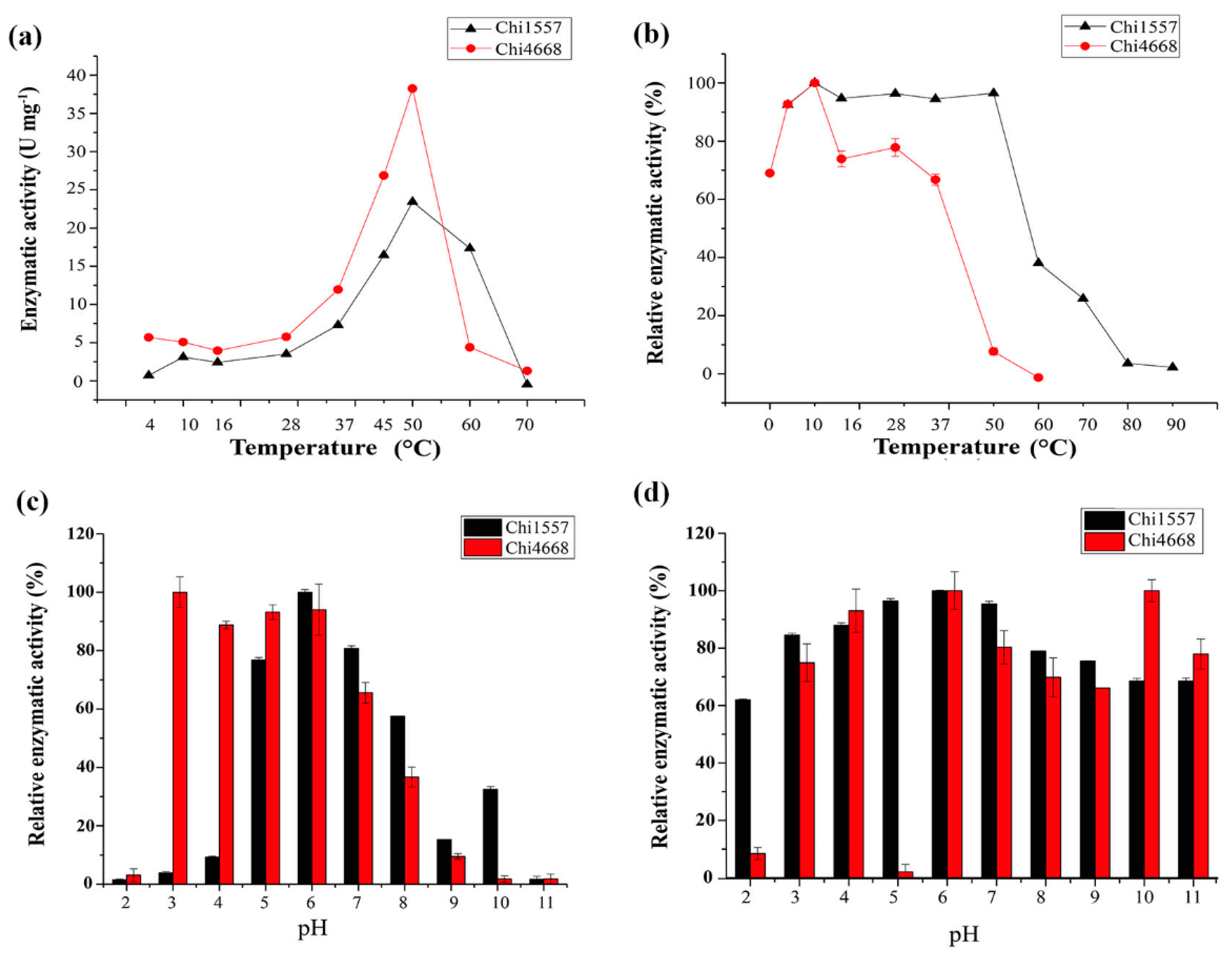
The optimum temperature of Chi1557 and Chi4668 are both 45–50 °C, but their activities varied greatly at higher temperature (Figure 2). Chi4668 is basically inactivated at 60 °C, while Chi1557 retains about 70% of its activity at 60 °C (Figure 2). For the stability of enzymes, Chi1557 retained more than 90% of its initial activity when incubated at 4–50 °C; the residual activity of the enzyme was reduced by 50% when incubated at 60 °C for one hour. Chi4668 retained only 45% enzymatic activity after incubation at 45 °C, and lost ~90% activity after incubation at 50 °C for an hour. Thus, it was indicated that the temperature stability of Chi4668 is worse than that of Chi1557. Different from Chi4668 and Chi1557, many other chitinases show maximum activities at lower temperatures, including chitinases from the Antarctic psychrotolerant bacterium Vibrio sp. Fi:7 (35 °C) [31], V. furnissii (35–37 °C) [32], V. proteolyticus (40 °C) [10] and Glaciozyma antarctica PI12 (15 °C) [33].
Similar with the other chitinases from Vibrio species [10,34,35,36,37], Chi1557 showed the highest activity at pH 5.0–7.0 (higher than 70%). It maintained relatively lower activity at pH 8.0 (~60%), pH 9.0 (~20%) and pH 10.0 (~30%), but almost lost its activity at pH 2.0–4.0 (<10%) (Figure 2c). For pH stability, Chi1557 could maintain more than 60% enzymatic activity within a broad range of pH (pH 2.0–11.0) after it was treated in different buffers for 1 h (Figure 2d), indicating that Chi1557 had extremely high pH stability. However, Chi4668 showed the maximum enzymatic activity at pH 3.0–6.0, and lost its activity at pH 9.0–11.0 (<10%) (Figure 2c). For pH stability, Chi4668 have extremely high pH stability and it could maintain more than 70% enzymatic activity within pH 3.0–11.0 except it basically deactivated after treated in 0.1 M citrate buffer at pH 5.0. These observations revealed that Chi1557 prefers alkaline environment whereas Chi4668 prefers acidic environment.

2.4. Metal Ions and Reductants on the Activity of Recombinant Chitinases

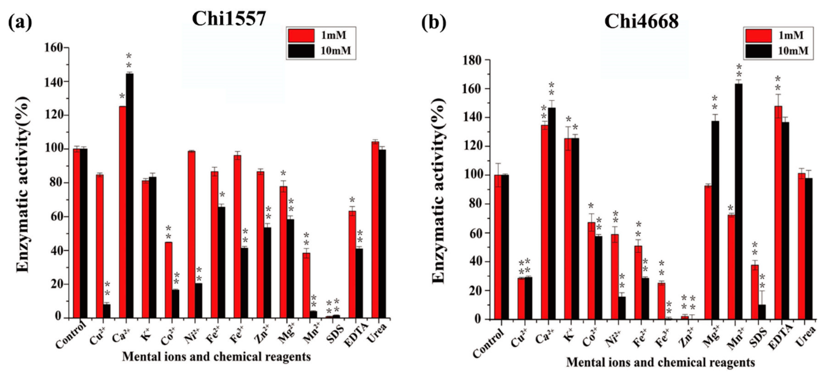
The effects of metal ions and chemical reagents (EDTA, SDS, and urea) on enzymatic activity were measured (Figure 3). The results show that only Ca2+ could increase the activity of Chi1557 up to 125% and 145% of initial activity at concentrations of 1 mM and 10 mM, respectively (Figure 3a). It was in common with chitinases from Vibrio sp. Fi:7 [31] and Sanguibacter antarcticus [38].
Ca2+, K+ and EDTA (1 mM and 10 mM) and Mg2+ and Mn2+ (10 mM) could improve the enzymatic activity of Chi4668 significantly (Figure 3b). 1 mM Cu2+, Ni2+, Fe2+, Fe3+, Zn2+ and urea had no obvious effect on Chi1557, but significantly inhibited the activity of Chi4668. Similarly, the activities of Chi1557 and Chi4668 were both inhibited with addition of 10 mM Cu2+, Co2+, Ni2+, Mn2+, Fe2+, Fe3+, Zn2+, and SDS.

2.5. The Kinetic Parameters and Hydrolysis Property of Recombinant Chitinases

The Michaelis-Menten constant (Km) values of Chi1557 and Chi4668 for colloidal chitin are individually 7.94 mg mL1 and 2.75 mg mL1. The Kcat/Km values of Chi1557 and Chi4668 for colloidal chitin are 0.40 s1M1 and 1.88 s1M1, respectively (Table 2).
The hydrolysis properties of recombinant chitinases on colloidal chitin and N-acetyl chitooligosaccharides (COSs) (DP 2-4) were investigated in detail. With the degradation of Chi1557, colloidal chitin was hydrolyzed into (GlcNAc)2 with a little GlcNAc (Figure 4a), and (GlcNAc)3-4 were hydrolyzed into (GlcNAc)2 (Figure S3a), indicating that Chi1557 is an endochitinase. This property was similar to many other chitinases in the family GH18, such as chitinase A from V. harveyi as a typical GH18-family chitinase could degrade chitin into (GlcNAc)2 [39], chitinases from V. cholerae [22], and Pa-Chi from V. parahaemolyticus [40]. Differently, the degradation products of Chi4668 were more diverse, it could hydrolyze chitin colloid into (GlcNAc)2 with a little (GlcNAc)3 and (GlcNAc)4 (Figure 4b), hydrolyzed (GlcNAc)3 into (GlcNAc)2 with GlcNAc and hydrolyzed (GlcNAc)4 into (GlcNAc)2 with little GlcNAc (Figure S3b). Like Chi4668, the diverse degradation products of Chitinase C1 and Chitinase C3 from strain V. alginolyticus H-8 are individually (GlcNAc)1–3 and (GlcNAc)1-6 [35].

2.6. Site-Directed Mutagenesis of Chi1557

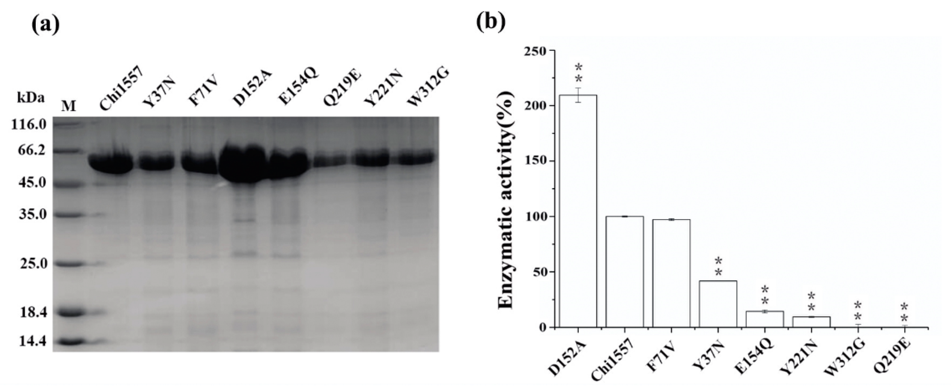
Site-directed mutagenesis was performed with Chi1557 to investigate the effect of some key residues in chitinase. Focusing on the conserved sites of chitinases from the family GH18, it has been reported that Asp152 and Glu154 were key sites of the catalytic domain [41]. Trp312 was the key amino acid residue for chitinase binding with chitin [42]. According to the three-dimensional structure of Chi1557 (Figure S4), Gln219 was close to the catalytic center Glu154, and it could maintain appropriately high pKa of catalytic amino acids [14]. Tyr37, Phe71, and Tyr221 were relatively conserved in the other chitinase of GH18 which were also located in the catalytic “pocket” and may keep a different strategy for chitin hydrolysis [42,43,44]. Hence, we selected seven potential key amino residues, Tyr37, Phe71, Asp152, Glu154, Gln219, Tyr221, and Trp312, in Chi1557 for mutation. In this study, we obtain seven purified mutant proteins which show different abilities in chitin degradation. The SDS-PAGE of seven mutant recombinant chitinases and the specific activity of them were tested (Figure 5a). Chitinase Chi1557 was almost completely inactive when Glu154 (mutated to Gln, E154Q), Gln219 (mutated to Glu, Q219E), Tyr221 (mutated to Asn, Y221N) and Trp312 (mutated to Gly, W312G) were mutated; Chi1557 retained ~50% and 100% of enzymatic activity after mutation of Tyr37 (mutated to Asn, Y37N) and Phe71 (mutated to Val, F71V), respectively. Besides, its activity was increased two times when Asp152 was mutated to Ala (D152A) (Figure 5b). It was suggested that Glu154, Gln219, Tyr221, Asp152, Trp312, and Tyr37 are key amino acid residues and absolutely required for Chi1557 activity (Figure 5b).

3. Discussion

In this study, two marine Vibrio strains were isolated from coastal of China, which could utilize chitin as the sole carbon source for growth and reproduction, indicating that they may play important roles in the marine chitin cycle around its habitat. V. rotiferianus WXL191 and V. harveyi WXL538 both contained a complete chitin metabolic pathway, including chitin-degrading genes and key transport systems, such as chitinases, glucosamine-1-phosphate N-acetyltransferase, β-N-acetylhexosaminidase and phosphotransferase system (PTS system) (Figure S6). Previous research showed that the genome of V. rotiferianus WXL191 (=V. rotiferianus B64D1) carried chitinase (EC 3.2.1.14) and β-N-acetylhexosaminidase (EC 3.2.1.52), which could completely hydrolyze chitin to monomer GlcNAc [24]. Here, two similar chitinases belonged to family GH18, Chi1557 and Chi4668, were obtained from V. rotiferianus WXL191 and V. harveyi WXL538 and expressed in E. coli system.
Most of Vibrio-derived chitinases were classified into family GH18 [24,31,32,37,39,40,45]. In addition to the catalytic domains, these enzymes usually contain one or more chitin-binding domains (ChtBD) [46,47,48] at N-terminus and may contain several polycystic kidney disease (PKD)-like domains [32,40,45]. Similarly, chitinases Chi1557 and Chi4668 in this study contained one catalytic domain, one ChtBD and two Ig-like domains, which may help them to improve the chitin degrading efficiency. The molecular mass of Vibrio-derived chitinases were between 30–120 kDa, the optimal temperature ranges for most of them were 45–55 °C, and the optimal pH values were between 6.0–8.0 [21,31,32,37,39,40,46,47,48]. However, Chi4668 show maximum enzymatic activity at acidic environment (pH 3.0 and 6.0). The only or main degradation product of these Vibrio-derived chitinases in family GH18, including chitinase Pa-Chi from V. parahaemolyticus [40] and chitinase A from V. carchariae (=V. harveyi) [39], was (GlcNAc)2. Other chitinases also yielded diverse degradation products, such as the degradation products of chitinase C1 and chitinase C3 from V. alginolyticus H-8 were (GlcNAc)1-3 and (GlcNAc)1-6, respectively [35]. Compared to Chi1557, more diverse degradation products of Chi4668 were identified, including (GlcNAc)2 with a little (GlcNAc)3 and (GlcNAc)4. The enzymatic activity of Vibrio-derived chitinases in family GH18 were lower than 10 U mg1 (ChiA from Vibrio sp. Fi: 7, 2–3 U mg1; chitinase C1 and chitinase C3 from V. alginolyticus H-8, 2.8–3.3 U mg1 and 4.6–5.8 U mg1), while chitinase from Vibrio sp. 11,211 had a higher enzymatic activity (36.5 U mg1) [31,35,37].
In this study, we analyzed and compared the amino acid composition and the enzymatic properties of Chi1557 and Chi4668. Even though the amino acids were much similar (identity is 97.33%), many enzymatic properties of Chi1557 and Chi4668 were different, including the enzymatic activity, the degradation products, and the responses to environmental conditions such as temperature and pH. According to the results of three-dimensional models’ prediction and native-PAGE (Figure 1), Chi1557 may exist as a dimer structure, while Chi4668 exists as a monomer structure. It was reported that the oligomeric structure of enzymes played an important role in biological processes, such as allosteric regulation, conformational stability, and thermal stability [49]. Fraser et al. [50] found that αE7 carboxylesterases are more prone to forming dimer or tetramer mutations at high temperatures to improve their stability. Schwab et al. [51] exposited that the monomeric enzyme obtained by polymerization has an enzymatic activity similar to that of the wild-type dimerize. However, its stability was significantly reduced. Hence, we speculated that oligomeric form of Chi1557 may be the reason for its stronger temperature and pH stabilities than Chi4668. Furthermore, a previous study [52] showed that the core domain of the protein containing more hydrophobic amino acids and hydrophobic residues determined the relative positions of secondary structures. Additionally, hydrophobic amino acid residues at key locations were closely associated with the stability of enzymes [53,54]. For Vibrio-derived chitinases in GH18 family, the chitin-binding domain is crucial for chitinase-chitin recognition and interactions [48,55]. Compared to Chi557, Chi4668 contains more hydrophobic amino acid residues, especially in the chitin binding domain (Table S1). Previous study [56] has shown that non-conservative substitution of tryptophan residue in chitin-binding domain nearly abolished its chitin-binding affinity. Here, we hypothesized that the hydrophobic tryptophan residues in the chitin-binding domain of Chi4668 may help it maintain the stability of the conformation and facilitate the combination with chitin, and keep high enzymatic activity under extreme conditions (pH = 3.0, 4.0, 6.0).

4. Materials and Methods

4.1. Bacterial Strains, Media and Growth Conditions

V. rotiferianus WXL191 (=V. rotiferianus B64D1) was isolated from the bottom water (17.5 m water depth) of Bohai Sea at 119.04°E, 38.23°N during the expedition on the R/V Dong Fang Hong 2 in August 2015 [57]. V. harveyi WXL538 was obtained from the East China Sea (at water depth of 25 m) at 122.56°E, 31.35°N during the expedition on the R/V Dong Fang Hong 2 in October 2015 [24]. Both strains were isolated using thiosulfate citrate bile salts sucrose (TCBS) agar (Hopebio, Qingdao, China) and demonstrated strong capacity to degrade chitin when growing on chitin agar plates. Then, these two purified strains were cultured on marine agar 2216E (Hopebio, Qingdao, China) plates at 28 °C for further research. The complete genome sequences of Vibrio rotiferianus WXL191 (=V. rotiferianus B64D1) and V. harveyi WXL538 have been deposited in NCBI GenBank server under the accession number CP018311 to CP018312 and CP045070 to CP045071, respectively. And E. coli BL21(DE3) was cultured on Luria-Bertani (LB) agar at 37 °C, and used as a host for expressing proteins whose encoding genes were cloned into pET24a (+) (Novagen, Beijing, China).

4.2. Sequence Analysis of Chitinase Genes

The extraction of total genomic DNA, prediction and annotation of chitinase genes were based on the methods of Lin et al. [24]. The bioinformatic analyses of chitinase sequences as follows: the amino acid sequences of chitinases were analyzed by BLASTP against protein data bank (pdb) database (https://blast.ncbi.nlm.nih.gov/) [58]; the closely related chitinases of Chi1557 and Chi4668 were obtained from NCBI, phylogenetic relationships between Chi1557, Chi4668, and the closely related chitinases were constructed using MEGA version 7.0 [59]; the three-dimensional models were predicted using SwissModel (https://www.swissmodel.expasy.org/interactive) and analyzed by PyMOL [60]; the Molecular mass and pI of chitinases were predicted by ExPASy database (https://web.expasy.org/compute_pi/) [30] and the signal peptide was predicted by the SignalP 4.1 server (http://www.cbs.dtu.dk/services/SignalP/) [61]. The GenBank accession number for the chitinase gene sequences of chi1557 and chi4668 are MN555466 and MN555465, respectively.

4.3. Preparation of Chitin Colloids

Colloidal chitin was prepared from commercial chitin by the method of Roberts and Selitrennikoff [62] with some modifications. Briefly, the chitin powder (Sigma-Aldrich, C7170, Munich, Germany) was rinsed in 1 mol L1 HCL and 1 mol L1 sodium hydroxide solutions five times in turn (about 2 h each time). And then the deposit was washed 4 times immersing in 95% ethanol, and then dry naturally. Sixty milliliters of concentrated HCl was added slowly into 5 g of processed chitin powder and left at 4 °C with vigorous stirring (about 4–5 h). The mixture was adjusted to pH 7.0 with 10 mol L1 sodium hydroxide and washed with 5 liters of ice-cold deionized water. The precipitant was collected by centrifugation at 5000 g for 10 min at 4 °C, and the colloidal chitin solution (5%) was prepared and stored at 4 °C until further applications.

4.4. Expression and Purification of Recombinant Chitinases

To obtain recombinant proteins without signal peptide, the putative gene was amplified with the primer pairs Chi1557F-Chi1557R, Chi4668F-Chi4668R (Table 1). The genomic DNA of strains WXL191 and WXL531 were used as templates for polymerase chain reaction (PCR) amplification of chi1557 and chi4668, respectively. The expression and purification of chitinases in E. coli BL21(DE3) were performed according to Tang et al. [63]. The purified recombinant chitinases were assessed by 12% sulfate-polyacrylamide gel electrophoresis (SDS-PAGE) according to the method of Laemmli et al. [64] and nondenaturing conditions PAGE (native PAGE) according to the method of Davis et al. [65].

4.5. Chitinase Activity Assay and Protein Quantification

Chitinase activity was detected by the methods of Lee et al. [66] with modification. Briefly, 190 μL of 1% (w/v) colloidal chitin was incubated at 50 °C for 10 min, then 10 μL purified enzyme was added with continued incubation at 50 °C for 30 min. The reducing sugars released were determined by the modified dinitrosalicylic acid (DNS) method [67]. One unit (U) of chitinase activity was defined as the amount of enzyme that released 1 μmol of reducing sugars per minute in 1 mL reaction mixture under the assay conditions by using GlcNAc as the standard. Protein concentration was measured by the Bradford method [68] using bovine serum albumin (BSA) as the standard.

4.6. Characterization of Purified Chitinases

The optimal pH of Chi1557 and Chi4668 were measured between pH 2.0–11.0 (at intervals of 1.0) using four kinds of buffer systems: 0.05 M Glycine-HCl (pH 2.0–4.0), 0.1 M citrate (pH 5.0–7.0), 0.05 M Tris-HCl (pH 8.0 and 9.0), and 0.05 M Glycine-NaOH (pH 10.0 and 11.0). To determine the pH stability of the chitinases, the purified enzyme samples were incubated in the above-mentioned buffers at 4 °C for 1 h. And the residual activities were tested at 50 °C. The optimal temperature of chitinases was studied by incubating the enzyme samples with substrate in optimal pH at 4, 10, 16, 28, 37, 45, 50, 60, and 70 °C. For detecting the thermostability, the enzyme samples were incubated in optimal pH at 4, 10, 16, 28, 37, and 50–90 °C (at interval of 10 °C) for 1 h, and then the residual activities were tested. To study the effects of metal ions and chemical reagents (EDTA, SDS and urea), the enzymatic activity was measured by standard methods in the presence of Na+, K+, Ca2+, Fe3+, Mn2+, Al3+, Co2+, Ni2+, Fe2+, Cu2+, Mg2+, Zn2+, and chemical reagents (EDTA, SDS, and urea) at final concentration of 1 mM and 10 mM. Also, the residual activities were tested.

4.7. Kinetic Parameters and Hydrolytic Properties of Chitinases

The kinetic parameters of chitinases in colloidal chitin was determined by measuring enzymatic activity at 50 °C in optimum pH for 30 min with substrate concentrations of 0.05%, 0.1%, 0.2%–0.8% (at interval of 0.2%), 1.0%, and 2.0%. The Km and Vmax values were calculated from kinetic data fitting of the Michaelis–Menten equation [69].
The hydrolysis products of chitinases were determined by thin layer chromatographic (TLC) method using colloidal chitin and (GlcNAc)2-4 as substrates [70]. Briefly, purified chitinase and 1% (w/v) colloidal chitin or (GlcNAc)2-4 were mixed in optimum pH, and then the mixtures were incubated at optimum temperature (50 °C) for 10 min, 30 min, 1 h, 2 h, or 4 h. The reaction products were spotted onto a Silica gel plate (Merck, Damsladt, Germany), and spread using butanol: water: ammonia water (6:3:1, v/v/v) as spreading agent. The plate was sprinkled with chromogenic agent (containing 4 g diphenylamine, 4 mL aniline, 2 mL concentrated hydrochloric acid, 20 mL phosphate, and 200 mL acetone).

4.8. Site-Directed Mutagenesis of Chi1557

Site-directed mutagenesis was used to determine the effects of several amino acid residues on enzyme activity of Chi1557. Here, based on the previous studies on key amino acid residues [13,42,71,72,73] and protein structure of family GH18 chitinases [59], as well as the predicted structure of protein Chi1557 in this study, we intended to alter the key amino acid residues Tyr37 by Asn, replace Phe71 by Val, replace Asp152 by Ala, replace Glu154 by Gln, replace Gln219 by Glu, replace Tyr221 by Asn, and replace Trp312 by Gly, respectively. The plasmid pET24a (+)-chi1557 was used as PCR template, and primers used to obtain each mutant are listed in Table 1. Positive mutations selected by cloning were confirmed by DNA sequencing. Each mutated protein was expressed and purified to measure its chitin-degrading activities, as described above.

Supplementary Materials

The following are available online at https://www.mdpi.com/1660-3397/18/3/139/s1. Supplementary data of this work can be found in online version of the paper, Figure S1: Neighbor-joining phylogenic tree based on amino acid sequences of Chi1557, Chi4668 and other chitinases from bacteria was built with CLUSTAL-X in the MEGA version 7.0, Figure S2: Multiple sequence alignment of the amino acid sequences of Chi1557 and Chi4668 by CLUSTAL-X in the MEGA version 7.0, Figure S3: The degradation productions of Chi1557 and Chi4668 for N-acetyl COSs (DP 2-4), Figure S4: Three-dimensional structure of Chi1557, Figure S5: Multiple sequence alignment of the amino acid sequences of Chi1557, Chi4668 and other family GH18 chitinases from different bacterial by MUSCLE program in the MEGA version 7.0 and enhanced by ESPript v3.0, Figure S6: The chitin metabolic pathway in Vibrio rotiferianus WXL191 and V. harveyi WXL538 annotated by KAAS (KEGG Automatic Annotation Server). Table S1: The confidently predicted domains, repeats, motifs and features of Chi1557 and Chi4668, Table S2: The different amino acids of chitinases Chi1557 and Chi4668.

Author Contributions

X.H. and M.Y. contributed equally to this work. Conceptualization, X.-H.Z.; Data curation, X.H. and Y.W.; Formal analysis, X.H. and M.Y.; Investigation, X.H., Y.W. and L.R.; Methodology, X.H., M.Y. and X.-H.Z.; Project administration, X.H. and X-H.Z.; Resources, X-H.Z.; Supervision, X-H.Z. and W.L.; Validation, X.H. and Y.W.; Visualization, X.H.; Writing—original draft, X.H.; Writing—review and editing, M.Y., W.L. and X.-H.Z.; Funding acquisition, X.-H.Z. All authors have read and agreed to the published version of the manuscript.

Funding

This work was funded by the National Natural Science Foundation of China (41730530 and 91751202), the National Key Research and Development Program of China (No. 2018YFE0124100), and the Marine Science & Technology Fund of Shandong Province for Pilot National Laboratory for Marine Science and Technology (Qingdao) (No. 2018SDKJ0406-4).

Acknowledgments

We thank Xiaolei Wang, Shun Zhou and Xiao-Yu Zhu from Ocean University of China for help during purification and identification of bacterial strains. We thank Brian Austin from Stirling University, U.K. for help with English modification.

Conflicts of Interest

The authors declare no conflict of interest.

Abbreviations

aa, amino acid; CBMs, carbohydrate-binding modules; ChtBD, chitin-binding domains; COSs, chitooligosaccharides; FnIII, fibronectin III; GH, glycoside hydrolase; Ig-like, immunoglobulin-like; PKD, polycystic kidney disease; SDS-PAGE, sulfate-polyacrylamide gel electrophoresis; TLC, thin layer chromatography.

References

  1. Hamed, I.; Özogul, F.; Regenstein, J.M. Industrial applications of crustacean by-products (chitin, chitosan, and chitooligosaccharides): A review. Trends Food Sci. Technol. 2016, 48, 40–50. [Google Scholar] [CrossRef]
  2. Beier, S.; Bertilsson, S. Bacterial chitin degradation-mechanisms and ecophysiological strategies. Front Microbiol. 2013, 4, 149. [Google Scholar] [CrossRef] [Green Version]
  3. Kirchman, D.L.; White, J. Hydrolysis and mineralization of chitin in the Delaware Estuary. Aquat. Microb. Ecol. 1999, 18, 187–196. [Google Scholar] [CrossRef]
  4. Howard, M.B.; Ekborg, N.A.; Weiner, R.M.; Hutcheson, S.W. Detection and characterization of chitinases and other chitin-modifying enzymes. J. Ind. Microbiol. Biotechnol. 2003, 30, 627–635. [Google Scholar] [CrossRef] [PubMed]
  5. Souza, C.P.; Almeida, B.C.; Colwell, R.R.; Rivera, I.N. The importance of chitin in the marine environment. Mar. Biotechnol. 2011, 13, 823. [Google Scholar] [CrossRef] [PubMed]
  6. Dahiya, N.; Tewari, R.; Hoondal, G.S. Biotechnological aspects of chitinolytic enzymes: A review. Appl. Microbiol. Biotechnol. 2006, 71, 773–782. [Google Scholar] [CrossRef] [PubMed]
  7. Karthik, N.; Binod, P.; Pandey, A. Purification and characterisation of an acidic and antifungal chitinase produced by a Streptomyces sp. Bioresour. Technol. 2015, 188, 195–201. [Google Scholar] [CrossRef]
  8. Stoykov, Y.M.; Pavlov, A.I.; Krastanov, A.I. Chitinase biotechnology: Production, purification, and application. Eng. Life Sci. 2015, 15, 30–38. [Google Scholar] [CrossRef]
  9. Tarentino, A.L.; Maley, F. Purification and properties of an endo-β-N-acetylglucosaminidase from Streptomyces griseus. J. Biol. Chem. 1974, 249, 811–817. [Google Scholar]
  10. Honda, Y.; Taniguchi, H.; Kitaoka, M. A reducing-end-acting chitinase from Vibrio proteolyticus belonging to glycoside hydrolase family 19. Appl. Microbiol. Biotechnol. 2008, 78, 627–634. [Google Scholar] [CrossRef]
  11. Vaaje-Kolstad, G.; Horn, S.J.; Sørlie, M.; Eijsink, V.G. The chitinolytic machinery of Serratia marcescens-a model system for enzymatic degradation of recalcitrant polysaccharides. FEBS J. 2013, 280, 3028–3049. [Google Scholar] [CrossRef]
  12. Toratani, T.; Kezuka, Y.; Nonaka, T.; Hiragi, Y.; Watanabe, T. Structure of full-length bacterial chitinase containing two fibronectin type III domains revealed by small angle X-ray scattering. Biochem. Biophys. Res. Commun. 2006, 348, 814–818. [Google Scholar] [CrossRef] [PubMed]
  13. Watanabe, T.; Ishibashi, A.; Ariga, Y.; Hashimoto, M.; Nikaidou, N.; Sugiyama, J.; Matsumoto, T.; Nonaka, T. Trp122 and Trp134 on the surface of the catalytic domain are essential for crystalline chitin hydrolysis by Bacillus circulans chitinase A1. FEBS Lett. 2001, 494, 74–78. [Google Scholar] [CrossRef] [Green Version]
  14. Synstad, B.; Gåseidnes, S.; Van Aalten, D.M.; Vriend, G.; Nielsen, J.E.; Eijsink, V.G. Mutational and computational analysis of the role of conserved residues in the active site of a family 18 chitinase. Eur. J. Biochem. 2004, 271, 253–262. [Google Scholar] [CrossRef] [PubMed] [Green Version]
  15. Madhuprakash, J.; El Gueddari, N.E.; Moerschbacher, B.M.; Podile, A.R. Catalytic efficiency of chitinase-D on insoluble chitinous substrates was improved by fusing auxiliary domains. PLoS ONE 2015, 10, e0116823. [Google Scholar] [CrossRef] [PubMed] [Green Version]
  16. Yan, Q.; Fong, S.S. Bacterial chitinase: Nature and perspectives for sustainable bioproduction. Bioresour. Bioprocess. 2015, 2, 1–9. [Google Scholar] [CrossRef] [Green Version]
  17. Hamid, R.; Khan, M.A.; Ahmad, M.; Ahmad, M.M.; Abdin, M.Z.; Musarrat, J.; Javed, S. Chitinases: An update. J. Pharm. BioAllied Sci. 2013, 5, 21. [Google Scholar]
  18. Shin-Hye, P. Purification and characterization of chitinase from a marine bacterium, Vibrio sp. 98CJ11027. J Microbiol. 2000, 38, 224–229. [Google Scholar]
  19. Annamalai, N.; Rajeswari, M.V.; Vijayalakshmi, S.; Balasubramanian, T. Purification and characterization of chitinase from Alcaligenes faecalis AU02 by utilizing marine wastes and its antioxidant activity. Ann. Microbiol. 2011, 61, 801–807. [Google Scholar] [CrossRef] [Green Version]
  20. Mander, P.; Cho, S.S.; Choi, Y.H.; Panthi, S.; Choi, Y.S.; Kim, H.M.; Yoo, J.C. Purification and characterization of chitinase showing antifungal and biodegradation properties obtained from Streptomyces anulatus CS242. Arch. Pharmacal. Res. 2016, 39, 878–886. [Google Scholar] [CrossRef]
  21. Itoh, T.; Sugimoto, I.; Hibi, T.; Suzuki, F.; Matsuo, K.; Fujii, Y.; Taketo, A.; Kimoto, H. Overexpression, purification, and characterization of Paenibacillus cell surface-expressed chitinase ChiW with two catalytic domains. Biosci. Biotechnol. Biochem. 2014, 78, 624–634. [Google Scholar] [CrossRef] [PubMed]
  22. Hunt, D.E.; Gevers, D.; Vahora, N.M.; Polz, M.F. Conservation of the chitin utilization pathway in the Vibrionaceae. Appl. Environ. Microbiol. 2008, 74, 44–51. [Google Scholar] [CrossRef] [PubMed] [Green Version]
  23. Meibom, K.L.; Li, X.B.; Nielsen, A.T.; Wu, C.-Y.; Roseman, S.; Schoolnik, G.K. The Vibrio cholerae chitin utilization program. Proc. Natl. Acad. Sci. USA 2004, 101, 2524–2529. [Google Scholar] [CrossRef] [PubMed] [Green Version]
  24. Lin, H.; Yu, M.; Wang, X.; Zhang, X.-H. Comparative genomic analysis reveals the evolution and environmental adaptation strategies of Vibrios. BMC Genomics 2018, 19, 135. [Google Scholar] [CrossRef] [PubMed] [Green Version]
  25. Saito, A.; Fujii, T.; Yoneyama, T.; REDENBACH, M.; OHNO, T.; WATANABE, T.; MIYASHITA, K. High-multiplicity of chitinase genes in Streptomyces coelicolor A3 (2). Biosci. Biotechnol. Biochem. 1999, 63, 710–718. [Google Scholar] [CrossRef] [Green Version]
  26. Suzuki, K.; SuGAwARA, N.; Suzuki, M.; Uchiyama, T.; Katouno, F.; Nikaidou, N.; Watanabe, T. Chitinases A, B, and C1 of Serratia marcescens 2170 produced by recombinant Escherichia coli: Enzymatic properties and synergism on chitin degradation. Biosci. Biotechnol. Biochem. 2002, 66, 1075–1083. [Google Scholar] [CrossRef]
  27. Suzuki, K.; Taiyoji, M.; Sugawara, N.; Nikaidou, N.; Henrissat, B.; Watanabe, T. The third chitinase gene (chiC) of Serratia marcescens 2170 and the relationship of its product to other bacterial chitinases. Biochem. J. 1999, 343, 587–596. [Google Scholar] [CrossRef]
  28. Svitil, A.L.; Chadhain, S.; Moore, J.A.; Kirchman, D.L. Chitin Degradation Proteins Produced by the Marine Bacterium Vibrio harveyi Growing on Different Forms of Chitin. Appl. Environ. Microbiol. 1997, 63, 408–413. [Google Scholar] [CrossRef] [Green Version]
  29. Malecki, P.H.; Raczynska, J.E.; Vorgias, C.E.; Rypniewski, W. Structure of a complete four-domain chitinase from Moritella marina, a marine psychrophilic bacterium. Acta Crystallogr. Sect. D Biol. Crystallogr. 2013, 69, 821–829. [Google Scholar] [CrossRef]
  30. Gasteiger, E.; Gattiker, A.; Hoogland, C.; Ivanyi, I.; Appel, R.D.; Bairoch, A. ExPASy: The proteomics server for in-depth protein knowledge and analysis. Nucleic Acids Res. 2003, 31, 3784–3788. [Google Scholar] [CrossRef] [Green Version]
  31. Bendt, A.; Hüller, H.; Kammel, U.; Helmke, E.; Schweder, T. Cloning, expression, and characterization of a chitinase gene from the Antarctic psychrotolerant bacterium Vibrio sp. strain Fi: 7. Extremophiles 2001, 5, 119–126. [Google Scholar] [CrossRef] [PubMed]
  32. Keyhani, N.O.; Roseman, S. The chitin catabolic cascade in the marine bacterium Vibrio furnissii molecular cloning, isolation, and characterization of a periplasmic chitodextrinase. J. Biol. Chem. 1996, 271, 33414–33424. [Google Scholar] [CrossRef] [PubMed] [Green Version]
  33. Ramli, A.N.; Mahadi, N.M.; Rabu, A.; Murad, A.M.; Bakar, F.D.; Illias, R.M. Molecular cloning, expression and biochemical characterisation of a cold-adapted novel recombinant chitinase from Glaciozyma antarctica PI12. Microb. Cell Fact. 2011, 10, 94. [Google Scholar] [CrossRef] [PubMed] [Green Version]
  34. Huang, L.; Shizume, A.; Nogawa, M.; Taguchi, G.; Shimosaka, M. Heterologous expression and functional characterization of a novel chitinase from the chitinolytic bacterium Chitiniphilus shinanonensis. Biosci. Biotechnol. Biochem. 2012, 1203012832. [Google Scholar]
  35. Ohishi, K.; Yamagishi, M.; Ohta, T.; Suzuki, M.; Izumida, H.; Sano, H.; Nishijima, M.; Miwa, T. Purification and properties of two chitinases from Vibrio alginolyticus H-8. J. Ferment. Bioeng. 1996, 82, 598–600. [Google Scholar] [CrossRef]
  36. Revathi, M.; Saravanan, R.; Shanmugam, A. Production and characterization of chitinase from Vibrio species, a head waste of shrimp Metapenaeus dobsonii (Miers, 1878) and chitin of Sepiella inermis Orbigny, 1848. Adv. Biosci. Biotechnol. 2012, 3, 392. [Google Scholar] [CrossRef] [Green Version]
  37. Zhou, S.; Yang, C.; Lu, Y.; Huang, L.; Cai, C.; Lin, Y. Isolation and characterization of chitinase from a marine bacterium Vibrio sp. World J. Microbiol. Biotechnol. 1999, 15, 745–746. [Google Scholar] [CrossRef]
  38. Park, H.J.; Kim, D.; Kim, I.H.; Lee, C.-E.; Kim, I.-C.; Kim, J.Y.; Kim, S.J.; Lee, H.K.; Yim, J.H. Characteristics of cold-adaptive endochitinase from Antarctic bacterium Sanguibacter antarcticus KOPRI 21702. Enzyme Microb. Technol. 2009, 45, 391–396. [Google Scholar] [CrossRef]
  39. Suginta, W.; Vongsuwan, A.; Songsiriritthigul, C.; Prinz, H.; Estibeiro, P.; Duncan, R.R.; Svasti, J.; Fothergill-Gilmore, L.A. An endochitinase A from Vibrio carchariae: Cloning, expression, mass and sequence analyses, and chitin hydrolysis. Arch. Biochem. Biophys. 2004, 424, 171–180. [Google Scholar] [CrossRef]
  40. Kadokura, K.; Rokutani, A.; Yamamoto, M.; Ikegami, T.; Sugita, H.; Itoi, S.; Hakamata, W.; Oku, T.; Nishio, T. Purification and characterization of Vibrio parahaemolyticus extracellular chitinase and chitin oligosaccharide deacetylase involved in the production of heterodisaccharide from chitin. Appl. Microbiol. Biotechnol. 2007, 75, 357. [Google Scholar] [CrossRef]
  41. Alam, M.; Nikaidou, N.; Tanaka, H.; Watanabe, T. Cloning and sequencing of chiC gene of Bacillus circulans WL-12 and relationship of its product to some other chitinases and chitinase-like proteins. J. Ferment. Bioeng. 1995, 80, 454–461. [Google Scholar] [CrossRef]
  42. Rush, C.L.; Schüttelkopf, A.W.; Hurtado-Guerrero, R.; Blair, D.E.; Ibrahim, A.F.; Desvergnes, S.; Eggleston, I.M.; van Aalten, D.M. Natural product-guided discovery of a fungal chitinase inhibitor. Chem. Biol. 2010, 17, 1275–1281. [Google Scholar] [CrossRef] [PubMed]
  43. Hahn, M.; Hennig, M.; Schlesier, B.; Höhne, W. Structure of jack bean chitinase. Acta Crystallogr. Sect. D Biol. Crystallogr. 2000, 56, 1096–1099. [Google Scholar] [CrossRef] [PubMed]
  44. Hollis, T.; Monzingo, A.F.; Bortone, K.; Ernst, S.; Cox, R.; Robertus, J.D. The X-ray structure of a chitinase from the pathogenic fungus Coccidioides immitis. Protein Sci. 2000, 9, 544–551. [Google Scholar] [CrossRef] [Green Version]
  45. Itoi, S.; Kanomata, Y.; Koyama, Y.; Kadokura, K.; Uchida, S.; Nishio, T.; Oku, T.; Sugita, H. Identification of a novel endochitinase from a marine bacterium Vibrio proteolyticus strain No. 442. Biochim. Biophys. Acta. 2007, 1774, 1099–1107. [Google Scholar] [CrossRef] [PubMed]
  46. Murao, S.; Kawada, T.; Itoh, H.; Oyama, H.; Shin, T. Purification and characterization of a novel type of chitinase from Vibrio alginolyticus TK-22. Biosci. Biotechnol. Biochem. 1992, 56, 368–369. [Google Scholar] [CrossRef]
  47. Pantoom, S.; Songsiriritthigul, C.; Suginta, W. The effects of the surface-exposed residues on the binding and hydrolytic activities of Vibrio carchariae chitinase A. BMC Biochem. 2008, 9, 2. [Google Scholar] [CrossRef] [Green Version]
  48. Suginta, W.; Sirimontree, P.; Sritho, N.; Ohnuma, T.; Fukamizo, T. The chitin-binding domain of a GH-18 chitinase from Vibrio harveyi is crucial for chitin-chitinase interactions. Int. J. Biol. Macromol. 2016, 93, 1111–1117. [Google Scholar] [CrossRef]
  49. Shallom, D.; Golan, G.; Shoham, G.; Shoham, Y. Effect of dimer dissociation on activity and thermostability of the α-glucuronidase from Geobacillus stearothermophilus: Dissecting the different oligomeric forms of family 67 glycoside hydrolases. J. Bacteriol. 2004, 186, 6928–6937. [Google Scholar] [CrossRef] [Green Version]
  50. Fraser, N.J.; Liu, J.-W.; Mabbitt, P.D.; Correy, G.J.; Coppin, C.W.; Lethier, M.; Perugini, M.A.; Murphy, J.M.; Oakeshott, J.G.; Weik, M. Evolution of protein quaternary structure in response to selective pressure for increased thermostability. J. Mol. Biol. 2016, 428, 2359–2371. [Google Scholar] [CrossRef] [Green Version]
  51. Schwab, T.; Skegro, D.; Mayans, O.; Sterner, R. A rationally designed monomeric variant of anthranilate phosphoribosyltransferase from Sulfolobus solfataricus is as active as the dimeric wild-type enzyme but less thermostable. J. Mol. Biol. 2008, 376, 506–516. [Google Scholar] [CrossRef]
  52. Betts, M.J.; Russell, R.B. Amino acid properties and consequences of substitutions. Bioinform. Genet. 2003, 317, 289. [Google Scholar]
  53. Shortle, D.; Stites, W.E.; Meeker, A.K. Contributions of the large hydrophobic amino acids to the stability of staphylococcal nuclease. Biochemistry 1990, 29, 8033–8041. [Google Scholar] [CrossRef] [PubMed]
  54. Yutani, K.; Ogasahara, K.; Tsujita, T.; Sugino, Y. Dependence of conformational stability on hydrophobicity of the amino acid residue in a series of variant proteins substituted at a unique position of tryptophan synthase alpha subunit. Proc. Natl. Acad. Sci. USA 1987, 84, 4441–4444. [Google Scholar] [CrossRef] [PubMed] [Green Version]
  55. Montgomery, M.T.; Kirchman, D.L. Role of chitin-binding proteins in the specific attachment of the marine bacterium Vibrio harveyi to chitin. Appl. Environ. Microbiol. 1993, 59, 373–379. [Google Scholar] [CrossRef] [PubMed] [Green Version]
  56. Hardt, M.; Laine, R.A. Mutation of active site residues in the chitin-binding domain ChBDChiA1 from chitinase A1 of Bacillus circulans alters substrate specificity: Use of a green fluorescent protein binding assay. Arch. Biochem. Biophys. 2004, 426, 286–297. [Google Scholar] [CrossRef]
  57. Liang, J.; Liu, J.; Wang, X.; Lin, H.; Liu, J.; Zhou, S.; Sun, H.; Zhang, X.-H. Spatiotemporal dynamics of free-living and particle-associated Vibrio communities in the northern Chinese marginal seas. Appl. Environ. Microbiol. 2019, 85, e00217–e00219. [Google Scholar] [CrossRef] [Green Version]
  58. Consortium, U. UniProt: A hub for protein information. Nucleic Acids Res. 2014, 43, D204–D212. [Google Scholar] [CrossRef]
  59. Kumar, S.; Stecher, G.; Tamura, K. MEGA7: Molecular evolutionary genetics analysis version 7.0 for bigger datasets. Mol. Biol. Evol. 2016, 33, 1870–1874. [Google Scholar] [CrossRef] [Green Version]
  60. Schwede, T.; Kopp, J.; Guex, N.; Peitsch, M.C. SWISS-MODEL: An automated protein homology-modeling server. Nucleic Acids Res. 2003, 31, 3381–3385. [Google Scholar] [CrossRef] [Green Version]
  61. Nielsen, H. Predicting secretory proteins with SignalP. In Protein Function Prediction; Springer Humana Press: New York, NY, USA, 2017; Volume 1611, pp. 59–73. [Google Scholar]
  62. Roberts, W.K.; Selitrennikoff, C.P. Plant and bacterial chitinases differ in antifungal activity. Microbiology 1988, 134, 169–176. [Google Scholar] [CrossRef] [Green Version]
  63. Tang, K.; Su, Y.; Brackman, G.; Cui, F.; Zhang, Y.; Shi, X.; Coenye, T.; Zhang, X.-H. MomL, a novel marine-derived N-acyl homoserine lactonase from Muricauda olearia. Appl. Environ. Microbiol. 2015, 81, 774–782. [Google Scholar] [CrossRef] [PubMed] [Green Version]
  64. Laemmli, U.K. Cleavage of structural proteins during the assembly of the head of bacteriophage T4. Nature 1970, 227, 680. [Google Scholar] [CrossRef] [PubMed]
  65. Davis, B. Gel for nondenature gel electrophoresis. Part II. Clinical applications. Method and application to human serum proteins. Ann. N. Y. Acad. Sci. 1964, 121, 405–427. [Google Scholar]
  66. Lee, Y.-S.; Park, I.-H.; Yoo, J.-S.; Chung, S.-Y.; Lee, Y.-C.; Cho, Y.-S.; Ahn, S.-C.; Kim, C.-M.; Choi, Y.-L. Cloning, purification, and characterization of chitinase from Bacillus sp. DAU101. Bioresour. Technol. 2007, 98, 2734–2741. [Google Scholar] [CrossRef]
  67. Miller, G.L. Use of dinitrosalicylic acid reagent for determination of reducing sugar. Anal. Chem. 1959, 31, 426–428. [Google Scholar] [CrossRef]
  68. Bradford, M.M. A rapid and sensitive method for the quantitation of microgram quantities of protein utilizing the principle of protein-dye binding. Anal. Biochem. 1976, 72, 248–254. [Google Scholar] [CrossRef]
  69. Raaijmakers, J.G. Statistical analysis of the Michaelis-Menten equation. Biometrics 1987, 793–803. [Google Scholar] [CrossRef]
  70. Songsiriritthigul, C.; Lapboonrueng, S.; Pechsrichuang, P.; Pesatcha, P.; Yamabhai, M. Expression and characterization of Bacillus licheniformis chitinase (ChiA), suitable for bioconversion of chitin waste. Bioresour. Technol. 2010, 101, 4096–4103. [Google Scholar] [CrossRef]
  71. Suginta, W.; Sritho, N. Multiple roles of Asp313 in the refined catalytic cycle of chitin degradation by Vibrio harveyi chitinase A. Biosci. Biotechnol. Biochem. 2012, 120559. [Google Scholar]
  72. Suginta, W.; Vongsuwan, A.; Songsiriritthigul, C.; Svasti, J.; Prinz, H. Enzymatic properties of wild-type and active site mutants of chitinase A from Vibrio carchariae, as revealed by HPLC-MS. FEBS J. 2005, 272, 3376–3386. [Google Scholar] [CrossRef] [PubMed]
  73. Vaaje-Kolstad, G.; Houston, D.R.; Rao, F.V.; Peter, M.G.; Synstad, B.; van Aalten, D.M.; Eijsink, V.G. Structure of the D142N mutant of the family 18 chitinase ChiB from Serratia marcescens and its complex with allosamidin. Biochim. Biophys. Acta Proteins Proteom. 2004, 1696, 103–111. [Google Scholar] [CrossRef] [PubMed]
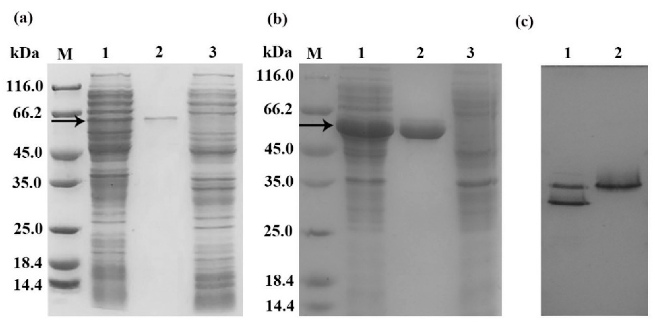
Figure 1. SDS-PAGE and native-PAGE of recombinant chitinases Chi1557 and Chi4668. (a) SDS-PAGE of purified Chi1557. M, molecular mass markers; lane 1, cell-free extracts of Escherichia coli BL21(DE3)/pET24(+)-chi1557; lane 2, purified Chi1557; lane 3, cell-free extracts of E. coli BL21 (DE3)/pET24a (+); (b) SDS-PAGE of purified Chi4668. M, molecular mass markers; lane 1, cell-free extracts of E. coli BL21 (DE3)/pET24 (+)-chi4668; lane 2, purified Chi4668; lane 3, cell-free extracts of E. coli BL21 (DE3)/pET24a (+). (c), native-PAGE of Chi1557 and Chi4668. lane 1, purified Chi1557; lane 2, purified Chi4668. Approximately 10 μL of samples were loaded onto each lane and stained by Coomassie brilliant blue. The band indicated by the arrow is the location of the target protein.
Marinedrugs 18 00139 g001
Figure 2. Response of Chi1557 and Chi4668 to temperature and pHs. (a) The optimum temperature of Chi1557 and Chi4668. They all show the highest enzyme activity at 50 °C, but the enzyme activity of Chi4668 is about 1.5 times higher than that of Chi1557. (b) The temperature stability of Chi1557 and Chi4668. Chi1557 is more stable at temperature between 37 °C to 50 °C. (c), The optimum pH of Chi1557 and Chi4668. The optimum pH of Chi1557 is 5.0–7.0, and the optimum pH of Chi4668 is 3.0–6.0. (d), the pH stability of Chi1557 and Chi4668.
Marinedrugs 18 00139 g002
Figure 3. Response of Chi1557 (a) and Chi4668 (b) to metal ions and reductants. (a) Effects of 1 mM and 10 mM metal ions and chemical reagents on the activity of Chi1557; (b) Effects of 1 mM and 10 mM metal ions and chemical reagents on the activity of Chi4668. (** P < 0.01; * P < 0.05).
Marinedrugs 18 00139 g003
Figure 4. Hydrolysis property for colloidal chitin of Chi1557 and Chi4668. (a) The degradation production of Chi1557 for 1% (w/v) colloidal chitin; (b) The degradation production of Chi4668 for 1% (w/v) colloidal chitin. Purified Chi1557 and 1% (w/v) colloidal chitin were incubated in 0.1M citrate buffer citrate buffer (pH 5) at 50 °C for different time intervals respectively, Purified Chi4668 and 1% (w/v) colloidal chitin were incubated in 0.1 M citrate buffer citrate buffer (pH 6) at 50 °C for different time intervals respectively, and the degradation products were determined by TLC.
Marinedrugs 18 00139 g004
Figure 5. SDS-PAGE and the specific activity of purified chitinases Chi1557 and seven mutant proteins. (a), SDS-PAGE of purified Chi1557 and seven mutant proteins. M: marker; 1, purified Chi1557; 2, purified mutant protein Tyr37 (Y37N); 3, purified mutant protein Phe71 (F71V); 4, purified mutant protein Asp152 (D152A); 5, purified mutant protein Glu154 (E154Q); 6, purified mutant protein Gln219 (Q219E); 7, purified mutant protein Tyr221 (Y221N); 8, purified mutant protein Trp312 (W312G). (b), The specific activity of Chi1557 and mutant proteins. Chi1557 only retained ~50% of enzymatic activity after mutate Tyr37; its activity was increased twice when mutate Asp152; and Chi1557 could retain original activity after mutate Phe71. (** P < 0.01).
Marinedrugs 18 00139 g005
Table 1. Primers used in cloning Chi1557, Chi4668, and the mutagenesis of Chi1557. Primers were designed using the Primer-primer 5 design tool. The cleavage sites are underlined, and the mutagenic nucleotides are represented in lowercase.
PrimerSequence (5’-3’) bRestriction Site and Mutant Amino Acid
Chi1557-F5-CGCGGATCCATGAATGAGATGGTGA-3BamH I
Chi1557-R5-TAGCTCGAGCAACTTATCCCACGCG-3Xho I
Chi4668-F5’-CGCGGATCCATGAATGAAATGGTGA-3’BamH I
Chi4668-R5’-CCCAAGCTTCAACTTATCCCATGCG-3’Hind III
Y37N-F5’-GTAGTCGGTaATTGGCATAACTGGT-3’Tyr37
Y37N-R5’-GACACCACTATCTGGATTCACCATC-3’
F71V-F5’-AACGTCTCCgTTATGAAGGTGT-3’Phe71
F71V-R5’-AACCACATTGTACATAGGATCAACT-3’
D152A-F5’-GGTCTGGACATCGcCTTAGAGCA-3’Asp152
D152A-R5’-ATCAAAGCCGAACTTGTCAGTCAGG-3’
E154Q-F5’-GACATCGACTTAcAGCAATCTGCAG-3’Glu154
E154Q-R5’-CAGACCATCAAAGCCGAACTTGT-3’
Q219E-F5’-ATCAACCCTgAATTTTACAACCAAG-3’Gln219
Q219E-R5’-CCAATCGTAGTACCCTTCTAATCCA-3’
Y221N-F5’-CCTCAATTTaACAACCAAGGTGG-3’Tyr221
Y221N-R5’-GTTGATCCAATCGTAGTACCCTTCT-3’
W312G-F5’-GTAATGACAgGGTCGGTGAACTGGG-3’Trp312
W312G-R5’-GCCACGAAGTGCCTGCCCTTG-3’
Table 2. The enzymology properties of Chi1557 and Chi4668.
Enzymology PropertiesChi1557-NiChi4668-Ni
The optimal temperature (°C)45–50~50
The optimal pH5.0–7.03.0–6.0
Total enzymatic activity (U)2.053.16
Total protein content (mg mL−1)0.130.18
Specific activity (U mg−1)23.4241.14
The kinetic parameters:
Vmax (mg U−1)2.94 6.21
Km (mg mL−1)7.94 2.75
Kcat (s−1)3.00 5.18
Kcat/Km (s−1M−1)0.401.88