Article

Bioactive Lactones from the Mangrove-Derived Fungus Penicillium sp. TGM112

1
Key Laboratory of Tropical Medicinal Resource Chemistry of Ministry of Education, College of Chemistry and Chemical Engineering, Hainan Normal University, Haikou, Hainan 571158, China
2
Key Laboratory of Tropical Medicinal Plant Chemistry of Hainan Province, Hainan Normal University, Haikou, Hainan 571158, China
*
Authors to whom correspondence should be addressed.
Mar. Drugs 2019, 17(8), 433; https://doi.org/10.3390/md17080433
Submission received: 12 June 2019 / Revised: 13 July 2019 / Accepted: 22 July 2019 / Published: 24 July 2019

Abstract

:
Three new lactones penicilactones A−C (13) were obtained from the mangrove-derived fungus Penicillium sp. TGM112. Their structures and absolute configurations were determined by detailed NMR, MS spectroscopic data, Mo2(OAc)4-induced electronic circular dichroism (ECD), and circular dichroism (CD) spectroscopy. Compound 1 showed antibacterial activity against Staphylococcus aureus with an MIC value of 6.25 μg/mL. Compound 2 showed insecticidal activity against newly hatched larvae of Culex quinquefasciatus with the LC50 value of 78.5 (±0.58) μg/mL.

1. Introduction

Natural products from marine-derived fungi have played an important role for leading compounds and have attracted interests from chemists and biologists [1]. An increasing number of structural novel and bioactive natural products have been obtained from mangrove-derived fungi [2]. Ordinarily, lactones with a 6,7-dihydroxyocta-2,4-dien skeleton were a small group of natural products. To date, only 12 ones, including cytotoxic goniobutenolodes A and B [3], 6,7-dihydroxy-2-propyl-2,4-octadien-4-olide [4], versicolactones A and B [5], antioxidant litchiol B [6], and plecmillins G-L [7], have been identified from natural resources. Moreover, goniobutenolodes A and B [8], versicolactones A and B and six stereoisomers [9], have been synthesized. As part of our continuing investigation into bioactive metabolites of mangrove-derived fungi [10,11,12,13], two new meroterpenoids and eight new isocoumarins have been identified from the fermentation of Penicillium sp. TGM112, a fungus obtained from the mangrove Bruguiera sexangula var. rhynchopetala [14]. Furthering our work on the remaining extract of the fungal strain Penicillium sp. TGM112, it was found that its fermentation extract showed antibacterial activity against Staphylococcus aureus with an MIC value of 20 μg/mL. The subsequent bioassay-guided fractionation and isolation were carried out and resulted in the characterization of three new lactone penicilactones A−C (13) (Figure 1). Herein, the isolation, structure elucidation, and bioactivities of these compounds are described.

2. Results and Discussion

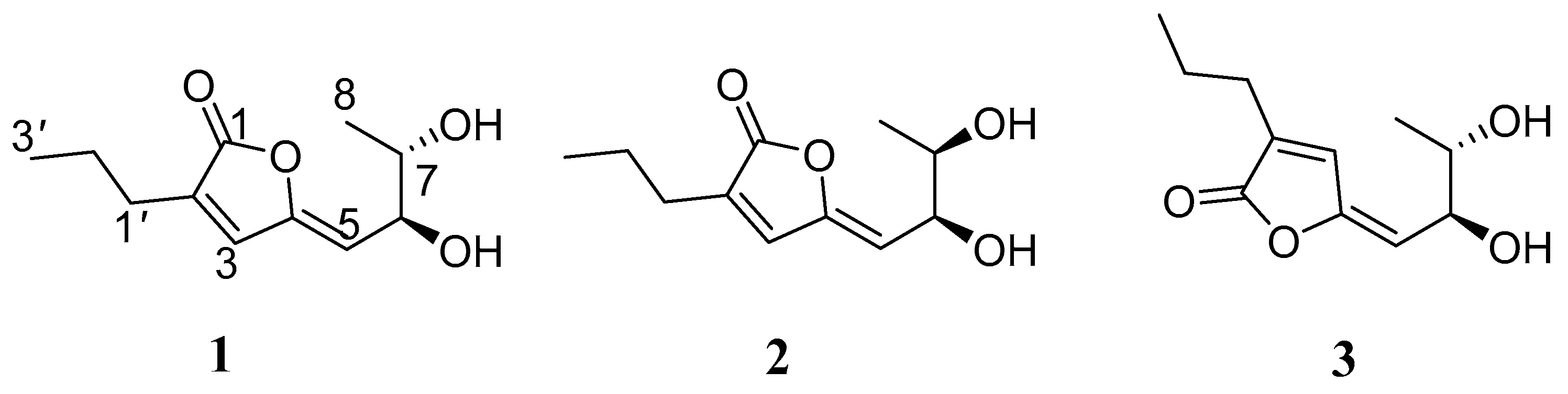
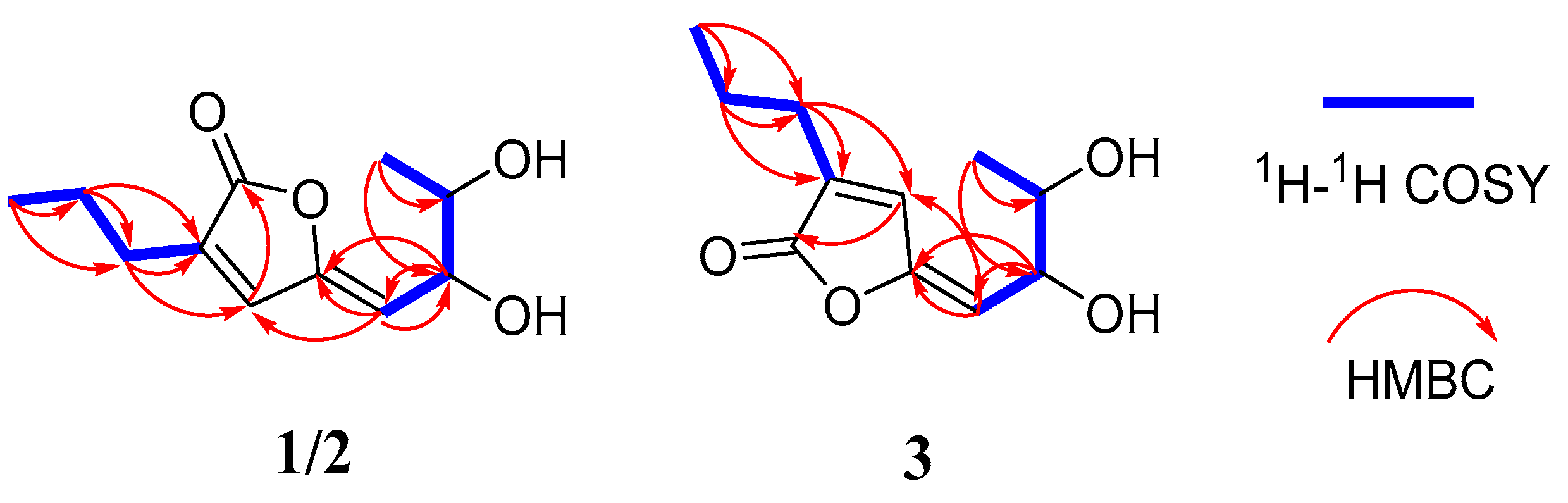
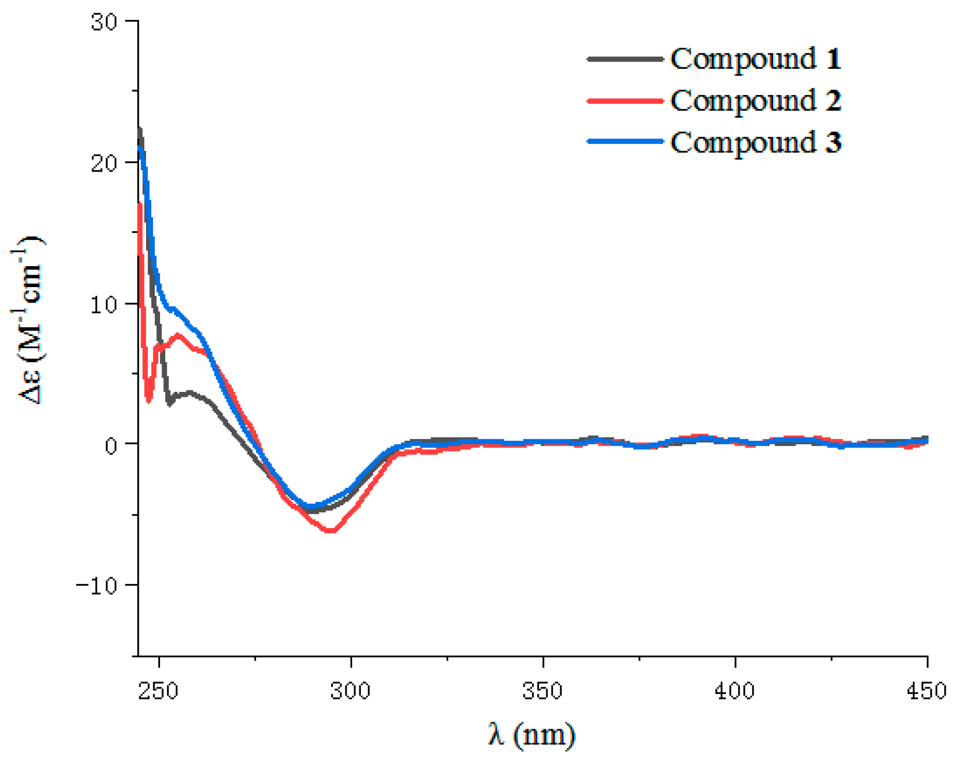
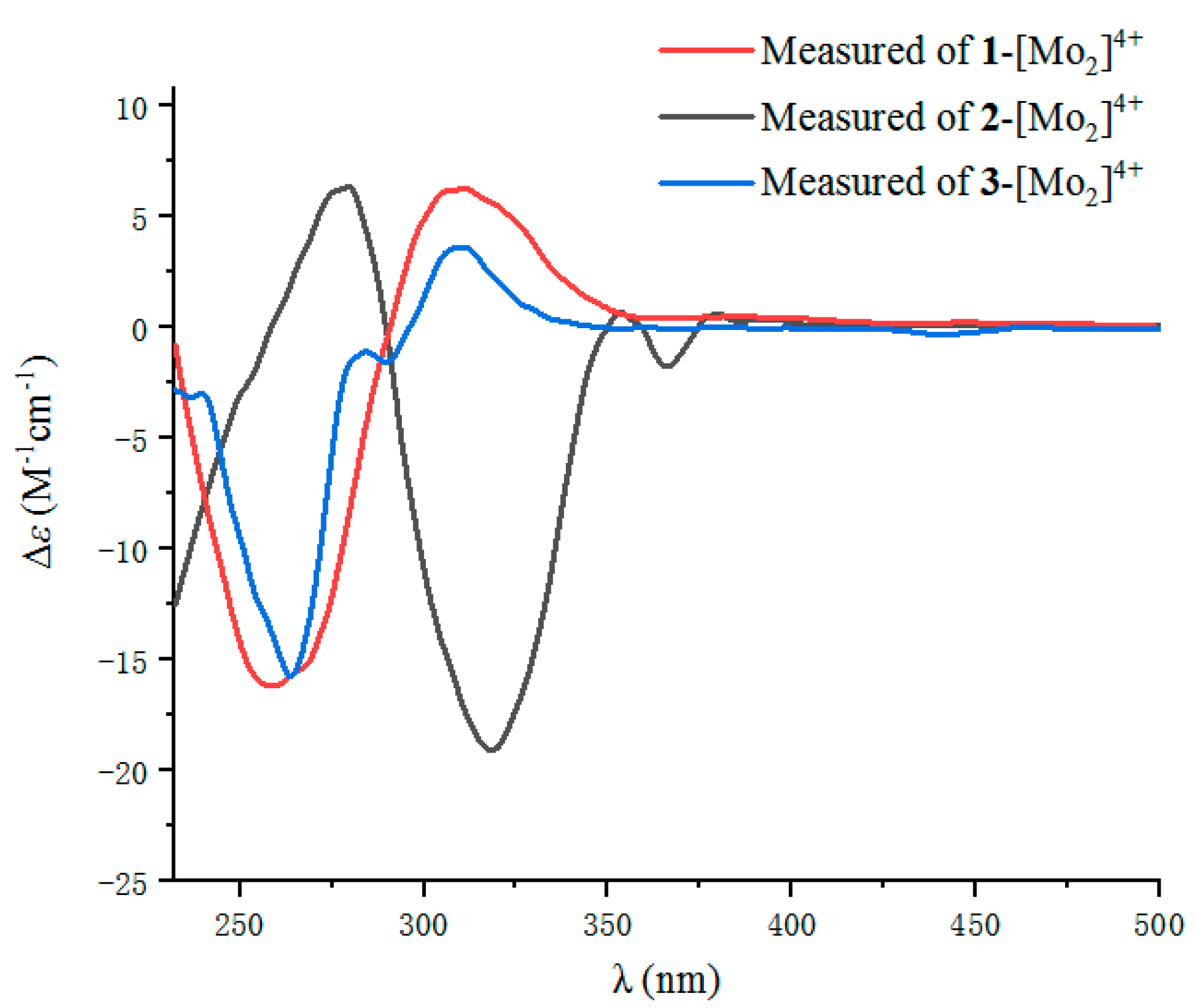
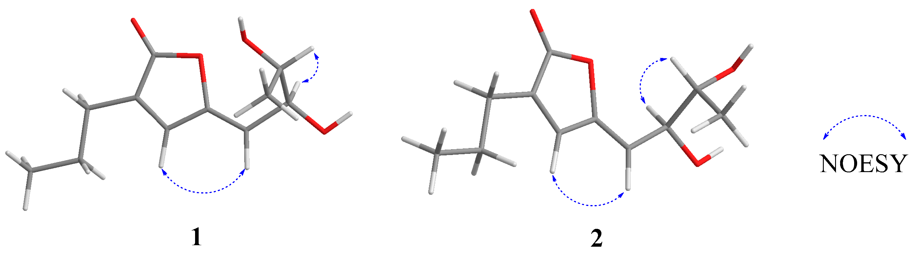
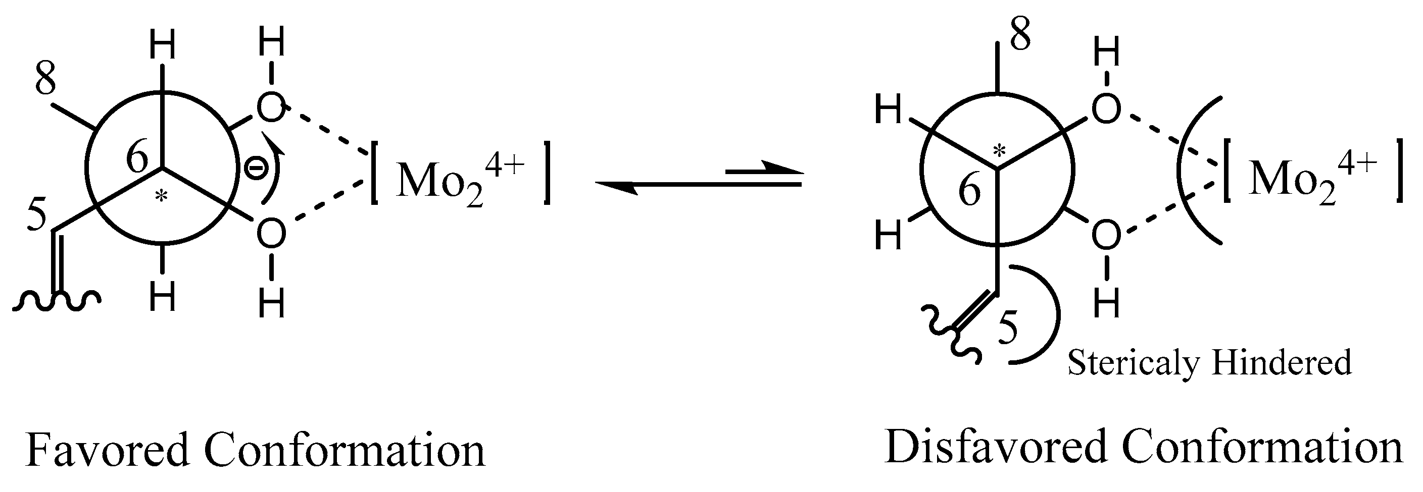
2.1. Structure Elucidation

Compound 1 was isolated as a yellow oil, with the molecular formula of C11H16O4 (four degrees of unsaturation) determined by the HRESIMS data. The 1H NMR data (Table 1 and Figure S1) showed two methyl signals at δH 1.13 (3H, d, J = 6.8 Hz, H-8) and 0.98 (3H, t, J = 7.2 Hz, H-3′), two methylene signals at δH 2.31 (2H, t, J = 6.8 Hz, H-1′) and 1.60 (2H, m, H-2′), two oxygenated methine groups at δH 4.45 (1H, dd, J = 6.0, 9.2 Hz, H-6) and 3.69 (1H, m, H-7), and two olefinic protons at δH 7.31 (1H, s, H-3) and 5.30 (1H, d, J = 9.2 Hz, H-5). The 13C NMR data (Table 1 and Figure S2) of 1 exhibits 11 carbon resonances corresponding to two methyl groups at δC 18.8 and 13.9, two methylene groups at δC 28.0 and 21.9, two oxygenated methine groups at δC 71.7 and 71.5, two pairs of olefinic carbons at δC 150.6, 139.0, 135.9 and 113.9, and one carbonyl carbon at δC 171.9. The 1H-1H COSY correlations of H-5/H-6/H-7/H-8 and H-1′/H-2′/H-3′ together with the key HMBC correlations from H-1′ to C-1/C-3 and from H-5 to C-3/C-4/C-6/C-7 confirmed the planar structure of 1 (Figure 2). The NOESY correlation of H-5 with H-3 indicated the Z configuration of the double bond in the side-chain (Figure 3). Thus, the structure of 1 was established to be an analogue of plecmillin G [7], with the main differences of the presence of an oxygenated methine group at (δH 3.87 (m) and δC 64.4 (CH)) for C-2′ in plecmillin G with a methylene group at (δH 1.60 (m) and δC 21.9 (CH2)) for C-2′ in 1.
The absolute configuration of C-6 in 1 was determined by circular dichroism (CD) spectroscopy (Figure 4). The same negative cotton effect observed at about 280 nm for 1 suggested the S configuration at C-6, by comparison with data for plecmillin G [7]. Furthermore, the absolute configurations at C-6 and C-7 in 1 were determined on the basis of the CD spectrum of the complex formed in situ upon the addition of Mo2(OAc)4 to a solution of 1 in DMSO (Figure 5), the metal complex generated as an auxiliary chromophore, with the inherent CD spectra subtracted [15,16,17,18,19]. According to the empirical rule proposed by Snatzke′s method [15], the observed induced CD (ICD) curve at around 310 nm showing the same sign with the O−C−C−O torsion angle in the favored conformation allows the assignment of the absolute configuration. The ICD spectrum of the metal complex of 1 showed a positive cotton effect at 308 nm (Δε + 7.08) (Figure 6). Therefore, the absolute configuration of 1 was identified as 6S, 7S, and named as penicilactone A.
Compound 2 was obtained as a yellow oil, with the molecular formula of C11H16O4 (four degrees of unsaturation) as deduced from the HRESIMS data. Its NMR data (Table 1) were almost identical to those of 1, the slight differences of the NMR chemical shifts (upshifted or downshifted) between 2 and 1 were clearly distinguished (Table 1). These data proposed that 2 and 1 could be sharing the same carbon skeleton, and the variations of chemical shifts for the carbons of (δC 113.6, 71.5, and 71.4) might be due to the presence of chiral carbons (C-6 and C-7). The planar structure was established by the HMBC and 1H-1H COSY correlations, as shown in Figure 2. The absolute configuration of C-6 in 2 was confirmed as 6S by the same negative cotton effect at about 280 nm as observed in (Z)-5-((2S,3R)-2,3-dihydroxybutylidene)furan-2(5H)-one (which was synthesized by Wang and Zhu in 2013) [7,9]. Likewise, the absolute configurations of C-6 and C-7 in 2 were also assigned using an in situ dimolybdenum CD method. The negative Cotton effect at 316 nm (Δε −19.3) confirmed the 6S, 7R configurations (Figure 6) [19]. Thus, compound 2 was determined and named penicilactone B.
Compound 3 was the isomer of 1, with the molecular formula of C11H16O4 from the HRESIMS data. Its 1D NMR data were very similar to those of 1, and the planar structure of 3 was assigned to be the same as that of 1 by analysis of its 1D and 2D NMR spectral data (Figure 2). The main differences between 1 and 3 were that H-3 and H-5 undergo large downfield shifts in 1 (δH-3 7.66 vs. 7.31, δH-5 5.68 vs 5.30, respectively), owing to a decrease in steric effect and C-3 experienced a large upfield shift (δC-3 136.4 vs. 139.0) due to steric effects between 6-CH(OH) and C-3, indicating that the double bond in the side-chain of 3 was of the E form [7]. The absolute configuration of C-6 in 3 was assigned as 6S by comparison of the specific rotation data and the cotton effects observed at about 280 nm for 3 and plecmillin I (Figure 4) [7]. The absolute configurations of C-6 and C-7 in 3 were also assigned using an in situ dimolybdenum CD method. The positive cotton effect at 308 nm (Δε + 4.01) (Figure 6) confirmed the 6S, 7S configurations for 3, and the compound named penicilactone C.

2.2. Biological Activity

Compounds 13 were evaluated for their antibacterial activities against six pathogenic bacteria. In addition, only compound 1 showed antibacterial activity against Staphylococcus aureus with an MIC value of 6.25 μg/mL (Table 2). All compounds were also tested for their insecticidal activity against newly hatched larvae of Culex quinquefasciatus, and only 2 showed insecticidal activity against newly hatched larvae of C. quinquefasciatus, with an LC50 value of 78.5 (±0.58) μg/mL (Table 2).

3. Materials and Methods

3.1. General Experimental Procedures

Optical rotations were measured on a JASCO P-1020 digital polarimeter (JASCO, Tokyo, Japan). Preparative HPLC were used for an Agilent 1260 prep-HPLC system with an Agilent Eclipse XDB-C18 column (9.4 × 250 mm, 7 µm, Agilent Corporation, Santa Clara, CA, USA). 1D and 2D NMR spectra were obtained on a Bruker AV-400 spectrometer (Bruker Corporation, Switzerland) (the temperature was 300 K, and the mix time of NOESY was 0.3 s) using TMS as an internal standard. The other experimental procedures were performed as reported previously [14].

3.2. Fungal Materials

The fungus TGM112 was isolated from the mangrove B. sexangula var. rhynchopetala, collected in the South China Sea in August, 2014, and was identified as Penicillium sp. [14]. This strain was deposited in China General Microbiological Culture Collection Center, Beijing, China, with the CGMCC code 16499. The fungal strain was cultivated in 20 L potato glucose liquid medium [14] at 25 C without shaking for 4 weeks.

3.3. Extraction and Isolation

The fungal cultures were extracted with EtOAc (20 L, 24 h each) at three different times to obtain the extracts. The extracts were then concentrated in vacuo to yield an oily residue (20.6 g). The residue was subjected to silica gel column chromatography (CC) (petroleum ether, EtOAc v/v, gradient 100:0–0:100) to generate five fractions (Fr. 1–Fr. 5). Fr. 3 (5 g) was separated by silica gel CC and eluted with petroleum ether-EtOAc (from 10:1 to 3:1) to afford four subfractions (Fr. 3a–3d). Then Fr. 3c was separated by semi-preparative HPLC (CH3CN–H2O, 45:55 for subfraction 3c v/v) to obtain 1 (5.1 mg, tR = 10.5 min), 2 (3.5 mg, tR = 17.5 min), and 3 (3.2 mg, tR = 29.1 min).
Penicilactone A (1): yellow oil. [α ] D 24 −20.1 (c = 0.10, CHCl3). UV (MeOH) λmax (log ε) 280 (3.02) nm; CD (c 2 × 10⁻⁴ mol/L, MeOH) λmax (Δε) 288 (−4.88) nm; IR (KBr) νmax 3315, 3230, 1710, 1432, 1082, 798, 718 cm−1; 1H and 13C NMR see Table 1; HRESIMS m/z 213.1121 [M + H]+ (calcd. for C11H17O4, 213.1121).
Penicilactone B (2): yellow oil. [α ] D 24 −34.3 (c = 0.10, CHCl3). UV (MeOH) λmax (log ε) 280 (3.00) nm; CD (c 2 × 10⁻⁴ mol/L, MeOH) λmax (Δε) 292 (−5.65) nm; IR (KBr) νmax 3314, 3231, 1707, 1430, 1080, 799, 720 cm−1; 1H and 13C NMR see Table 1; HRESIMS m/z 213.1124 [M + H]+ (calcd. for C11H17O4, 213.1121).
Penicilactone C (3): yellow oil. [α ] D 24 −40.7 (c = 0.10, CHCl3). UV (MeOH) λmax (log ε) 280 (3.05) nm; CD (c 2 × 10⁻⁴ mol/L, MeOH) λmax (Δε) 287(−4.98) nm; IR (KBr) νmax 3313, 3228, 1705, 1422, 1092, 799, 716 cm−1; 1H and 13C NMR see Table 1; HRESIMS m/z 213.1122 [M + H]+ (calcd. for C11H17O4, 213.1121).

3.4. Computational Section

According to the published procedure [18,19,20,21], compounds 13 (0.5 mg) were dissolved in a dry solution of the stock Mo2(OAc)4 complex (1.1 mg) in DMSO and then recorded immediately for the first induced ECD spectra, and its time evolution was monitored until stationary (about 30 min after mixing).

3.5. Antibacterial Activities

Antibacterial activity was determined against six pathogenic bacteria, Escherichia coli (ATCC 25922), Staphylococcus aureus (ATCC 25923), S. albus (ATCC 8799), Micrococcus luteus (ATCC 10240), Vibrio parahaemolyticus (ATCC 17802) and V. alginolyticus (ATCC 17749) by the microplate assay method [22]. Ciprofloxacin was used as the positive control.

3.6. Insecticidal Activities against Newly Hatched Larvae of Culex Quinquefasciatus

In the test, each containing 10 neonate larvaes of C. quinquefasciatus in 6-well microtiter plates, the examined compounds were dissolved in DMSO at the concentration of 1 mg/mL [14]. DMSO was used as the negative control, Azadirachtin was used as the positive control, and 10 mL dechlorinated water was used as the blank control. The number of dead larvae was recorded on the 1st, 2nd, 3rd, and 4th day after treatment, respectively [23].

4. Conclusions

In summary, three new lactones, penicilactones A−C (13) with a 6,7-dihydroxyocta-2,4-dien skeleton, were isolated from the mangrove-derived fungus Penicillium sp. TGM112. Their absolute configurations were determined by Mo2(OAc)4-induced electronic circular dichroism (ECD) and circular dichroism (CD) spectroscopy. Compound 1 showed antibacterial activity against Staphylococcus aureus with an MIC value of 6.25 μg/mL. Compound 2 showed insecticidal activity against newly hatched larvae of C. quinquefasciatus with an LC50 value of 78.5 (±0.58) μg/mL.

Supplementary Materials

The following are available online at https://www.mdpi.com/1660-3397/17/8/433/s1, Figure S1: 1H NMR (CD3OD, 400 MHz) spectrum of 1. Figure S2: 13C NMR (CD3OD, 100MHz) spectrum of 1. Figure S3: DEPT (CD3OD, 100 MHz) spectrum of 1. Figure S4: HMQC spectrum of 1. Figure S5: HMBC spectrum of 1. Figure S6: 1H-1H COSY spectrum of 1. Figure S7: NOESY spectrum of 1. Figure S8: HRESIMS spectrum of 1. Figure S9: 1H NMR (CD3OD, 400 MHz) spectrum of 2. Figure S10: 13C NMR (CD3OD, 100 MHz) spectrum of 2. Figure S11: DEPT (CD3OD, 100 MHz) spectrum of 2. Figure S12: HMQC spectrum of 2. Figure S13: HMBC spectrum of 2. Figure S14: 1H-1H COSY spectrum of 2. Figure S15: NOESY spectrum of 2. Figure S16: HRESIMS spectrum of 2. Figure S17: 1H NMR (CD3OD, 400 MHz) spectrum of 3. Figure S18: 13C NMR (CD3OD, 100 MHz) spectrum of 3. Figure S19: DEPT (CD3OD, 100 MHz) spectrum of 3. Figure S20: HMQC spectrum of 3. Figure S21: HMBC spectrum of 3. Figure S22: 1H-1H COSY spectrum of 3. Figure S23: NOESY spectrum of 3. Figure S24: HRESIMS spectrum of 3.

Author Contributions

M.B. performed the experiments for the isolation, structure elucidation, insecticidal activity and antimicrobial evaluation, and prepared the manuscript; Y.-P.L. and Q.-R.M. performed the 1D and 2D NMR experiments; X.-H.N. contributed to part of the structure determination; G.-L.H. and B.W. contributed the optimization of fermentation; G.-Y.C. contributed to part of the structure determination and jointly supervised the research; C.-J.Z. supervised the research work and revised the manuscript.

Funding

This work was supported by the National Natural Science Foundation of China (Nos. 31760093, 21462015, 41866005 and 21662012), Key Research and Development Program of Hainan Province (No. ZDYF2018183), the National Natural Science Foundation of Hainan Province (No. 218MS045), Program for Innovative Research Team in University (No. IRT-16R19).

Conflicts of Interest

The authors declare no competing financial interest.

References

  1. Yu, G.; Wang, Q.; Liu, S.; Zhang, X.; Che, Q.; Zhang, G.; Zhu, T.; Gu, Q.; Li, D. Methylsulfonylated Polyketides Produced by Neosartorya udagawae HDN13-313 via Exogenous Addition of Small Molecules. J. Nat. Prod. 2019, 82, 998–1001. [Google Scholar] [CrossRef] [PubMed]
  2. Zhang, P.; Meng, L.-H.; Mándi, A.; Kurtán, T.; Li, X.-M.; Liu, Y.; Li, X.; Li, C.-S.; Wang, B.-G. Brocaeloids A-C, 4-Oxoquinoline and Indole Alkaloids with C-2 Reversed Prenylation from the Mangrove-Derived Endophytic Fungus Penicillium brocae. Eur. J. Org. Chem. 2014, 2014, 4029–4036. [Google Scholar] [CrossRef]
  3. Fang, X.-P.; Anderson, J.E.; Chang, C.-J.; McLaughlin, J.L. Three new bioactive styryllactones from Goniothalamus giganteus (Annonaceae). Tetrahedron 1991, 47, 9751–9758. [Google Scholar] [CrossRef]
  4. Findlay, J.A.; Miller, J.D.; Womiloju, T.O.; Li, G. Insect toxins from spruce endophytes. Can. J. Chem. 2003, 81, 284–292. [Google Scholar] [CrossRef]
  5. Zhuang, Y.; Teng, X.; Wang, Y.; Liu, P.; Wang, H.; Li, J.; Li, G.; Zhu, W. Cyclopeptides and polyketides from coral-associated fungus, Aspergillus versicolor LCJ-5-4. Tetrahedron 2011, 67, 7085–7089. [Google Scholar] [CrossRef]
  6. Wang, L.J.; Luo, G.D.; Ma, Z.J.; Liu, X.M. Chemical constituents with antionxident activities from litchi (Litchi chinensis sonn.) seeds. Food Chem. 2011, 126, 1081–1087. [Google Scholar] [CrossRef]
  7. Wang, K.; Bao, L.; Ma, K.; Qi, W.; Song, F.H.; Yao, Y.J.; Yin, W.B.; Zhang, L.X.; Huang, Y.; Han, J.J.; et al. Bioactive spirobisnaphthalenes and lactones from a cup fungus Plectania sp. collected in the Tibet Platesu Region. Eur. J. Org. Chem. 2016, 25, 4338–4346. [Google Scholar] [CrossRef]
  8. Xu, D.Q.; Sharpless, K.B. Synthesis and stereochemical assignments for Goniobutenolodes A and B. Tetrahedron Lett. 1994, 35, 4685–4688. [Google Scholar] [CrossRef]
  9. Wang, L.P.; Zhu, W.M. Versicolactones A and B: Total synthesis and structure revision. Tetrahedron Lett. 2013, 54, 6729–6731. [Google Scholar] [CrossRef]
  10. Zheng, C.-J.; Bai, M.; Zhou, X.-M.; Huang, G.-L.; Shao, T.-M.; Luo, Y.-P.; Niu, Z.-G.; Niu, Y.-Y.; Chen, G.-Y.; Han, C.-R. Penicilindoles A–C, Cytotoxic Indole Diterpenes from the Mangrove-Derived Fungus Eupenicillium sp. HJ002. J. Nat. Prod. 2018, 81, 1045–1049. [Google Scholar] [CrossRef]
  11. Zhou, X.-M.; Li, G.-N.; Zheng, C.-J.; Chen, G.-Y.; Song, X.-P.; Han, C.-R.; Fu, Y.-H.; Chen, W.-H.; Niu, Z.-G. Bioactive Anthraquinone Derivatives from the Mangrove-Derived Fungus Stemphylium sp. 33231. J. Nat. Prod. 2014, 77, 2021–2028. [Google Scholar] [CrossRef] [PubMed]
  12. Huang, G.-L.; Zhou, X.-M.; Bai, M.; Liu, Y.-X.; Zhao, Y.-L.; Luo, Y.-P.; Niu, Y.-Y.; Zheng, C.-J.; Chen, G.-Y. Dihydroisocoumarins from the Mangrove-Derived Fungus Penicillium citrinum. Mar. Drugs 2016, 14, 177. [Google Scholar] [CrossRef] [PubMed]
  13. Mei, R.; Huang, G.; Wang, B.; Bai, M.; Luo, Y.; Chen, G.; Zheng, C. Two New Isocoumarins Isolated from a Mangrove-Derived Fungus Penicillium citrinum HL-5126. Chin. J. Org. Chem. 2019, 39, 1479–1482. [Google Scholar] [CrossRef]
  14. Bai, M.; Zheng, C.-J.; Huang, G.-L.; Mei, R.-Q.; Wang, B.; Luo, Y.-P.; Zheng, C.; Niu, Z.-G.; Chen, G.-Y. Bioactive Meroterpenoids and Isocoumarins from the Mangrove-Derived Fungus Penicillium sp. TGM112. J. Nat. Prod. 2019, 82, 1155–1164. [Google Scholar] [CrossRef] [PubMed]
  15. Snatzke, G.; Wagner, U.; Wolff, H.P. Cirulardichroism-LXXV 1: Cottonogenic derivatives of chiral bidentate ligands with the comples [Mo2(OAc)4]. Tetrahedron 1981, 37, 349–361. [Google Scholar] [CrossRef]
  16. Di Bari, L.; Pescitelli, G.; Pratelli, C.; Pini, D.; Salvadori, P. Determination of absolute configuration of acyclic 1,2-diols with Mo2(OAc)4. 1. Snatzke′s method revisited. J. Org. Chem. 2001, 66, 4819–4825. [Google Scholar] [CrossRef]
  17. Dong, S.-H.; Zhang, C.-R.; Dong, L.; Wu, Y.; Yue, J.-M. Onoceranoid-Type Triterpenoids fromLansium domesticum. J. Nat. Prod. 2011, 74, 1042–1048. [Google Scholar] [CrossRef]
  18. Zhang, L.-B.; Ji, J.; Lei, C.; Wang, H.-Y.; Zhao, Q.-S.; Hou, A.-J. Isoprenylated Flavonoid and Adipogenesis-Promoting Constituents of Dodonaea viscosa. J. Nat. Prod. 2012, 75, 699–706. [Google Scholar] [CrossRef]
  19. Gorecki, M.; Kaminska, A.; Ruskowska, P.; Suszczynska, A.; Frelek, J. Dimolybdenum method for determination of the absolute configuration of vic-diols-foundations and developments. Pol. J. Chem. 2006, 80, 523–534. [Google Scholar]
  20. Elnaggar, M.S.; Ebrahim, W.; Mándi, A.; Kurtán, T.; Müller, W.E.G.; Kalscheuer, R.; Singab, A.; Lin, W.; Liu, Z.; Proksch, P. Hydroquinone derivatives from the marine-derived fungus Gliomastix sp. RSC Adv. 2017, 7, 30640–30649. [Google Scholar] [CrossRef]
  21. Ren, J.; Ding, S.-S.; Zhu, A.; Cao, F.; Zhu, H.-J. Bioactive Azaphilone Derivatives from the Fungus Talaromyces aculeatus. J. Nat. Prod. 2017, 80, 2199–2203. [Google Scholar] [CrossRef] [PubMed]
  22. Pierce, C.G.; Uppuluri, P.; Tristan, A.R.; Wormley, F.L.; Mowat, E.; Ramage, G.; Lopez-Ribot, J.L. A simple and reproducible 96-well plate-based method for the formation of fungal biofilms and its application to antifungal susceptibility testing. Nat. Protoc. 2008, 3, 1494–1500. [Google Scholar] [CrossRef] [PubMed]
  23. Zhang, W.; Crickmore, N.; George, Z.; Xie, L.; He, Y.-Q.; Li, Y.; Tang, J.-L.; Tian, L.; Wang, X.; Fang, X. Characterization of a new highly mosquitocidal isolate of Bacillus thuringiensis—An alternative to Bti? J. Invertebr. Pathol. 2012, 109, 217–222. [Google Scholar] [CrossRef] [PubMed]
Figure 1. The structures of compounds 13.
Marinedrugs 17 00433 g001
Figure 2. 1H-1H COSY correlations and key HMBC correlations for compounds 13.
Marinedrugs 17 00433 g002
Figure 3. Key NOESY correlations for compounds 1 and 2.
Marinedrugs 17 00433 g003
Figure 4. Experimental CD Spectra of 1–3.
Marinedrugs 17 00433 g004
Figure 5. Conformations of the Mo24+ complex of 1.
Marinedrugs 17 00433 g005
Figure 6. Experimental ECD spectra of the Mo24+ complex of 13 with the inherent CD spectrum subtracted.
Marinedrugs 17 00433 g006
Table 1. 1H NMR and 13C NMR Data (δ) for 13 (400/100 MHz) (δ in ppm, J in Hz) in CD3OD.
Position123
δC, typeδH (J in Hz)δC, typeδH (J in Hz)δC, typeδH (J in Hz)
1171.9, C 172.0, C 172.1, C
2135.9, C 135.7, C 136.0, C
3139.0, CH7.31, s139.0, CH7.32, s136.4, CH7.66, s
4150.6, C 150.6, C 152.0, C
5113.9, CH5.30, d (9.2)113.6, CH5.33, d (9.2)113.7, CH5.68, d (8.8)
671.7, CH4.45, dd (6.0, 9.2)71.5, CH4.51, dd (4.4, 9.2)72.7, CH4.27, dd (5.2, 8.8)
771.5, CH3.69, m71.4, CH3.80, m71.6, CH3.75, m
818.8, CH31.13, d (6.8)18.8, CH31.15, d (6.4)19.1, CH31.17, d (6.4)
1′28.0, CH22.31, t (6.8)28.0, CH22.32, t (6.8)28.2, CH22.33, t (6.8)
2′21.9, CH21.60, m21.9, CH21.62, m21.9, CH21.62, m
3′13.9, CH30.98, t (7.2)13.9, CH30.98, t (7.2)13.9, CH30.98, t (7.2)
Table 2. Biological activities of 13.
CompoundsMIC (µg/mL)LC50 (µg/mL)
S. aureusC. quinquefasciatus
16.25>80
2>20.078.5 (±0.58)
3>20.0>80
Ciprofloxacin a0.39
Azadirachtin b 9.8 (±0.58)
a Ciprofloxacin was used as a positive control. b Azadirachtin was used as a positive control.

