Article

Dimeric 1,4-Benzoquinone Derivatives with Cytotoxic Activities from the Marine-Derived Fungus Penicillium sp. L129

1
Department of Natural Medicinal Chemistry and Pharmacognosy, School of Pharmacy, Qingdao University, Qingdao 266021, China
2
Key Laboratory of Chemical Biology of Ministry of Education, Department of Natural Product Chemistry, School of Pharmaceutical Sciences, Shandong University, Jinan 250012, China
*
Authors to whom correspondence should be addressed.
Mar. Drugs 2019, 17(7), 383; https://doi.org/10.3390/md17070383
Submission received: 12 June 2019 / Revised: 21 June 2019 / Accepted: 24 June 2019 / Published: 26 June 2019

Abstract

:
Two new dimeric 1,4-benzoquinone derivatives, peniquinone A (1) and peniquinone B (2), a new dibenzofuran penizofuran A (3), and a new pyrazinoquinazoline derivative quinadoline D (4), together with 13 known compounds (517), were isolated from a marine-derived fungus Penicillium sp. L129. Their structures, including absolute configurations, were elucidated by extensive spectroscopic data and electronic circular dichroism calculations. Compound 1 exhibited cytotoxicity against the MCF-7, U87 and PC3 cell lines with IC50 values of 12.39 µM, 9.01 µM and 14.59 µM, respectively, while compound 2 displayed relatively weak cytotoxicity activities against MCF-7, U87 and PC3 cell lines with IC50 values of 25.32 µM, 13.45 µM and 19.93 µM, respectively. Furthermore, compound 2 showed weak quorum sensing inhibitory activity against Chromobacterium violaceum CV026 with an MIC value of 20 μg/well.

Graphical Abstract

1. Introduction

Marine-derived fungi are an important resource of structurally novel and unique substances. These secondary metabolites often possess diverse biological activities, enabling producers to adapt to extremely challenging environments and contributing to the complex association with other organisms in certain ecological niches [1,2]. Enthusiasm for biologically active and novel molecules has presented opportunities in the realm of marine fungi-derived secondary metabolites. A novel diketopiperazine was characterized from the mangrove-derived Penicillium brocae. It exhibited potent cytotoxicity against both sensitive and cisplatin-resistant human ovarian cancer cells and established strong antimicrobial activity against pathogenic Staphylococcus aureus [3]. Two novel lipopeptidyl benzophenones from the fungus Aspergillus sp. collected from marine-submerged decaying wood displayed significant cytotoxicity on diverse cancer cells [4]. A novel dimeric nitrophenyl trans-epoxyamide was obtained from the deep-sea-derived fungus Penicillium chrysogenum and showed an inhibitory effect on the production of the proinflammatory cytokine IL-17 [5]. Marine-derived fungi hold great potential for the discovery of new bioactive natural products (NPs).
During our search for bioactive metabolites from marine-derived fungi, Penicillium sp. L129 was isolated from the rhizosphere-soil of Limonium sinense ( Girald ) Kuntze collected in Yangkou Beach in Qingdao, China, and was rationally selected for chemical investigation after chemical and biological evaluation of its crude extract. Penicillium is a productive fungal genus and has been reported to produce many new NPs, such as citrinin dimer derivatives [6,7], quinazoline [8,9] and indole diterpenoids [10]. Extensive chromatographic separation of the EtOAc extract of the fermented cultures resulted in the isolation of two new dimeric 1,4-benzoquinone derivatives, peniquinone A (1) and peniquinone B (2), a new dibenzofuran derivative, penizofuran A (3) and a new pyrazinoquinazoline derivative quinadoline D (4), along with 13 known compounds, i.e., quinadoline A (5) [11], 3,4-dimethoxy-5-methylphenol (6) [12], orcinol (7) [13], 1,3,5,6-tetrahydroxy-8-methylxanthone (8) [14], mucorisocoumarins A (9) [15], penicillic acid (10) [16], dihydropenicillic acid (11) [16], dechlorogriseofulvin (12) [17], griseofulvin (13) [18], isogriseofulvin (14) [19], dehydrogriseofulvin (15) [20], trans-capsaicin (16) [21], and dihydrocapsaicin (17) [22] (Figure 1). Their structures were elucidated through several spectroscopic methods. Herein we report the structures and bioactivities of these compounds.

2. Results and Discussion

2.1. Structural Elucidation

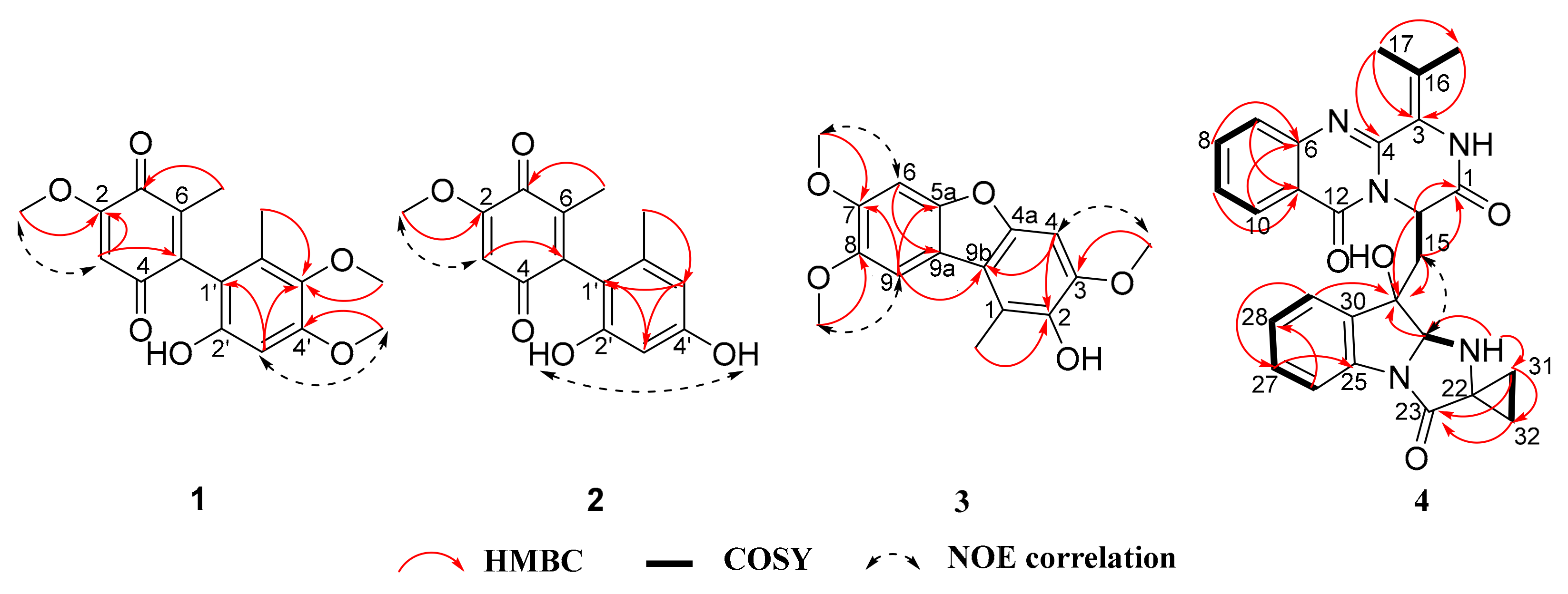
Peniquinone A (1) was obtained as a red oil. Its molecular formula was deduced as C17H18O6 with nine degrees of unsaturation according to the HRESIMS peak at m/z 319.1171 [M + H]+ (Figure S6). Analysis of the combined 1D NMR spectral data (Table 1) established that 1 possessed two aromatic methyl groups (6-CH3 and 6’-CH3), three methoxy groups (2-OCH3, 4′-OCH3 and 5′-OCH3), two aromatic protons (C-3 and C-3’) as well as a benzene ring. The connection mode and substitution pattern on the 1,4-benzoquinone and benzene rings were determined by key HMBC and NOSEY correlations (Figure 2). Key HMBC connections of 2-OCH3/C-2, 4’-OCH3/C-4’, 5’-OCH3/C-5’, 6’-CH3/C-1’, 6’-CH3/C-5’, and 6’-CH3/C-6’ and the chemical shift of C-2’ established that the phenol fragment of compound 1 was 3,4-dimethoxy-5-methylphenol (6) [12]. The 1,4-benzoquinone ring system was also confirmed by HMBC and NOESY correlations, which was further supported by comparing its NMR data with those of reported derivatives [23,24]. Compound 1, as a new compound containing a dimeric 1,4-benzoquinone motif and a phenol fragment, was thus elucidated as 2’-hydroxy-2,4’,5’-trimethoxy-6,6’-dimethyl-(5,1’-biphenyl)-1,4-dione.
Peniquinone B (2) was isolated as a red oil. The molecular formula was determined to be C15H14O5 on the basis of HRESIMS m/z 273.0767 [M − H] (Figure S14), along with the 1H and 13C NMR data (Table 1), indicating nine degrees of unsaturation. Its 1D NMR spectra were similar to those of 1, except for an additional hydroxyl signal and an aromatic proton (Table 1, Figures S8 and S9). In addition, two aromatic proton signals exhibited a small coupling constant (J = 1.9 Hz) (Table 1). The above analysis indicated that there was resorcinol substructure in compound 2, which was further supported by HMBC correlations (Figure 2, Figures S11 and S13). Therefore, compound 2 was constructed by a 1,4-benzoquinone moiety linking with a resorcinol group. The resorcinol fragment of compound 2 was orcinol (7) [13], which was also a known structure. Thus, compound 2 was elucidated as 2’,4’-dihydroxy-2-methoxy-6,6’-dimethyl-(5,1’-biphenyl)-1,4-dione.
Penizofuran A (3) was obtained as a brown powder, and its molecular formula C16H16O5 was established from the HRESIMS m/z 289.1079 [M + H]+ (Figure S21). Nine degrees of unsaturation were indicated besides the 1H and 13C NMR data (Table 2). The 1H NMR spectrum (Table 2) of 3 showed signals corresponding to one methyl (1-CH3), three methoxys (3-OCH3, 7-OCH3 and 8-OCH3) and three aromatic protons (C-4, C-6 and C-9). The 13C NMR spectrum showed sixteen carbon signals including twelve aromatic, one methyl, and three methoxyl groups. As two aromatic rings accounted for eight degrees of unsaturation, compound 3 was inferred to have an additional ring. Therefore, it was assumed that 3 has a dibenzofuran skeleton [25]. The position of the methyl, methoxy and hydroxyl in the dibenzofuran nucleus was established by 2D NMR data (Figure 2, Figures S19 and S20), and the key HMBC connections of 3-OCH3/C-3, 7-OCH3/C-7, 8-OCH3/C-8, 1-CH3/C-1, 1-CH3/C-2, and 1-CH3/C-9b, and NOESY connections between 3-OCH3 and C-4, 7-OCH3 and C-6, 8-OCH3 and C-9. Compound 3, as a new compound containing the parent nucleus of dibenzofuran, was thus elucidated as 3,7,8-trimethoxy-1-methyldibenzo[b,d]furan-2-ol.
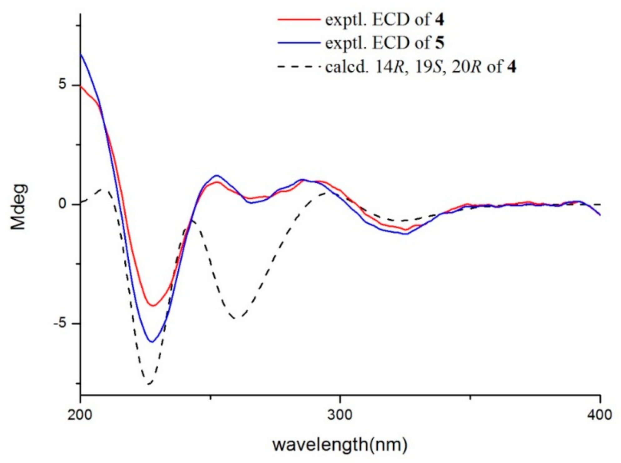
Quinadoline D (4) was obtained as a yellow powder. Its molecular formula was determined as C27H25N5O4 according to the HRESIMS peak at m/z 484.1966 [M + H]+ (Figure S30). The IR and UV spectra of 4 were very similar to those of quinadoline A (5) [11]. This suggested that compound 4 was a pyrazinoquinazoline derivative. Comparing its MS and NMR data with those in compound 5 (Table 3, Figures S23 and S24), compound 4 has 2 amu less than 5 in the MS spectrum, and it showed vicinal-coupling methylene groups (31-CH2 and 32-CH2) in 4, instead of two methyl groups in 5. The above cyclopropane fragment was further observed in the COSY spectrum (Figure 2), and verified by the key HMBC correlations from both H-31 and H-32 to C-22 and C-23. The NOE correlation observed between H-20 and H-15a, and absence of an NOE correlation between H-20 and OH-19 (Figure S29) revealed that H-20 and OH-19 were cofacial. The ECD spectrum of compound 4 was matched to 5 (Figure 3). It was hypothesized that compound 4 has the same absolute configuration as that of 5. ECD calculations were further employed to determine the absolute configuration. The predicted ECD spectrum was obtained by the TDDFT [mPW1PW91/6-311G(d)] method, and was subsequently compared with the experimental data (Figure S32). As shown in Figure 3, the calculated ECD spectrum was in good accordance with the experimental curve, confirming the absolute configuration of compound 4 as 14R, 19S, 20R (Figure 3).

2.2. The Bioactivities of Compounds

Compounds 14 were tested for their cytotoxicity against MCF-7, A549, U87 and PC3 cancer cell lines using the 3-(4,5)-dimethylthiahiazo(-z-y1)-3,5-di-phenytetrazoliumromide (MTT) method [26] (Table 4). Adriamycin was employed as the positive control in the cytotoxicity test. Compound 1 showed cytotoxicity against the MCF-7, U87 and PC3 cell lines with IC50 values of 12.39 µM, 9.01 µM and 14.59 µM, respectively. Compound 2 also exhibited cytotoxicity against MCF-7, U87 and PC3 cell lines with IC50 values of 25.32 µM, 13.45 µM and 19.93 µM, respectively. Compounds 14 were also evaluated for their antibacterial activities against the bacteria Staphylococcus aureus, Bacillus subtilis, Pseudomonas aeruginosa and Escherichia coli, by applying the disk diffusion method, according to the Clinical and Laboratory Standards Institute (CLSI) [27]. Unfortunately, none of these compounds were effective against the tested microorganisms.
All of the compounds were evaluated for their QS inhibitory activity against C. violaceum CV026. C30 was applied as the positive control in the QS inhibitory activity test. Compounds 2 and 8 displayed weak QS inhibitory activity against C. violaceum CV026 both with an MIC value of 20 μg/well (Figure S33). Compound 10 was a classical QS inhibitory active compound, penicillic acid. By comparing compounds 10 and 11 with structurally related metabolites, it was revealed that alterations to the alkene bond side chain substitutions could significantly influence their QS inhibitory activity. In addition, all of the compounds were tested for their antifungal activities against four agricultural pathogens Colletotrichum musae (ACCC 31244), Colletotrichum coccodes (ACCC 36067), Colletotrichum asianum T0408 and Magnaporthe grisea (ACCC 37631), using the disk diffusion method according to the CLSI [27,28]. The results showed that compounds 1215 had potent inhibitory activities against C. musae (ACCC 31244), and compound 14 also showed inhibitory activity against C. coccodes (ACCC 36067) (Table 5 and Figure S34). The positive control used in the antifungal activity test was cycloheximide.

3. Materials and Methods

3.1. General Experimental Procedures

Optical rotations were measured with a JASCO P-1020 digital polarimeter (JASCO Corporation, Tokyo, Japan). ECD spectra were measured on a JASCO J-715 (JASCO) or Chirascan CD (Applied Photophysics) spectropolarimeter. IR spectra were recorded on a Nicolet iS5 (Thermo Fisher Scientific, USA) in KBr discs. NMR spectra were obtained on a Bruker Advance spectrometer operating at 500 (1H) and 125 (13C) MHz using TMS as an internal standard, with chemical shifts recorded as δ values. Mass spectra were obtained on an LTQ-Orbitrap spectrometer equipped with an ESI source. Flash chromatography was performed on a Teledyne ISCO CombiFlash Rf 200 system. The high performance liquid chromatography (HPLC) system (Tianjin Bonna-Agela Technologies Co., Ltd., China) was composed of two HP-Q-P050 high pressure pumps equipped with a HP-Q-UV100S variable UV detector, a ATS-051-H10 automatic sampler, a FL-C100B fraction collector, and a Innoval column (10 × 250 mm, 10μm). The semi-preparative HPLC system (Agilent 1260 Infinity II; Agilent technologies, Germany) was equipped with a 1260 Quat Pump VL, a 1260 Vialsampler, a 1260 MCT, a 1260 DAD WR, and an ZORBAX SB-C18 column (5 μm, 9.4 × 250 mm). Thin-layer chromatography (TLC) was performed with silica gel GF254 plates (Qingdao Haiyang Chemical Co., Ltd., China). Column chromatography was performed on silica gel (200–300 mesh, Qingdao Haiyang Chemical Co., Ltd., China). Size exclusion chromatography was performed using Sephadex LH-20 (25–100 μm; Pharmacia, Uppsala, Sweden).

3.2. Fungal and Plant Material

The fungal strain Penicillium sp. L129 was isolated from the rhizosphere-soil of Limonium sinense (Girald) Kuntze collected in Yangkou Beach, Qingdao, China. It was identified according to its morphological characteristics and the analysis of the internal transcribed spacer (ITS) region of 16S rDNA (Genbank access No. MK625482). The fungus was deposited at the School of Pharmacy, Qingdao University, China, and was maintained at −80 °C.

3.3. Fermentation and Extraction

For large-scale fermentation, the fresh mycelia of Penicillium sp. L129 were cultured on potato dextrose agar (PDA) medium at 28 ± 2 °C for five days. The agar plugs were cut into small pieces under aseptic conditions, and 150 pieces were used to inoculate 50 flasks (500 mL) each containing 80 g rice, 120 mL seawater, and 0.24 g peptone. The cultures were inoculated at 28 ± 2 °C for 40 days. After 40 days of cultivation, the fermented cultures were extracted three times with ethyl acetate (EtOAc). The organic solvent was evaporated under reduced pressure to afford the crude extract (40.7 g).

3.4. Purification and Identification

The obtained EtOAc crude extract (40.7 g) based on TLC analysis was fractionated into six fractions (Fr.1 to Fr.6) by column chromatography on silica gel, eluting with a gradient of CH2Cl2–MeOH (100–50%). Fr.2 (24.5 g) was fractionated by silica gel column chromatography with a gradient of EtOAc–petroleum ether (3%–100%) to give eight subfractions (Fr.2.1 to Fr.2.8). Fr.2.2 (283.0 mg) was purified by HPLC (MeOH/H2O, 50/50, 4 mL/min) to yield 1 (2.0 mg, tR 14.5 min), 6 (7.0 mg, tR 8.3 min), and 9 (1.1 mg, tR 22.2 min). Fr.2.3 (1.9 g) was separated through a Sephadex LH-20 chromatograph (MeOH as eluent) to give Fr.2.3.1 and Fr.2.3.2. Fr.2.3.1 (1.4 g) was separated by a CombiFlash Rf 200 purification system (eluting with MeOH–H2O, 5% MeOH for 5 min, a gradient of 5–100% MeOH over 20 min, 100% MeOH for 10 min), which obtained four subfractions (Fr.2.3.1.1 to Fr.2.3.1.4), and Fr.2.3.1.4 (21.8 mg) was further purified by semi-preparative HPLC (MeOH/H2O, 70/30, 2 mL/min) to give compounds 3 (1.7 mg, tR 14.6 min), 16 (0.8 mg, tR 19.2 min), and 17 (0.6 mg, tR 26.7 min). Fr.2.3.2 (64.6 mg) was applied to Sephadex LH-20 (MeOH as eluent) to yield four subfractions (Fr.2.3.2.1 to Fr.2.3.2.4), and Fr.2.3.2.2 (12.0 mg) was further purified by semi-preparative HPLC (MeOH/H2O, 55/45, 2 mL/min) to give compound 2 (4.8 mg, tR 9.3 min), while Fr.2.3.2.3 (25.6 mg) was further purified by semi-preparative HPLC (MeOH/H2O, 50/50, 2 mL/min) to give compound 7 (11.5 mg, tR 8.0 min). Part of Fr.2.5 (250.0 mg) was separated through a Sephadex LH-20 chromatograph (MeOH as eluent) to give Fr.2.5.1 to Fr.2.5.2, and Fr.2.5.1 (200.0 mg) was purified through HPLC (MeOH/H2O, 40/60, 4 mL/min) to obtain compounds 10 (150.0 mg, tR 7.5 min) and 11 (6.0 mg, tR 10.5 min). Fr.2.7 (128.2 mg) was purified through HPLC (MeOH/H2O, 55/45, 4 mL/min) to provide compounds 12 (4.3 mg, tR 14.7 min), 13 (3.7 mg, tR 22.9 min), 14 (0.6 mg, tR 26.0 min), and 15 (0.9 mg, tR 20.8 min). Fr.2.8 (0.5 g) was subjected to semi-preparative HPLC (MeOH/H2O, 65/35, 2 mL/min) to yield compounds 4 (1.7 mg, tR 17.6 min) and 5 (2.2 mg, tR 20.5 min). Then, Fr.3 (3.7 g) was fractionated by a CombiFlash Rf 200 purification system (eluting with MeOH–H2O, 5% MeOH for 5 min, a gradient of 5–100% MeOH over 20 min, 100% MeOH for 10 min) to give four subfractions (Fr.3.1 to Fr.3.4), and Fr.3.4 (700 mg) was subjected to semi-preparative HPLC (MeOH/H2O, 60/40, 2 mL/min) to yield compound 8 (9.2 mg, tR 18.7 min).
Compound 1. Red oil; UV (CH3OH) λmax (log ε) 200, 274, 352 nm; IR (KBr) νmax 3431, 2927, 1682, 1617, 1229, 1040 cm−1; HRESIMS m/z 319.1171 ([M + H] +, calcd. for C17H18O6 319.1176); 13C NMR and 1H NMR (Table 1).
Compound 2. Red oil; UV (CH3OH) λmax (log ε) 216, 270, 380 nm; IR (KBr) νmax 3431, 2921, 2850, 1621, 1463, 1377, 1231, 720 cm−1; HRESIMS m/z 273.0767 ([M − H], calcd. for C15H13O5 273.0768); 13C NMR and 1H NMR (Table 1).
Compound 3. Brown powder; UV (CH3OH) λmax (log ε) 226, 252, 316 nm; IR (KBr) νmax 3438, 2922, 2852, 1632, 1463, 1384, 1261, 1119, 585 cm−1; HRESIMS m/z 289.1079 ([M + H]+, calcd. for C16H17O5 289.1071); 13C NMR and 1H NMR (Table 2).
Compound 4. Yellow powder; UV (CH3OH) λmax (log ε) 210, 230, 292 nm; IR (KBr) νmax 3432, 2923, 2853, 1628, 1464, 1378, 1128 cm−1; [ α ] D 25 −6. (c 0.1, MeOH); HRESIMS m/z 484.1966 ([M + H]+, calcd. for C27H26N5O4 484.1979); 13C NMR and 1H NMR (Table 3).

3.5. QS inhibitory Activity Essay

The in vitro QS inhibitory activity assay was carried out against the C. violaceum CV026. The strain C. violaceum CV026 was inoculated in a 20 mL LB broth medium (NaCl 10 g/L, Tryptone 10 g/L, Yeast Extract 5 g/L) at 37 °C on a rotary shaker (220 rpm) overnight. The culture (0.2 mL) was further mixed with 15 mL of warm molten LB agar (~40 °C) containing kanamycin (Sigma, 0.72mg) and N-hexanoyl-L-homoserine-lactone (C6-HSL) (Sigma, 1.5 μg). The agar was poured into a Petri dish and then punched with a sterile cork borer (Φ 5mm). The methanol solution (10 μL) of tested compounds at the concentration of 40 μg/well were pipetted into each well. Furanone C30 at the concentration of 10 μg/well was employed as the positive control while methanol was used as the negative control. The plates were then incubated overnight at 37 °C.

3.6. Cytotoxic Activity Essay

The MCF-7 and PC3 cell lines were obtained from the American Type Culture Collection (Manassas, VA) [29]. The A549 cell line was purchased from the Shanghai Institute for Biological Sciences, China Academy of Sciences (China) [30]. The U87 cell line was obtained from Dr. Bing Yan (Shan Dong University) [31].

4. Conclusions

We identified 17 compounds from the fermentation of Penicillium sp. L129, including two new dimeric 1,4-benzoquinone, peniquinone A (1) and peniquinone B (2), a new dibenzofuran derivative penizofuran A (3) and a new pyrazinoquinazoline derivative quinadoline D (4). Compounds 1–4 were screened for their cytotoxic activity against MCF-7, A549, U87 and PC3 cancer cell lines, and antibacterial activities against Gram-positive bacteria S. aureus and B. subtilis and Gram-negative bacteria P. aeruginosa and E. coli. All the compounds were tested for their QS inhibitory activity against C. violaceum CV026 and antifungal activities against C. musae (ACCC 31244), C. coccodes (ACCC 36067), M. grisea (ACCC 37631) and C. asianum T0408. Compound 1 showed cytotoxicity against the MCF-7, U87 and PC3 cell lines with IC50 values of 12.39 µM, 9.01 µM and 14.59 µM, respectively, while compound 2 exhibited cytotoxicity against MCF-7, U87 and PC3 cell lines with IC50 values of 25.32 µM, 13.45 µM and 19.93 µM, respectively. The known compounds 13 and 14 had potent antifungal activities against C. musae (ACCC 31244), both with an MIC values of 0.1 µg/scrip, and compound 14 also showed antifungal activity against C. coccodes (ACCC 36067) with an MIC value of 0.1 µg/scrip. Compounds 2 and 8 displayed weak QS inhibitory activities against C. violaceum CV026 both with an MIC value of 20µg/well. Compound 10 was penicillic acid, showing obvious QS inhibitory activity. The results suggested that the marine-derived fungi are an important source of new bioactive substances.

Supplementary Materials

The following are available online at https://www.mdpi.com/1660-3397/17/7/383/s1, Figures S1–S36: HRESIMS, IR and NMR spectra of the new compounds 14, the quantum calculation details are supplied, the QS inhibitory and antifungal activities, the 16S rDNA gene sequence of Penicillium sp. L129, and the HPLC data of compounds 14 are supplied.

Author Contributions

H.-M.Z. contributed to compounds purification, structural elucidation, bioassay and the preparation of the paper. C.-X.J. isolated and identified the strain. G.L. was responsible for the analysis of ECD calculations. Y.S contributed to the cytotoxicity test. Y.P. and Y.-X.L. were responsible for the strain preservation and fermentation. H.-X.L. and X.-P.P. designed the work and revised the paper.

Funding

This work was supported financially by the National Natural Science Foundation of China [grant number 40218030034], China Postdoctoral Science Foundation [grant number 2019M652309] and the Natural Science Foundation of Shandong Province [grant number ZR2018BC001].

Acknowledgments

We gratefully acknowledge Dr. Wen-Xuan Wang (School of pharmaceutical Science, South-Central University of Nationalities) for the quantum chemistry calculations and valuable discussions.

Conflicts of Interest

The authors declare no conflict of interest.

References

  1. Carroll, A.R.; Copp, B.R.; Davis, R.A.; Keyzers, R.A.; Prinsep, M.R. Marine natural products. Nat. Prod. Rep. 2019, 36, 122–173. [Google Scholar] [CrossRef] [PubMed] [Green Version]
  2. Liu, S.; Su, M.; Song, S.J.; Jung, J.H. Marine-derived Penicillium species as producers of cytotoxic metabolites. Mar. Drugs 2017, 15, 329. [Google Scholar] [CrossRef] [PubMed]
  3. Meng, L.H.; Wang, C.Y.; Mandi, A.; Li, X.M.; Hu, X.Y.; Kassack, M.U.; Kurtan, T.; Wang, B.G. Three diketopiperazine alkaloids with spirocyclic skeletons and one bisthiodiketopiperazine derivative from the mangrove-derived endophytic fungus Penicillium brocae MA-231. Org. Lett. 2016, 18, 5304–5307. [Google Scholar] [CrossRef]
  4. Liao, L.; Bae, S.Y.; Won, T.H.; You, M.; Kim, S.H.; Oh, D.C.; Lee, S.K.; Oh, K.B.; Shin, J. Asperphenins A and B, lipopeptidyl benzophenones from a marine-derived Aspergillus sp. Fungus. Org. Lett. 2017, 19, 2066–2069. [Google Scholar] [CrossRef] [PubMed]
  5. Chen, S.; Wang, J.; Lin, X.; Zhao, B.; Wei, X.; Li, G.; Kaliaperumal, K.; Liao, S.; Yang, B.; Zhou, X.; et al. Chrysamides A-C, three dimeric nitrophenyl trans-Epoxyamides produced by the deep-sea-derived fungus Penicillium chrysogenum SCSIO41001. Org. Lett. 2016, 18, 3650–3653. [Google Scholar] [CrossRef] [PubMed]
  6. Wang, W.; Liao, Y.; Zhang, B.; Gao, M.; Ke, W.; Li, F.; Shao, Z. Citrinin monomer and dimer derivatives with antibacterial and cytotoxic activities isolated from the deep sea-derived fungus Penicillium citrinum NLG-S01-P1. Mar. Drugs 2019, 17, 46. [Google Scholar] [CrossRef]
  7. Lu, Z.Y.; Lin, Z.J.; Wang, W.L.; Du, L.; Zhu, T.J.; Fang, Y.C.; Gu, Q.Q.; Zhu, W.M. Citrinin dimers from the halotolerant fungus Penicillium citrinum B-57. J. Nat. Prod. 2008, 71, 543–546. [Google Scholar] [CrossRef]
  8. Leong, S.L.; Schnurer, J.; Broberg, A.; Verrucine, F. A quinazoline from Penicillium verrucosum. J. Nat. Prod. 2008, 71, 1455–1457. [Google Scholar] [CrossRef]
  9. Michael, J.P. Quinoline, quinazoline and acridone alkaloids. Nat. Prod. Rep. 2008, 25, 166–187. [Google Scholar] [CrossRef]
  10. Hu, X.Y.; Meng, L.H.; Li, X.; Yang, S.Q.; Li, X.M.; Wang, B.G. Three new indole diterpenoids from the sea-anemone-derived fungus Penicillium sp. AS-79. Mar. Drugs 2017, 15, 137. [Google Scholar] [CrossRef]
  11. Huang, L.H.; Xu, M.Y.; Li, H.J.; Li, J.Q.; Chen, Y.X.; Ma, W.Z.; Li, Y.P.; Xu, J.; Yang, D.P.; Lan, W.J. Amino acid-directed strategy for inducing the marine-derived fungus Scedosporium apiospermum F41-1 to maximize alkaloid diversity. Org. Lett. 2017, 19, 4888–4891. [Google Scholar] [CrossRef] [PubMed]
  12. Tadross, P.M.; Gilmore, C.D.; Bugga, P.; Virgil, S.C.; Stoltz, B.M. Regioselective reactions of highly substituted arynes. Org. Lett. 2010, 12, 1224–1227. [Google Scholar] [CrossRef] [PubMed]
  13. Rojas, I.S.; Lotina-Hennsen, B.; Mata, R. Effect of lichen metabolites on thylakoid electron transport and photophosphorylation in isolated spinach chloroplasts. J. Nat. Prod. 2000, 63, 1396–1399. [Google Scholar] [CrossRef] [PubMed]
  14. Belofsky, G.N.; Gloer, K.B.; Gloer, J.B.; Wicklow, D.T.; Dowd, P.F. New p-terphenyl and polyketide metabolites from the sclerotia of Penicillium raistrickii. J. Nat. Prod. 1998, 61, 1115–1119. [Google Scholar] [CrossRef] [PubMed]
  15. Feng, C.C.; Chen, G.D.; Zhao, Y.Q.; Xin, S.C.; Li, S.; Tang, J.S.; Li, X.X.; Hu, D.; Liu, X.Z.; Gao, H. New isocoumarins from a cold-adapted fungal strain Mucor sp. and their developmental toxicity to zebrafish embryos. Chem. Biodivers. 2014, 11, 1099–1108. [Google Scholar] [CrossRef] [PubMed]
  16. Kimura, Y.; Nakahara, S.; Fujioka, S. Aspyrone, a nematicidal compound isolated from the fungus, Aspergillus melleus. Biosci. Biotechnol. Biochem. 1996, 60, 1375–1376. [Google Scholar] [CrossRef]
  17. Shang, Z.; Li, X.M.; Li, C.S.; Wang, B.G. Diverse secondary metabolites produced by marine-derived fungus Nigrospora sp. MA75 on various culture media. Chem. Biodivers. 2012, 9, 1338–1348. [Google Scholar] [CrossRef]
  18. Zhao, J.H.; Zhang, Y.L.; Wang, L.W.; Wang, J.Y.; Zhang, C.L. Bioactive secondary metabolites from Nigrospora sp. LLGLM003, an endophytic fungus of the medicinal plant Moringa oleifera lam. World J. Microb. Biot. 2012, 28, 2107–2112. [Google Scholar] [CrossRef]
  19. Levine, S.G.; Hicks, R.E.; Gottlieb, H.E.; Wenkert, E. Carbon-13 nuclear magnetic resonance spectroscopy of naturally occurring substances. XXX. Griseofulvin. J. Org. Chem. 1975, 40, 2540–2542. [Google Scholar] [CrossRef]
  20. Wubshet, S.G.; Nyberg, N.T.; Tejesvi, M.V.; Pirttila, A.M.; Kajula, M.; Mattila, S.; Staerk, D. Targeting high-performance liquid chromatography-high-resolution mass spectrometry-solid-phase extraction-nuclear magnetic resonance analysis with high-resolution radical scavenging profiles-bioactive secondary metabolites from the endophytic fungus Penicillium namyslowskii. J. Chromatogr. A. 2013, 1302, 34–39. [Google Scholar]
  21. Lin, L.Z.; West, D.P.; Cordell, G.A. NMR assignments of cis- and trans-capsaicin. Nat. Prod. Lett. 2006, 3, 5–8. [Google Scholar] [CrossRef]
  22. Gomez-Calvario, V.; Garduno-Ramirez, M.L.; Leon-Rivera, I.; Rios, M.Y. 1H and 13C NMR data on natural and synthetic capsaicinoids. Magn. Reson. Chem. 2016, 54, 268–290. [Google Scholar] [PubMed]
  23. Liu, H.; Zhao, F.; Yang, R.; Wang, M.; Zheng, M.; Zhao, Y.; Zhang, X.; Qiu, F.; Wang, H. Dimeric 1,4-benzoquinone derivatives and a resorcinol derivative from Ardisia gigantifolia. Phytochemistry 2009, 70, 773–778. [Google Scholar] [CrossRef] [PubMed]
  24. Fukuyama, Y.; Kiriyama, Y.; Okino, J.; Kodama, M. Belamcandaquinones A and B, novel dimeric 1,4-benzoquinone derivatives possessing cyclooxygenase inhibitory activity. Tetrahedron Lett. 1993, 34, 7633–7636. [Google Scholar] [CrossRef]
  25. Ko, R.K.; Lee, S.; Hyun, C.G.; Lee, N.H. New dibenzofurans from the branches of Distylium racemosum Sieb. et Zucc. Bull. Korean Chem. Soc. 2009, 30, 1376–1378. [Google Scholar]
  26. Xu, K.; Gao, Y.; Li, Y.L.; Xie, F.; Zhao, Z.T.; Lou, H.X. Cytotoxic p-terphenyls from the endolichenic fungus Floricola striata. J. Nat. Prod. 2018, 81, 2041–2049. [Google Scholar] [CrossRef]
  27. Michael, A.; Pfaller, M.D.; Ronald, N.; Jones, M.D. Performance accuracy of antibacterial and antifungal susceptibility test methods: Report from the College of American Pathologists (CAP) microbiology surveys program (2001-2003). Arch. Pathol. Lab. Med. 2006, 130, 767–778. [Google Scholar]
  28. Li, G.; Kusari, S.; Lamshoft, M.; Schuffler, A.; Laatsch, H.; Spiteller, M. Antibacterial secondary metabolites from an endophytic fungus, Eupenicillium sp. LG41. J. Nat. Prod. 2014, 77, 2335–2341. [Google Scholar] [CrossRef]
  29. Zheng, H.; Dong, Y.; Li, L.; Sun, B.; Liu, L.; Yuan, H.; Lou, H. Novel benzo[a]quinolizidine analogs induce cancer cell death through paraptosis and apoptosis. J. Med. Chem. 2016, 59, 5063–5076. [Google Scholar] [CrossRef]
  30. Sun, B.; Liu, J.; Gao, Y.; Zheng, H.B.; Li, L.; Hu, Q.W.; Yuan, H.Q.; Lou, H.X. Design, synthesis and biological evaluation of nitrogen-containing macrocyclic bisbibenzyl derivatives as potent anticancer agents by targeting the lysosome. Eur. J. Med. Chem. 2017, 136, 603–618. [Google Scholar] [CrossRef]
  31. Zhu, F.H.; Liu, Y.Q.; Lou, H.X. Bisbibenzyl-induced autophagy delays cell death in PC-3 cells. J. Shandong Univ. Health Sci. 2013, 51, 11–17. [Google Scholar]
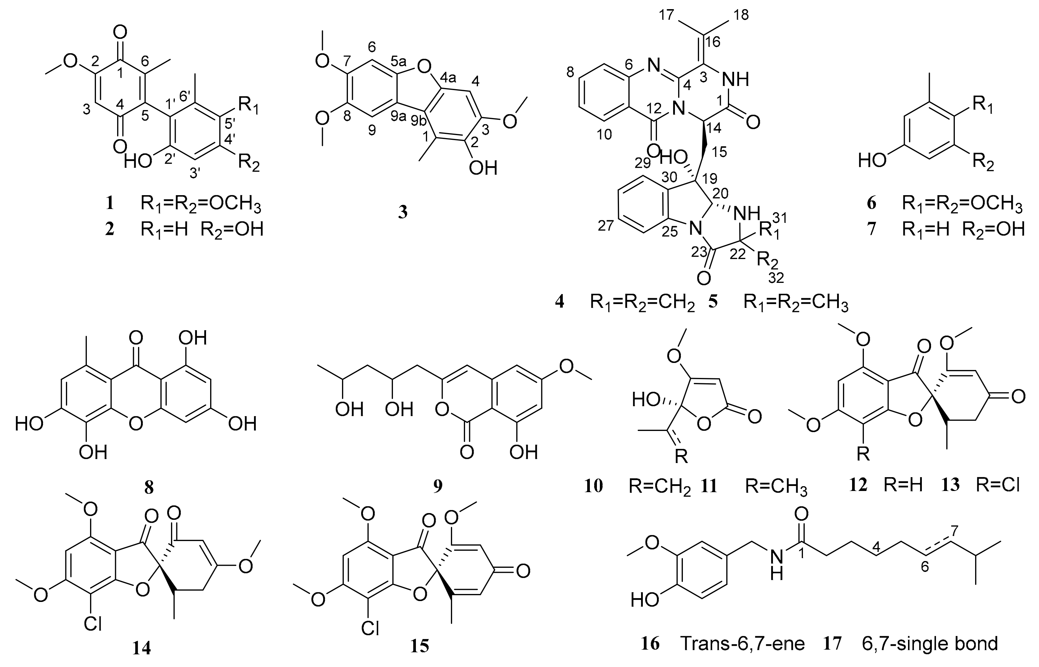
Figure 1. Chemical structures of compounds 117.
Marinedrugs 17 00383 g001
Figure 2. The key 2D NMR correlations for compounds 14.
Marinedrugs 17 00383 g002
Figure 3. Comparison of measured and calculated ECD spectrums of 4 and 5 (Calcd. ECD curves with a bathochromic shift for 10 nm).
Marinedrugs 17 00383 g003
Table 1. 1H and 13C NMR Data for 1 and 2 (1H 500 MHz, 13C 125 MHz, TMS, δ ppm).
Position1 a2 b
δC, TypeδH (J in Hz)δC, TypeδH (J in Hz)
1182.6, C 182.3, C
2158.8, C 158.3, C
3107.7, CH6.01, s107.6, CH6.11, s
4186.5, C 186.1, C
5140.9, C 141.7, C
6143.0, C 140.7, C
1′112.7, C 111.7, C
2′148.7, C 155.1, C
3′98.8, CH6.36, s99.9, CH6.16, d (1.9)
4′153.7, C 157.9, C
5′141.6, C 107.8, CH6.13, d (1.9)
6′131.0, C 137.3, C
6-CH313.6, CH31.88, s13.0, CH31.69, s
6′-CH313.4, CH31.96, s19.5, CH31.82, s
2-OCH356.4, OCH33.85, s56.3, OCH33.80, s
4′-OCH355.8, OCH33.83, s
5′-OCH360.6, OCH33.75, s
2′-OH 9.06, s
4′-OH 9.24, s
a Recorded in CDCl3; b Recorded in DMSO-d6.
Table 2. 1H and 13C NMR Data for 3 (1H 500 MHz, 13C 125 MHz, TMS, δ ppm).
Position3a
δC, TypeδH (J in Hz)
1116.6, C
2140.1, C
3147.0, C
493.1, CH7.13, s
4a149.1, C
5a150.3, C
696.2, CH7.30, s
7148.1, C
8145.5, C
9104.1, CH7.45, s
9a116.3, C
9b115.5, C
1-CH312.3, CH32.56, s
3-OCH356.2, OCH33.87, s
7-OCH356.0, OCH33.84, s
8-OCH356.3, OCH33.86, s
a Recorded in DMSO-d6.
Table 3. 1H and 13C NMR Data for 4 (1H 500 MHz, 13C 125 MHz, TMS, δ ppm).
Position4a
δC, TypeδH (J in Hz)
1166.5, C
3121.5, C
4146.8, C
6146.8, C
7127.1, CH7.66, d (8.2)
8134.6, CH7.85, t (7.7)
9126.9, CH7.55, t (7.4)
10126.3, CH8.16, d (8.3)
11119.6, C
12159.9, C
1452.5, CH5.43, t (6.5)
1537.5, CH215a: 2.48, dd (8.2, 14.8)
15b: 2.57, dd (5.4, 14.8)
16130.8, C
1721.5, CH32.31, s
1821.1, CH31.96, s
1975.1, C
2080.5, CH5.25, d (9.5)
2246.6, C
23173.5, C
25138.3, C
26124.3, CH7.10, m
27129.7, CH7.33, m
28114.4, CH7.33, m
29124.6, CH7.35, m
30137.1, C
3114.3, CH20.84, m
3210.9, CH21.01, m; 0.93, m
19-OH 5.68, br s
2-NH 10.04, br s
21-NH 3.57, d (9.4)
a Recorded in DMSO-d6.
Table 4. Cytotoxic activities of compounds 14.
CompoundsIC50
MCF-7A549U87PC3
112.39 ± 2.43>409.01 ± 2.3614.59 ± 2.75
225.32 ± 3.22>4013.45 ± 3.1219.93 ± 3.48
3>40>40>40>40
4>40>40>40>40
Adriamycin2.03 ± 0.421.53 ± 0.371.21 ± 0.500.98 ± 0.23
Table 5. Minimum inhibitory concentrations (MIC) of the compounds 1215 against fungal pathogensa.
Organism12131415
Colletotrichum musae (ACCC 31244)10.1<10>1
Colletotrichum coccodes (ACCC 36067)NANA<10NA
a All values are in μg/scrip and derived from experiments in triplicate; NA: no activity at the concentration of 10 µg/scrip; Compound 14 did not have enough mass to get MIC data.