Article

Slow-Binding Inhibition of Tyrosinase by Ecklonia cava Phlorotannins

1
College of Pharmacy, Chungnam National University, Daejeon 34134, Korea
2
Biospectrum Life Science Institute, A-1805, U-TOWER, 767, Sinsu-ro, Suji-gu, Yongin-si, Gyeonggi-do 16827, Korea
*
Authors to whom correspondence should be addressed.
These authors contributed equally to this work.
Mar. Drugs 2019, 17(6), 359; https://doi.org/10.3390/md17060359
Submission received: 29 May 2019 / Revised: 13 June 2019 / Accepted: 14 June 2019 / Published: 16 June 2019

Abstract

:
Tyrosinase inhibitors improve skin whitening by inhibiting the formation of melanin precursors in the skin. The inhibitory activity of seven phlorotannins (17), triphlorethol A (1), eckol (2), 2-phloroeckol (3), phlorofucofuroeckol A (4), 2-O-(2,4,6-trihydroxyphenyl)-6,6′-bieckol (5), 6,8′-bieckol (6), and 8,8′-bieckol (7), from Ecklonia cava was tested against tyrosinase, which converts tyrosine into dihydroxyphenylalanine. Compounds 3 and 5 had IC50 values of 7.0 ± 0.2 and 8.8 ± 0.1 μM, respectively, in competitive mode, with Ki values of 8.2 ± 1.1 and 5.8 ± 0.8 μM. Both compounds showed the characteristics of slow-binding inhibitors over the time course of the enzyme reaction. Compound 3 had a single-step binding mechanism and compound 5 a two-step-binding mechanism. With stable AutoDock scores of −6.59 and −6.68 kcal/mol, respectively, compounds 3 and 5 both interacted with His85 and Asn260 at the active site.

1. Introduction

Melanin is produced in skin epidermal cells to protect the skin from ultraviolet radiation [1]. The overproduction and accumulation of melanin causes age spots, freckles, melisma, and hyperpigmentation [1,2]. Tyrosinase (EC 1.14.18.1), which belongs to the type 3 copper protein family, is a key enzyme in melanogenesis [1,3]. It is a multifunctional oxide that contains a copper in each of two sets of three histidine residues in the active site. [4]. Furthermore, it catalyzes the hydroxylation of l-tyrosine to l-3,4-dihydroxyphenylalanine (l-DOPA), and the subsequent oxidation of l-DOPA to DOPA quinone [3,5]. These products are used in melanin biosynthesis [4]. Recently, the tyrosinase inhibitors kojic acid and arbutin were developed to improve skin whitening and as anti-hyperpigmentation agents [3,6]. However, they had undesirable side effects, including dermatitis, skin irritation, and DNA damage [1,4]. Much research has examined a variety of natural plants, with the aim of developing new inhibitors [7]. This study also sought alternative inhibitors without adverse effects.
Ecklonia cava, in the family Laminariaceae, grows along the coast of Jeju Island, Korea [8] and is widely used in Korea as a food ingredient, animal feed, and medicine [9]. E. cava contains phlorotannins in polymerized phloroglucinol units [8]. Phlorotannins are secondary metabolites that are important to human health [10]. These compounds have antioxidant, anti-cancer, anti-allergy, and anti-HIV bioactivities [10,11]. In addition, the compounds eckol, phlorofucofureckol A, and dieckol, from the edible brown alga Ecklonia stolonifera, exhibit anti-tyrosinase activity at macromolar concentrations [12].
This study evaluated the tyrosinase inhibitory activity of minor components from E. cava. Among seven isolated compounds (17), two minor compounds (3 and 5) had IC50 values of less than 10 μM, and also exhibited competitive and slow-binding inhibition of tyrosinase. This study also shows how these compounds interact with the catalytic site of tyrosinase via molecular docking.

2. Results and Discussion

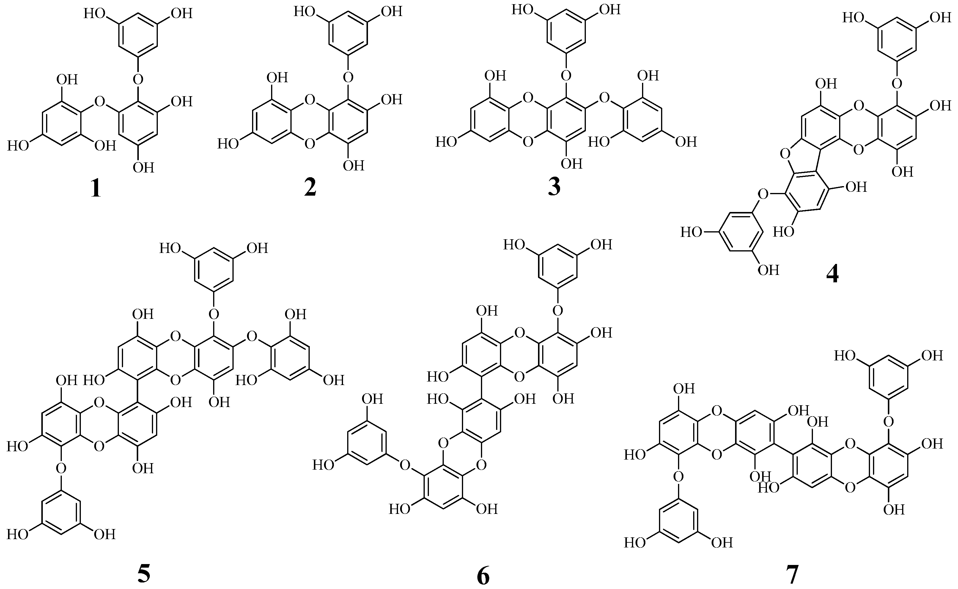
2.1. Isolation and Identification

The ethyl acetate fraction of ethanol extract of E. cava was known to suppress the catalytic reaction of tyrosinase [13]. Phlorotannins, eckol, triphlorethol-A, phlorofucofuroeckol A, and dieckol, were reported to be isolated from this fraction [14,15]. To isolate phlorotannins which have not been identified as tyrosinase inhibitors, dried E. cava powder (1.1 kg) was extracted by reflux with 80% ethanol for 72 h. The concentrated (80%) ethanol extract (290.0 g) was progressively divided into n-hexane, ethyl acetate, and butanol layers. The ethyl acetate layer (54.9 g) was subjected to open column chromatography with silica gel and C-18 resin to obtain seven phlorotannins (17). By comparing spectroscopic data with reported results, the compounds were identified as triphlorethol A (1) [16], eckol (2) [8], 2-phloroeckol (3) [16], phlorofucofuroeckol A (4) [12], 2-O-(2,4,6-trihydroxyphenyl)-6,6′-bieckol (5) [17], 6,8′-bieckol (6) [18], and 8,8′-bieckol (7) [8] (Figure 1).

2.2. Inhibition of Phlorotannins on Tyrosinase

Phlorotannins of E. cava and E. stolonifera were reported to have the inhibitory activity on mushroom tyrosinase [12,19]. 7-phloroeckol and dieckol were revealed to be the potential inhibitors within micromole concentration [13,19]. This study evaluated the ability of the isolated phlorotannins 17 to suppress the catalytic reaction of tyrosinase over time, in the absence or presence of inhibitor. Their inhibitory activity was calculated using equation (1). A commercial tyrosinase inhibitor was used as a positive control (kojic acid; IC50 = 25.0 ± 0.4 μM). To identify potent inhibitors, the inhibitory activity of all of the isolated compounds at 100 μM was tested against tyrosinase in vitro (Table 1). Of these, compounds 25 were confirmed to have inhibitory activity exceeding 50%. Serial dilutions were used to calculate the IC50 values. The tyrosinase inhibitory activity increased in a dose-dependent fashion (Figure 2A). Compounds 25 showed inhibitory activity, with IC50 values of 7.0 ± 0.2 to 66.4 ± 0.1 μM (Table 1). Of these, compounds 3 and 5 had inhibitory activity at < 10 μM. Interestingly, the structure of compound 5 contained the moiety of compound 3.
Our studies and reported results showed that phlorotannins, 2-phloroeckol, 7-phloroeckol dieckol, and 2-O-(2,4,6-trihydroxyphenyl)-6,6′-bieckol, substituted with phloroglucinol in eckol increased the inhibitory effect on tyrosinase compared to other phlorotannins [12,19]. Recently, secondary metabolites from terrestrial plants have been studied primarily for tyrosinase inhibitors [4,5,6,7]. Morin, norartocarpetin, artogomezianone, and (-)-N-formylanonaine have been revealed as the potential inhibitors [4,5,6,7]. Interest in the components from seaweed led to the study of the isolation of phlorotannin. It has been proven that they improve skin whitening [12,13,19]. These results suggest that 2-phloroeckol and 2-O-(2,4,6-trihydroxyphenyl)-6,6′-bieckol are tyrosinase inhibitors that can replace phenolic compounds [4,5,6,7].
To gain insight into the interaction of the enzyme with phlorotannins, as tyrosinase inhibitors, the products of the catalytic reaction were determined by ultraviolet-visible photometry after the inhibitor was added to the enzyme solution. The IC50 values of compounds 3 and 5 increased linearly with [S]/Km (Figure 2B). Furthermore, compounds 3 and 5 were produced with a set of the family liners by various inhibitors at substrate concentrations ranging from 0.156 to 2.50 mM. As shown in Lineweaver–Burk plots (Figure 2C,D), they had different values of −1/Km and the same 1/Vmax. The interactions with tyrosinase were competitive, with respective Ki values of 8.2 ± 1.1 and 5.8 ± 0.8 μM based on Dixon plots (Figure 2E,F, Table 1). Phlorotannins from seaweeds have been used to develop the non-competitive tyrosinase inhibitors 7-phloroeckol, dieckol, and phlorofucofuroeckol A, and the competitive inhibitors phloroglucinol and eckstolonol [12,13]. We found that compounds 3 and 5 competitively inhibited the catalysis of tyrosinase and might be useful for improving whitening.

2.3. Slow-Binding Inhibition

To confirm time-dependent inhibition by the potential inhibitors (compounds 3 and 5), the substrate was added into a mixture of ligand (3.1 μM) that was preincubated with tyrosinase. Over time, their inhibitory activities increased. To calculate the slow-binding parameters (k3, k4, k5, k6, and kapp i), the progress curves were analyzed using equation (2), with increasing concentrations of compounds 3 and 5 (Figure 3C,D); a replot of kobs was obtained as [I] (Figure 3E,F). The replot of compound 3 was a straight line that fit equation (3). When forming an encounter complex (EI) of the receptor with the ligand, compound 3 slowly binds to the active site of tyrosinase according to slow-binding mechanism A (a single-step binding mechanism) (Figure 3E) [20]. In comparison, the replot of kobs for compound 5 fit a hyperbolic equation (5) based on mechanism B (a two-step binding mechanism). This indicates that enzyme isomerization (E*I) results in slow bonding after the ligand rapidly interacts with the receptor [20,21]. Table 2 shows the kinetic parameters for the time-dependent inhibition of tyrosinase by the inhibitors. The two strongest inhibitors had different slow-binding mechanisms. Inhibitor 5, which has a higher molecular weight, induced a new conformational state of the enzyme.

2.4. Molecular Docking

Molecular docking was performed to determine how the ligand interacts with the active site of tyrosinase. The best fit of their complex was considered to have the lowest energy, as calculated using Autodock 4.2. As indicated in Figure 4A–E and Table 3, the AutoDock scores of compounds 3 and 5 showed a trend similar to their IC50 values in vitro. They covered the hole and area around the active site, with calculated energies of −6.59 and −6.68 kcal/mol, respectively (Figure 4A). Compound 3 was held by six hydrogen bonds with three amino acids (Glu256 2.82 Å, Asn260 2.73 Å, and Met280 2.68 Å) and hydrophobic interactions with 15 amino acids (Figure 4B,C, Table 3). The phloroglucinol moiety of compound 3 had a π–π bond interaction with His85 at 9.4Å distance (Figure 4E). For compound 5, seven hydroxyl groups were close to eight amino acids: Lys79 (2.95Å), Asn81 (2.77 and 2.85Å), His85 (3.01, 3.17, and 2.79Å), Cys83 (2.71Å), Asn260 (2.69Å), Gly281 (2.69Å), His244 (3.30Å), and Glu322 (2.68Å). Furthermore, there were hydrophobic interactions with 18 amino acids (Figure 4D,E, Table 3).

3. Materials and Methods

3.1. General Experimental Procedures

NMR experiments were conducted on an ECA500 (JEOL, Tokyo, Japan), with the chemical shift referenced to the residual solvent signals, using methanol-d4 and DMSO-d6 as a solvent. TLC analysis was performed on silica-gel 60 F254 and RP-18 F254S plates (both 0.25 mm layer thickness, Merck, Darmstadt, Germany). Compounds were visualized by dipping plates into 10% (v/v) H2SO4 reagent and then air heat treated at 300 °C for 15 s. Silica gel (Merck 60A, 70–230 or 230–400 mesh ASTM) and reversed-phase silica gel (YMC Co., ODS-A 12 nm S-150, S-75 μm) were used for open column chromatography. Tyrosinase (T3824), kojic acid (K3125) and l-tyrosine (T3754) were purchased from Sigma-Aldrich (ST. Louis, Mo, USA). UV–vis and fluorescence spectra were measured by TECAN infinite 200 PRO® spectrophotometer (Zurich, Switzerland).

3.2. Plant Material

E. cava was purchased from a herbal market in Jeju Island, Korea, on May 2015. One of the author (Prof. Y.H. Kim) identified this brown algal species. A voucher specimen (CNU-15005) was deposited at the Herbarium, College of Pharmacy, Chungnam National University (CNU).

3.3. Extraction and Isolation

The dried powder (1.0 kg) of E. cava was refluxed with 80% EtOH (16 L) for 72 h, and the ethanol extract was concentrated under vacuum to yield a dark green residue (290.0 g). The residue (290.0 g) was suspended in H2O (2.0 L), and the aqueous layer was partitioned with n-hexane, ethyl acetate, and n-butanol. The ethyl acetate layer (54.9 g) was subjected to silica gel column eluted with CH2Cl2:MeOH (8:1→2:1) to obtain four fractions (5A–5D). Fraction 5B was chromatographed on silica gel column eluting with CHCl3:MeOH:H2O (7:1:0.1→1:1:0.1) to afford six fractions (5BA–5BF). Fraction 5BA was chromatographed on C-18 column eluting with MeOH:H2O (1:3→1:1) to isolate compound 1 (304.4 mg). Fraction 5BB was chromatographed on silica gel column eluting with CHCl3:MeOH:H2O (4:1:0.1→1:1:0.1) to obtain two fractions (5BBA–5BBB). Fraction 5BBB was subjected to C-18 column eluting with (CH3)2CO:MeOH:H2O (1:1:3→1:1:1) to isolate compound 3 (69.0 mg). Fraction 5BD was subjected to C-18 column eluted with (CH3)2CO:H2O (1:3→1:1) to obtain compound 2 (128.5 mg). Fraction 5BE was subjected to C-18 column using MeOH:H2O (1:3→1:1) to afford compound 4 (230.2 mg). The fraction 5BF was subjected to C-18 column eluted with MeOH:H2O (1:4→1:1) to obtain compound 5 (52.7 mg). Fraction 5C was subjected to C-18 column eluted with (CH3)2CO:H2O (1:5→1:1) to obtain compound 7 (110.5 mg). Fraction 5D was chromatographed on C-18 column eluting with (CH3)2CO:H2O (1:4→1:1) to obtain compound 6 (67.5 mg).

Compound 1

White powder; ESI-MS: m/z 373.2 [M − H]; 1H-NMR (methanol-d4, 400 MHz): δ 6.02 (1H, d, J = 2.8 Hz, H-5), 5.97 (2H, d, J = 2.1 Hz, H-6″, H-2″), 5.89 (1H, t, J = 2.1 Hz, H-4″), 5.86 (2H, s, H-5′, H-3′), 5.72 (1H, d, J = 2.8 Hz, H-3). 13C-NMR (methanol-d4, 100 MHz): δ 162.5 (C-1″), 160.4 (C-3″, C-5″), 156.5 (C-4′), 156.2 (C-4), 153.8 (C-6), 152.7 (C-2), 152.2 (C-6′, C-2′), 125.7 (C-1), 124.7 (C-1′), 98.0 (C-3), 97.5 (C-4″), 96.2 (C-5′, C-3′), 95.4 (C-2″, C-6″), 95.0 (C-5).

Compound 2

Light brown powder; ESI-MS: m/z 371.2 [M − H]; 1H-NMR (DMSO-d6, 300 MHz): δ 9.54 (1H, s, OH-9), 9.48 (1H, s, OH-4), 9.21 (2H, s, OH-2, OH-7), 9.17 (2H, s, OH-3′, OH-5′), 6.14 (1H, s, H-3), 5.97 (1H, d, J = 2.5 Hz, H-8), 5.81 (1H, d, J = 1.7 Hz, H-4′), 5.80 (1H, d, J = 2.5 Hz, H-6), 5.72 (2H, d, J = 1.7 Hz, H-2′, H-6′). 13C-NMR (DMSO-d6, 75 MHz): δ 160.4 (C-1′), 158.9 (C-3′, C-5′), 153.1 (C-7), 146.1 (C-9), 146.0 (C-2), 142.6 (C-5a), 142.0 (C-4), 137.2 (C-10a), 123.2 (C-1), 122.7 (C-9a), 122.2 (C-4a), 98.5 (C-8), 98.2 (C-3), 96.3 (C-4′), 93.9 (C-6), 93.7 (C-2′, C-6′).

Compound 3

Light brown powder; ESI-MS: m/z 495.2 [M − H]; 1H NMR (DMSO-d6, 400 MHz): δ 9.52 (1H, s, OH-9), 9.41 (1H, s, OH-4), 9.19 (1H, s, OH-7), 9.13 (2H, s, OH-3′, OH-5′), 9.08 (2H, s, OH-2″, OH-6″), 8.98 (1H, s, OH-4″), 5.98 (1H, d, J = 2.2 Hz, H-8), 5.87 (2H, d, J = 1.8 Hz, H-2′, H-6′), 5.85 (2H, s, H-3″, H-5″), 5.84 (1H, t, J = 1.8 Hz, H-4′), 5.83 (1H, s, H-3), 5.80 (1H, d, J = 2.2 Hz, H-5). 13C NMR (DMSO-d6, 100 MHz): δ 160.5 (C-1′), 158.9 (C-3′, C-5′), 154.9 (C-4″), 153.2 (C-7), 151.3 (C-2″, C-6″), 147.9 (C-2), 146.3 (C-9), 142.6 (C-5a), 141.7 (C-4), 137.2 (C-10a), 124.4 (C-4a), 122.7 (C-1), 122.0 (C-9a), 98.7 (C-8), 96.4 (C-4′), 96.0 (C-3), 94.9 (C-3″, C-5″), 94.1 (C-2′, C-6′), 93.9 (C-6).

Compound 4

Amorphous powder; ESI-MS: m/z 601.2 [M − H]; 1H-NMR (DMSO-d6, 300 MHz): δ 10.14 (1H, s, OH-14), 9.87 (1H, s, OH-4), 9.85 (1H, s, OH-10), 9.44 (1H, s, OH-2), 9.21 (2H, s, OH-3′, OH-5′), 9.18 (2H, s, OH-3″, OH-5″), 8.22 (1H, s, OH-8), 6.73 (1H, s, H-13), 6.44 (1H, s, H-9), 6.31 (1H, s, H-3), 5.84 (2H, d, J = 1.8 Hz, H-4′, H-4″), 5.78 (2H, d, J = 1.8 Hz, H-2′, H-6′), 5.73 (2H, d, J = 1.8 Hz, H-2″, H-6″). 13C-NMR (DMSO-d6, 75 MHz): δ 160.2 (C-1′), 160.0 (C-1″), 159.0 (C-3″, C-5″), 158.8 (C-3′, C-5′), 150.8 (C-12a), 150.4 (C-10), 149.5 (C-11a), 147.0 (C-2), 146.5 (C-8), 144.8 (C-14), 142.0 (C-4), 136.8 (C-15a), 134.0 (C-5a), 126.4 (C-14a), 122.6 (C-4a), 122.5 (C-1), 120.1 (C-11), 103.4 (C-7), 103.2 (C-6), 99.1 (C-9), 98.3 (C-3), 96.5 (C-4″), 96.4 (C-4′), 94.8 (C-13), 93.7 (C-2′, C-6′), 93.5 (C-2″, C-6″).

Compound 5

Amorphous powder; ESI-MS: m/z 865.3 [M − H]; 1H NMR (DMSO-d6, 400 MHz): δ 9.29 (1H, s, OH-9), 9.26 (1H, s, OH-9′), 9.15 (5H, s, OH-3″, OH-5″, OH-3‴, OH-5‴, OH-2′), 9.09 (IH, s, OH-4), 9.07 (1H, s, OH-4′), 9.05 (2H, s, OH-2⁗, OH-6⁗), 8.93 (1H, s, OH-4⁗), 8.66 (IH, s, OH-7), 8.62 (1H, s, OH-7′), 6.08 (1H, s, H-3′), 6.06 (1H, s, H-8), 6.04 (1H, s, H-8′), 5.90 (2H, s, J = 2.1 Hz, H-2″, H-6″), 5.83 (1H, d, J = 2.1 Hz, H-4″), 5.83 (2H, s, H-3‴, H-5‴), 5.80 (1H, s, H-3), 5.79 (1H, d, J = 3.0 Hz, H-4⁗), 5.74 (2H, d, J = 3.0 Hz, H-2⁗, H-6⁗). 13C NMR (DMSO-d6, 100 MHz): δ 160.6 (C-1″, C-1‴), 158.9 (C-3″, C-3‴, C-5″, C-5‴), 154.9 (C-4⁗), 151.5 (C-7, C-7′), 151.3 (C-2⁗, C-6⁗), 147.4 (C-2′), 145.6 (C-2), 144.7 (C-9, C-9′), 142.0 (C-4′), 141.8 (C-4), 141.5 (C-5a′), 141.5 (C-5a), 137.4 (C-10a′), 137.3 (C-10a), 124.8 (C-1⁗), 123.8 (C-1″), 122.9 (C-9a, C-9a′), 122.5 (C-1), 122.1 (C-4a, C-4a′), 99.8 (C-6′), 99.7 (C-6), 98.0 (C-8, C-8′), 97.8 (C-3′), 96.4 (C-4‴, C-4″), 96.3 (C-3), 94.9 (C-3⁗, C-5⁗), 94.2 (C-2″, C-2‴), 93.8 (C-″, C-6‴).

Compound 6

Brown amorphous powder; ESI-MS: m/z 741.2 [M − H]; 1H NMR (DMSO-d6, 400 MHz): δ 9.46 (1H, s, OH-2′), 9.28 (1H, s, OH-9′), 9.17 (3H, s, OH-3‴, OH-5‴, OH-4′), 9.12 (3H, s, OH-3″, OH-5″, OH-2), 9.07 (1H, s, OH-4), 8.76 (1H, s, OH-7′), 8.65 (1H, s, OH-7), 7.90 (1H, s, OH-9), 6.19 (1H, d, H-8), 6.10 (1H, s, H-3′), 6.06 (1H, s, H-3), 5.97 (1H, s, H-6′), 5.81 (2H, d, J = 2.0 Hz, H-4″, H-4‴), 5.78 (2H, d, J = 2.0 Hz, H-2‴, H-6‴), 5.76 (2H, d, J = 2.0 Hz, H-2″, H-6″). 13C NMR (DMSO-d6, 100 MHz): δ 160.4 (C-1″, C-1‴), 158.8 (C-3″, C-3‴, C-5″, C-5‴), 151.7 (C-7), 151.5 (C-7′), 145.8 (C-2), 145.4 (C-2′), 144.6 (C-9), 144.3 (C-9′), 141.8 (C-4), 141.6 (C-4′), 141.2 (C-5a), 140.9 (C-5a′), 137.2 (C-10a), 137.2 (C-10a′), 123.5 (C-1), 123.2 (C-1′), 123.0 (C-9a), 122.8 (C-9a′), 122.3 (C-4a), 121.8 (C-4a′), 104.1 (C-8′), 99.3 (C-6), 98.0 (C-8), 96.1 (C-4″), 96.0 (C-4‴), 93.8 (C-2‴, C-6‴), 93.7 (C-2″, C-6″), 93.6 (C-6′).

Compound 7

Brown amorphous powder; ESI-MS: m/z 741.2 [M − H]; 1H NMR (DMSO-d6, 300 MHz): δ 9.46 (1H, s, OH-4), 9.18 (1H, s, OH-2), 9.13 (2H, s, OH-3′, OH-5′), 8.77 (1H, s, OH-9), 7.91 (1H, s, OH-7), 6.18 (1H, s, H-3), 5.98 (1H, s, H-6), 5.81 (1H, t, J = 1.8 Hz, H-4′), 5.76 (2H, d, J = 1.8 Hz, H-2′, H-6′). 13C NMR (DMSO-d6, 75 MHz): δ 160.9 (C-1′), 159.2 (C-3′, C-5′), 152.2 (C-7), 146.3 (C-2), 145.1 (C-9), 142.3 (C-4), 141.8 (C-5a), 137.7 (C-10a), 123.8 (C-1), 123.6 (C-9a), 122.9 (C-4a), 104.7 (C-8), 98.6 (C-3), 96.7 (C-4′), 94.4 (C-2′, C-6′), 94.3 (C-6).

3.4. Tyrosinase Assay

To evaluate inhibitory activity on tyrosinase with isolated compounds, 130 μL of tyrosinase in 0.05 mM phosphate buffer (pH 6.8) was divided in 96 well plates [19]. 20 μL of compound concentrations ranging from 1–0.015 mM was added. 50 μL of 1.5 mM L-tyrosine in phosphate buffer was diluted into the mixture for calculating the inhibitory activity. 50 μL of 10–0.62 mM L-tyrosine in buffer was added to analyze initial velocity (v0). After starting their reaction for 20 min, an amount of the product was detected at UV–vis 475 nm. The inhibitory activity was analyzed according to Equation (1)
Inhibitory activity rate (%) = [(ΔC − ΔS)/ΔC] × 100

3.5. Slow-Binding Inhibition Analysis

The progress curves were calculated to Equation (2) by Morrison according to time course for 420 s.
[P] = vst + [(v0vs)/kobs(1 − ekobst)]
where t is time, [P] is product intensity, v0 and vs are the initial and steady-state reaction velocities, and kobs is the apparent first-order rate concentration.
kobs = k4 + k3[I]
K i a p p = k 4 / k 3
This is a linear Equation (3) for slow-binding mechanism A. K i a p p is the apparent value of an inhibitor by Equation (4). Where [I] is inhibitor concentration.
k obs = k 6 + [ ( k 5 × [ I ] ) / ( K i a p p + [ I ] )
Equation (5) of hyperbola is for the calculation of rate constant in slow-binding mechanism B.

3.6. Molecular Docking

This simulation was performed as described previously. The 3D structure of protein coded with 2Y9X was achieved from RCSB homepage [22]. This was added hydrogen atoms and assigned with gasteiger charges by using Autodocktools. Ligands were built and minimized energy by MM2 with Chem3D Pro. They were established the grid containing activity site with number of points (X: 80, Y: 80, Z: 80). DPS file was contained to set up Lamarckian genetic algorithm for docking of the ligand into receptor (runs 50 and the maximum number was set as long). Their results were represented with Chimera (version 3.13.1, San Francisco, CA, USA) and LigPlot (version 4.5.3, Cambridge, UK).

4. Conclusions

Seven phlorotannins (17) from E. cava were isolated using column chromatography and their structures were identified by spectral analysis. Of these, compounds 3 and 5 potentially inhibited the catalytic reaction of tyrosinase by blocking the entrance to the active site. Moreover, compounds 3 and 5 altered the turnover rate from substrate to the enzyme product over time. The former underwent simple reversible slow binding with the enzyme, while the latter initially formed a complex (EI) with the enzyme quickly, followed by an isomerization step to create a new enzyme-type complex (E*I). Phlorotannins containing flexible phloroglucinol to eckol backbone were found the facts bound easily into active site of tyrosinase. The findings suggested the phloroglucinol may be key moiety for induced fit of inhibitor with enzyme flexibility. The molecular simulation study indicated that the two inhibitors were docked at the active site of tyrosinase with AutoDock scores or −6.59 and −6.68 kcal/mol, respectively. Both compounds used the amino acids His85 and Asn260 to maintain their connection with tyrosinase. These findings imply that 2-phloroeckol (3) and 2-O-(2,4,6-trihydroxyphenyl)-6,6′-bieckol (5) might inhibit the production of melanin products.

Author Contributions

Y.H.K. and S.Y.Y. participated in the research design. S.P. and J.S.P. conducted the experiments. J.H.K. and S.L. contributed to the writing of the manuscript and the experiments.

Funding

This research was supported by Basic Science Research Program through the National Research Foundation of Korea (NRF) funded by the Ministry of Education, Science and Technology (NRF-2018R1A6A3A11047338).

Conflicts of Interest

The authors declare no conflict of interest.

References

  1. Şöhretoğlu, D.; Sari, S.; Barut, B.; Özel, A. Tyrosinase inhibition by some flavonoids: Inhibitory activity, mechanism by in vitro and in silico studies. Bioorg. Chem. 2018, 81, 168–174. [Google Scholar] [CrossRef] [PubMed]
  2. Bukhari, S.N.A.; Jantan, I.; Tan, O.U.; Sher, M.; Naeem-ul-Hassan, M.; Qin, H.-L. Biological activity and molecular docking studies of curcumin-related α,β-unsaturated carbonyl-based synthetic compounds as anticancer agents and mushroom tyrosinase inhibitors. J. Agric. Food Chem. 2014, 62, 5538–5547. [Google Scholar] [CrossRef] [PubMed]
  3. Ha, Y.M.; Park, Y.J.; Lee, J.Y.; Park, D.; Choi, Y.J.; Lee, E.K.; Kim, J.M.; Kim, J.-A.; Park, J.Y.; Lee, H.J.; et al. Design, synthesis and biological evaluation of 2-(substituted phenyl)thiazolidine-4-carboxylic acid derivatives as novel tyrosinase inhibitors. Biochimie 2012, 94, 533–540. [Google Scholar] [CrossRef] [PubMed]
  4. Wang, Y.; Zhang, G.; Yan, J.; Gong, D. Inhibitory effect of morin on tyrosinase: Insights from spectroscopic and molecular docking studies. Food Chem. 2014, 163, 226–233. [Google Scholar] [CrossRef] [PubMed]
  5. Lan, W.-C.; Tzeng, C.-W.; Lin, C.-C.; Yen, F.-L.; Ko, H.-H. Prenylated flavonoids from Artocarpus altilis: Antioxidant activities and inhibitory effects on melanin production. Phytochemistry 2013, 89, 78–88. [Google Scholar] [CrossRef] [PubMed]
  6. Wang, H.-M.; Chen, C.-Y.; Chen, C.-Y.; Ho, M.-L.; Chou, Y.-T.; Chang, H.-C.; Lee, C.-H.; Wang, C.-Z.; Chu, I.-M. (-)-N-Formylanonaine from Michelia alba as a human tyrosinase inhibitor and antioxidant. Bioorg. Med. Chem. 2010, 18, 5241–5247. [Google Scholar] [CrossRef] [PubMed]
  7. Zolghadri, S.; Bahrami, A.; Khan, M.T.H.; Munoz-Munoz, J.; Garcia-Molina, F.; Garcia-Canovas, F.; Saboury, A.A. A comprehensive review on tyrosinase inhibitors. J. Enzyme Inhib. Med. Chem. 2019, 34, 279–309. [Google Scholar] [CrossRef] [Green Version]
  8. Lee, J.; Jun, M. Dual BACE1 and cholinesterase inhibitory effects of phlorotannins from Ecklonia cava-An in vitro and in silico study. Mar. Drugs 2019, 17, 91. [Google Scholar] [CrossRef]
  9. Yang, H.; Lee, S.Y.; Lee, S.R.; Pyun, B.-J.; Kim, H.J.; Lee, Y.H.; Kwon, S.W.; Suh, D.H.; Lee, C.H.; Hong, E.-J.; et al. Therapeutic effect of Ecklonia cava extract in letrozole-induced polycystic ovary syndrome rats. Front. Pharmacol. 2018, 9, 1325. [Google Scholar] [CrossRef]
  10. Oh, S.; Son, M.; Lee, H.S.; Kim, H.-S.; Jeon, Y.-J.; Byun, K. Protective effect of pyrogallol-phloroglucinol-6,6-bieckol from Ecklonia cava on monocyte-associated vascular dysfunction. Mar. Drugs 2018, 16, 441. [Google Scholar] [CrossRef]
  11. Yang, Y.-I.; Woo, J.-H.; Seo, Y.-J.; Lee, K.-T.; Lim, Y.; Choi, J.-H. Protective effect of brown alga not italic phlorotannins against hyperinflammatory responses in lipopolysaccharide-induced sepsis models. J. Agric. Food Chem. 2016, 64, 570–578. [Google Scholar] [CrossRef] [PubMed]
  12. Kang, H.S.; Kim, H.R.; Byun, D.S.; Son, B.W.; Nam, T.J.; Choi, J.S. Tyrosinase inhibitors isolated from the edible brown alga Eckloina stolonifera. Arch. Pharmcal. Res. 2004, 27, 1226–1232. [Google Scholar] [CrossRef]
  13. Yoon, N.Y.; Eom, T.-K.; Kim, M.-M.; Kim, S.-K. Inhibitory effect of phlorotannins isolated from Ecklonia cava on mushroom tyrosinase activity and melanin formation in mouse B16F10 melanoma cells. J. Agric. Food Chem. 2009, 57, 4124–4129. [Google Scholar] [CrossRef] [PubMed]
  14. Wijesinghe, W.A.J.P.; Ko, S.-C.; Jeon, Y.-J. Effect of phlorotannins isolated from Ecklonia cava on angiotensin enzyme (ACE) inhibitory activity. Nutr. Res. Pract. 2011, 5, 93–100. [Google Scholar] [CrossRef] [PubMed]
  15. Lee, J.H.; Ko, J.Y.; Oh, J.Y.; Kim, C.Y.; Lee, H.J.; Kim, J.; Jeon, Y.J. Preparative isolation and purification of phlorotannins from Ecklonia cava using centrifugal partition chromatography by one-step. Food Chem. 2014, 158, 433–437. [Google Scholar] [CrossRef]
  16. Park, J.Y.; Kim, J.H.; Kwon, J.M.; Kwon, H.J.; Jeong, H.J.; Kim, Y.M.; Kim, D.; Lee, W.S.; Ryu, Y.B. Dieckol, a SARS-CoV 3CL pro inhibitor, isolated from the edible brown algae Ecklonia cava. Bioorg. Med. Chem. 2013, 21, 3730–3737. [Google Scholar] [CrossRef] [PubMed]
  17. Fukuyama, Y.; Kodama, M.; Miura, I.; Kinzyo, Z.; Mori, H.; Nakayama, Y.; Takahashi, M. Anti-plasmin Inhibitor. V.: Structures of novel dimeric eckols isolated from the brown alga Ecklonia kurome OKAMURA. Chem. Pharm. Bull. 1989, 37, 2438–2440. [Google Scholar] [CrossRef]
  18. Sugiura, Y.; Matsuda, K.; Yamada, Y.; Nishikawa, M.; Shioya, K.; Katsuzaki, H.; Imai, K.; Amano, H. Anti-allergic phlorotannins from the edible brown alga, Eisenia arborea. Food Sci. Technol. Res. 2007, 13, 54–60. [Google Scholar] [CrossRef]
  19. Kang, S.-M.; Heo, S.-J.; Kim, K.-N.; Yang, S.-H.; Kim, A.-D.; Jeon, Y.-J. Molecular docking studies of a phlorotannin, dieckol isolated from Eckloina cava with tyrosinase inhibitory activity. Bioorg. Med. Chem. 2012, 20, 311–316. [Google Scholar] [CrossRef]
  20. Masson, P.; Lushchekina, S.V. Slow-binding inhibition of cholinesterase, pharmacological and toxicological relevance. Arch. Biochem. Biophys. 2016, 593, 60–68. [Google Scholar] [CrossRef]
  21. Menon, V.; Rao, M. Slow-tight binding inhibition of pepsin by an aspartic protease inhibitor from Streptomyces sp. MBR04. Inter. J. Biol. Macromol. 2012, 51, 165–174. [Google Scholar] [CrossRef] [PubMed]
  22. Xiong, S.-L.; Lim, G.T.; Yin, S.-J.; Lee, J.; Si, Y.-X.; Yang, J.-M.; Park, Y.-D.; Qian, G.-Y. The inhibitory effect of pyrogallol on tyrosinase activity and structure: Integration study of inhibition kinetics with molecular dynamics simulation. Int. J. Biol. Macromol. 2019, 121, 463–471. [Google Scholar] [CrossRef] [PubMed]
Figure 1. Structure of compounds 17 from E. cava.
Marinedrugs 17 00359 g001
Figure 2. Inhibitory activity of compounds on tyrosinase (A). Effects of S/Km ratio on the IC50 values (B). Lineweaver–Burk (C,D) and Dixon (E,F) plots of tyrosinase inhibition by compounds, respectively.
Marinedrugs 17 00359 g002aMarinedrugs 17 00359 g002b
Figure 3. Progress curves for slow-binding inhibition (A,B) and dependence of the values of kobs on the concentration of compounds 3 and 5 (C,D) of tyrosinase. Mechanism of slow binding inhibitor (E,F).
Marinedrugs 17 00359 g003
Figure 4. The binding pose (A) of two compounds 3 and 5 into tyrosinase (orange: 3, green: 5). Hydrogen bonds (B,C), hydrophobic interactions (D,E), and 9.4 Å π-π interaction (F) of inhibitor with the enzyme.
Marinedrugs 17 00359 g004
Table 1. Tyrosinase inhibitory activities of compounds 17.
Inhibitory Activity of Compounds on Tyrosinase a
100 μM (%)IC50 (μM)Binding Mode (Ki, μM)
126.7 ± 1.0N.T. cN.T. c
286.0 ± 4.713.5 ± 0.1Reported as non-competitive d
394.7 ± 1.47.0 ± 0.2Competitive (8.2 ± 1.1)
457.1 ± 3.366.4 ± 0.1N.T. c
586.9 ± 0.98.8 ± 0.1Competitive (5.8 ± 0.8)
641.2 ± 4.3N.T. cN.T. c
743.3 ± 1.5N.T. cN.T. c
Kojic acid b 25.0 ± 0.4
a All compounds examined in a set of triplicated experiment; b Positive control; c Not tested; d Reference [12].
Table 2. Kinetics parameters of tyrosinase by compounds 3 and 5.
CompoundK3 (mMs−1)k4 (s−1)K5 (s−1)K6 (s−1)kapp i (μM)
30.00020.0013--6.5 μM
5--0.00470.00034.4 μM
Table 3. Hydrogen bonding interactions and Autodock score between tyrosinase and inhibitors
CompoundHydrogen Bonds (Å)Binding Energy (kcal/mol)
3Glu256(2.82), Asn260(2.73), Met280(2.68)−6.59
5Lys79(2.95), Asn81(2.85), 2.77), Cys83(2.71), His85(2.77, 3.17, 2.79), His244(3.30), Asn260(2.69), Gly281(2.69), Glu322(2.68)−6.68

