Article

Aspergixanthones I–K, New Anti-Vibrio Prenylxanthones from the Marine-Derived Fungus Aspergillus sp. ZA-01

1
Key Laboratory of Pharmaceutical Quality Control of Hebei Province, Key Laboratory of Medicinal Chemistry and Molecular Diagnostics of Education Ministry of China, College of Pharmaceutical Sciences, Hebei University, Baoding 071002, China
2
Shandong Provincial Key Laboratory of Synthetic Biology, CAS Key Laboratory of Biofuels, Qingdao Institute of Bioenergy and Bioprocess Technology, Chinese Academy of Sciences, Qingdao 266101, China
*
Authors to whom correspondence should be addressed.
These authors contributed equally to this work.
Mar. Drugs 2018, 16(9), 312; https://doi.org/10.3390/md16090312
Submission received: 15 August 2018 / Revised: 28 August 2018 / Accepted: 31 August 2018 / Published: 4 September 2018
(This article belongs to the Special Issue Isolation and Structure Elucidation of Marine Secondary Metabolites)

Abstract

:
Marine-derived fungi are a rich source of structurally diverse metabolites. Fungi produce an array of compounds when grown under different cultivation conditions. In the present work, different media were used to cultivate the fungus Aspergillus sp. ZA-01, which was previously studied for the production of bioactive compounds, and three new prenylxanthone derivatives, aspergixanthones I–K (13), and four known analogues (47) were obtained. The absolute configuration of 1 was assigned by ECD experiment and the Mo2(AcO)4 ICD spectrum of its methanolysis derivative (1a). All the compounds (17) were evaluated for their anti-Vibrio activities. Aspergixanthone I (1) showed the strongest anti-Vibrio activity against Vibrio parahemolyticus (MIC = 1.56 μM), Vibrio anguillarum (MIC = 1.56 μM), and Vibrio alginolyticus (MIC = 3.12 μM).

Graphical Abstract

1. Introduction

Xanthones, usually obtained from many marine-derived fungi, are a class of secondary metabolites containing a polysubstituted 9H-xanthen-9-one skeleton [1]. They are described as “privileged structures” in the field of modern medicine [2], due to their pronounced pharmacological activities, including antibacterial [3], antifungal [4], cancer chemopreventive [5,6], and cytotoxic activities [7]. Among them, prenylxanthones have been mainly isolated from the fungi of the genus Aspergillus/Emericella [8,9,10]. The first prenylxanthone derivative, tajixanthone, was isolated from the fungus Aspergillus variecolor by Chexal et al. in 1974 [11]. Since then, about 20 bioactive prenylxanthone analogues have been obtained, including ruguloxanthones A–C [12] and emerixanthones A–D [10].
In our previous investigation on the marine-derived fungus Aspergillus sp. ZA-01, several new cytotoxic 14,15-hydroxylated prenylxanthones, aspergixanthones A–H were obtained from cultures grown in rice solid medium [9]. Fungal strains are reported to produce an array of constituents when grown under different cultivation conditions [13], including variations in the composition of culture medium, period of cultivation, the pH, and the temperature. Different HPLC-UV profiles of the EtOAc extract were obtained when fermentation of strain ZA-01 was carried out using a shaken Czapek-Dox medium. Further systematic chemical exploration of this extract led to the isolation of three new prenylxanthone derivatives, aspergixanthones I–K (13), and four known analogues: aspergixanthone A (4) [9], 15-acetyl tajixanthone hydrate (5) [14], tajixanthone hydrate (6) [15], and 16-chlorotajixanthone (7) [15] (Figure 1). Herein, we report the isolation, structure elucidation, absolute configurations, and anti-Vibrio activities of these compounds (17).

2. Results

Aspergixanthone I (1) was obtained as a yellow powder, which showed five maximum UV absorbance bands at 228, 242, 264, 285, and 385 nm, indicating a prenylxanthone nucleus for 1 [8,9,10]. The molecular formula of C27H30O8 for 1 was deduced from the molecular ion peak [M + Na]+ at m/z 505.1827 (calculated (calcd.) for C27H30O8Na, 505.1833) in positive HRESIMS, which corresponded to 13 degrees of unsaturation. The 1H NMR and 13C NMR data of 1 (Table 1 and Table 2), showed the presence of four methyl signals (δH 2.38 (3H, s, H-24), 1.86 (3H, s, H-23), 1.38 (3H, s, H-18), and 1.34 (3H, s, H-17); δC 26.9 (C-17), 25.3 (C-18), 22.5 (C-23), and 17.4 (C-24)), one oxygen-bearing methylene signal (δH 4.46 (1H, brd, J = 10.8 Hz, H-19a) and 4.35 (1H, dd, J = 12.0, 10.8 Hz, H-19b); δC 64.1 (C-19)), three aromatic methine signals (δH 7.41 (1H, d, J = 8.4 Hz, H-3), 7.29 (1H, s, H-5), and 6.71 (1H, d, J = 8.4 Hz, H-2); δC 137.9 (C-3), 119.5 (C-5), and 109.5 (C-2)), and one keto carbonyl signal (δC 184.5 (C-13)), confirming the prenylxanthone skeleton of 1 [8,9,10]. In fact, the structure of 1 was closely related to that of compound epitajixanthone hydrate, a prenylxanthone derivative that was previously isolated from the endophytic fungus Emericella sp. XL029 [8]. Additional signals for an acetoxy (δH 1.99 (3H, s); δC 170.4 and 20.7) were present in the NMR spectra of 1, implicating an epitajixanthone hydrate analogue bearing an additional acetoxy group for 1. The position of this 15-OAc unit was deduced from the proton spin system of H-14/H-15 from the 1H-1H COSY spectrum (Figure S4), and the long-range couplings of H-15/15-COCH3 and H-18/C-15 in the HMBC spectrum (Figure S5) of 1 (Figure 2). Thus, 1 was the 15-acetyl derivative of epitajixanthone hydrate.
In order to define the relative and absolute configurations of 1, the methanolysis derivative of 1 (1a) was prepared using K2CO3 in anhydrous MeOH. The NMR data of 1a were identical to those of epitajixanthone hydrate, suggesting that 1a and epitajixanthone hydrate were the same compound, and that 1 and epitajixanthone hydrate had the same stereoconfiguration. This deduction was verified by the NOESY correlation (Figure S6) between H-20 and H-25 in 1, and the positive specific rotation value ([α] 20 D = +42.5 (c 0.10, MeOH)) of 1 [8,9]. Additionally, the same ECD cotton effects of 1 and epitajixanthone hydrate (1a) (Figure 3a) indicated that 1 had the same stereoconfiguration as epitajixanthone hydrate (1a), whose relative configuration was determined using crystal data (Mo Kα radiation) [8]. To assign the absolute configuration of 1a, the dimolybdenum tetraacetate (Mo2(AcO)4) ICD procedure (Snatzke’s method) was used. The positive ICD cotton effects at 300 (0.10) and 400 (0.34) nm of 1 gave the Newman form of the Mo-complexes of 1 (Figure 3b), which showed a clockwise rotation, and suggested a 15S configuration for 1a [16,17]. Based on the above data analysis, the absolute configuration of 1 could be defined as 15S,20R,25R.
Aspergixanthone J (2) showed an [M + Na]+ ion peak at m/z 517.1826, indicating a molecular formula of C28H30O8. The NMR data of 2 (Table 1 and Table 2) closely resembled those of aspergixanthone A (4) [9], except for the signals for the 17-Me in aspergixanthone A (4) being replaced by those for an olefinic methylene (δH 4.65 (1H, brs, H-17a) and 4.62 (1H, brs, H-17b); δC 114.8 (C-17)), indicating the presence of a double bond between C-16 and C-17 in 2. Analysis of HMBC correlations from H-17 to C-15/C-16/C-18 demonstrated the elucidation of the plane structure of 2. The NOESY correlations (Figure S16), the coupling constants, the negative specific rotation value of 2, and the similarity of the ECD spectra of 2 and 4 (Figure 4) suggested that 2 had the same absolute configuration as 4, which was previously assigned as 14R,15R,20S,25R by a combined analysis of ECD, ORD, and VCD methods [9]. In particular, the absolute configuration at C-15 in 4 was demonstrated to be R, using Snatzke’s method (Figure 3b), unambiguously, which was opposite to 1.
Aspergixanthone K (3) was determined to have a molecular formula of C26H28O7 using HRESIMS analysis. The 1D and 2D NMR data of 3 (Table 1 and Table 2) revealed that 3 represents a structural analogue of 2, but it is missing the acetoxy group at C-25. The unambiguous 1H-1H COSY cross-peaks of 25-OH/H-25/H-20/H-19 confirmed the postulated 25-deacetylation homologue of 2. Similar NOESY correlations (Figure S23) and ECD spectra of 2 and 3 implied that they had the same stereoconfigurations.
Prenylxanthone derivatives (17) are a class of bioactive natural compounds that belong to the family of naturally occurring xanthones [1]. These prenylxanthones with a C-4 terpenoid-derived side chain were mainly isolated from fungi of the genus Aspergillus/Emericella [8,9,10]. It was an interesting and challenging task to define the stereoconfigurations of the C-4 side chain for these prenylxanthone derivatives. In particular, the absolute configuration at C-15 in prenylxanthone derivatives was often assigned by comparison of the specific rotation with that of previous reports [8,14], which was inappropriate, since the absolute configuration of C-15 had nothing to do with specific rotation [9]. In this work, two possible absolute configurations for C-15 were present in different prenylxanthone derivatives, which were assigned using Snatzke′s method.
Vibrio spp., such as Vibrio anguillarum, Vibrio parahemolyticus, and Vibrio alginolyticus, are a class of Gram-negative halophilic bacteria that usually occur in marine and coastal environments throughout the world, which could lead to vibriosis in crustaceans and cause serious damage to mariculture production [18,19]. However, there is no effective vaccine to prevent vibriosis due to lacking adaptive immunity in crustacean species. In the past few decades, searching for anti-Vibrio agents from marine-derived fungi for controlling vibriosis has become one of the research trends. Therefore, the anti-Vibrio activities against V. parahemolyticus, V. anguillarum, and V. alginolyticus of 17 were tested. All of the compounds (17) showed anti-Vibrio activities to three pathogenic Vibrio spp., with MIC values between 1.56 and 25.0 μM (Table 3). Among them, aspergixanthone I (1) exhibited the strongest anti-Vibrio activity, indicating that the propenyl at C-20 with α-stereoconfiguration may play an important role for the anti-Vibrio activity.

3. Experimental Section

3.1. General Experimental Procedures

Specific rotations: AA-55 series polarimeter (Optical Activity Ltd., Cambridgeshire, UK). UV spectra: a multiskan go microplate spectrophotometer (Thermo Scientific Co., Waltham, MA, USA). Electronic circular dichroism curves: J-815 spectropolarimeter (JASCO Electric Co., Ltd., Tokyo, Japan). IR spectra: Nicolet NEXUS 470 spectrophotometer (Thermo Electron Co., Madison, WI, USA) using KBr pellets. 1D and 2D NMR spectra: Bruker AVIII 600 MHz NMR spectrometer (Bruker BioSpin GmbH Co., Rheinstetten, Germany), using the residual solvent resonance as an internal standard. Semi-preparative HPLC: Shimadzu LC-20AT system with a SPD-M20A photodiode array detector (Shimadzu Co., Kyoto, Japan), and Waters RP-18 (XBridge OBD, 5 μm, 10 mm × 250 mm).

3.2. Isolation of the Fungal Material

The fungus Aspergillus sp. ZA-01 has been previously described [9]. Liquid fermentation of the fungus Aspergillus sp. ZA-01 using shaken Czapek-Dox medium (150 rpm, 30 L, 1 L Erlenmeyer flasks each containing 500 mL of culture broth) was performed at 30 °C for 14 days. The culture was filtered to separate the culture broth from the mycelia and was repeatedly extracted using EtOAc (10 L) at room temperature six times, which yielded a crude extract (3.2 g). The extract was then chromatographed on a silica gel column using a stepwise gradient of petroleum ether (PE)/EtOAc (100:0 to 0:100) to produce six fractions: Fr.1–Fr.6. Fr.3 was further purified by silica gel CC (PE:EtOAc = 2:1), Sephadex LH-20 (CH2Cl2:MeOH = 1:1), and preparative HPLC using a C18 column (CH3OH:H2O = 73:27) to provide 1 (5.2 mg, tR 20.5 min), 2 (2.0 mg, tR 28.4 min), 3 (2.3 mg, tR 25.1 min), 4 (4.6 mg, tR 13.6 min), 5 (6.2 mg, tR 16.2 min), 6 (5.0 mg, tR 11.0 min), and 7 (4.1 mg, tR 22.3 min).
Aspergixanthone I (1): yellow, amorphous powder; [α] 20 D = +42.5 (c 0.10, MeOH); UV (MeOH) λmax (log ε) 230 (4.5), 243 (4.0), 266 (4.7), 285 (2.1), 382 (1.9) nm; IR (KBr) νmax 3451, 2930, 2356, 1637, 1593, 1462, 1257, 1081, 903 cm–1; NMR data, see Table 1 and Table 2; HRESIMS m/z 505.1827 [M + Na]+, (calcd. for C27H30O8Na, 505.1833).
Aspergixanthone J (2): yellow, amorphous powder; [α] 20 D = −78.2 (c 0.1, MeOH); UV (MeOH) λmax (log ε) 233 (4.7), 242 (4.1), 265 (5.0), 287 (2.2), 383 (2.0) nm; IR (KBr) νmax 3449, 2920, 2362, 1651, 1579, 1428, 1274, 1040, 867 cm–1; NMR data, see Table 1 and Table 2; HRESIMS m/z 517.1826 [M + Na]+, (calcd. for C28H30O8Na, 517.1833).
Aspergixanthone K (3): yellow, amorphous powder; [α] 20 D = −94.0 (c 0.1, MeOH); UV (MeOH) λmax (log ε) 232 (4.0), 243 (3.8), 267 (4.3), 286 (1.7), 384 (1.5) nm; IR (KBr) νmax 3439, 2954, 2371, 1663, 1543, 1460, 1269, 1069, 935 cm–1; NMR data, see Table 1 and Table 2; HRESIMS m/z 453.1912 [M + Na]+, (calcd. for C26H29O7, 453.1908).

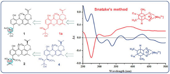
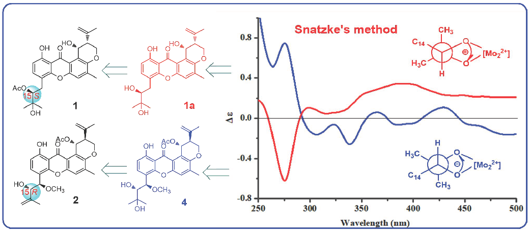
3.3. Preparation of the Methanolysis Derivative (1a) of 1

A solution of 1 (3.0 mg) and K2CO3 (10.0 mg) in anhydrous MeOH (3 mL) was stirred at room temperature for 5 h. The mixture was evaporated to dryness, and then purified using a silica gel column (PE/EtOAc, 1:1) to give the methanolysis derivative 1a (2.5 mg).
Methanolysis derivative (1a): yellow, amorphous powder; 1H NMR (CDCl3, 600 MHz) δ 12.59 (1H, s, 1-OH), 7.52 (1H, d, J = 7.8 Hz, H-3), 7.23 (1H, s, H-5), 6.75 (1H, d, J = 7.8 Hz, H-2), 5.48 (1H, brs, H-25), 5.05 (1H, s, H-22a), 4.78 (1H, s, H-22b), 4.46 (1H, dd, J = 9.6, 1.8 Hz, H-19a), 4.32 (1H, dd, J = 10.2, 9.6 Hz, H-19b), 3.75 (1H, d, J = 9.6 Hz, H-15), 3.19 (1H, d, J = 13.8 Hz, H-14a), 2.68 (1H, dd, J = 13.8, 9.6 Hz, H-14b), 2.55 (1H, brd, J = 12.0 Hz, H-20), 2.36 (3H, s, H-24), 1.98 (3H, s, H-23), 1.43 (3H, s, H-18), and 1.35 (3H, s, H-17); 13C NMR (CDCl3, 150 MHz) δ 184.4 (C, C-13), 160.6 (C, C-1), 153.3 (C, C-10), 151.9 (C, C-11), 149.5 (C, C-7), 142.3 (C, C-21), 138.8 (C, C-6), 138.3 (CH, C-3), 121.7 (C, C-8), 119.4 (CH, C-5), 116.9 (C, C-12), 116.3 (C, C-4), 111.8 (CH2, C-22), 110.1 (CH, C-2), 109.4 (C, C-9), 77.9 (CH, C-15), 73.2 (C, C-16), 64.2 (CH2, C-19), 61.5 (CH, C-25), 44.1 (CH, C-20), 32.1 (CH2, C-14), 26.7 (CH3, C-18), 23.7 (CH3, C-17), 22.6 (CH3, C-23), and 17.6 (CH3, C-24); HRESIMS m/z 441.1906 [M + H]+, (calcd. for C25H29O7, 441.1908).

3.4. Snatzke’s Method

The ICD spectra of 1a and 4 were obtained after addition of Mo2(OAc)4 following a previously referenced procedure [16,17].

3.5. Anti-Vibrio Activity Assays

Anti-Vibrio activity was evaluated by the conventional broth dilution assay [20]. Three pathogenic Vibrio strains, Vibrio parahemolyticus, Vibrio anguillarum, and Vibrio alginolyticus were used, and ciprofloxacin was used as a positive control with MIC values of 0.078 μM, 0.312 μM, and 0.625 μM, respectively. Replicates were maintained for each test bacteria.

4. Conclusions

Seven prenylxanthone derivatives, including three new compounds (13), were obtained from the marine-derived fungus Aspergillus sp. ZA-01 by using a shaken Czapek-Dox medium. The absolute configuration of 1 was determined by the Mo2(AcO)4 ICD method. This work suggested that the OSMAC approach was an active pathway for the exploration of new bioactive molecules.

Supplementary Materials

The following are available online at https://www.mdpi.com/1660-3397/16/9/312/s1. Figures S1–S24: 1D and 2D NMR, and mass spectra of 13.

Author Contributions

A.Z. and X.-W.Z. contributed to the fermentation, extraction, and isolation; M.Z. contributed to the ECD test; W.L. contributed to the bioactivities test; Z.-Y.M. contributed to the MS test; F.C. contributed to manuscript preparation; H.-J.Z. was the project leader, organizing and guiding the experiments and manuscript writing.

Funding

This work was supported by the National Natural Science Foundation of China (Nos. 41606174; 21877025), the Natural Science Foundation of Hebei Province of China (No. B2017201059, H2018201273, H2017201075,), and the High Performance Computer Center of Hebei University.

Conflicts of Interest

The authors declare no conflict of interest.

References

  1. Masters, K.S.; Bräse, S. Xanthones from fungi, lichens, and bacteria: The natural products and their synthesis. Chem. Rev. 2012, 112, 3717–3776. [Google Scholar] [CrossRef] [PubMed]
  2. Lesch, B.; Bräse, S. A short, atom-economical entry to tetrahydroxanthenones. Angew. Chem. Int. Ed. 2004, 43, 115–118. [Google Scholar] [CrossRef] [PubMed]
  3. Li, H.L.; Li, X.M.; Liu, H.; Meng, L.H.; Wang, B.G. Two new diphenylketones and a new xanthone from Talaromyces islandicus EN-501, an endophytic fungus derived from the marine red alga Laurencia okamurai. Mar. Drugs 2016, 14, 223. [Google Scholar] [CrossRef] [PubMed]
  4. Shaala, L.A.; Youssef, D.T. Identification and bioactivity of compounds from the fungus Penicillium sp. CYE-87 isolated from a marine tunicate. Mar. Drugs 2015, 13, 1698–1709. [Google Scholar] [CrossRef] [PubMed]
  5. Anja, K.; Stefan, K.; Clarissa, G.; Karin, K.; Martin, N.; Armin, M.; Heinz, H.F.; Iuliana, A.; Gerhard, R.; Jörg, F.; et al. Potential cancer chemopreventive in vitro activities of monomeric xanthone derivatives from the marine algicolous fungus Monodictys putredinis. J. Nat. Prod. 2007, 70, 353–360. [Google Scholar] [CrossRef]
  6. Pontius, A.; Krick, A.; Mesry, R.; Kehraus, S.; Foegen, S.E.; Müller, M.; Klimo, K.; Gerhäuser, C.; König, G.M. Monodictyochromes A and B, dimeric xanthone derivatives from the marine algicolous fungus Monodictys putredinis. J. Nat. Prod. 2008, 71, 1793–1799. [Google Scholar] [CrossRef] [PubMed]
  7. Wu, G.W.; Yu, G.H.; Kurtán, T.; Mándi, A.; Peng, J.; Mo, X.M.; Liu, M.; Li, H.; Sun, X.H.; Li, J.; et al. Versixanthones A–F, cytotoxic xanthone-chromanone dimers from the marine-derived fungus Aspergillus versicolor HDN1009. J. Nat. Prod. 2015, 78, 2691–2698. [Google Scholar] [CrossRef] [PubMed]
  8. Wu, X.; Fang, L.Z.; Liu, F.L.; Pang, X.J.; Qin, H.L.; Zhao, T.; Xu, L.L.; Yang, D.F.; Yang, X.L. New prenylxanthones, polyketide hemiterpenoid pigments from the endophytic fungus Emericella sp. XL029 and their anti-agricultural pathogenic fungal and antibacterial activities. RSC Adv. 2017, 7, 31115–31122. [Google Scholar] [CrossRef]
  9. Zhu, A.; Yang, M.Y.; Zhang, Y.H.; Shao, L.L.; Wang, C.Y.; Hu, L.D.; Cao, F.; Zhu, H.J. Absolute configurations of 14,15-hydroxylated prenylxanthones from a marine-derived Aspergillus sp. fungus by chiroptical methods. Sci. Rep. 2018, 8, 10621. [Google Scholar] [CrossRef] [PubMed]
  10. Fredimoses, M.; Zhou, X.F.; Lin, X.P.; Tian, X.P.; Ai, W.; Wan, J.F.; Liao, S.R.; Liu, J.; Yang, B.; Yang, X.W.; et al. New prenylxanthones from the deep-sea derived fungus Emericella sp. SCSIO 05240. Mar. Drugs 2014, 1, 3190–3202. [Google Scholar] [CrossRef] [PubMed]
  11. Chexal, K.K.; Fouweather, C.; Holker, J.S.E.; Simpson, T.J.; Young, K. The biosynthesis of fungal metabolites. Ш. Structure of shamixanthone and tajixanthone, metabolites of Aspergillus variecolor. J. Chem. Soc. Perkin. Trans. 1 1974, 5, 1584–1593. [Google Scholar] [CrossRef]
  12. Moosophon, P.; Kanokmedhakul, S.K.; Soytong, K. Prenylxanthones and a bicyclo[3.3.1]nona-2,6-diene derivative from the fungus Emericella rugulosa. J. Nat. Prod. 2009, 72, 1442–1446. [Google Scholar] [CrossRef] [PubMed]
  13. Ren, J.; Ding, S.S.; Zhu, A.; Cao, F.; Zhu, H.J. Bioactive azaphilone derivatives from the fungus Talaromyces aculeatus. J. Nat. Prod. 2017, 80, 2199–2203. [Google Scholar] [CrossRef] [PubMed]
  14. González, A.M.; Rivera, C.J.; Sosa, P.A.; Figueroa, M.; Rodríguez-Sotres, R.; Mata, R. Development of the fluorescent biosensor h Calmodulin (h CaM) L39C-monobromobimane (mBBr)/V91C-mBBr, a novel tool for discovering new calmodulin inhibitors and detecting calcium. J. Med. Chem. 2011, 54, 3875–3884. [Google Scholar] [CrossRef] [PubMed]
  15. Figueroa, M.; Gonźalez, M.C.; Rodŕıguez-Sotres, R.; Sosa-Peinado, A.; Gonźalez-Andrade, M.; Cerda-Garćıa-Rojas, C.M.; Mata, R. Calmodulin inhibitors from the fungus Emericella sp. Bioorg. Med. Chem. 2009, 17, 2167–2174. [Google Scholar] [CrossRef] [PubMed]
  16. Sun, X.P.; Cao, F.; Shao, C.L.; Chen, M.; Liu, H.J.; Zheng, C.J.; Wang, C.Y. Subergorgiaols A–L, 9,10-secosteroids from the South China Sea gorgonian Subergorgia rubra. Steroids 2015, 94, 7–14. [Google Scholar] [CrossRef] [PubMed]
  17. Di Bari, L.; Pescitelli, G.; Pratelli, C.; Pini, D.; Salvadori, P. Determination of absolute configuration of acyclic 1,2-diols with Mo2(OAc)4. Snatzke’s method revisited. J. Org. Chem. 2001, 66, 4819–4825. [Google Scholar] [CrossRef] [PubMed]
  18. Moreno, E.; Parks, M.; Pinnell, L.J.; Tallman, J.J.; Turner, J.W. Draft genome sequence of a Vibrio harveyi strain associated with vibriosis in Pacific white shrimp (Litopenaeus vannamei). Genome Announc. 2017, 5, e01662-16. [Google Scholar] [CrossRef] [PubMed]
  19. Vezzulli, L.; Pezzati, E.; Brettar, I.; Höfle, M.; Pruzzo, C. Effects of global warming on Vibrio ecology. Microbiol. Spectr. 2015, 3, VE-0004-2014. [Google Scholar] [CrossRef] [PubMed]
  20. Appendino, G.; Gibbons, S.; Giana, A.; Pagani, A.; Grassi, G.; Stavri, M.; Smith, E.; Rahman, M.M. Antibacterial cannabinoids from Cannabis sativa: A structure-activity study. J. Nat. Prod. 2008, 71, 1427–1430. [Google Scholar] [CrossRef] [PubMed]
Figure 1. Chemical structures of 17.
Marinedrugs 16 00312 g001
Figure 2. COSY and key HMBC correlations of 1.
Marinedrugs 16 00312 g002
Figure 3. (a) Experimental ECD spectra for 1 and 1a; (b) ICD spectra of Mo-complexes of 1a (red) and 4 (blue) recorded in DMSO.
Marinedrugs 16 00312 g003
Figure 4. Experimental ECD spectra for 24.
Marinedrugs 16 00312 g004
Table 1. 1H NMR data (δ) of 13 (600 MHz, δ in ppm, CDCl3, J in Hz).
Position123
26.71, d (8.4)6.80, d (8.4)6.85, d (8.4)
37.41, d (8.4)7.60, d (8.4)7.65, d (8.4)
57.29, s7.27, s7.24, s
143.33, dd (14.4, 2.4)4.82, d (8.4)4.83, d (8.4)
2.91, dd (14.4, 10.8)
155.15, dd (10.8, 2.4)4.19, d (8.4)4.19, d (8.4)
171.34, s4.65, brs4.64, brs
4.62, brs4.60, brs
181.38, s1.76, s1.77, s
194.46, brd (10.8)4.56, brd (11.4)4.43, dd (10.8, 3.0)
4.35, dd (12.0, 10.8)4.32, dd (11.4, 3.0)4.35, dd (10.8, 3.0)
202.55, d (12.0)2.72, brs2.72, d (3.0)
225.06, s4.81, s4.81, s
4.78, s4.76, s4.59, s
231.86, s1.89, s1.85, s
242.38, s2.36, s2.37, s
255.50, brs6.90, brs5.43, brs
1-OH12.63, brs13.06, brs12.83, brs
14-OCH3-3.28, s3.30, s
15-OAc1.99, s--
25-OH4.51, brs-4.96, d (4.2)
25-OAc-2.10, s-
Table 2. 13C NMR data (δ) of 13 (150 MHz, δ in ppm, CDCl3).
Position123
1161.1, C162.0, C161.8, C
2109.5, CH110.7, CH110.7, CH
3137.9, CH134.7, CH135.1, CH
4115.1, C115.4, C115.8, C
5119.5, CH120.4, CH119.1, CH
6139.0, C138.0, C139.0, C
7149.6, C150.4, C149.9, C
8121.8, C115.0, C121.4, C
9109.2, C109.0, C108.8, C
10153.3, C153.5, C153.7, C
11151.8, C151.8, C152.0, C
12116.9, C116.4, C116.9, C
13184.5, C183.4, C184.5, C
1429.7, CH278.7, CH78.8, CH
1578.6, CH80.0, CH80.0, CH
1672.5, C141.7, C142.5, C
1726.9, CH3114.8, CH2114.8, CH2
1825.3, CH318.2, CH318.2, CH3
1964.1, CH263.9, CH264.8, CH2
2044.1, CH42.6, CH45.1, CH
21142.3, C142.5, C142.7, C
22111.7, CH2112.9, CH2112.4, CH2
2322.5, CH322.6, CH322.7, CH3
2417.4, CH317.5, CH317.7, CH3
2561.0, CH65.7, CH63.3, CH
14-OCH3-57.2, CH357.2, CH3
15-OAc170.4, C--
20.7, CH3
25-OAc-170.2, C-
21.4, CH3
Table 3. Tests of anti-Vibrio activities for compounds 17.
StrainsCompounds [MIC (μM)]
1234567Ciprofloxacin
V. parahemolyticus1.566.253.1225.012.56.2525.00.078
V. anguillarum1.5625.025.025.025.06.256.250.312
V. alginolyticus3.1225.012.525.012.512.525.00.625

